Review

Oxidative Stress, Bone Marrow Failure, and Genome Instability in Hematopoietic Stem Cells

Department of Biological Sciences, UNC Charlotte, 9201 University City Blvd., Woodward Hall Room 386B, Charlotte, NC 28223, USA
*
Author to whom correspondence should be addressed.
Int. J. Mol. Sci. 2015, 16(2), 2366-2385; https://doi.org/10.3390/ijms16022366
Submission received: 19 December 2014 / Revised: 6 January 2015 / Accepted: 16 January 2015 / Published: 22 January 2015
(This article belongs to the Special Issue DNA Damage and Repair in Degenerative Diseases 2014)

Abstract

:
Reactive oxygen species (ROS) can be generated by defective endogenous reduction of oxygen by cellular enzymes or in the mitochondrial respiratory pathway, as well as by exogenous exposure to UV or environmental damaging agents. Regulation of intracellular ROS levels is critical since increases above normal concentrations lead to oxidative stress and DNA damage. A growing body of evidence indicates that the inability to regulate high levels of ROS leading to alteration of cellular homeostasis or defective repair of ROS-induced damage lies at the root of diseases characterized by both neurodegeneration and bone marrow failure as well as cancer. That these diseases may be reflective of the dynamic ability of cells to respond to ROS through developmental stages and aging lies in the similarities between phenotypes at the cellular level. This review summarizes work linking the ability to regulate intracellular ROS to the hematopoietic stem cell phenotype, aging, and disease.

1. Introduction

Organisms in aerobic environments are continuously exposed to reactive oxygen species (ROS). ROS include oxygen molecules (O2), superoxide anion radicals (O2), hydroxyl free radicals (OH), singlet oxygen (1O2), and hydrogen peroxide (H2O2), and can be generated by imbalanced endogenous reduction of oxygen by cellular enzymes or in mitochondrial respiratory pathway, as well as by exogenous exposure to UV or environmental damaging agents [1]. Regulation of intracellular ROS levels and ROS-mediated signaling is central maintaining the balance between self-renewal, proliferation, and differentiation of normal stem and progenitor cells in the hematopoietic and neuronal compartments, as well as the early embryonic stem (ES) cell compartment. Increases in ROS above normal concentrations can lead to oxidative stress (OS) and DNA damage [2]. OS is thought to damage approximately 10,000 bases per day per human cell and be one of the major causes of DNA damage and mutation [3]. The majority of these damaged sites are repaired through base excision repair (BER), although persistent damage or damage on two opposing strands at nearby nucleotides may result in double-strand breaks (DSBs) requiring repair by non-homologous end-joining (NHEJ) or homologous recombination (HR). OS is also known to induce higher order chromatin single strand DNA (ssDNA) nicks characteristic of those generated during programmed cell death. ROS and the DNA damage that may result are both capable of triggering multiple DNA damage response pathways. Results of signaling and cellular response to ROS range from cell proliferation and survival to growth arrest, senescence, and cell death depending on the level of ROS and cell type studied. A growing body of evidence indicates that the inability to regulate high levels of ROS leading to alteration of cellular homeostasis or defective repair of ROS-induced damage lies at the root of diseases characterized by both neurodegeneration and bone marrow failure [4,5,6,7]. Further, high levels of ROS appear to be a distinct feature of acute myeloid leukemia (AML) [8,9,10]. That these diseases may be reflective of the dynamic ability of cells to respond to ROS through developmental stages and aging lies in the similarities between phenotypes at the cellular level.

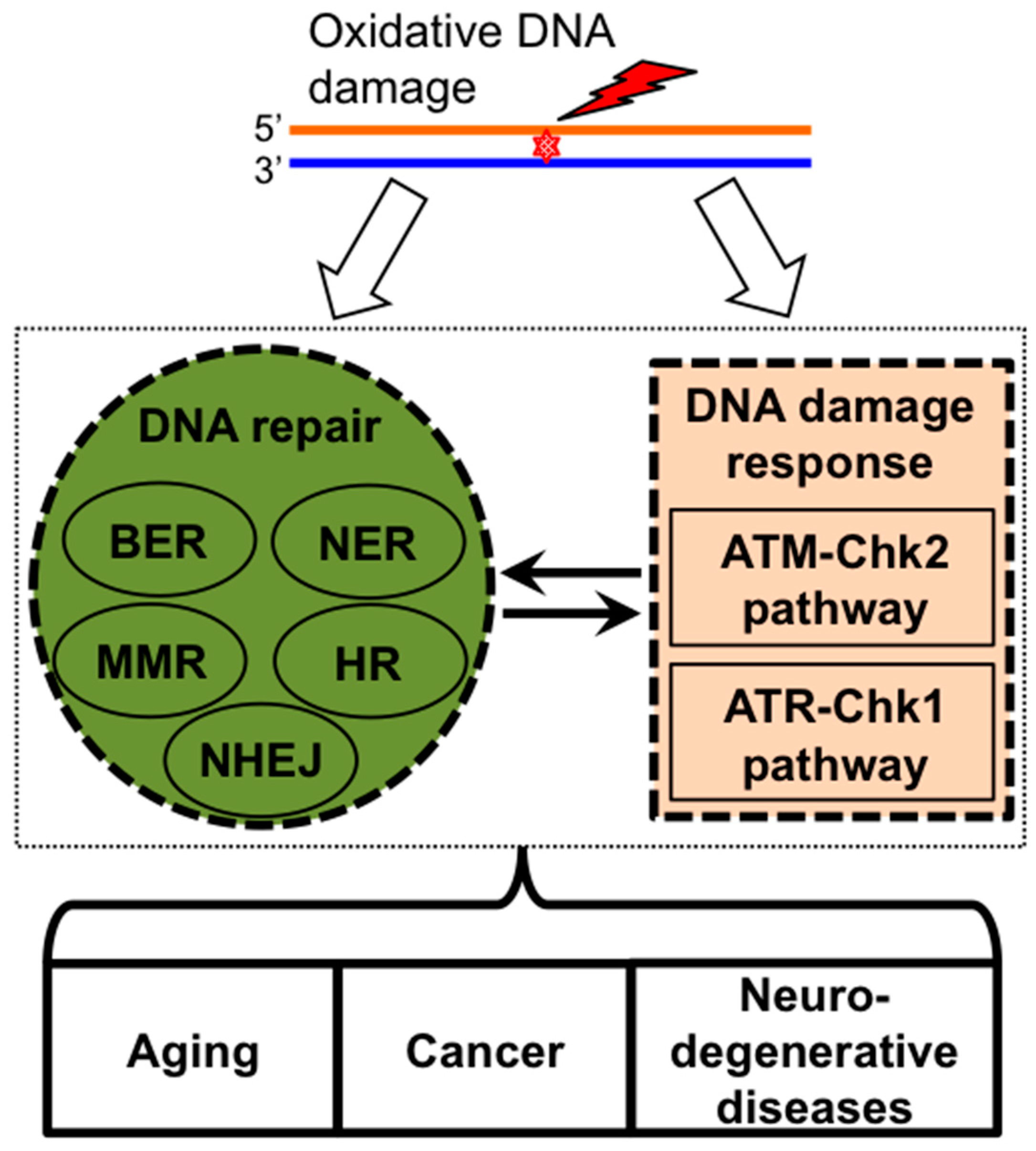
2. ROS Induces DNA Damage Signaling and Repair

2.1. Oxidative Stress and Oxidative DNA Damage

Genomes of all organisms are exposed to endogenous and exogenous stresses, such as OS. OS refers to an imbalance between antioxidant defenses and production of ROS [11,12,13,14]. ROS are generated endogenously from cellular metabolism, such as oxidative phosphorylation in mitochondria and long-chain fatty acids oxidation in peroxisomes, or exogenously by environmental toxins, such as ionizing radiation (IR), ultraviolet (UV) radiation, and DNA damaging/chemotherapeutic agents [15,16,17,18,19,20]. When ROS generation exceeds antioxidant defense capacity in cells, ROS may react with almost all macromolecules including DNA, RNA, proteins, and lipids. Notably, oxidative DNA damage may represent the major type of DNA damage, evidenced that approximately 10,000 DNA alterations are generated per mammalian cell per day [21,22,23,24]. Oxidative DNA damage includes oxidized base (purine and pyrimidine) damage, oxidized sugar moiety damage, apurinic/apyrimidinic (AP) sites, DNA single-strand breaks (SSBs), DSBs, DNA intrastrand crosslinks, DNA intrastrand crosslinks, DNA interstrand crosslinks (ICLs), protein-DNA crosslinks, mismatched pairs with damaged bases, stalled DNA replication forks, and oxidatively-generated clustered DNA lesions (OCDLs) [21,22,23,24,25]. OS has been implicated in the pathogeneses of multiple diseases, such as bone marrow failure, cancer, and neurodegenerative disorders [26,27,28].

2.2. DNA Damage Response Pathways and DNA Repair

To maintain genomic integrity, DNA repair and DNA damage response (DDR) pathways are employed in cellular responses to oxidative DNA damage [22,23,29,30] (Figure 1). The majority of these damaged sites are repaired through BER. However, depending on the extent of oxidative DNA damage processes, such as nucleotide excision repair (NER), mismatch repair (MMR), HR, and NHEJ, may be employed in the repair processes to protect cells from OS [17,22]. Additionally, Ataxia-telangiectasia mutated (ATM)-Checkpoint kinase 2 (Chk2) and ATM- and Rad3-related (ATR)-Checkpoint kinase 1 (Chk1) checkpoints are the two major DDR pathways induced by oxidative DNA damage in order to coordinate DNA repair process with cell cycle progression, transcription, apoptosis, and senescence [23,30,31,32,33,34]. ATM modulates cell cycle in response to multiple types of cell stress and DNA damage including oxidative stress. In response to DSBs, ATM auto-phosphorylates its own Ser1981, leading to dimer dissociation into a monomer and the full activation of ATM [35,36]. ATM is also directly activated after disulfide bond-dependent conformation change induced by OS in the absence of direct DNA damage [37,38,39]. In support of a critical role for ATM in protecting hematopoietic stem cells (HSC) from OS, the HSC compartment of ATM-deficient mice present with increased levels of ROS-dependent phosphorylation of the downstream effector mitogen activated protein kinase (MAPK) p38 protein that is known to activate both cell cycle signaling and apoptosis in response to multiple cellular stressors including OS, UV, and inflammatory cytokines. This loss of ATM signaling and increase in p38 phosphorylation is in turn associated with a loss of HSC quiescence [40]. Notably, ATR is activated by oxidative DNA damage after a distinct 3'–5' SSB end resection by a BER protein APE2 [41]. Functional interplay between various DNA repair programs and ATM-Chk2- and ATR-Chk1-dependent DDR pathways in response to oxidative stress is summarized in a recent review article [28]. A chronic impairment of the DDR would be expected to result in accumulation of DNA damage, a mutator effect, and genome instability as observed in normal aging, cancer, and neurodegenerative disease (Figure 1). Elevated intracellular ROS promotes DNA DSBs and altered NHEJ repair leading to chromosomal deletions, translocations, and tumorigenesis [42,43,44]. Interestingly, multiple studies have identified elevated ROS production in the mitochondrial compartment of apoptotic cells. However, the extent of interplay, if any, between damage signaling pathways or DNA repair pathways in the mitochondrial and nuclear compartments is unclear.
Figure 1. Cellular responses to oxidative DNA damage. DNA repair pathways (BER, NER, MMR, HR, and NHEJ) and DNA damage response pathways (ATM-Chk2 and ATR-Chk1) are integrating into an interacting network in response to OS. Defective DNA repair and response pathways are associated with aging, cancers, and neurodegenerative diseases. BER (base excision repair); NER (nucleotide excision repair); MMR (mismatch repair); NHEJ (non-homologous end-joining); HR (homologous recombination); ATM-Chk2 (Ataxia-telangiectasia mutated-Checkpoint kinase 2); ATR-Chk1 (ATM- and Rad3-related-Checkpoint kinase 1); OS (oxidative stress).
Ijms 16 02366 g001

3. Bone Marrow Failure Syndromes

Bone marrow failure is characterized by the progressive absence of one or more hematopoietic cell lineages. These syndromes are characterized as inherited or acquired. The best-characterized inherited syndromes include Fanconi anemia (FA), as well as the telomere diseases dyskeratosis conegenitia (DC), Diamond-Blackfin anemia (DBA), and Shwachman-Diamond syndrome, while the most prevalent acquired syndrome is aplastic anemia (AA). There is increasing evidence linking elevated ROS levels and impaired DNA repair mechanisms to contribute to bone marrow failure observed in these diseases. On the other hand, neurodegeneration corresponds to any pathological condition that primarily affects neurons and neurodegenerative disorders including Alzheimer disease, Parkinson disease, Huntington disease, and amyotrophic lateral sclerosis [45]. However, both systems appear particularly sensitive to the control of intracellular ROS and OS, and altered responses to these “leading to DNA damage have been implicated in the pathogeneses of both bone marrow failure and neurodegenerative disorders [4,6,7,26,27,28,46,47].

3.1. Fanconi Anemia

FA is an autosomal X-linked disease that is characterized mainly by bone marrow failure, anemia, developmental abnormalities, cancer susceptibility, and altered mitochondrial function. It is considered a rare disease affecting approximately 1 in every 100,000 people [48]. FA is a chronic and degenerative disorder with the probability of patients developing leukemia, squamous cell carcinomas, or liver tumors increasing by 40% by age 30, 50% by age 45 and 76% by age 60 [49]. Progressive bone marrow failure and myeloid malignancies account for 90% of deaths in FA patients. At the molecular level, there are 15 complementation groups that been identified, and mutations in any of these genes can lead to FA or a similar disorders. FA proteins that make up the core complex are FancA, FancB, FancC, FancE, FancF, FancG, FancL, and FancM, and defects in any are known to cause FA in humans. This core complex acts to ubiquitylate downstream targets FancD2 and FancI that then act in parallel with the remaining FA proteins (FancD1, FancJ, FancN, FancO, and FancP) to repair ICLs. Several mouse models have provided insight into the pathology and etiology of the disease.
There is some discordance between humans with FA and FA mouse models, most notably the absence of bone marrow failure, anemia, and developmental abnormalities in mice that are the defining characteristics of the disease in humans. These differences, however, could be in part due to human reliance on the FA pathway for detoxification and tumor suppression to ensure normal development whereas laboratory mice lack the same genotoxic stressors that may trigger the disease [50]. Almost all FA mouse models so far have exhibited reduced fertility, and cells derived from these mice display sensitivity to ICL inducing agents [50]. FancA−/− mice, for instance, do not display hematopoietic abnormalities or congenital defects; however, they have hypergonadism and mouse embryonic fibroblasts (MEFs) derived from these mice are hypersensitive to mitomycin C (MMC) [51]. Interestingly, bone marrow-derived megakaryocyte progenitors demonstrate impaired proliferation and all bone marrow cells derived from FancA−/− mice display increased apoptosis indicating a possible HSC defect [52]. Also, both FancA−/− and FancG−/− mice have increased apoptosis in neural stem and progenitor cells that leads to microcephaly [53].
FancC−/− mice born do not display overt hematopoietic abnormalities, however they are born with sub-Mendelian frequency and have a significantly increased incidence of micropthalmia in C57BL/6J mice [54]. HSC derived from FancC−/− mice have impaired differentiation and self-renewal capacity [55], a defect that is rescued by retroviral-mediated gene transfer of FancC, indicating that FancC is required for properly functioning HSCs. Also, FancC−/− mice exposed to low doses of MMC develop progressive bone marrow failure and chromosomal aberrations [56]. Differences in humans with FA and the mouse models may be due to the differences in environmental and dietary exposures.
FancD1/Brca2 is downstream from the FA core complex. Brca2 is required for HR-directed repair of DNA DSBs. FancD1 knockout in mice leads to embryonic lethality, but a mouse model has been created with homozygous deletion of exon 27, which allows expression of the protein but prevents FancD1 from interacting with FancD2 [57]. FancD1∆27/∆27 mice display similar phenotypes to FancA−/− and FancC−/− mice, though there is some evidence that the hematopoietic abnormalities are more severe than with the other phenotypes. For instance, FancD1∆27/∆27 mice are significantly more sensitive to MMC and acquire spontaneous chromosomal aberrations and early loss of colony forming hematopoietic progenitor cells compared to the other phenotypes [58]. This severity would be expected as FancD1 is necessary not only for ICL repair, but also for DSB repair via HR. Notably, unlike the other FA mouse models, FancD1∆27/∆27 mice do not have impaired fertility indicating that it is dispensable for meiosis unlike the other FA proteins, but is important for somatic cell HR [57].
FancD2−/− mice are born with sub-Mendelian frequency and also display growth retardation, a phenotype that varies based on the genetic background of the mice. These mice have an increased incidence of tumor formation similar to that of FancD1 haploinsufficiency [59]. Also, the phenotype of these mice is more severe than those with deleted FA core proteins, indicating alternate activity outside of the FA pathway, possibly protecting against oxidative DNA damage including DSBs [60].
Cells derived from FA mutant mice are hypersensitive to OS. It has recently been shown that impaired or reduced mitochondrial function in FA cells is coupled with increased intracellular ROS, and inactivation of enzymes essential for energy production [61]. Intriguingly these defects, as well as hypersensitivity to MMC can be partially rescued by overexpression of superoxide dismutase (SOD1) [61]. Bone marrow progenitor cells derived from FancC−/− mice undergo replicative senescence following exposure to hypoxic/hyperoxic conditions [62]. Interestingly, the FancC−/−/Sod1−/− mouse model was created to increase endogenous OS. These mice demonstrate loss of hematopoietic progenitor cells in the bone marrow leading to anemia and leucopenia [63], strongly indicating that OS is a key factor in the initiation of bone marrow failure in FA. Also, the FancD2−/−/Aldh2−/− mice, which can no longer detoxify aldehydes by oxidizing them to carboxylic acids, present with severe phenotypes including the development of acute lymphoblastic leukemia (ALL) after three to six months. Exposure to ethanol in utero results in development of physical abnormalities and death; and surviving mutant mice have a vast reduction in bone marrow cellularity [64]. Taken together, these models demonstrate that FA phenotypes that are hallmarks of the disease may indeed require stressors, such as aldehydes or ROS, in order to initiate the disease phenotype, and it is the absence of these stressors in the laboratory environment that prevents the FA mouse models from accurately mimicking the disease in humans.

3.2. Additional Degenerative Excision Repair Defects

Cockayne syndrome (CS) and xeroderma pigmentosum (XP) are characterized by defects in DNA repair of damage by crosslinking agents or elevated ROS due to mutation of any one of the excision repair pathway cross complementation (ERCC) group genes. Whereas CS patients are defective in the transcription coupled NER (TC-NER) pathway and present with neurological abnormalities, XP patients are deficient in the global genome NER (GG-NER) pathway and are hypersensitive to sun and susceptible to carcinoma [65]. Mutant mouse models have been generated for most of the proteins involved in this pathway that mimic the genotypes and phenotypes of patients with CS or XP.
CS is caused by defects primarily in Csa and Csb, which are the core proteins essential for TC-NER. Csa−/− or Csbm/m mice genetically mimic the disease, and both mutants present with UV sensitivity, loss of photoreceptors, increased sensitivity to γ-irradiation, and mild neurodegeneration [66]. Cells from Csbm/m mice but not Csa−/− mice show hypersensitivity to γ-IR and paraquat which generates superoxide anions and H2O2 in cells indicating that Csb plays a role in the repair of oxidative damage [67,68]. Although Csa−/− mice do not show an increased sensitivity to OS, human cells deficient in CSA are more sensitive to H2O2 than wild-type cells [69], and primary fibroblasts and keratinocytes from CSA patients are hypersensitive to potassium bromate, a known inducer of oxidative damage [70]. This indicates that the role of the CSA protein may differ between species and that it is likely involved in the oxidative damage response in humans but not mice [71].
XP is caused by defects in core NER proteins as well as proteins in the GG-NER pathway. Xpc functions primarily in the GG-NER pathway and Xpc−/− mice have a reduced lifespan and have a propensity for developing spontaneous tumors Also, these mice have been demonstrated to have a very large increase in lung tumor incidence compared to controls thought to be a result of chronic OS [72]. Also, fibroblasts derived from Xpc−/− mice have demonstrated OS sensitivity compared to controls by mutation accumulation and decreased survival [73]. Analysis of bone marrow cells from Xpc−/− mice shows reduced cellular viability, hypocellularity of most lineages, reduced numbers of CFU (colony-forming units), and increased sensitivity to carboplatin [74]. Since XP patients develop skin cancers by the age of 20, and the average life expectancy is early 30s, the findings in bone marrow of the Xpc−/− mice suggest the possibility that if patients were to live to later age, they could exhibit myelosuppression or bone marrow failure. In contrast to the Xpc−/− mice, Xpa−/− mice have a significant increase in tumors in the liver, but not in the lungs and do not have an increase in mutation load like the Xpc−/− mice [73]. However, cells derived from XPA patients exhibit defective repair in 8,5'-(S)-cyclo-2'-deoxyadenosine, a DNA lesion induced by ROS indicating that XPA may play a role in the repair of oxidative bulky lesions [75].
Cells from both CS and XP individuals affected by disease and from multiple mouse models have elevated ROS and defective mitochondrial function [65,75]. Interestingly, a similar set of defects are observed in lymphoblasts from DC patients, namely elevated ROS production, impaired DNA response leading to apoptosis and a proliferative defect [76], further connecting elevated ROS with bone marrow failure outcome.
Additional proteins in the ERCC complement group are now being identified as central to the etiology of other bone marrow failure syndromes. Through Next-Gen sequencing, ERCC6L2 was recently reported as deficient in two patient samples both presenting with bone marrow failure and neurodegeneration [77]. Loss of function of the same gene in both cases demonstrates the link between these two degenerative processes. Reduction of ERCC6L2 induced by siRNA knockdown in cells results in significantly increased intracellular ROS, effective DNA repair response shown by γ-H2AX foci, reduced cell viability and increased sensitivity to a panel of DNA damaging agents [77]. The similarities in phenotypes from ERCC genes thus far suggest that additional mutations in ERCC genes will be identified as a paradigm for bone marrow failure syndromes.

4. Hematopoietic Stem Cell Response to ROS

4.1. HSC Response to ROS

ROS are important for fate determination of normal stem cells [78]. Normal HSC are primarily in a quiescent state within the BM niche [79]. HSC exposed to elevated ROS exhibit altered characteristics and undergo both proliferation and differentiation, typically after mobilization to the oxygen rich bloodstream [80], but also to senescence and apoptosis in a dose-dependent manner [78]. This impact of ROS is evolutionarily conserved and observable in analogous Drosophila systems [81].
A number of transcriptional microarray, exome, and proteomic approaches have been utilized in an effort to understand the signal transduction pathways activated in response to ROS [82,83,84,85,86,87,88,89,90,91]; however, few have centered on specific HSC populations. Despite this, the p53, Akt, MAPK, and ATM pathways have all been implicated in HSC response to ROS. MDM2 promotes survival of hematopoietic progenitors by promoting p53 degradation [92]. The Forkhead box O (FoxO) family of transcription factors has known roles in metabolism, proliferation, and OS response and resistance (recently reviewed in [93]). Single FoxO3a deficiency in HSCs is associated with elevated intracellular ROS [94]. Triple knock-out FoxO1−/− FoxO3a−/− FoxO4−/− mice exhibit reduced numbers of both HSC and progenitor cell populations [95]. Recent molecular analysis suggests that the co-factor KDM5 activates OS resistance genes through interaction with the lysine deacetylase HDAC4 to in turn promote FoxO deacetylation and alter specificity of target gene binding [96]. Drosophila also demonstrates signaling paradigms with increases in ROS signaling through JNK pathway, FoxO activation, and polycomb downregulation [81]. In conjunction with elevated ROS, depletion of the Meis1 transcription factor in mice leads to apoptosis, loss of HSC quiescence, and loss of transplantation capacity [97].
Differences in the levels of ROS also impact the observed cellular response of stem cells [98]. Elevated levels of OS induced by mutations of metabolic enzyme genes, ischemia/reperfusion, chemotherapy, or chronic exposure to 10–100 μM H2O2 induces multiple effects ranging from cell cycle arrest to death, depending on the cell type [99,100]. At submicromolar concentrations, ROS and H2O2 act as proliferation and growth signaling molecules. Low concentration exposure (100 μM) of stem cells is sufficient to promote hyperphosphorylation of Akt and RAF1, activation of insulin signaling pathways, and phosphorylation of MAPK p38 within 1 h [98]. This initial rapid growth is then temporally followed 4–24 h later by induction of cell cycle checkpoints characterized by dephosphorylation of Rb, hyperphosphorylation of cdc2, and down-regulation of E2F targets, such as FoxD3 [98].
To directly link ROS to genetic instability in HSC, we genetically engineered murine HSC cells to contain two transgene reporters at unlinked loci—each with either a 5' or 3' exon of the green fluorescent protein (GFP). This scheme enables us to score for chromosomal translocations following DNA damage and repair by interchromosomal NHEJ repair that results in a repair event joining the two exons onto the same DNA helix to form an intact GFP (Figure 2A). Following exposure of the engineered HSC to H2O2 (Figure 2A, Figure 2B left image), surviving cells were then allowed to proliferate into myeloid CFU by standard protocols [101] (Figure 2B middle image), and those CFU expressing GFP readily identified by fluorescent microscopy (Figure 2B right image). This approach demonstrated that a single 30 min exposure of HSC to 100 μM H2O2 or 5 mM H2O2 is sufficient to promote the appearance of GFP+ colonies in a dose-dependent manner at average frequencies of 6 × 10⁻⁶ and 10 × 10−6, respectively (Figure 2C). This in vitro system is analogous to the outgrowth of transformed myeloid leukemic cells and connects ROS and genome rearrangements in HSC and leukemia.
It is now well established that IR promotes differentiation, short-term apoptosis, and long-term senescence of hematopoietic stem cells [78,102]. Several studies demonstrated that IR of CD34+ hematopoietic progenitor cells induces indirect effects on DNA damage through elevated OS [102,103]. IR of CD34+ cells induces early increase of superoxide anion radicals and hydrogen peroxide, followed by elevated MnSOD expression by 24 h, followed by elevated persistent intracellular ROS over longer culture periods associated with reduced clonogenic potential [104,105]. By contrast γ-H2AX expression and foci associated with direct DNA DSBs are elevated in short periods following IR but return to baseline by 24 h [105]. Presence of insulin-like growth factor 1 (IGF-1) in irradiated CD34+ cells can inhibit the mitochondria-mediated apoptosis and rescue the clonogenic potential defect [104]. This role for IGF-1 in response to ROS mirrors transcriptional and proteomic profiling of stem cells demonstrating induction of insulin regulated pathways in response to ROS [98]. Further, sublethal total body IR of mice leads to a persistent elevation of ROS in HSC associated with sustained increases in oxidative DNA damage and DSBs, inhibition of HSC clonogenic potential, and induction of HSC senescence, at least in part due to upregulation of nicotinamide adenine dinucleotide phosphate oxidase 4 (NOX4) [103].
Figure 2. Direct connection between exposure of hematopoietic stem cells (HSC) to reactive oxygen species (ROS) and genome rearrangements. (A) Scheme of exposure of genetically engineered HSC to ROS and scoring of GFP+ cells indicative of translocations; (B) Left image—HSC colony by contrast microscopy (100×); Middle image—GFP+ derivative myeloid CFU (colony-forming units) by contrast microscopy (100×); Right image—Same GFP+ CFU colony shown in middle by fluorescence microscopy (100×); and (C) Bar graph showing dose dependent appearance of GFP+ myeloid CFU derived from HSC exposed to 0, 100 μM, or 5 mM H2O2 for 30 min, then returned to normal conditions. 100 μM resulted in an average frequency of CFU at 6.5 × 10⁻⁶; 5 mM resulted in an average frequency of CFU at 10.4 × 10⁻⁶. * denotes statistically significant stimulation of chromosomal translocations by H2O2 (students t-test p-value < 0.037). A few colonies were observed in untreated samples that by contrast microscopy but appeared to be unviable and auto-fluorescent rather than bona fide GFP+ CFU; however, further analysis was not performed on them so they are reported here.
Ijms 16 02366 g002

4.2. HSC Response to ROS and Aging

Control of ROS in ES cells is now understood to be critical to maintenance of self-renewal phenotype. Molecular evidence suggests this is due, at least in part, to ROS activation of miR-29b that targets Sirt1 to control ES cell self-renewal [106]. Differentiation of ES cells leads to increased intracellular ROS [61,107]. Interestingly, during differentiation of ES cells, superoxide production, cellular levels of intracellular ROS, and DNA damage levels increase although expression of major antioxidant genes and genes involved in multiple DNA repair pathways are downregulated [107], and DNA repair by HR is reduced [101].
Aging is associated with reduced expression of multiple DNA repair proteins, reduced DNA repair efficiency and elevated levels of genome instability in hematopoietic cells [6,108,109,110]. Hematopoietic cells in older mice and humans are characterized by reduced regenerative potential, lineage bias toward myeloid cells, and increased potential for outgrowth of genetically unstable cells and malignancy. Serial transplantation of human HSC into immunodeficient mice is associated with replication stress, elevated intracellular ROS, accumulation of DNA damage, and reduced self-renewal potential that can be further aggravated by addition of a glutathione synthetase inhibitor [80]. The connection between natural HSC aging and intracellular ROS was made by the ability to separate the CD34−, Lin− (lineage cocktail negative), Sca-1+, c-kit+ HSC compartment into two fractions based on intracellular ROS levels [111], and the observation that the ROSlow fraction has higher self-renewal capacity and serial transplantation capability. Conversely, ROShi fraction has reduced capacity for self-renewal and in addition is biased toward myeloid differentiation, similar to aged mice in vivo [111]. Intriguingly, the observed phenotype can be partially rescued following inhibition of MAPK p38 suggesting that p38 is a potential therapeutic target [40,111].
ROS seem well tolerated in “young” cells with highly proficient DNA repair, and thus have few if any long-term deleterious effects. However, “older” cells with less efficient repair become more sensitive to ROS levels, which lead to increased genome instability. Recent work demonstrated that quiescent HSC (analogous to the ROSlow fraction) repair DNA damage less efficiently than progenitors leading to accumulation of DNA damage and impaired function, suggesting a mechanism for increased presentation of myeloid malignancies in aged populations [112].
HSC reactivity to ROS during aging in vivo combines cell-intrinsic and cell-extrinsic mechanisms [113]. A relationship between the HSC compartment and supporting cells within the bone marrow niche is central to homeostasis and changes during not only aging but also other pathophysiologic processes including atherosclerosis, hypertension, and diabetes [114,115]. HSC and progenitors are typically exposed to low levels of ROS in the bone marrow niche, and hypoxia in turn helps preserve HSC and progenitor characteristics. HIF1α, HIF2α have been shown to be major regulators in modulating the response of HSC to ROS in this microenvironment [103,116,117,118]. ROS derived from NAPH oxidase and mitochondria in response to injury and stress is elevated in supportive bone marrow microenvironment cells, leading to hypoxia and HIF1α expression, and HSC progenitor expansion and differentiation [114]. Similarly, HIF2α depletion in HSC leads to increased ROS, activation of endoplasmic reticulum (ER) stress and apoptosis [119].

4.3. Leukemic Cells and ROS

Defective signaling in response to ROS occurs in multiple leukemic stem cell populations to promote proliferation, differentiation, genomic, and epigenetic alterations, immune evasion, and survival [120]. ROS act as signaling molecules to regulate redox-sensitive transcriptional factors, enzymes, oncogenes, and other downstream effectors, and thus AML cells are thought to be addicted to elevated ROS [120]. Extracellular ROS, but not intracellular mitochondrial ROS, have been found to be significantly elevated in both AML cell lines and cells derived from patients [8,9,10] and correlates with constitutive activation of NOX, reduced levels of glutathione, and attenuated p38 MAPK responsiveness, all postulated to promote proliferation of AML blasts [121]. AKT negatively regulates the FoxO proteins, which target expression of several anti-oxidant enzymes, including the GPx, catalase (CAT) and SODs. Akt is constituitively activated in most primary AML cells, leading to FoxO inhibition, and increased ROS levels [95,122]. Activated RAS is a feature of a portion of AML patients, and upregulates ROS levels through NOX to promote proliferation of hematopoietic cells [121]. Similarly, FLT3 plays a role in H2O2 production in AML cells through NOX p22 protein, while knockdown of p22 leads to immediate reduction of H2O2 within 24 h [123]. Activating mutations of FLT3 by internal tandem duplications or activating mutations of the FLT3 receptor are seen in a large percentage of AML patients associated with increased DNA DSBs, genome instability, and poor prognosis [124]. Interestingly FLT3 activation in AML cells leads to inactivation of tumor suppressor PTP, DEP1, and reactivation of DEP1 is sufficient to decrease ROS. Elevated ROS DNA damage occurs both in the nuclear fraction and mitochondria. Mitochondria have more limited DNA repair functions and mitochondrial DNA genome instability has been noted in AML [125]. HIF2α activity may also promote proliferation and inhibition of apoptosis of AML blasts in response to ROS [119].
Philadelphia-negative chronic myeloproliferative neoplasms (MPNs; essential thrombocythemia, polycythemia vera, and myelofibrosis) have recently been shown to be associated with chronic inflammation, OS and accumulation of ROS. Transcriptional profiling of these revealed several OS modulating genes significantly deregulated in MPNs [126]. Interestingly the transcriptional factor Nrf2, which is upregulated in multiple AML cells, was found to be downregulated in this study [126,127]. In addition, downregulation of ATM and p53 were also observed, although the impact of these is likely to be broader than through OS signaling. OS may also play a role in mutation accumulation and progression of myelodysplastic syndromes (MDS) to AML as suggested by studies in a mouse model of MDS that displays increased ROS in Lin− bone marrow cells, increased DNA breaks, and increased mutation frequency over time, although to date these studies have been correlative and not causative [128].
Elevated ROS in in AML cells may be exploited as a therapeutic strategy. A recent report outlines the design of an H2O2 activatable anti-AML agent based on a conjugate addition strategy [129]. In this, a dual pharmacophore was developed in which the first is a peroxidase acceptor and the second is a pendant amine which react via conjugate addition following H2O2 oxidation within the tumor cell, and demonstrated 10-fold increased activity of the pendant amine agent in AML cells over normal CD34+ cells.

5. Conclusions

A growing body of evidence indicates that the inability to regulate high levels of ROS leading to alteration of cellular homeostasis or defective repair of ROS-induced damage lies at the root of diseases characterized by both neurodegeneration and bone marrow failure [4,5,6,7,26,27,28,46,47]. That these diseases may be reflective of the dynamic ability of cells to respond to ROS through developmental stages and aging lies in the similarities between phenotypes at the cellular level. It appears that neuronal or hematopoietic progenitors are particularly susceptible to altered ROS and mutation of any gene within these systems that alters ROS levels is more likely to trigger signaling pathways to promote cell death and hypocellularity within the tissue compartments. Natural aging in the human population is associated with multiple neurodegenerative phenotypes as well as a loss of the HSC population, genome instability, and cancer. These outcomes are likely also reflective of the sensitivity of progenitors in both these compartments to ROS homeostasis. The coupled reduction of DNA repair protein expression and DNA repair efficiency in neuronal stem and HSC compartments would also put these cells at higher risk. However, why these two organ systems appear to be particularly susceptible to ROS remains unclear and warrants further study. It is intriguing that several recent studies have demonstrated some amelioration to elevated ROS levels through add-back of DNA repair proteins to diseased cells; whether these will be harnessed for personalized medicine remains to be seen.

Acknowledgments

Christine Richardson is funded in part by NIH/NCI (R01 CA100159). Shan Yan is supported in part by a grant from NIH/NIGMS (R15 GM101571). We apologize to colleagues whose studies were not referred due to space limitations. We thank Noelle Cornelio and Mary Adkins for technical support.

Author Contributions

Christine Richardson conceived and wrote and edited the paper and generated Figure 2; Shan Yan wrote and edited the paper and generated Figure 1; and C. Greer Vestal wrote and edited the paper and generated Figure 2.

Conflicts of Interest

The authors declare no conflict of interest.

References

  1. Jackson, A.L.; Loeb, L.A. The contribution of endogenous sources of DNA damage to the multiple mutations in cancer. Mutat. Res. 2001, 477, 7–21. [Google Scholar] [CrossRef] [PubMed]
  2. Davies, K.J. Oxidative stress: The paradox of aerobic life. Biochem. Soc. Symp. 1995, 61, 1–31. [Google Scholar] [PubMed]
  3. Beckman, K.B.; Ames, B.N. Oxidative decay of DNA. J. Biol. Chem. 1997, 272, 19633–19636. [Google Scholar] [CrossRef] [PubMed]
  4. Olinski, R.; Gackowski, D.; Foksinski, M.; Rozalski, R.; Roszkowski, K.; Jaruga, P. Oxidative DNA damage: Assessment of the role in carcinogenesis, atherosclerosis, and acquired immunodeficiency syndrome. Free Radic. Biol. Med. 2002, 33, 192–200. [Google Scholar] [CrossRef] [PubMed]
  5. Wang, M.C.; Bohmann, D.; Jasper, H. Jnk signaling confers tolerance to oxidative stress and extends lifespan in drosophila. Dev. Cell 2003, 5, 811–816. [Google Scholar] [CrossRef] [PubMed]
  6. Rossi, D.J.; Jamieson, C.H.; Weissman, I.L. Stems cells and the pathways to aging and cancer. Cell 2008, 132, 681–696. [Google Scholar] [CrossRef] [PubMed]
  7. Trushina, E.; McMurray, C.T. Oxidative stress and mitochondrial dysfunction in neurodegenerative diseases. Neuroscience 2007, 145, 1233–1248. [Google Scholar] [CrossRef] [PubMed]
  8. Er, T.K.; Tsai, S.M.; Wu, S.H.; Chiang, W.; Lin, H.C.; Lin, S.F.; Wu, S.H.; Tsai, L.Y.; Liu, T.Z. Antioxidant status and superoxide anion radical generation in acute myeloid leukemia. Clin. Biochem. 2007, 40, 1015–1019. [Google Scholar] [CrossRef] [PubMed]
  9. Li, L.; Li, M.; Sun, C.; Francisco, L.; Chakraborty, S.; Sabado, M.; McDonald, T.; Gyorffy, J.; Chang, K.; Wang, S.; et al. Altered hematopoietic cell gene expression precedes development of therapy-related myelodysplasia/acute myeloid leukemia and identifies patients at risk. Cancer Cell 2011, 20, 591–605. [Google Scholar] [CrossRef]
  10. Zhou, F.L.; Zhang, W.G.; Wei, Y.C.; Meng, S.; Bai, G.G.; Wang, B.Y.; Yang, H.Y.; Tian, W.; Meng, X.; Zhang, H.; et al. Involvement of oxidative stress in the relapse of acute myeloid leukemia. J. Biol. Chem. 2010, 285, 15010–15015. [Google Scholar] [CrossRef]
  11. Sies, H. Oxidative stress: Oxidants and antioxidants. Exp. Physiol. 1997, 82, 291–295. [Google Scholar] [CrossRef] [PubMed]
  12. Betteridge, D.J. What is oxidative stress? Metabolism 2000, 49, 3–8. [Google Scholar] [CrossRef] [PubMed]
  13. Jones, D.P. Redefining oxidative stress. Antioxid. Redox Signal. 2006, 8, 1865–1879. [Google Scholar] [CrossRef] [PubMed]
  14. De Bandeira, M.S.; da Fonseca, L.J.; da Guedes, S.G.; Rabelo, L.A.; Goulart, M.O.; Vasconcelos, S.M. Oxidative stress as an underlying contributor in the development of chronic complications in diabetes mellitus. Int. J. Mol. Sci. 2013, 14, 3265–3284. [Google Scholar] [CrossRef] [PubMed]
  15. Agnez-Lima, L.F.; Melo, J.T.; Silva, A.E.; Oliveira, A.H.; Timoteo, A.R.; Lima-Bessa, K.M.; Martinez, G.R.; Medeiros, M.H.; di Mascio, P.; Galhardo, R.S.; et al. DNA damage by singlet oxygen and cellular protective mechanisms. Mutat. Res. 2012, 751, 15–28. [Google Scholar] [CrossRef]
  16. Ames, B.N.; Shigenaga, M.K.; Hagen, T.M. Oxidants, antioxidants, and the degenerative diseases of aging. Proc. Natl. Acad. Sci. USA 1993, 90, 7915–7922. [Google Scholar] [CrossRef] [PubMed]
  17. Berquist, B.R.; Wilson, D.M., 3rd. Pathways for repairing and tolerating the spectrum of oxidative DNA lesions. Cancer Lett. 2012, 327, 61–72. [Google Scholar] [CrossRef] [PubMed]
  18. Riley, P.A. Free radicals in biology: Oxidative stress and the effects of ionizing radiation. Int. J. Radiat. Biol. 1994, 65, 27–33. [Google Scholar] [CrossRef] [PubMed]
  19. Dizdaroglu, M. Oxidatively induced DNA damage: Mechanisms, repair and disease. Cancer Lett. 2012, 327, 26–47. [Google Scholar] [CrossRef] [PubMed]
  20. Cook, J.A.; Gius, D.; Wink, D.A.; Krishna, M.C.; Russo, A.; Mitchell, J.B. Oxidative stress, redox, and the tumor microenvironment. Semin. Radiat. Oncol. 2004, 14, 259–266. [Google Scholar] [CrossRef] [PubMed]
  21. Lindahl, T. Instability and decay of the primary structure of DNA. Nature 1993, 362, 709–715. [Google Scholar] [CrossRef] [PubMed]
  22. Friedberg, E.C. DNA damage and repair. Nature 2003, 421, 436–440. [Google Scholar] [CrossRef] [PubMed]
  23. Ciccia, A.; Elledge, S.J. The DNA damage response: Making it safe to play with knives. Mol. Cell 2010, 40, 179–204. [Google Scholar] [CrossRef] [PubMed]
  24. Hoeijmakers, J.H. DNA damage, aging, and cancer. N. Engl. J. Med. 2009, 361, 1475–1485. [Google Scholar] [CrossRef] [PubMed]
  25. Cadet, J.; Ravanat, J.L.; TavernaPorro, M.; Menoni, H.; Angelov, D. Oxidatively generated complex DNA damage: Tandem and clustered lesions. Cancer Lett. 2012, 327, 5–15. [Google Scholar] [CrossRef] [PubMed]
  26. Gutowski, M.; Kowalczyk, S. A study of free radical chemistry: Their role and pathophysiological significance. Acta Biochim. Pol. 2013, 60, 1–16. [Google Scholar] [PubMed]
  27. Rubattu, S.; Mennuni, S.; Testa, M.; Mennuni, M.; Pierelli, G.; Pagliaro, B.; Gabriele, E.; Coluccia, R.; Autore, C.; Volpe, M.; et al. Pathogenesis of chronic cardiorenal syndrome: Is there a role for oxidative stress? Int. J. Mol. Sci. 2013, 14, 23011–23032. [Google Scholar] [CrossRef] [PubMed]
  28. Yan, S.; Sorrell, M.; Berman, Z. Functional interplay between ATM/ATR-mediated DNA damage response and DNA repair pathways in oxidative stress. Cell. Mol. Life Sci. CMLS 2014, 71, 3951–3967. [Google Scholar] [CrossRef]
  29. Barzilai, A.; Yamamoto, K. DNA damage responses to oxidative stress. DNA Repair (Amst) 2004, 3, 1109–1115. [Google Scholar] [CrossRef]
  30. Chen, B.P.; Li, M.; Asaithamby, A. New insights into the roles of ATM and DNA-PKCs in the cellular response to oxidative stress. Cancer Lett. 2012, 327, 103–110. [Google Scholar] [CrossRef] [PubMed]
  31. Cimprich, K.A.; Cortez, D. ATR: An essential regulator of genome integrity. Nat. Rev. Mol. Cell Biol. 2008, 9, 616–627. [Google Scholar] [CrossRef] [PubMed]
  32. Jackson, S.P.; Bartek, J. The DNA-damage response in human biology and disease. Nature 2009, 461, 1071–1078. [Google Scholar] [CrossRef] [PubMed]
  33. Marechal, A.; Zou, L. DNA damage sensing by the ATM and ATR kinases. Cold Spring Harb. Perspect. Biol. 2013. [Google Scholar] [CrossRef]
  34. Branzei, D.; Foiani, M. Maintaining genome stability at the replication fork. Nat. Rev. Mol. Cell Biol. 2010, 11, 208–219. [Google Scholar] [CrossRef] [PubMed]
  35. Bakkenist, C.J.; Kastan, M.B. Initiating cellular stress responses. Cell 2004, 118, 9–17. [Google Scholar] [CrossRef] [PubMed]
  36. Bakkenist, C.J.; Kastan, M.B. DNA damage activates atm through intermolecular autophosphorylation and dimer dissociation. Nature 2003, 421, 499–506. [Google Scholar] [CrossRef] [PubMed]
  37. Guo, Z.; Kozlov, S.; Lavin, M.F.; Person, M.D.; Paull, T.T. ATM activation by oxidative stress. Science 2010, 330, 517–521. [Google Scholar] [CrossRef] [PubMed]
  38. Ambrose, M.; Gatti, R.A. Pathogenesis of ataxia-telangiectasia: The next generation of ATM functions. Blood 2013, 121, 4036–4045. [Google Scholar] [CrossRef] [PubMed]
  39. Bhatti, S.; Kozlov, S.; Farooqi, A.A.; Naqi, A.; Lavin, M.; Khanna, K.K. ATM protein kinase: The linchpin of cellular defenses to stress. Cell. Mol. Life Sci. CMLS 2011, 68, 2977–3006. [Google Scholar] [CrossRef]
  40. Ito, K.; Hirao, A.; Arai, F.; Takubo, K.; Matsuoka, S.; Miyamoto, K.; Ohmura, M.; Naka, K.; Hosokawa, K.; Ikeda, Y.; et al. Reactive oxygen species act through p38 MAPK to limit the lifespan of hematopoietic stem cells. Nat. Med. 2006, 12, 446–451. [Google Scholar] [CrossRef] [PubMed]
  41. Willis, J.; Patel, Y.; Lentz, B.L.; Yan, S. APE2 is required for ATR-chk1 checkpoint activation in response to oxidative stress. Proc. Natl. Acad. Sci. USA 2013, 110, 10592–10597. [Google Scholar] [CrossRef] [PubMed]
  42. Clerkin, J.S.; Naughton, R.; Quiney, C.; Cotter, T.G. Mechanisms of ros modulated cell survival during carcinogenesis. Cancer Lett. 2008, 266, 30–36. [Google Scholar] [CrossRef] [PubMed]
  43. Panayiotidis, M. Reactive oxygen species (ROS) in multistage carcinogenesis. Cancer Lett. 2008, 266, 3–5. [Google Scholar] [CrossRef] [PubMed]
  44. Sallmyr, A.; Fan, J.; Rassool, F.V. Genomic instability in myeloid malignancies: Increased reactive oxygen species (ROS), DNA double strand breaks (DSBs) and error-prone repair. Cancer Lett. 2008, 270, 1–9. [Google Scholar] [CrossRef] [PubMed]
  45. Przedborski, S.; Vila, M.; Jackson-Lewis, V. Neurodegeneration: What is it and where are we? J. Clin. Investig. 2003, 111, 3–10. [Google Scholar] [CrossRef] [PubMed]
  46. Radak, Z.; Zhao, Z.; Goto, S.; Koltai, E. Age-associated neurodegeneration and oxidative damage to lipids, proteins, and DNA. Mol. Asp. Med. 2011, 32, 305–315. [Google Scholar] [CrossRef]
  47. Rulten, S.L.; Caldecott, K.W. DNA strand break repair and neurodegeneration. DNA Repair 2013, 12, 558–567. [Google Scholar] [CrossRef] [PubMed]
  48. Rosenberg, P.S.; Tamary, H.; Alter, B.P. How high are carrier frequencies of rare recessive syndromes? Contemporary estimates for Fanconi anemia in the United States and Israel. Am. J. Med. Genet. A 2011, 155A, 1877–1883. [Google Scholar] [CrossRef]
  49. Alter, B.P.; Greene, M.H.; Velazquez, I.; Rosenberg, P.S. Cancer in Fanconi anemia. Blood 2003, 101, 2072. [Google Scholar] [CrossRef] [PubMed]
  50. Bakker, J.L.; van Mil, S.E.; Crossan, G.; Sabbaghian, N.; de Leeneer, K.; Poppe, B.; Adank, M.; Gille, H.; Verheul, H.; Meijers-Heijboer, H.; et al. Analysis of the novel Fanconi anemia gene SLX4/FancP in familial breast cancer cases. Hum. Mutat. 2013, 34, 70–73. [Google Scholar] [CrossRef] [PubMed]
  51. Cheng, N.C.; van de Vrugt, H.J.; van der Valk, M.A.; Oostra, A.B.; Krimpenfort, P.; de Vries, Y.; Joenje, H.; Berns, A.; Arwert, F. Mice with a targeted disruption of the Fanconi Anemia homolog FancA. Hum. Mol. Genet. 2000, 9, 1805–1811. [Google Scholar] [CrossRef] [PubMed]
  52. Hanna, L.A.; Foreman, R.K.; Tarasenko, I.A.; Kessler, D.S.; Labosky, P.A. Requirement for FoxD3 in maintaining pluripotent cells of the early mouse embryo. Genes Dev. 2002, 16, 2650–2661. [Google Scholar] [CrossRef] [PubMed]
  53. Sii-Felice, K.; Barroca, V.; Etienne, O.; Riou, L.; Hoffschir, F.; Fouchet, P.; Boussin, F.D.; Mouthon, M.A. Role of fanconi DNA repair pathway in neural stem cell homeostasis. Cell Cycle 2008, 7, 1911–1915. [Google Scholar] [CrossRef] [PubMed]
  54. Carreau, M. Not-so-novel phenotypes in the Fanconi anemia group D2 mouse model. Blood 2004, 103, 2430. [Google Scholar] [CrossRef] [PubMed]
  55. Aube, M.; Lafrance, M.; Charbonneau, C.; Goulet, I.; Carreau, M. Hematopoietic stem cells from FancC−/− mice have lower growth and differentiation potential in response to growth factors. Stem Cells 2002, 20, 438–447. [Google Scholar] [CrossRef] [PubMed]
  56. Whitney, M.A.; Royle, G.; Low, M.J.; Kelly, M.A.; Axthelm, M.K.; Reifsteck, C.; Olson, S.; Braun, R.E.; Heinrich, M.C.; Rathbun, R.K.; et al. Germ cell defects and hematopoietic hypersensitivity to γ-interferon in mice with a targeted disruption of the Fanconi anemia c gene. Blood 1996, 88, 49–58. [Google Scholar] [PubMed]
  57. Atanassov, B.S.; Barrett, J.C.; Davis, B.J. Homozygous germ line mutation in exon 27 of murine Brca2 disrupts the FancD2-Brca2 pathway in the homologous recombination-mediated DNA interstrand cross-links’ repair but does not affect meiosis. Genes Chromosomes Cancer 2005, 44, 429–437. [Google Scholar] [CrossRef] [PubMed]
  58. Navarro, S.; Meza, N.W.; Quintana-Bustamante, O.; Casado, J.A.; Jacome, A.; McAllister, K.; Puerto, S.; Surralles, J.; Segovia, J.C.; Bueren, J.A.; et al. Hematopoietic dysfunction in a mouse model for Fanconi anemia group D1. Mol. Ther. 2006, 14, 525–535. [Google Scholar] [CrossRef] [PubMed]
  59. Houghtaling, S.; Timmers, C.; Noll, M.; Finegold, M.J.; Jones, S.N.; Meyn, M.S.; Grompe, M. Epithelial cancer in Fanconi anemia complementation group D2 (FancD2) knockout mice. Genes Dev. 2003, 17, 2021–2035. [Google Scholar] [CrossRef] [PubMed]
  60. Godthelp, B.C.; Wiegant, W.W.; Waisfisz, Q.; Medhurst, A.L.; Arwert, F.; Joenje, H.; Zdzienicka, M.Z. Inducibility of nuclear Rad51 foci after DNA damage distinguishes all Fanconi anemia complementation groups from D1/Brca2. Mutat. Res. 2006, 594, 39–48. [Google Scholar] [CrossRef] [PubMed]
  61. Kumari, U.; Ya Jun, W.; Huat Bay, B.; Lyakhovich, A. Evidence of mitochondrial dysfunction and impaired ROS detoxifying machinery in fanconi anemia cells. Oncogene 2014, 33, 165–172. [Google Scholar] [CrossRef] [PubMed]
  62. Zhang, X.; Li, J.; Sejas, D.P.; Pang, Q. Hypoxia-reoxygenation induces premature senescence in FA bone marrow hematopoietic cells. Blood 2005, 106, 75–85. [Google Scholar] [CrossRef] [PubMed]
  63. Hadjur, S.; Ung, K.; Wadsworth, L.; Dimmick, J.; Rajcan-Separovic, E.; Scott, R.W.; Buchwald, M.; Jirik, F.R. Defective hematopoiesis and hepatic steatosis in mice with combined deficiencies of the genes encoding FancC and Cu/Zn superoxide dismutase. Blood 2001, 98, 1003–1011. [Google Scholar] [CrossRef] [PubMed]
  64. Langevin, F.; Crossan, G.P.; Rosado, I.V.; Arends, M.J.; Patel, K.J. FancD2 counteracts the toxic effects of naturally produced aldehydes in mice. Nature 2011, 475, 53–58. [Google Scholar] [CrossRef] [PubMed]
  65. Kraemer, K.H.; Patronas, N.J.; Schiffmann, R.; Brooks, B.P.; Tamura, D.; DiGiovanna, J.J. Xeroderma pigmentosum, trichothiodystrophy and Cockayne syndrome: A complex genotype-phenotype relationship. Neuroscience 2007, 145, 1388–1396. [Google Scholar] [CrossRef] [PubMed]
  66. Van der Horst, G.T.; Meira, L.; Gorgels, T.G.; de Wit, J.; Velasco-Miguel, S.; Richardson, J.A.; Kamp, Y.; Vreeswijk, M.P.; Smit, B.; Bootsma, D.; et al. UVb radiation-induced cancer predisposition in Cockayne syndrome group A (Csa) mutant mice. DNA Repair (Amst) 2002, 1, 143–157. [Google Scholar] [CrossRef]
  67. De Waard, H.; de Wit, J.; Gorgels, T.G.; van den Aardweg, G.; Andressoo, J.O.; Vermeij, M.; van Steeg, H.; Hoeijmakers, J.H.; van der Horst, G.T. Cell type-specific hypersensitivity to oxidative damage in Csb and XPA mice. DNA Repair (Amst) 2003, 2, 13–25. [Google Scholar] [CrossRef]
  68. De Waard, H.; de Wit, J.; Andressoo, J.O.; van Oostrom, C.T.; Riis, B.; Weimann, A.; Poulsen, H.E.; van Steeg, H.; Hoeijmakers, J.H.; van der Horst, G.T.; et al. Different effects of Csa and Csb deficiency on sensitivity to oxidative DNA damage. Mol. Cell. Biol. 2004, 24, 7941–7948. [Google Scholar] [CrossRef] [PubMed]
  69. Spivak, G.; Hanawalt, P.C. Host cell reactivation of plasmids containing oxidative DNA lesions is defective in Cockayne syndrome but normal in UV-sensitive syndrome fibroblasts. DNA Repair (Amst) 2006, 5, 13–22. [Google Scholar] [CrossRef]
  70. D’Errico, M.; Parlanti, E.; Teson, M.; Degan, P.; Lemma, T.; Calcagnile, A.; Iavarone, I.; Jaruga, P.; Ropolo, M.; Pedrini, A.M.; et al. The role of Csa in the response to oxidative DNA damage in human cells. Oncogene 2007, 26, 4336–4343. [Google Scholar] [CrossRef] [PubMed]
  71. Melis, J.P.; van Steeg, H.; Luijten, M. Oxidative DNA damage and nucleotide excision repair. Antioxid. Redox Signal. 2013, 18, 2409–2419. [Google Scholar] [CrossRef] [PubMed]
  72. Hollander, M.C.; Philburn, R.T.; Patterson, A.D.; Velasco-Miguel, S.; Friedberg, E.C.; Linnoila, R.I.; Fornace, A.J., Jr. Deletion of XPC leads to lung tumors in mice and is associated with early events in human lung carcinogenesis. Proc. Natl. Acad. Sci. USA 2005, 102, 13200–13205. [Google Scholar] [CrossRef] [PubMed]
  73. Melis, J.P.; Wijnhoven, S.W.; Beems, R.B.; Roodbergen, M.; van den Berg, J.; Moon, H.; Friedberg, E.; van der Horst, G.T.; Hoeijmakers, J.H.; Vijg, J.; et al. Mouse models for xeroderma pigmentosum group a and group C show divergent cancer phenotypes. Cancer Res. 2008, 68, 1347–1353. [Google Scholar] [CrossRef] [PubMed]
  74. Fischer, J.L.; Kumar, M.A.; Day, T.W.; Hardy, T.M.; Hamilton, S.; Besch-Williford, C.; Safa, A.R.; Pollok, K.E.; Smith, M.L. The Xpc gene markedly affects cell survival in mouse bone marrow. Mutagenesis 2009, 24, 309–316. [Google Scholar] [CrossRef] [PubMed]
  75. Hayashi, M. Roles of oxidative stress in xeroderma pigmentosum. Adv. Exp. Med. Biol. 2008, 637, 120–127. [Google Scholar] [PubMed]
  76. Walne, A.J.; Dokal, I. Advances in the understanding of dyskeratosis congenita. Br. J. Haematol. 2009, 145, 164–172. [Google Scholar] [CrossRef] [PubMed]
  77. Tummala, H.; Kirwan, M.; Walne, A.J.; Hossain, U.; Jackson, N.; Pondarre, C.; Plagnol, V.; Vulliamy, T.; Dokal, I. ERCC6L2 mutations link a distinct bone-marrow-failure syndrome to DNA repair and mitochondrial function. Am. J. Hum. Genet. 2014, 94, 246–256. [Google Scholar] [CrossRef] [PubMed]
  78. Zhou, D.; Shao, L.; Spitz, D.R. Reactive oxygen species in normal and tumor cells. Adv. Cancer Res. 2014, 122, 1–67. [Google Scholar] [PubMed]
  79. Yahata, T.; Muguruma, Y.; Yumino, S.; Sheng, Y.; Uno, T.; Matsuzawa, H.; Ito, M.; Kato, S.; Hotta, T.; Ando, K.; et al. Quiescent human hematopoietic stem cells in the bone marrow niches organize the hierarchical structure of hematopoiesis. Stem Cells 2008, 26, 3228–3236. [Google Scholar] [CrossRef] [PubMed]
  80. Yahata, T.; Takanashi, T.; Muguruma, Y.; Ibrahim, A.A.; Matsuzawa, H.; Uno, T.; Sheng, Y.; Onizuka, M.; Ito, M.; Kato, S.; et al. Accumulation of oxidative DNA damage restricts the self-renewal capacity of human hematopoietic stem cells. Blood 2011, 118, 2941–2950. [Google Scholar] [CrossRef] [PubMed]
  81. Owusu-Ansah, E.; Banerjee, U. Reactive oxygen species prime drosophila haematopoietic progenitors for differentiation. Nature 2009, 461, 537–541. [Google Scholar] [CrossRef] [PubMed]
  82. Allen, R.G.; Tresini, M. Oxidative stress and gene regulation. Free Radic. Biol. Med. 2000, 28, 463–499. [Google Scholar] [CrossRef] [PubMed]
  83. Amundson, S.A.; Do, K.T.; Vinikoor, L.; Koch-Paiz, C.A.; Bittner, M.L.; Trent, J.M.; Meltzer, P.; Fornace, A.J., Jr. Stress-specific signatures: Expression profiling of p53 wild-type and -null human cells. Oncogene 2005, 24, 4572–4579. [Google Scholar] [CrossRef] [PubMed]
  84. Anantharam, V.; Lehrmann, E.; Kanthasamy, A.; Yang, Y.; Banerjee, P.; Becker, K.G.; Freed, W.J.; Kanthasamy, A.G. Microarray analysis of oxidative stress regulated genes in mesencephalic dopaminergic neuronal cells: Relevance to oxidative damage in Parkinson’s disease. Neurochem. Int. 2007, 50, 834–847. [Google Scholar] [CrossRef] [PubMed]
  85. Birrell, G.W.; Brown, J.A.; Wu, H.I.; Giaever, G.; Chu, A.M.; Davis, R.W.; Brown, J.M. Transcriptional response of saccharomyces cerevisiae to DNA-damaging agents does not identify the genes that protect against these agents. Proc. Natl. Acad. Sci. USA 2002, 99, 8778–8783. [Google Scholar] [CrossRef] [PubMed]
  86. Chuang, Y.Y.; Chen, Y.; Gadisetti; Chandramouli, V.R.; Cook, J.A.; Coffin, D.; Tsai, M.H.; DeGraff, W.; Yan, H.; Zhao, S.; et al. Gene expression after treatment with hydrogen peroxide, menadione, or t-butyl hydroperoxide in breast cancer cells. Cancer Res. 2002, 62, 6246–6254. [Google Scholar] [PubMed]
  87. Islaih, M.; Halstead, B.W.; Kadura, I.A.; Li, B.; Reid-Hubbard, J.L.; Flick, L.; Altizer, J.L.; Thom Deahl, J.; Monteith, D.K.; Newton, R.K.; et al. Relationships between genomic, cell cycle, and mutagenic responses of TK6 cells exposed to DNA damaging chemicals. Mutat. Res. 2005, 578, 100–116. [Google Scholar] [CrossRef] [PubMed]
  88. Purdom-Dickinson, S.E.; Lin, Y.; Dedek, M.; Morrissy, S.; Johnson, J.; Chen, Q.M. Induction of antioxidant and detoxification response by oxidants in cardiomyocytes: Evidence from gene expression profiling and activation of Nrf2 transcription factor. J. Mol. Cell. Cardiol. 2007, 42, 159–176. [Google Scholar] [CrossRef] [PubMed]
  89. Weigel, A.L.; Handa, J.T.; Hjelmeland, L.M. Microarray analysis of H2O2-, HNE-, or tBH-treated ARPE-19 cells. Free Radic. Biol. Med. 2002, 33, 1419–1432. [Google Scholar] [CrossRef] [PubMed]
  90. Yu, Q.; He, M.; Lee, N.H.; Liu, E.T. Identification of Myc-mediated death response pathways by microarray analysis. J. Biol. Chem. 2002, 277, 13059–13066. [Google Scholar] [CrossRef] [PubMed]
  91. Zhang, Y.; Fong, C.C.; Wong, M.S.; Tzang, C.H.; Lai, W.P.; Fong, W.F.; Sui, S.F.; Yang, M. Molecular mechanisms of survival and apoptosis in Raw 264.7 macrophages under oxidative stress. Apoptosis 2005, 10, 545–556. [Google Scholar] [CrossRef] [PubMed]
  92. Abbas, H.A.; Maccio, D.R.; Coskun, S.; Jackson, J.G.; Hazen, A.L.; Sills, T.M.; You, M.J.; Hirschi, K.K.; Lozano, G. Mdm2 is required for survival of hematopoietic stem cells/progenitors via dampening of ROS-induced p53 activity. Cell Stem Cell 2010, 7, 606–617. [Google Scholar] [CrossRef] [PubMed]
  93. Wang, Y.; Zhou, Y.; Graves, D.T. FoxO transcription factors: Their clinical significance and regulation. BioMed Res. Int. 2014. [Google Scholar] [CrossRef]
  94. Miyamoto, K.; Araki, K.Y.; Naka, K.; Arai, F.; Takubo, K.; Yamazaki, S.; Matsuoka, S.; Miyamoto, T.; Ito, K.; Ohmura, M.; et al. FoxO3a is essential for maintenance of the hematopoietic stem cell pool. Cell Stem Cell 2007, 1, 101–112. [Google Scholar] [CrossRef] [PubMed]
  95. Tothova, Z.; Kollipara, R.; Huntly, B.J.; Lee, B.H.; Castrillon, D.H.; Cullen, D.E.; McDowell, E.P.; Lazo-Kallanian, S.; Williams, I.R.; Sears, C.; et al. FoxOs are critical mediators of hematopoietic stem cell resistance to physiologic oxidative stress. Cell 2007, 128, 325–339. [Google Scholar] [CrossRef] [PubMed]
  96. Liu, X.; Greer, C.; Secombe, J. KDM5 interacts with foxO to modulate cellular levels of oxidative stress. PLoS Genet. 2014, 10, e1004676. [Google Scholar] [CrossRef] [PubMed]
  97. Kocabas, F.; Zheng, J.; Thet, S.; Copeland, N.G.; Jenkins, N.A.; DeBerardinis, R.J.; Zhang, C.; Sadek, H.A. Meis1 regulates the metabolic phenotype and oxidant defense of hematopoietic stem cells. Blood 2012, 120, 4963–4972. [Google Scholar] [CrossRef] [PubMed]
  98. Mouzannar, R.; McCafferty, J.; Benedetto, G.; Richardson, C. Transcriptional and phospho-proteomic screens reveal stem cell activation of insulin-resistance and transformation pathways following a single minimally toxic episode of ROS. Int. J. Genomics Proteomics 2011, 2, 34–49. [Google Scholar] [CrossRef] [PubMed]
  99. Burdon, R.H. Superoxide and hydrogen peroxide in relation to mammalian cell proliferation. Free Radic. Biol. Med. 1995, 18, 775–794. [Google Scholar] [CrossRef] [PubMed]
  100. Burdon, R.H.; Alliangana, D.; Gill, V. Hydrogen peroxide and the proliferation of BHK-21 cells. Free Radic. Res. 1995, 23, 471–486. [Google Scholar] [CrossRef] [PubMed]
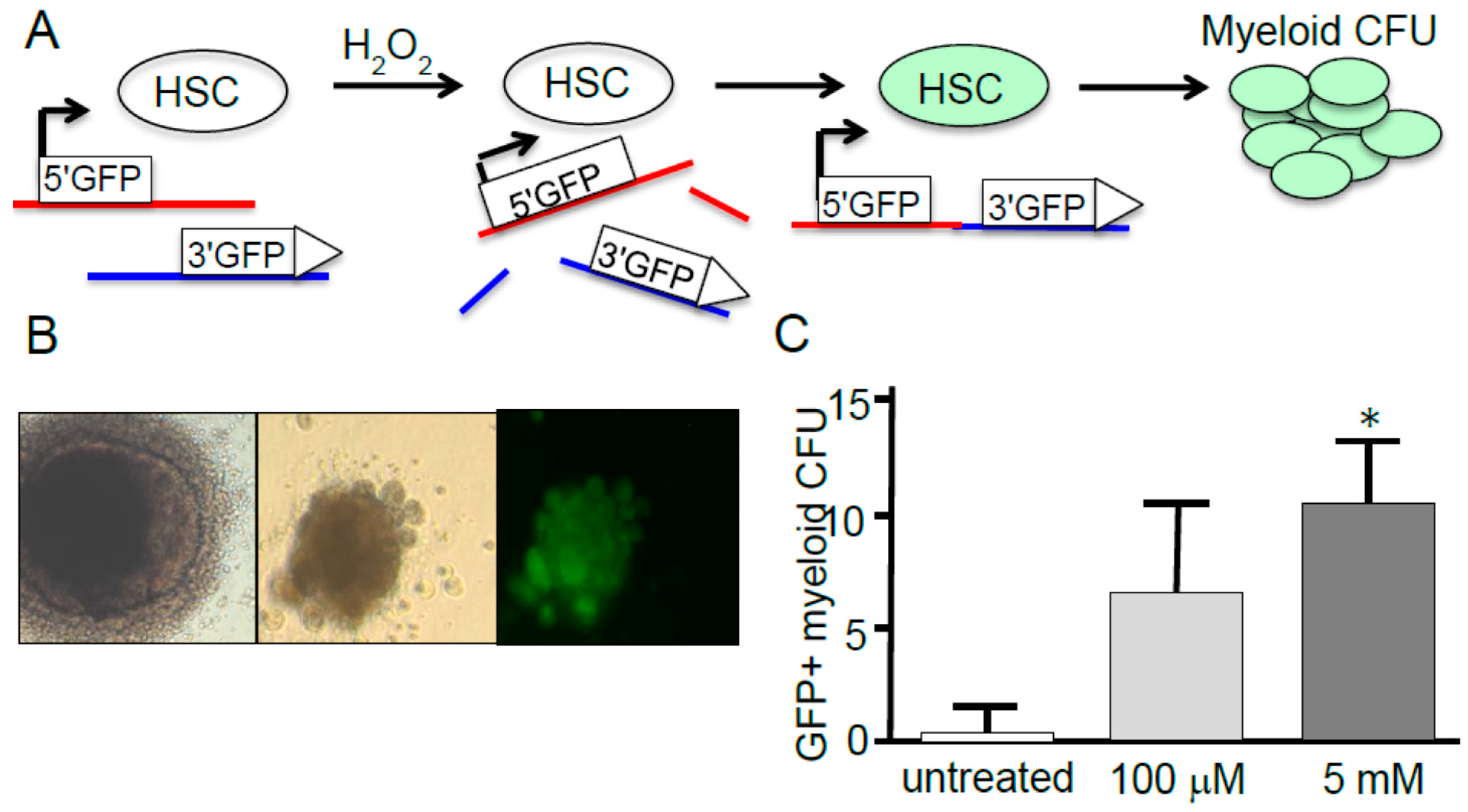
  101. Francis, R.; Richardson, C. Multipotent hematopoietic cells susceptible to alternative double-strand break repair pathways that promote genome rearrangements. Genes Dev. 2007, 21, 1064–1074. [Google Scholar] [CrossRef] [PubMed]
  102. Shao, L.; Luo, Y.; Zhou, D. Hematopoietic stem cell injury induced by ionizing radiation. Antioxid. Redox Signal. 2014, 20, 1447–1462. [Google Scholar] [CrossRef] [PubMed]
  103. Wang, Y.; Liu, L.; Pazhanisamy, S.K.; Li, H.; Meng, A.; Zhou, D. Total body iradiation causes residual bone marrow injury by induction of persistent oxidative stress in murine hematopoietic stem cells. Free Radic. Biol. Med. 2010, 48, 348–356. [Google Scholar] [CrossRef] [PubMed]
  104. Floratou, K.; Giannopoulou, E.; Antonacopoulou, A.; Karakantza, M.; Adonakis, G.; Kardamakis, D.; Matsouka, P. Oxidative stress due to radiation in CD34+ hematopoietic progenitor cells: Protection by IGF-1. J. Radiat. Res. 2012, 53, 672–685. [Google Scholar] [CrossRef] [PubMed]
  105. Yamaguchi, M.; Kashiwakura, I. Role of reactive oxygen species in the radiation response of human hematopoietic stem/progenitor cells. PLoS One 2013, 8, e70503. [Google Scholar] [CrossRef] [PubMed]
  106. Wang, B.; Li, W.; Liu, H.; Yang, L.; Liao, Q.; Cui, S.; Wang, H.; Zhao, L. miR-29b suppresses tumor growth and metastasis in colorectal cancer via downregulating Tiam1 expression and inhibiting epithelial-mesenchymal transition. Cell Death Dis. 2014, 5, e1335. [Google Scholar] [CrossRef] [PubMed]
  107. Saretzki, G.; Armstrong, L.; Leake, A.; Lako, M.; von Zglinicki, T. Stress defense in murine embryonic stem cells is superior to that of various differentiated murine cells. Stem Cells 2004, 22, 962–971. [Google Scholar] [CrossRef] [PubMed]
  108. Ergene, U.; Cagirgan, S.; Pehlivan, M.; Yilmaz, M.; Tombuloglu, M. Factors influencing engraftment in autologous peripheral hematopoetic stem cell transplantation (PBSCT). Transfus. Apher. Sci. 2007, 36, 23–29. [Google Scholar] [CrossRef] [PubMed]
  109. Rossi, D.J.; Bryder, D.; Seita, J.; Nussenzweig, A.; Hoeijmakers, J.; Weissman, I.L. Deficiencies in DNA damage repair limit the function of haematopoietic stem cells with age. Nature 2007, 447, 725–729. [Google Scholar] [CrossRef] [PubMed]
  110. Rossi, D.J.; Seita, J.; Czechowicz, A.; Bhattacharya, D.; Bryder, D.; Weissman, I.L. Hematopoietic stem cell quiescence attenuates DNA damage response and permits DNA damage accumulation during aging. Cell Cycle 2007, 6, 2371–2376. [Google Scholar] [CrossRef] [PubMed]
  111. Jang, Y.Y.; Sharkis, S.J. A low level of reactive oxygen species selects for primitive hematopoietic stem cells that may reside in the low-oxygenic niche. Blood 2007, 110, 3056–3063. [Google Scholar] [CrossRef] [PubMed]
  112. Beerman, I.; Seita, J.; Inlay, M.A.; Weissman, I.L.; Rossi, D.J. Quiescent hematopoietic stem cells accumulate DNA damage during aging that is repaired upon entry into cell cycle. Cell Stem Cell 2014, 15, 37–50. [Google Scholar] [CrossRef] [PubMed]
  113. Oh, J.; Lee, Y.D.; Wagers, A.J. Stem cell aging: Mechanisms, regulators and therapeutic opportunities. Nat. Med. 2014, 20, 870–880. [Google Scholar] [CrossRef] [PubMed]
  114. Urao, N.; Ushio-Fukai, M. Redox regulation of stem/progenitor cells and bone marrow niche. Free Radic. Biol. Med. 2013, 54, 26–39. [Google Scholar] [CrossRef] [PubMed]
  115. Ushio-Fukai, M.; Rehman, J. Redox and metabolic regulation of stem/progenitor cells and their niche. Antioxid. Redox Signal. 2014, 21, 1587–1590. [Google Scholar] [CrossRef] [PubMed]
  116. Picolli, C.; D’Aprile, A.; Ripoli, M.; Scrima, R.; Boffoli, D.; Tabilo, A.; Capitanio, N. The hypoxia-inducible factor is stabilized in circulating hematopoietic stem cells under normoxic conditions. FEBS Lett. 2007, 581, 3111–3119. [Google Scholar] [CrossRef] [PubMed]
  117. Picolli, C.; D’Aprile, A.; Ripoli, M.; Scrima, R.; Lecce, L.; Boffoli, D.; Tabilo, A; Capitanio, N. Bone-marrow derived hematopoietic stem/progenitor cells express multiple isoforms of NADPH oxidase and produce constituitively reactive oxygen species. Biochem. Biophys. Res. Commun. 2007, 353, 965–972. [Google Scholar] [CrossRef] [PubMed]
  118. Picolli, C.; Ria, R.; Ripoli, M.; Scrima, R.; Cela, O.; D’Aprile, A.; Boffoli, D.; Falzetti, F.; Tabilo, A; Capitanio, N.; et al. Characterization of mitochondrial and extra-mitochondrial oxygen consuming reactions in human hematopoietic stem cells: Novel evidence of the occurrence of NAD(P)H oxidase activity. J. Biol. Chem. 2005, 280, 26467–26476. [Google Scholar] [CrossRef] [PubMed]
  119. Rouault-Pierre, K.; Lopez-Onieva, L.; Foster, K.; Anjos-Afonso, F.; Lamrissi-Garcia, I.; Serrano-Sanchez, M.; Mitter, R.; Ivanovic, Z.; de Verneuil, H.; Gribben, J.; et al. HIF-2α protects human hematopoietic stem/progenitors and acute myeloid leukemic cells from apoptosis induced by endoplasmic reticulum stress. Cell Stem Cell 2013, 13, 549–563. [Google Scholar] [CrossRef] [PubMed]
  120. Zhou, F.; Shen, Q.; Claret, F.X. Novel roles of reactive oxygen species in the pathogenesis of acute myeloid leukemia. J. Leukoc. Biol. 2013, 94, 423–429. [Google Scholar] [CrossRef] [PubMed]
  121. Hole, P.S.; Zabkiewicz, J.; Munje, C.; Newton, Z.; Pearn, L.; White, P.; Marquez, N.; Hills, R.K.; Burnett, A.K.; Tonks, A.; et al. Overproduction of NOX-derived ROS in AML promotes proliferation and is associated with defective oxidative stress signaling. Blood 2013, 122, 3322–3330. [Google Scholar] [CrossRef] [PubMed]
  122. Juntilla, M.M.; Patil, V.D.; Calamito, M.; Joshi, R.P.; Birnbaum, M.J.; Koretzky, G.A. AKT1 and AKT2 maintain hematopoietic stem cell function by regulating reactive oxygen species. Blood 2010, 115, 4030–4038. [Google Scholar] [CrossRef] [PubMed]
  123. Woolley, J.F.; Naughton, R.; Stanicka, J.; Gough, D.R.; Bhatt, L.; Dickinson, B.C.; Chang, C.J.; Cotter, T.G. H2O2 production downstream of FLT3 is mediated by p22phox in the endoplasmic reticulum and is required for STAT5 signalling. PLoS One 2012, 7, e34050. [Google Scholar] [CrossRef] [PubMed]
  124. Sallmyr, A.; Fan, J.; Datta, K.; Kim, K.T.; Grosu, D.; Shapiro, P.; Small, D.; Rassool, F. Internal tandem duplication of FLT3 (FLT3/ITD) induces increased ros production, DNA damage, and misrepair: Implications for poor prognosis in AML. Blood 2008, 111, 3173–3182. [Google Scholar] [CrossRef] [PubMed]
  125. He, L.; Luo, L.; Proctor, S.J.; Middleton, P.G.; Blakely, E.L.; Taylor, R.W.; Turnbull, D.M. Somatic mitochondrial DNA mutations in adult-onset leukaemia. Leukemia 2003, 17, 2487–2491. [Google Scholar] [CrossRef] [PubMed]
  126. Hasselbalch, H.C.; Thomassen, M.; Hasselbalch Riley, C.; Kjaer, L.; Stauffer Larsen, T.; Jensen, M.K.; Bjerrum, O.W.; Kruse, T.A.; Skov, V. Whole blood transcriptional profiling reveals deregulation of oxidative and antioxidative defence genes in myelofibrosis and related neoplasms: Potential implications of downregulation of Nrf2 for genomic instability and disease progression. PLoS One 2014, 9, e112786. [Google Scholar] [CrossRef] [PubMed]
  127. Rushworth, S.A.; MacEwan, D.J. HO-1 underlies resistance of aml cells to TNF-induced apoptosis. Blood 2008, 111, 3793–3801. [Google Scholar] [CrossRef] [PubMed]
  128. Chung, Y.J.; Robert, C.; Gough, S.M.; Rassool, F.V.; Aplan, P.D. Oxidative stress leads to increased mutation frequency in a murine model of myelodysplastic syndrome. Leuk. Res. 2014, 38, 95–102. [Google Scholar] [CrossRef] [PubMed]
  129. Vadukoot, A.K.; AbdulSalam, S.F.; Wunderlich, M.; Pullen, E.D.; Landero-Figueroa, J.; Mulloy, J.C.; Merino, E.J. Design of a hydrogen peroxide-activatable agent that specifically targets cancer cells. Bioorg. Med. Chem. 2014, 22, 6885–6892. [Google Scholar] [CrossRef] [PubMed]

