Review

Biochemical Properties and Possible Roles of Ectophosphatase Activities in Fungi

1
Institute of Medical Biochemistry, Federal University of Rio de Janeiro (UFRJ), CCS, Bloco H, University City, Fundão Island, Rio de Janeiro, RJ 21941-590, Brazil
2
Institute of National Science and Technology of Structural Biology and Bioimage (INCTBEB), CCS, Bloco H, University City, Fundão Island, Rio de Janeiro, RJ 21941-590, Brazil
*
Authors to whom correspondence should be addressed.
Int. J. Mol. Sci. 2014, 15(2), 2289-2304; https://doi.org/10.3390/ijms15022289
Submission received: 17 October 2013 / Revised: 27 November 2013 / Accepted: 14 January 2014 / Published: 6 February 2014
(This article belongs to the Section Biochemistry)

Abstract

:
Ectophosphatases are surface membrane-bound proteins whose active sites face the extracellular medium. These enzymes have been reported in several microorganisms including a large number of medically relevant fungal species. An effective technique for identifying ectophosphatases is performing phosphatase activity assays using living intact cells. Biochemical characterization of these activities has shown their differential modulation by classical phosphatase inhibitors, divalent metals and pH range. The physiological roles of ectophosphatases are not well established; however, it has been suggested that these enzymes play important roles in nutrition, proliferation, differentiation, adhesion, virulence and infection. Adhesion to host cells is the first step in establishing a fungal infection and ectophosphatases may be one of the first parasite proteins that come into contact with the host cells. Several results indicate that ectophosphatase activities increase the capacity of fungi to adhere to the host cells. In this context, the present review provides an overview of recent discoveries related to the occurrence and possible roles of ectophosphatase activities in fungal cells.

1. Introduction

Protein phosphorylation is undoubtedly the most common and also the best-studied post-translational modification. Indeed, most proteins in the cell can be regulated, directly or indirectly, through this mechanism. The phosphorylation status of any given protein is controlled by both protein kinases and phosphatases [1].
Protein phosphatases remove phosphate groups from various phosphorylated amino acids. The most common phosphorylation sites in eukaryotic cells are detected on serine, threonine and tyrosine residues [2]. Phosphorylation and dephosphorylation of these residues can trigger conformational changes in the protein that alter its properties [3,4].
It is noteworthy that most of the knowledge related to the nature and function of phosphatases and kinases is due to genetic analysis of these enzymes in fungi. Thus, phosphorylation and dephosphorylation of proteins is related to several fundamental biological processes in fungi, such as the cell cycle, transcription and cell differentiation [5,6]. Furthermore, these enzymes are involved in other cellular processes, such as cell wall synthesis [7], formation of hyphae [8] and maintenance of cellular integrity in stress situations [911].

2. Occurrence of Ectophosphatase Activities in Fungi Cells

Protein phosphatases may exist in soluble [4,12] or secreted forms [1320], or even remain attached to the outer surface of the inner membrane [2123] or the cell wall [24,25]. In this context, the ectophosphatases are surface membrane-bound proteins whose active site faces the extracellular medium [2628]. It is noteworthy that some authors have designated secreted phosphatase activities as ectophosphatases because both activities are involved with extracellular metabolism.
Ectophosphatase activities have been reported in several microorganisms, including protozoa such as Leishmania [23,29], Trypanosoma [21,22,30], and bacteria, such as Mycobacterium bovis [31].
In fungi, ectophosphatases have been described in a large number of species, including Saccharomyces cerevisiae [25], Sporothrix schenckii [32], Paracocciodiodes brasiliensis [24], Candida parapsilosis [33,34], Kluyveromyces marxianus [35], Aspergillus fumigatus [36], Fosencaea pedrosoi [37,38], Cryptococcus neoformans [39], Pseudallescheria boydii [40], Candida albicans [41], Rhinocladiella aquaspersa [42] and Metarhizium anisopliae [43]. It is noteworthy that this review focuses on medically relevant fungal species.

2.1. Ectophosphatases

An effective technique for identifying ectophosphatases is performing phosphatase activity assays using intact cells. The artificial substrate p-nitrophenyl phosphate (p-NPP) is commonly used. The p-NPP hydrolysis generates inorganic phosphate (Pi) and p-nitrophenol (p-NP), which turns yellow under alkaline conditions and can be quantified spectrophotometrically. This approach was performed with S. cerevisiae [25], C. parapsilosis [33,34], F. pedrosoi [37,38], C. neoformans [39], P. boydii [40], C. albicans [41], R. aquaspersa [42] and M. anisopliae [43].
Transmission electron microscopy has been used to confirm the localization of ectophosphatase activity in F. pedrosoi [37,38], C. parapsilosis [34] and P. boydii [40]. Cerium phosphate deposits were observed on the cell wall of the three organisms. Furthermore, in C. parapsilosis, precipitates were also observed in the plasma membrane and in intracellular structures, resembling vesicles [34].
In K. marxianus, an acid phosphatase called Pho610 was the first example of a naturally occurring cell wall protein that has enzyme activity. Pho610 was cloned in S. cerevisiae and the deduced amino acid sequence exhibited hydrophobic stretches at both the N- and C-termini, which is characteristic of the precursors of glycophosphatidylinositol (GPI)-anchored proteins. The results also showed that a significant portion of the Pho610 polypeptide was covalently linked to the cell wall glucan [35]. Similarly, an acid phosphatase (PhoAp) was identified as a cell wall component of A. fumigatus. As observed with K. marxianus, this is a GPI-anchored protein. However, it was not possible to determine if the type of association between PhoAp and the cell wall glucans is covalent or not [36].

2.2. Secreted Phosphatases

Cell-free extracts of mycelia, conidia and yeast of S. schenckii were submitted to polyacrylamide gel electrophoresis that showed the presence of an acid phosphatase [32]. A secreted phosphatase has also been identified in S. cerevisiae in a study involving both cell-free preparations and intact cells [25]. Recently, it was observed that conidial forms of M. anisopliae express both ectophosphatase and secreted phosphatase activity [43]. This ectophosphatase activity has been fully characterized and comparison with a secreted phosphatase activity reported in the mycelial form of the same fungus indicates that they have different biochemical properties [43,44].

3. Biochemical Properties of Fungal Ectophosphatases

Ectophosphatases are hydrolases that act on ester bonds and generates Pi as final product. For this reason, they are classified as phosphomonoesterases (EC 3.1.3). These phosphatases can be classified according to their cellular location, comprising intracellular enzymes [4,12], secreted enzymes [14,17,18] or surface enzymes that can be associated with the plasma membrane [2123] or cell wall [24,25]. Other important tools for the classification of phosphatases are the use of inhibitors, divalent cations and different pH ranges [45]. The various activities present in fungal membranes have shown to be differently modulated by those factors.

3.1. Substrate Specificity of Ectophosphatase Activities

Phosphatases that act specifically on phosphoserine and phosphothreonine are known as phospho-serine/threonine phosphatases (PPs). Their mechanism of catalysis involves a nucleophilic attack from a water molecule to the phosphorylated residue, which occurs in a single step without the transference of the phosphate to the enzyme. The activation of the water molecule depends on the presence of metallic ions [46].
In contrast, enzymes that specifically hydrolyze phosphotyrosine are known as phosphotyrosine phosphatases (PTPs). Unlike PPs, PTPs do not require metal ions for catalysis.
The mechanism of catalysis for PTPs involves the formation of a phosphorylated cysteine intermediate that occurs due to the nucleophilic attack of its thiol group [47].
A third group of phosphatases, dual-specific phosphatases, are capable of hydrolyzing phosphotyrosine as well as phosphoserine and phosphothreonine residues. However, these enzymes are commonly classified as a subfamily of PTPs because of the similarities in their active site and mechanism of catalysis [47].
In general, fungal ectophosphatase activities are acid phosphatases that present high affinity for phosphotyrosine analog substrates such as p-NPP [25,3239,42,43,48]. Hydrolysis of phosphorylated amino acids was examined in the mycelial and conidial forms of F. pedrosoi [37,38], C. parapsilosis [34] and P. boydii [40]. Phosphotyrosine was the best substrate in most cases, except for conidial forms of F. pedrosoi, whose ectophosphatase activity was increased three-fold when phosphothreonine was used as the substrate [38].

3.2. pH Influence on Ectophosphatase Activities

In yeast, acid phosphatases are localized mostly in the cell wall and the periplasmic space [49]. This is in contrast with the alkaline phosphatases, which are considered intracellular enzymes [50,51]. Accordingly, acid ectophosphatases occur much more commonly than alkaline phosphatases. Studies with S. cerevisiae [25], S. schenckii [32], C. parapsilosis [33,34], K. marxianus [35], A. fumigatus [36], F. pedrosoi [37,38], C. neoformans [39], P. boydii [40], R. aquaspersa [42] and M. anisopliae [43] have described acid phosphatases in these species.
Despite being classified as intracellular enzymes, alkaline phosphatases have also been reported as ecto-enzymes in P. brasiliensis [24], C. parapsilosis [33] and P. boydii [40]. In a study with C. parapsilosis, 24 isolates showed acid ectophosphatase activities; however, five of them also exhibited alkaline phosphatase activities. It is uncertain whether this phosphatase activity was located on the cell wall of C. parapsilosis or whether the enzyme seeped into a subsurface or exterior location during cell growth and maturation. However, it is possible to confirm that they are ectophosphatase activities because they have been quantified on intact cells [33].
Intact mycelial forms of P. boydii have been shown to express both acid and alkaline ectophosphatases. Transmission electron microscopy has confirmed that both phosphatase activities are located in the cell wall, though alkaline phosphatase activity seemed to be predominant because a large area of cerium phosphate precipitates was exhibited at pH 8 [40].

3.3. Modulation of Ectophosphatase Activities by Divalent Metals

It is well known that ectophosphatase activities can be positively and/or negatively modulated by cations [52]. Positive modulations were observed in F. pedrosoi [37,38], C. parapsilosis [34], P. boydii [40] and R. aquaspersa [42].
Conidial forms of F. pedrosoi express a Fe3+-activated ectophosphatase activity. However, the ectophosphatase activity from conidial forms that have grown in the absence of Pi in culture medium is insensitive to any metals. These results indicate that different ectophosphatases are expressed in response to exogenous Pi content [38].
The alkaline ectophosphatase activity of P. boydii has been shown to be stimulated by the following three cations: Mg2+, Mn2+ and Zn2+ [40]. In R. aquaspersa, Co2+ was able to promote an increase in ectophosphatase activity in a dose-dependent manner [42]. A Co2+-activated ectophosphatase activity was also reported in T. brucei [53]. Furthermore, cobalt is known to be capable of inducing the expression of alkaline phosphatase in bacteria [54].
Ectophosphatase activity in C. parapsilosis has been stimulated by Cu2+ and inhibited by Zn2+, both acting in a dose-dependent manner [34]. Zn2+ is also an ectophosphatase inhibitor in A. fumigatus [36], C. neoformans [39] and M. anisopliae [43]. Ectophosphatase activity in the intact mycelial forms of P. boydii and the conidial forms of M. anisopliae has been inhibited by Cd2+ and Cu2+ [40,43]. The inhibitory effects of Zn2+, Cd2+ and Cu2+ could be explained by the oxidizing effects that these metals can cause for the enzyme, as has been shown for other ectophosphatase activities [34,43,55].

3.4. Sensibility of Ectophosphatase Activities to Classical Phosphatase Inhibitors

Sodium orthovanadate is a phosphate analog. It acts as an irreversible inhibitor that binds to enzymes by adopting a trigonal bi-pyramidal structure observed in transition state during phosphoryl transfer [56]. This inhibitor can affect different biological processes [57]; however, its major biological activity in living cells is on the cell surface, as the oxidation-reduction reactions that take place in the cytoplasm diminish its inhibitory effect [56,58].
Sodium orthovanadate was able to inhibit ectophosphatase activities in a dose-dependent manner in F. pedrosoi [37], C. neoformans [39], C. parapsilosis [34], P. boydii [40] and R. aquaspersa [42]. Ectophosphatase activity from A. fumigatus was completely abolished by treatment with 10 mM of sodium orthovanadate [36]. Moreover, treatment with 1 mM of sodium orthovanadate inhibited 90% and 85% of ectophosphatase activities of C. albicans [41] and M. anisopliae [43], respectively. In F. pedrosoi [37,38], C. neoformans [39], C. parapsilosis [34], P. boydii [40], C. albicans [41] and R. aquaspersa [42], sodium orthovanadate inhibition of ectophosphatase activities was shown to be irreversible because removal of the inhibitor does not rescue the basal activity.
Ammonium molybdate and sodium fluoride are classical acid phosphatase inhibitors [59] that were also tested on several fungal ectophosphatase activities. In F. pedrosoi [37], C. neoformans [39], C. parapsilosis [34], P. boydii [40] and R. aquaspersa [42], both ammonium molybdate and sodium fluoride promote dose-dependent inhibition of ectophosphatase activities. In A. fumigatus, 10 mM ammonium molybdate and sodium fluoride were able to inhibit 85% and 60% of ectophosphatase activity, respectively [36]. Interestingly, C. albicans ectophosphatase activity was also inhibited 85% and 60% by, ammonium molybdate and sodium fluoride, respectively [41]. However, in this case, the concentration of the inhibitors was 1 mM, 10 times lower than the concentration used in A. fumigatus [36]. Only the inhibition of ectophosphatase activity by ammonium molybdate in P. boydii was shown to be irreversible [40].
Tartrate is a classical inhibitor for secreted phosphatases that can be used to confirm localization of the observed activity [20]. Indeed, tartrate was able to inhibit the secreted phosphatases present in S. schenckii [32]; however, this inhibitor was unable to modulate several ectophosphatase activities, as observed in A. fumigatus [36], F. pedrosoi [37,38], C. neoformans [39] and R. aquaspersa [42].
Levamisole is another classical alkaline phosphatase inhibitor [60] and 1 mM of this compound was able to inhibit approximately 40% of the alkaline ectophosphatase activity in P.boydii [40]. In contrast, acid ectophosphatase activities seemed to be insensitive to levamisole, as observed in F. pedrosoi [37,38], C. neoformans [39], C. parapsilosis [34], R. aquaspersa [42] and M. anisopliae [43].
Pi is considered a highly specific inhibitor of phosphatase activities because it is the natural product of reactions catalyzed by phosphatases [37,38]. Inhibition by Pi in phosphatase reaction medium was verified in S. cerevisiae [25], S. schenckii [32], F. pedrosoi [37,38], C. neoformans [39], C. parapsilosis [34], P. boydii [40], C. albicans [41], R. aquaspersa [42] and M. anisopliae [43]. The Pi content in the culture medium is also involved in the modulation of several ectophosphatase activities, as will be discussed in the following section.

3.5. Modulation of Ectophosphatase Activities by Exogenous Pi Content

The induction of phosphatase activity in response to Pi starvation is an important phenomenon because these enzymes are able to hydrolyze phosphorylated substrates and supply a Pi source during nutrient shortages [61].
The existence of phosphatase activities regulated by phosphate in the culture medium has been reported in a variety of microorganisms, including prokaryotes species [62,63], parasitic protozoa [64,65] and fungal species including Neurospora crassa [66], Aspergillus niger [67], Yarrowia lipolytica [68], Pichia pastoria [69], Penicillium chrysogenum [70,71] and S. cerevisiae [72].
In S. cerevisiae, the phosphate signal transduction pathway, known as the PHO pathway, regulates the expression of several genes involved in the availability and absorption of Pi from extracellular sources [73]. Consequently, the transcription of genes encoding acid and alkaline phosphatases and the Pi transporters is coordinately repressed and de-repressed depending on the Pi concentration in the culture medium [74].
Most of the phosphatases synthesized under Pi-limiting conditions are either located extracellularly or are associated with the plasma membrane or cell wall [75]. The PhoAp activity of A. fumigatus was significantly repressed by the presence of increasing Pi concentrations in culture medium [36]. Phosphate is essential for the growth of all fungi [76], and A. fumigatus was unable to grow at a Pi concentration lower than 10 μM. Because of the regulation by phosphates, it is possible to presume that PhoAp is part of an enzymatic arsenal that allows A. fumigatus to utilize Pi from its environment [36].
In F. pedrosoi, cultivation of conidia in the absence of exogenous Pi resulted in a generation of fungal cells expressing an ectophosphatase activity 130-fold higher than that expressed by fungi cultivated in the presence of Pi. The depletion of Pi from the culture medium apparently induced the expression of a different ectophosphatase, as suggested by: the differences in the affinity for the artificial substrate p-NPP; by the fact that both activities are differently modulated by metals; and by the differential sensitivity to classical phosphatase inhibitors [37,38]. Recently, similar results were obtained with R. aquaspersa. Removal of Pi from the culture medium of conidial forms resulted in a 121-fold increase in their ectophosphatase activities, when compared with the activities of conidia grown in the presence of Pi [42].

4. Possible Roles of Fungal Ectophosphatase Activities

Despite having been described in several microorganisms, the physiological roles of ectophosphatases in these cells are not well established. It has been suggested that these enzymes play important roles in nutrition, proliferation, differentiation, adhesion, virulence and infection [45].
The ectophosphatases may provide fungal cells with a source of orthophosphate by hydrolyzing organic phosphates [77]. In other microorganism, it was observed that the process of adhesion to mammalian cells requires energy [78]. Since Pi is the only phosphorous source readily available to be taken up and metabolized by microorganisms, ectophosphatase activity could play a pivotal role in the release of Pi during the first stages of infection, a moment that is not easy to exactly define and when the fungus probably is still not fully metabolically active [43,79].
Moreover, ectophosphatases may protect the cells from acidic conditions by buffering the periplasmic space with phosphates released from polyphosphates [80]. There is also evidence that ectophosphatases are involved in cell wall synthesis, remodeling and degradation [81].
In this section, we will discuss some recent reports that describe possible roles for ectophosphatase activity in fungal cells.

4.1. Differential Expression of Fungal Ectophosphatase Activities

The differentiation process is a fundamental event during the fungal life cycle. Interestingly, distinct levels of ectophosphatase activities can be observed in the different fungal morphological forms. Sclerotic bodies, conidial and mycelial forms of F. pedrosoi and R. aquaspersa, two etiological agents in chromoblastomycosis (CBM), have been shown to be able to hydrolyze the artificial substrate p-NPP. In R. aquaspersa, the conidial form presented higher levels of ectophosphatase activity when compared with the other two forms. In contrast, F. pedrosoi sclerotic bodies exhibited the highest ectophosphatase activities. As conidial forms are the putative infectious propagules in CBM and sclerotic bodies are the prevalent fungal forms observed in the tissue lesions, it is probable that ectophosphatase activity is associated with parasitism [37,42].
The in vitro differentiation of F. pedrosoi mycelia to sclerotic forms, induced by platelet-activating factor, concomitantly stimulates the ectophosphatase activity [48]. Furthermore, a strain recently isolated from a human case of CBM also demonstrated increased ectophosphatase activity suggesting that the expression of ectophosphatases in F. pedrosoi may be stimulated by interaction with the host [37].
The expression and/or activity of ectophosphatases in fungal species may vary in the different isolates. Different cryptococcal isolates have presented ectophosphatase activity, indicating that this enzyme is commonly expressed in C. neoformans strains; however, considerable variation in expression levels were observed between the different isolates [39].
Candida species usually exist as commensals in the oral, gastrointestinal and genital mucosa of healthy individuals. However, these fungi are also opportunistic pathogens that may cause a wide variety of superficial and systemic infections if the host immune system is compromised [82]. Interestingly, a strong association has been identified between the ectophosphatase activities of C. albicans and HIV infection [41].
Superficial isolates from C. parapsilosis exhibited significantly greater ectophosphatase activity when compared with their systemic counterparts. Moreover, the adhesion to human buccal epithelial cells was also increased in assays performed with superficial isolates of C. parapsilosis. The determination that the survival of the surface dwellers is critically dependent on their ability to attach suggests that ectophosphatases are involved in metabolic events on the interface between the host and the yeast and thereby play a role in candidal adhesion to host surfaces [33].

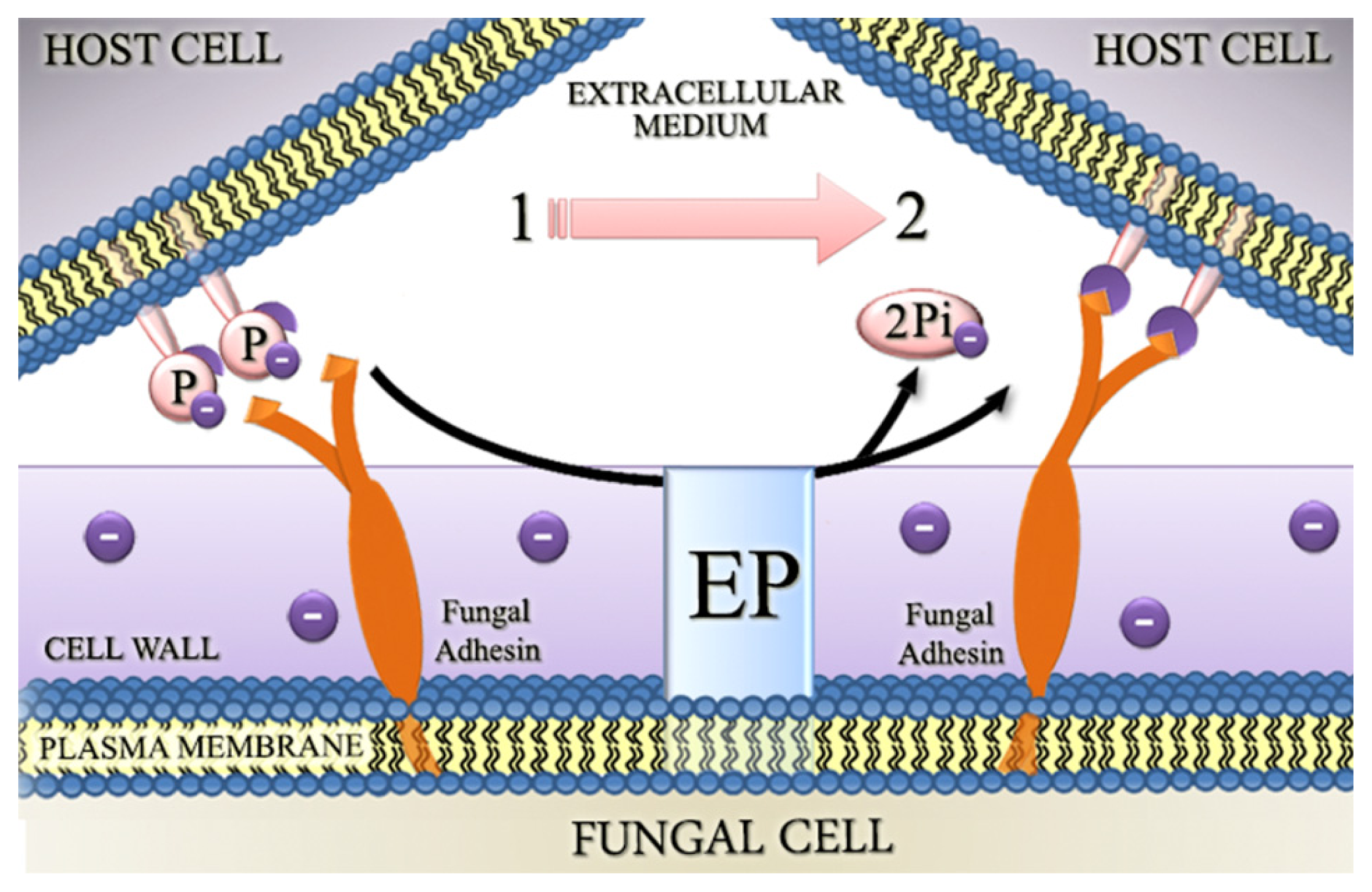
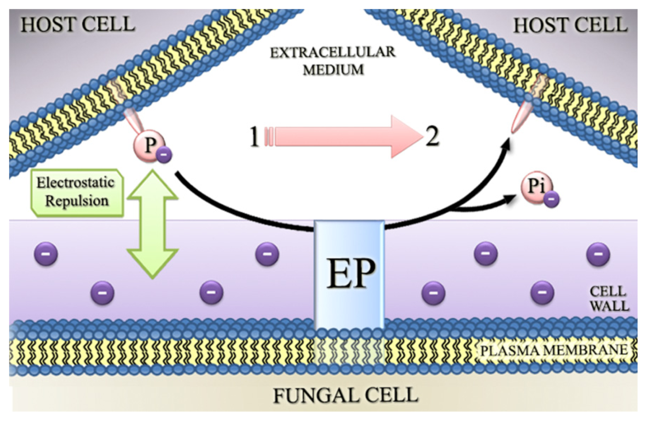
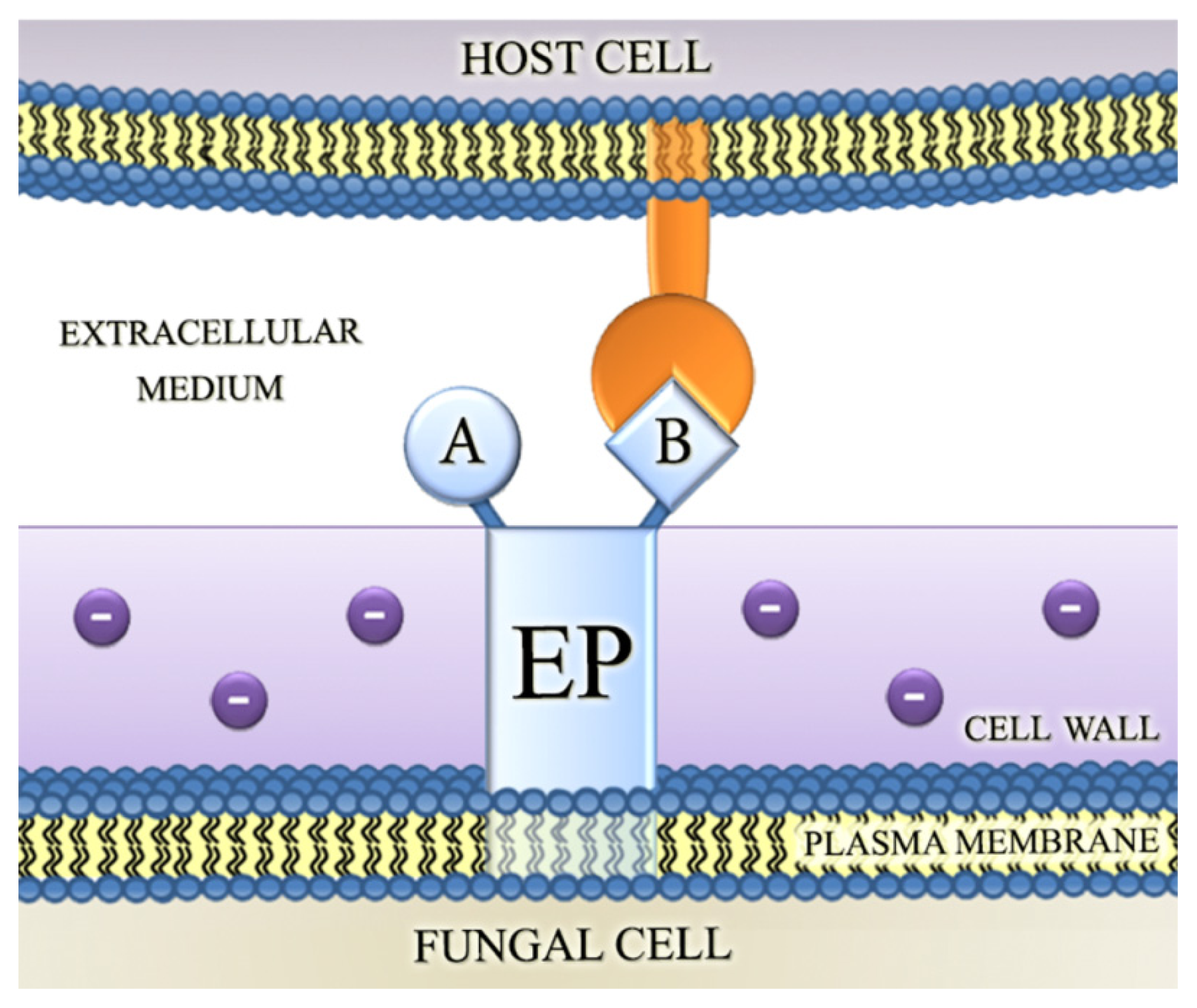
4.2. Involvement of Fungal Ectophosphatase Activities in Host-Parasite Interactions

To establish an infection, pathogens have to evade the immune system, survive and divide in the host environment and spread to new tissues [83,84]. Ecto-enzymes are located in the plasma membrane with their active sites facing the external environment, and could be one of the parasite proteins that initially come into contact with the host cells in the invasion process [27]. This peculiar location suggests that these enzymes may be involved in virulence and infection [27,45].
The irreversible profile of enzyme inhibition produced by sodium orthovanadate in F. pedrosoi [38], C. neoformans [39], C. parapsilosis [34], C. albicans [41], R. aquaspersa [42] and M. anisopliae [43] has allowed the comparison of the ability of these fungi to attach to host cells when ectophosphatase activity was fully functional and when surface enzyme activity was inhibited by pretreatment of fungal cells with this inhibitor. This pretreatment of fungal cells with orthovanadate inhibited adhesion to epithelial cells in F. pedrosoi and R. aquaspersa, two etiological agents in CBM [38,42], C. neoformans, the causative agent in cryptococcosis [39], and C. parapsilosis and C. albicans, two important agents in human candidiasis [33,41]. In F. pedrosoi, pretreatment with orthovanadate also decreased the association of the conidial forms to fibroblasts [38].
M. anisopliae is an entomopathogenic fungus with the ability to infect a broad range of arthropods, from ticks and agricultural insect pests to vectors of human diseases. Corroborating the results observed in human pathogens, pretreatment with phosphatase inhibitors (orthovanadate and molybdate) led to a decrease in the adhesion of M. anisopliae to the model host Dysdercus peruvianus fly wings. Moreover, bioassays have revealed an increase in the survival of D. peruvianus infected by M. anisopliae when conidial forms were pre-incubated with different phosphatase inhibitors, suggesting that ectophosphatase activity plays an important role in fungal virulence [43]. Conidial forms of F. pedrosoi and R. aquaspersa cultured in Pi-depleted medium presented higher levels of ectophosphatase activity and also showed greater adherence to cultured fibroblasts or epithelial cells than the cells grown in complete medium [38,42].
All the reported adhesion assays have shown that cells expressing high ectophosphatase activities were significantly more capable of adhering to host cells. These mechanisms seem to be conserved not only in fungi but also in protozoa. Recently, the involvement of ectophosphatases in host-pathogen interaction was described in Trypanosoma rangeli [30] and Strigomonas culicis [85].
To explain the involvement of ectophosphatase in host-parasite interaction, the following possible roles for this enzyme were proposed: (1) the removal of phosphate groups from surface proteins in host cells could result in conformational transitions and in an attenuated electrostatic repulsion between fungal and host cells (Figure 1); (2) the removal of Pi could expose additional sites for the interaction of infectious agents with the host surface (Figure 2); and (3) ectophosphatases may contain adhesive domains that could directly promote the attachment of fungal cells to their hosts, therefore functioning similarly to the well-characterized microbial adhesins (Figure 3) [34,38,39,4143].

5. Conclusion

Throughout this review, several studies were reported describing the occurrence of ectophosphatase activities in the plasma membranes of fungal cells. The biochemical characterization of these activities has shown their differential modulation by classical phosphatase inhibitors, divalent metals and pH. Furthermore, several results indicated that ectophosphatase activity increases the capacity of fungi adherence on host cells. Clearly, more studies are needed to determine the extent of ectophosphatase participation in fungal physiology, as well as in host-pathogen interactions. The possible correlations between the expression of these enzymes and the clinical manifestation of diseases make them a potential therapeutic target.

Acknowledgments

This work was supported by grants from the Brazilian National Research Council (CNPq); the Carlos Chagas Filho Rio de Janeiro State Research Foundation (FAPERJ); the Brazilian Federal Agency for Support and Evaluation of Graduate Education (CAPES); and the National Institutes of Science and Technology, Brazil.

Conflicts of Interest

The authors have declared that no competing interests exist.

References

  1. Andreeva, A.V.; Kutuzov, M.A. Protozoan protein tyrosine phosphatases. Int. J. Parasitol 2008, 38, 1279–1295. [Google Scholar]
  2. Szöör, B. Trypanosomatid protein phosphatases. Mol. Biochem. Parasitol 2010, 173, 53–63. [Google Scholar]
  3. Cohen, P. The structure and regulation of protein phosphatases. Annu. Rev. Biochem 1989, 58, 453–508. [Google Scholar]
  4. Zhan, X.L.; Hong, Y.; Zhu, T.; Mitchell, A.P.; Deschenes, R.J.; Guan, K.L. Essential functions of protein tyrosine phosphatase Ptp2 and Ptp3 and Rim11 tyrosine phosphorylation in Saccharomyces cerevisiae meiosis and sporulation. Mol. Biol. Cell 2000, 11, 663–676. [Google Scholar]
  5. Da-Silva, A.M.; Zapella, P.D.; Andrioli, L.P.; Campanhã, R.B.; Fiorini, L.C.; Etchebehere, L.C.; Da-Costa-Maia, J.C.; Terenzi, H.F. Searching for the role of protein phosphatases in eukaryotic microorganisms. Braz. J. Med. Biol. Res 1999, 32, 835–839. [Google Scholar]
  6. Madhani, H.D.; Fink, G.R. The control of filamentous differentiation and virulence in fungi. Trends Cell Biol 1998, 8, 348–353. [Google Scholar]
  7. Suresh, K.; Subramanyam, C. A putative role for calmodulin in the activation of Neurospora crassa chitin synthase. FEMS Microbiol. Lett 1997, 150, 95–100. [Google Scholar]
  8. Yatzkan, E.; Szöor, B.; Fehér, Z.; Dombrádi, V.; Yarden, O. Protein phosphatase 2A is involved in hyphal growth of Neurospora crassa. Mol. Genet. Genomics 1998, 259, 523–531. [Google Scholar]
  9. Borgia, P.T. Roles of orlA, tseE and bimG genes of Aspergillus nidulans in chitin synthesis. J. Bacteriol 1992, 174, 384–389. [Google Scholar]
  10. Csank, C.; Makris, C.; Meloche, S.; Schröppel, K.; Röllinghoff, M.; Dignard, D.; Thomas, D.Y.; Whiteway, M. Derepressed hyphal growth and reduced virulence in VH1 family-related protein phosphatase mutant of the human pathogen Candida albicans. Mol. Biol. Cell 1997, 8, 2539–2551. [Google Scholar]
  11. Winkler, A.; Arkind, C.; Mattison, C.P.; Burkholder, A.; Knoche, K.; Ota, I. Heat stress activated the yeast high-osmolarity glycerol mitogen-activated protein-kinase pathway, and protein tyrosine phosphatases are essential under heat stress. Eukaryot. Cell 2002, 1, 163–173. [Google Scholar]
  12. Andrews, P.D.; Stark, M.J.R. Type 1 protein phosphatase is required for maintenance of cell wall integrity, morphogenesis and cell cycle progression in Saccharomyces cerevisiae. J. Cell Sci 2000, 113, 507–520. [Google Scholar]
  13. De Jesus, J.B.; Ferreira, M.A.; Cuervo, P.; Britto, C.; Silva-Filho, F.C.; Meyer-Fernandes, J.R. Iron modulates ecto-phosphohydrolase activities in pathogenic trichomonads. Parasitol. Int 2006, 55, 285–290. [Google Scholar]
  14. Dutra, P.M.; Dias, F.A.; Rodrigues, C.O.; Romeiro, A.; Attias, M.; de Souza, W.; Lopes, A.H.; Meyer-Fernandes, J.R. Platelet-activating factor modulates a secreted phosphatase activity of the trypanosomatid parasite Herpetomonas muscarum muscarum. Curr. Microbiol 2001, 43, 288–292. [Google Scholar]
  15. Dutra, P.M.; Dias, F.A.; Santos, M.A.A.; Rodrigues, C.O.; Romeiro, A.; Attias, M.; de Souza, W.; Lopes, A.H.; Meyer-Fernandes, J.R. Secreted phosphatase activities in trypanossomatid parasites of plants modulated by patelet-activating factor. Phytophathology 2001, 1, 408–414. [Google Scholar]
  16. Fernandes, A.C.; Soares, D.C.; Saraiva, E.M.; Meyer-Fernandes, J.R.; Souto-Padrón, T. Different secreted phosphatase activities in Leishmania amazonensis. FEMS Microbiol. Lett 2013, 340, 117–128. [Google Scholar]
  17. Joliviet, P.; Queiroz-Claret, C.; Bergeron, E.; Meunier, J.C. Characterization of an exocellular protein phosphatase with dual substrate specificity from the yeast Yarrowia lipolytica. Int. J. Biochem. Cell Biol 1998, 30, 783–796. [Google Scholar]
  18. Justino, A.; Nosowa, S.R.; Maccheroni, W.; May, G.S.; Martinez-Rossi, N.M.; Rossi, A. The Aspergillus nidulans pyrG89 mutation alters glycosylation of secreted acid phosphatase. Fungal Genet. Biol 2001, 32, 113–120. [Google Scholar]
  19. Rodrigues, C.O.; Dutra, P.M.; Barros, F.S.; Souto-Padrón, T.; Meyer-Fernandes, J.R.; Lopes, A.H.C.S. Platelet-activating factor induction of secreted phosphatase activity in Trypanosoma cruzi. Biochem. Biophys. Res. Commun 1999, 266, 36–42. [Google Scholar]
  20. Santos, A.L.S.; Souto-Padrón, T.; Alviano, C.S.; Lopes, A.H.; Soares, R.M.; Meyer-Fernandes, J.R. Secreted phosphatase activity induced by dimethylsulfoxide in Herpetomonas samuelpessoai. Arch. Biochem. Biophys 2002, 405, 191–198. [Google Scholar]
  21. Fernandes, E.C.; Meyer-Fernandes, F.R.; Silva-Neto, M.A.C.; Vercesi, A.E. Trypanosoma brucei: Ecto-phosphatase activity on the surface of intact procyclic forms. Z. Naturforschung 1997, 52C, 351–358. [Google Scholar]
  22. Meyer-Fernandes, J.R.; da Silva-Neto, M.A.; Dos Santo Soares, M.; Fernandes, E.; Vercesi, A.E.; de Oliveira, M.M. Ecto-phosphatase activities on the cell surface of the amastigote forms of Trypanosoma cruzi. Z. Naturforschung 1999, 54, 977–984. [Google Scholar]
  23. Remaley, A.T.; Glew, R.H.; Kuhns, D.B.; Basford, R.E.; Waggoner, A.S.; Ernst, L.A.; Pope, M. Leishmania donovani: Surface membrane acid phosphatase blocks neutrophils oxidative metabolite production. Exp. Parasitol 1985, 60, 331–341. [Google Scholar]
  24. Campo-Aasen, I.; Albornoz, M.C. Alkaline phosphatase at the cell wall of the yeast phase of Paracoccidioides brasiliensis. Mycopathologia 1994, 127, 69–71. [Google Scholar]
  25. Mildner, P.; Ries, B.; Barbaric, S. Acid phosphatase and adenosine triphosphatase activities in the cell wall of baker’s yeast. Biochim. Biophys. Acta 1975, 391, 67–74. [Google Scholar]
  26. Furuya, T.; Zhong, L.; Meyer-Fernandes, J.R.; Lu, H.G.; Moreno, S.N.; Docampo, R. Ecto-protein tyrosine phosphatase activity in Trypanosoma cruzi infective stages. Mol. Biochem. Parasitol 1998, 92, 339–348. [Google Scholar]
  27. Meyer-Fernandes, J.R. Ecto-ATPases in protozoa parasites: Looking for a function. Parasitol. Int 2002, 51, 299–303. [Google Scholar]
  28. Meyer-Fernandes, J.R.; Lanz-Mendoza, H.; Gondim, K.C.; Willott, E.; Wells, M.A. Ectonucleotide diphosphohydrolase activities in hemocytes of larval Manduca sexta. Arch. Biochem. Biophys 2000, 382, 152–159. [Google Scholar]
  29. De Almeida-Amaral, E.E.; Belmont-Firpo, R.; Vannier-Santos, M.A.; Meyer-Fernandes, J.R. Leishmania amazonensis: Characterization of an ecto-phosphatase activity. Exp. Parasitol 2006, 114, 334–340. [Google Scholar]
  30. Dos-Santos, A.L.; Dick, C.F.; Alves-Bezerra, M.; Silveira, T.S.; Paes, L.S.; Gondim, K.C.; Meyer-Fernandes, J.R. Interaction between Trypanosoma rangeli and the Rhodnius prolixus salivary gland depends on the phosphotyrosine ecto-phosphatase activity of the parasite. Int. J. Parasitol 2012, 42, 819–827. [Google Scholar]
  31. Braibant, M.; Content, J. The cell surface associated phosphatase activity of Mycobacterium bovis BCG is not regulated by enviromental inorganis phosphate. FEMS Microbiol. Lett 2001, 195, 121–126. [Google Scholar]
  32. Arnold, W.N.; Mann, L.C.; Sakai, K.H.; Garrison, R.G.; Coleman, P.D. Acid phosphatases of Sporothrix schenckii. J. Gen. Microbiol 1986, 132, 3421–3432. [Google Scholar]
  33. Fernanado, P.H.; Panagoda, G.J.; Samaranayake, L.P. The relationship between the acid and alkaline phosphatase activity and the adherence of clinical isolates of Candida parapsilosis to human buccal epithelial cells. APMIS 1999, 107, 1034–1042. [Google Scholar]
  34. Kiffer-Moreira, T.; de Sá Pinheiro, A.A.; Alviano, W.S.; Barbosa, F.M.; Souto-Padrón, T.; Nimrichter, L.; Rodrigues, M.L.; Alviano, C.S.; Meyer-Fernandes, J.R. An ectophosphatase activity in Candida parapsilosis influences the interaction of fungi with epithelial cells. FEMS Yeast Res 2007, 7, 621–628. [Google Scholar]
  35. Yoda, K.; Ko, J.H.; Nagamatsu, T.; Lin, Y.; Kaibara, C.; Kawada, T.; Tomishige, N.; Hashimoto, H.; Noda, Y.; Yamasaki, M. Molecular characterization of a novel yeast cell-wall acid phosphatase cloned from Kluyveromyces marxianus. Biosci. Biotechnol. Biochem 2000, 64, 142–148. [Google Scholar]
  36. Bernard, M.; Mouyna, I.; Dubreucq, G.; Debeaupuis, J.P.; Fontaine, T.; Vorgias, C.; Fuglsang, C.; Latgé, J.P. Characterization of a cell-wall acid phosphatase (PhoAp) in Aspergillus fumigatus. Microbiology 2002, 148, 2819–2829. [Google Scholar]
  37. Kneipp, L.F.; Palmeira, V.F.; Pinheiro, A.A.; Alviano, C.S.; Rozental, S.; Travassos, L.R.; Meyer-Fernandes, J.R. Phosphatase activity on the cell wall of Fonsecaea pedrosoi. Med. Mycol 2003, 41, 469–477. [Google Scholar]
  38. Kneipp, L.F.; Rodrigues, M.L.; Holandino, C.; Esteves, F.F.; Souto-Padrón, T.; Alviano, C.S.; Travassos, L.R.; Meyer-Fernandes, J.R. Ectophosphatase activity in conidial forms of Fonsecaea pedrosoi is modulated by exogenous phosphate and influences fungal adhesion to mammalian cells. Microbiology 2004, 150, 3355–3362. [Google Scholar]
  39. Collopy-Junir, I.; Esteves, F.F.; Nimrichter, L.; Rodrigues, M.L.; Alviano, C.S.; Meyer-Fernandes, J.R. An ectophosphatase activity in Cryptococcus neoformans. FEMS Yeast Res 2006, 6, 1010–1017. [Google Scholar]
  40. Kiffer-Moreira, T.; Pinheiro, A.A.; Pinto, M.R.; Esteves, F.F.; Souto-Padrón, T.; Barreto-Bergter, E.; Meyer-Fernandes, J.R. Mycelial forms of Pseudallescheria boydii present ectophosphatase activities. Arch. Microbiol 2007, 188, 159–166. [Google Scholar]
  41. Portela, M.B.; Kneipp, L.F.; Ribeiro de Souza, I.P.; Holandino, C.; Alviano, C.S.; Meyer-Fernandes, J.R.; de Araújo Soares, R.M. Ectophosphatase activity in Candida albicans influences fungal adhesion: study between HIV-positive and HIV-negative isolates. Oral Dis 2010, 16, 431–437. [Google Scholar]
  42. Kneipp, L.F.; Magalhães, A.S.; Abi-Chacra, E.A.; Souza, L.O.; Alviano, C.S.; Santos, A.L.; Meyer-Fernandes, J.R. Surface phosphatase in Rhinocladiella aquaspersa: biochemical properties and its involvement with adhesion. Med. Mycol 2012, 50, 570–578. [Google Scholar]
  43. Cosentino-Gomes, D.; Rocco-Machado, N.; Santi, L.; Broetto, L.; Vainstein, M.H.; Meyer-Fernandes, J.R.; Schrank, A.; Beyes-da-Silva, W.O. Inhibition of ecto-phosphatase activity in conidia reduces adhesion and virulence of Metarhizium anisopliae on the host insect Dysdercus peruvianus. Curr. Microbiol 2013, 66, 467–474. [Google Scholar]
  44. Li, Z.; Wang, Z.; Peng, G.; Yin, Y.; Zhao, H.; Cao, Y.; Xia, Y. Purification and characterization of a novel thermostable extracellular protein tyrosine phosphatase from Metarhizium anisopliae strain CQMa102. Biosci. Biotechnol. Biochem 2006, 70, 1961–1968. [Google Scholar]
  45. Gomes, M.T.; Lopes, A.H.; Meyer-Fernandes, J.R. Possible roles of ectophosphatases in host-parasite interactions. J. Parasitol. Res 2011, 2011, 1–7. [Google Scholar]
  46. Egloff, M.P.; Cohen, P.T.; Reinemer, P.; Barford, D. Crystal structure of the catalytic subunit of human protein phosphatase 1 and its complex with tugstate. J. Mol. Biol 1995, 254, 942–959. [Google Scholar]
  47. Denu, J.M.; Stuckey, J.A.; Saper, M.A.; Dixon, J.E. Form and function in protein dephosphorylation. Cell 1996, 87, 361–364. [Google Scholar]
  48. Alviano, D.S.; Kneipp, L.F.; Lopes, A.H.; Travassos, L.R.; Meyer-Fernandes, J.R.; Rodrigues, M.L.; Alviano, C.S. Differentiation of Fonsecaea pedrosoi mycelial forms into sclerotic cells is induced by platelet-activating factor. Res. Microbiol 2003, 154, 689–695. [Google Scholar]
  49. Arnold, W.N. Enzymes. In Yeast Cell Envelopes: Biochemistry, Biophysics and Ultrastructure; Arnold, W.N., Ed.; CRC Press: Boca Raton, FL, USA, 1981; Volume 2, pp. 1–46. [Google Scholar]
  50. Onishi, M.R.; Tkacz, J.S.; Lampen, J.O. Glycoprotein nature of yeast alkaline phosphatase: Formation of active enzyme in the presence of tunicamycin. J. Biol. Chem 1979, 254, 11943–11952. [Google Scholar]
  51. Schurr, A.; Yagil, E. Regulation and characterization of acid and alkaline phosphatase in yeast. J. Gen. Microbiol 1971, 65, 291–303. [Google Scholar]
  52. Cosentino-Gomes, D.; Meyer-Fernandes, J.R. Ecto-phosphatases in protozoan parasites: Possible roles in nutrition, growth and ROS sensing. J. Bioenerg. Biomembr 2011, 43, 89–92. [Google Scholar]
  53. Fernandes, E.C.; Granjeiro, J.M.; Aoyama, H.; Fonseca, F.V.; Meyer-Fernandes, J.R.; Vercesi, A.E. A metallo phosphatase activity present on the surface of Trypanosoma brucei procyclic forms. Vet. Parasitol 2003, 118, 19–28. [Google Scholar]
  54. Spencer, D.B.; Chen, C.P.; Hulett, F.M. Effect of cobalt on synthesis and activation of Bacillus licheniformis alkaline phosphatase. J. Bacteriol 1981, 145, 926–933. [Google Scholar]
  55. De Sá Pinheiro, A.A.; Amazonas, J.N.; Barros, F.S.; de Menezes, L.F.; Batista, E.J.; Silva, E.F.; de Souza, W.; Meyer-Fernandes, J.R. Entamoeba histolytica: An ectophosphatase activity regulated by oxidation-reduction reactions. Exp. Parasitol 2007, 115, 352–358. [Google Scholar]
  56. Huyer, G.; Liu, S.; Kelly, J.; Moffat, J.; Payette, P.; Kennedy, B.; Tsaprailis, G.; Gresser, M.J.; Ramachandran, C. Mechanism of inhibition of protein-tyrosine phosphatases by vanadate and pervanadate. J. Biol. Chem 1997, 272, 843–851. [Google Scholar]
  57. Klarlund, J. Transformation of cells by an inhibitor of phosphatases acting on phosphotyrosine in proteins. Cell 1985, 41, 707–717. [Google Scholar]
  58. Cantley, L.C., Jr.; Aisen, P. The fate of cytoplasmic vanadium. Implications on (Na,K)-ATPase inhibition. J. Biol. Chem 1979, 254, 1781–1784. [Google Scholar]
  59. Dutra, P.M.; Rodrigues, C.O.; Jesus, J.B.; Lopes, A.H.; Souto-Padrón, T.; Meyer-Fernandes, J.R. A novel ecto-phosphatase activity of Herpetomonas muscarum muscarum inhibited by platelet-activating factor. Biochem. Biophys. Res. Commun 1998, 253, 164–169. [Google Scholar]
  60. Van Belle, H. Alkaline phosphatase. I. Kinetics and inhibition by levamisole of purified isoenzymes from humans. Clin. Chem 1976, 22, 972–976. [Google Scholar]
  61. Dick, C.F.; Dos-Santos, A.L.; Meyer-Fernandes, J.R. Inorganic phosphate as an important regulator of phosphatases. Enzym. Res 2011, 2011, 1–7. [Google Scholar]
  62. Hulett, F.M. The signal-transduction network for PHO regulation in Bacillus subtilis. Mol. Microbiol 1996, 19, 933–939. [Google Scholar]
  63. Torriani-Gorini, A.; Silver, S.; Yagil, E. Phosphate in Microorganisms: Cellular and Molecular Biology; ASM Press: Washington D.C., WA, USA, 1994. [Google Scholar]
  64. Dick, C.F.; Dos-Santos, A.L.; Fonseca-de-Souza, A.L.; Rocha-Ferreira, J.; Meyer-Fernandes, J.R. Trypanosoma rangeli: Differential expression of ecto-phosphatase activities in response to inorganic phosphate starvation. Exp. Parasitol 2010, 124, 386–393. [Google Scholar]
  65. Fonseca-de-Souza, A.L.; Dick, C.F.; Dos Santos, A.L.; Meyer-Fernandes, J.R. A Mg2+-dependent ecto-phosphatase activity on the external surface of Trypanosoma rangeli modulated by exogenous inorganic phosphate. Acta Trop 2008, 107, 153–158. [Google Scholar]
  66. Jacob, M.M.; Nyc, J.F.; Brown, D.M. Isolation and chemical properties of a repressible acid phosphatase in Neurospora crassa. J. Biol. Chem 1971, 246, 1419–1425. [Google Scholar]
  67. MacRae, W.D.; Buxton, F.P.; Sibley, S.; Garven, S.; Gwynne, D.I.; Davies, R.W.; Arst, H.N. A phosphate-repressible acid phosphatase gene from Aspergillus niger: Its cloning, sequencing and transcriptional analysis. Gene 1988, 71, 339–348. [Google Scholar]
  68. González, F.J.; Fauste, C.; Burguillo, F.J.; Domínguez, A. Kinetic behaviour of a repressible acid phosphatase from yeast Yarrowia lipolytica: A comparative study between the solubilized enzyme, the enzyme bound to cell-wall fragments and the enzyme bound to intact cells. Biochim. Biophys. Acta 1993, 1162, 17–27. [Google Scholar]
  69. Payne, W.E.; Gannon, P.M.; Kaiser, C.A. An inducible acid phosphatase from the yeast Pichia pastoris: Characterization of the gene and its product. Gene 1995, 163, 19–26. [Google Scholar]
  70. Haas, H.; Redl, B.; Friedlin, E.; Stoöffler, G. Isolation and analysis of the Penicillium chrysogenum phoA gene encoding a secreted phosphate-repressible acid phosphatase. Gene 1992, 113, 129–133. [Google Scholar]
  71. Haas, H.; Redl, B.; Leitner, E.; Stoöffler, G. Penicillium chrysogenum extracellular acid phosphatase: Purification and biochemical characterization. Biochim. Biophys. Acta 1991, 1074, 392–397. [Google Scholar]
  72. Ogawa, N.; DeRisi, J.; Brown, P.O. New components of a system for phosphate accumulation and polyphosphate metabolism in Saccharomyces cerevisiae revealed by genomic expression analysis. Mol. Biol. Cell 2000, 11, 4309–4321. [Google Scholar]
  73. Auesukaree, C.; Homma, T.; Tochio, H.; Shirakawa, M.; Kaneko, Y.; Harashima, S. Intracellular phosphate serves as a signal for the regulation of the PHO pathway in Saccharomyces cerevisiae. J. Biol. Chem 2004, 279, 17289–17294. [Google Scholar]
  74. Oshima, Y.; Ogawa, N.; Harashima, S. Regulation of phosphatase synthesis in Saccharomyces cerevisiae— A review. Gene 1996, 179, 171–177. [Google Scholar]
  75. Metzenberg, R.L. Implications of some genetic control mechanisms in Neurospora. Microbiol. Rev 1979, 43, 361–383. [Google Scholar]
  76. Jennings, D.H. Phosphorus. In The Physiology of Fungal Nutrition; Jennings, D.H., Ed.; Cambridge University Press: Cambridge, MA, USA, 1995; pp. 252–287. [Google Scholar]
  77. Toh-e, A. Phosphorus Regulation in Yeast. In Yeast Genetic Engineering; Barr, P.J., Brake, A.J., Valenzuela, P., Eds.; Butterworths: Boston, MA, USA, 1989; pp. 41–52. [Google Scholar]
  78. Schenkman, S.; Robbins, E.S.; Nussenzweig, V. Attachment of Trypanosoma cruzi to mammalian cells requires parasite energy, and invasion can be independent of the target cell cytoskeleton. Infect. Immun 1991, 59, 645–654. [Google Scholar]
  79. Manners, J.G. Assessment of Germination. In The fungus spore; Madelin, M.F., Ed.; Butterworths: London, UK, 1966; pp. 165–173. [Google Scholar]
  80. Touati, E.; Dassa, E.; Dassa, J.; Boquet, P.L. Acid phosphatase (pH 2.5) of Escherichia coli: regulatory characteristics. In Phosphate Metabolism and Cellular Regulation in Microorganisms; Torriani-Gorini, A., Rothman, F.G., Silver, S., Wright, A., Yagil, E., Eds.; ASM Press: Washington D.C., WA, USA, 1987; pp. 31–40. [Google Scholar]
  81. Novick, P.; Ferro, S.; Schekman, R. Order of events in the yeast secretory pathway. Cell 1981, 25, 461–469. [Google Scholar]
  82. Miceli, M.H.; Díaz, J.A.; Lee, S.A. Emerging opportunistic yeast infections. Lancet. Infect. Dis 2011, 11, 142–151. [Google Scholar]
  83. Naglik, J.R.; Challacombe, S.J.; Hube, B. Candida albicans secreted aspartyl proteinases in virulence and pathogenesis. Microbiol. Mol. Biol. Rev 2003, 67, 400–428. [Google Scholar]
  84. Yang, Y.L. Virulence factors of Candida species. J. Microbiol. Immunol. Infect 2003, 36, 223–228. [Google Scholar]
  85. Catta-Preta, C.M.; Nascimento, M.T.; Garcia, M.C.; Saraiva, E.M.; Motta, M.C.; Meyer-Fernandes, J.R. The presence of a symbiotic bacterium in Strigomonas culicis is related to differential ecto-phosphatase activity and influences the mosquito-protozoa interaction. Int. J. Parasitol 2013, 43, 571–577. [Google Scholar]
Figure 1. The removal of phosphate groups from surface proteins in host cells could result in attenuation of the electrostatic repulsion between fungal and host cells. The fungal cell surface is electronegative and the presence of phosphate residues on the surface of the host cell increases the electrostatic repulsion between them. Ectophosphatases may contribute to the success of fungal infection by removing phosphate residues of the host cell plasma membrane and thereby attenuating this electrostatic repulsion. Abbreviations: EP, ectophosphatase; P, phosphate residue; Pi, inorganic phosphate; 1, before ectophosphatase activity; and 2, after ectophosphatase activity.
Ijms 15 02289f1
Figure 2. The removal of inorganic phosphate could expose additional sites for the interaction of infectious agents with the host surface. Phosphorylated proteins may contain binding sites for fungal adhesins. Dephosphorylation promoted by ectophosphatase activity could therefore expose these sites, thus contributing for fungal adhesion and infection. Abbreviations: EP, ectophosphatase; P, phosphate residue; Pi, inorganic phosphate; 1, before ectophosphatase activity; and 2, after ectophosphatase activity.
Ijms 15 02289f2
Figure 3. Ectophosphatase may contain adhesive domains that could directly promote the attachment of fungal cells to their hosts. In addition to the catalytic domain, ectophosphatase may also exhibit an adhesion domain, thus functioning similarly to the well-characterized microbial adhesins. Abbreviations: EP, ectophosphatase; A, catalytic domain; and B, adhesion domain.
Ijms 15 02289f3

