Article

MAEWEST Expression in Flower Development of Two Petunia Species

1
Laboratory of Molecular Evolution, Department of Genetics, Universidade Federal do Rio Grande do Sul, P.O. Box 15053, 91501-970 Porto Alegre, RS, Brazil
2
Laboratory of Genomes and Plant Population, Center of Biotechnology and Department of Biophysics, Universidade Federal do Rio Grande do Sul, P.O. Box 15053, 91501-970 Porto Alegre, RS, Brazil
3
Department of Plant Biology, Institute of Biology, Universidade Estadual de Campinas, P.O. Box 6109, 13418-970 Campinas, SP, Brazil
*
Author to whom correspondence should be addressed.
Int. J. Mol. Sci. 2013, 14(7), 13796-13807; https://doi.org/10.3390/ijms140713796
Submission received: 3 May 2013 / Revised: 14 June 2013 / Accepted: 28 June 2013 / Published: 3 July 2013
(This article belongs to the Special Issue Molecular Research in Plant Secondary Metabolism)

Abstract

:
Changes in flower morphology may influence the frequency and specificity of animal visitors. In Petunia (Solanaceae), adaptation to different pollinators is one of the factors leading to species diversification within the genus. This study provides evidence that differential expression patterns of MAWEWEST (MAW) homologs in different Petunia species may be associated with adaptive changes in floral morphology. The Petunia × hybrida MAW gene belongs to the WOX (WUSCHEL-related homeobox) transcription factor family and has been identified as a controller of petal fusion during corolla formation. We analyzed the expression patterns of P. inflata and P. axillaris MAW orthologs (PiMAW and PaMAW, respectively) by reverse transcriptase polymerase chain reaction (RT-PCR), reverse transcription–quantitative PCR (qRT-PCR) and in situ hybridization in different tissues and different developmental stages of flowers in both species. The spatial expression patterns of PiMAW and PaMAW were similar in P. inflata and P. axillaris. Nevertheless, PaMAW expression level in P. axillaris was higher during the late bud development stage as compared to PiMAW in P. inflata. This work represents an expansion of petunia developmental research to wild accessions.

Graphical Abstract

1. Introduction

The great morphological variation seen in flowers is associated with the different pollinators of each species. Changes in flower color and shape may cause changes in frequency and specificity of animal visitors, contributing to speciation [1]. Even a mutation in a single gene may have a central role in pollinator shifts [2,3].
Although it is known that some floral characteristics (such as those resulting from the fusion of floral parts, shape and size) arise after organ initiation, the developmental mechanisms associated with these processes are poorly understood [4]. Post-initiation fusion occurs in petals, carpels, and stamen filaments of Petunia flowers: the petal primordia partially fuse to form the corolla tube, the stamen filaments also fuse to the proximal part of the corolla tube, and the two carpel primordia fuse to form the pistil [5,6]. In addition, the flowers of the different species have distinct shape and size [6]. MAEWEST (MAW) gene is associated with organ fusion and lateral growth in Petunia × hybrida [7]. MAW belongs to the WOX (Wuschel-related homeobox) family of transcription factors and has functions that are partially similar to the Arabidopsis thaliana genes WOX1 and PRESEED FLOWER (PRS/WOX3) involved in lateral organ development [811]. Accordingly, maw mutants showed defects in petal fusion, carpel fusion and lamina growth. Morphological analysis suggested that the phenotype could be the result of polarity defects along the adaxial/abaxial axis, which also affects leaves, bracts and sepals [7]. Here, we chose two wild Petunia species, Petunia inflata and Petunia axillaris, to analyze the expression patterns of their respective MAW orthologs. These are closely related species but nevertheless show great differences in their morphologies (Figure 1 and Table 1). Petunia inflata has a short and campanulate corolla tube, considered a small flower, while P. axillaris has a long and salveriform corolla tube and is a big flower. The differences in the size of the flower are pronounced and are considered to be adaptations to different pollinators. While P. axillaris is pollinated by nocturnal hawkmoths, P. inflata is pollinated by bees [1214]. Until now, no gene involved in controlling the difference in flower shape and size between these two species has been identified. Expression patterns of MAW orthologs in both Petunia species were analyzed by using reverse transcriptase polymerase chain reaction (RT-PCR), reverse transcription-quantitative PCR (qRT-PCR), and in situ hybridization; these experiments were aimed at investigating whether MAW might be involved in determining flower morphological differences in these two species. Our results suggested an association between MAW orthologs expression and flower development in these Petunia species.

2. Results

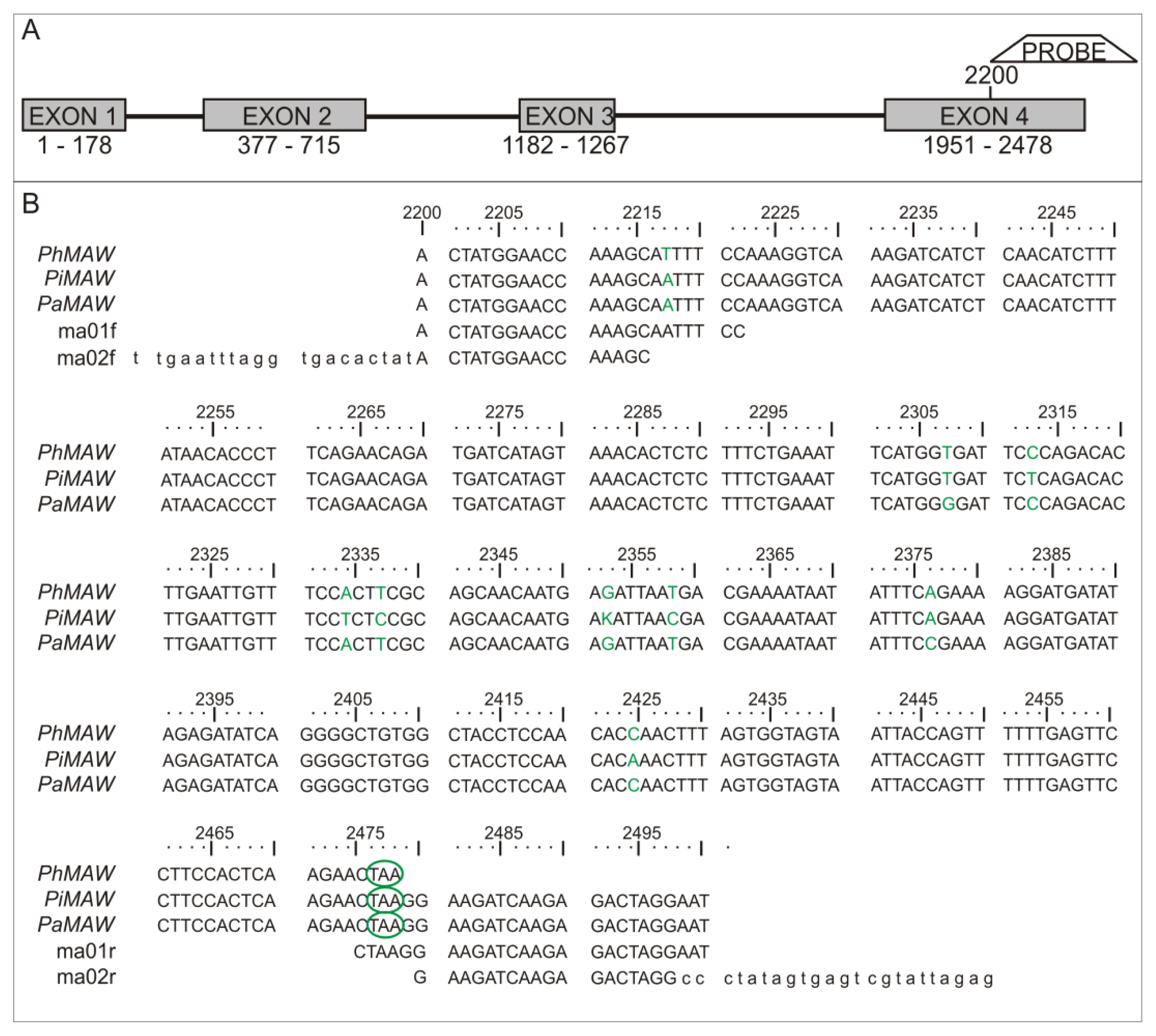
2.1. Sequence Analysis

Petunia × hybrida MAW (PhMAW) orthologs were identified in wild P. axillaris and P. inflata and were denoted PaMAW and PiMAW, respectively. The PaMAW and PiMAW genomic sequences were deposited in GenBank as JQ438842 and JQ438843, respectively. The sequence similarities among PaMAW, PiMAW and PhMAW were obtained using BLASTX [15]. The sequence alignment between the MAW homologs described in this work is shown in Figure 2. Sequence similarity among the P. × hybrida, P. inflata and P. axillaris MAW homologs could be considered high (BLASTX E-values ≈ 4 × 10⁻³²). Moreover, the phylogenetic analysis of WOX partial sequences showed orthology among P. axillaris, P. inflata and P. × hybrida MAW genes (Figures S1 and S2). Nine positions in a 301 bp region presented nucleotide mutations likely due to the recent diversification of the genus Petunia, though selective pressure cannot be ruled out.

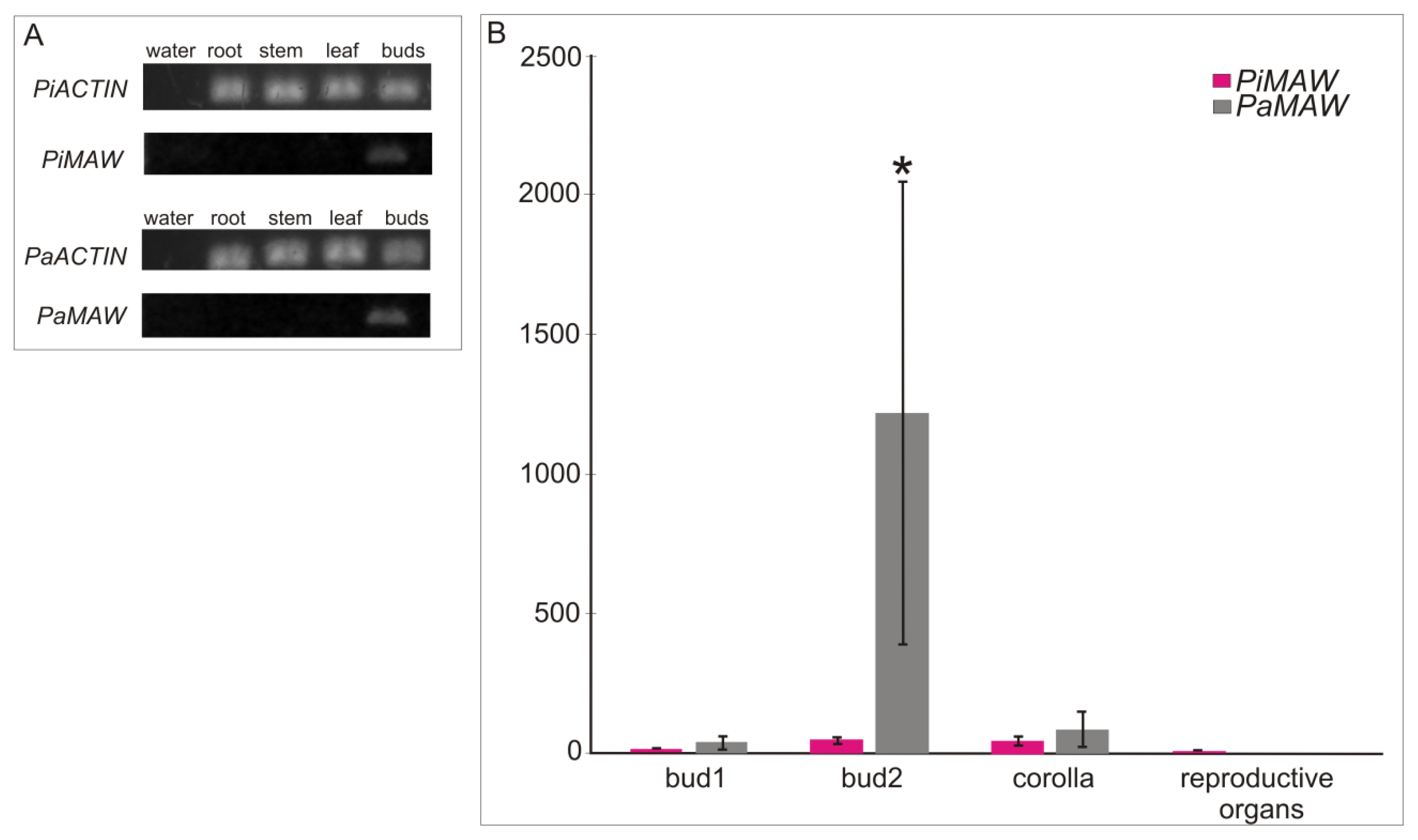
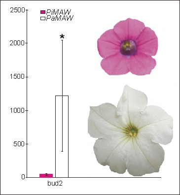
2.2. RT-PCR and qRT-PCR

PiMAW and PaMAW expression patterns are shown in Figure 3. According to the RT-PCR results, MAW transcripts were not detected in mature roots, stems or leaves (Figure 3A) but were detected in floral buds in both P. axillaris and P. inflata (Figure 3B). The use of qRT-PCR, a more sensitive and quantitative method, allowed for detection of a higher expression level of PaMAW in 5 mm or larger (bud 2) buds (Figure 3B). It is important to note that the high variance observed in the relative expression values among biological replicates. This kind of deviation is expected in real-time experiments [16], especially those using samples from natural populations that present high intrinsic genetic diversity. The expression level difference seen between the reproductive organs of the different species was not statistically significant due to this high variance (Figure 3B). In bud 2, the difference in PaMAW expression compared to PiMAW expression was statistically significant.

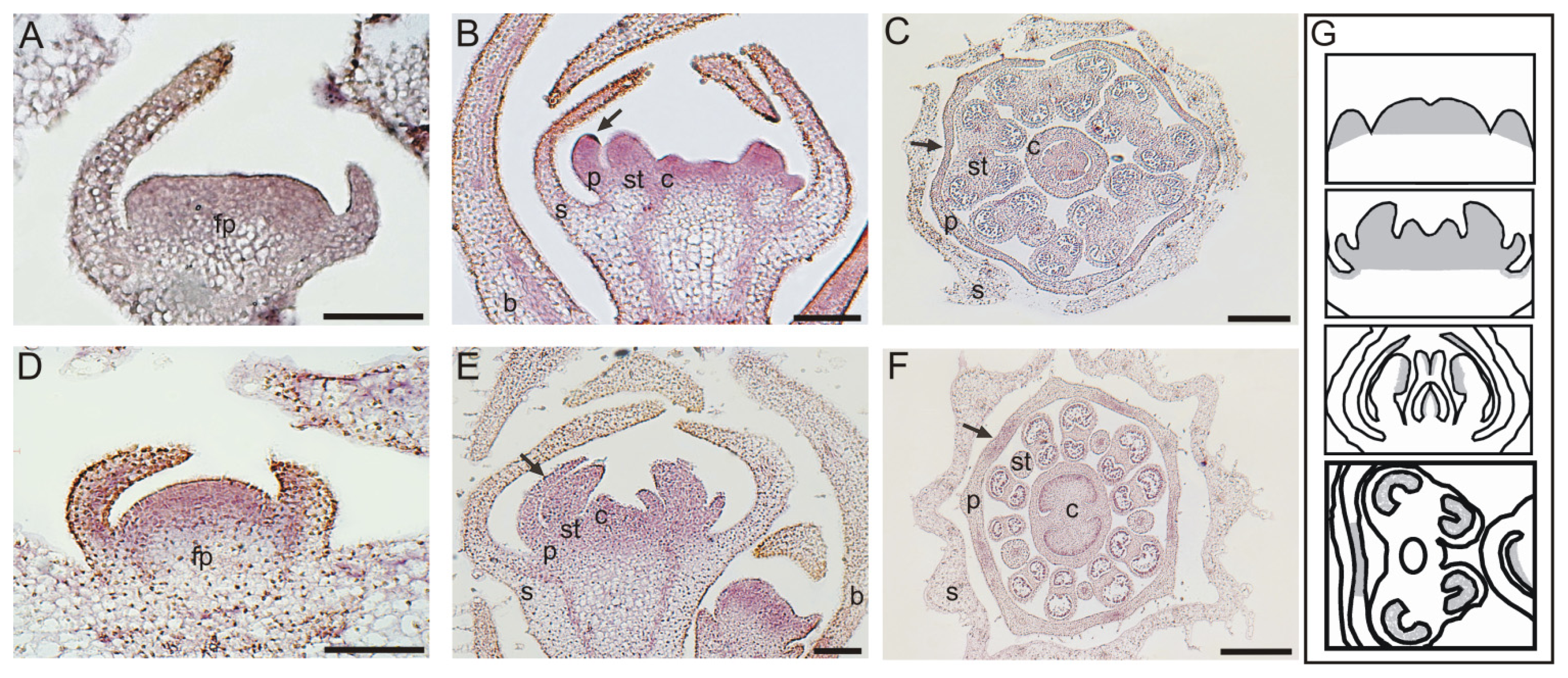
2.3. In Situ Hybridization

The local PaMAW expression pattern was similar to that observed for PiMAW, and both were similar to the expression pattern for P. × hybrida MAW [7]. During early floral meristem development, both PaMAW and PiMAW transcripts were detected in all emerging floral organ primordia (Figure 4A,D). As development proceeded, expression decreased in sepal primordia (Figure 4B,E) until it could no longer be detected (Figure 4C,F). In petal primordia, transcripts of both PaMAW and PiMAW were initially distributed uniformly (arrows in Figure 4B,E), but they became restricted to the distal ends of the primordia, corresponding to the points of organ fusion in later developmental stages (arrows in Figure 4C,F). The hybridization signal was also detected at the onset of stamen primordia and was detected only in stamen loculi at a later developmental stage (Figure 4C,F). Both PaMAW and PiMAW were evenly expressed in carpel primordia, and in later stages, the hybridization signal was stronger where the two carpels fused. Transcripts of both genes could also be detected in developing ovules (Figure 4C,F). The schematic representation in Figure 4G illustrates the stereotyped expression patterns of PaMAW and PiMAW during Petunia flower development, regions of concentrated expression are colored gray. This representation demonstrates that changes in MAW expression during the development progress becoming mainly restricted to regions where organ fusion occurs.

3. Discussion

The flower shape and size are considered defining characteristics for different pollinators in Petunia. Understanding the evolution of this trait may help us to elucidate the complex evolutionary history of the genus. Until now, no phylogenetic study could reliably clarify the ancestral and derived characteristics of Petunia species [6]. Speciation events must be related to the evolution of adaptations to different pollinators, and therefore, this is one potential path to unraveling the evolutionary history of the Petunia genus [3,17,18].
The broad expression of MAW orthologs detected by our RT-PCR, qRT-PCR and in situ hybridization in both P. inflata and P. axillaris flowers corroborates the role of this gene in the formation of reproductive organs, as in P. × hybrida [7]. While we detected expression of the MAW orthologs in P. inflata and P. axillaris in floral buds of sizes evaluated, P. axillaris showed a higher expression level later in flower development, indicating a possible connection between the differently sized flowers between these species. The in situ experiments confirmed the maintenance of PaMAW expression during later developmental stages of P. inflata and P. axillaris flowers, specifically in parts that underwent a process of post-initiation organ fusion as petals and carpels. In P. × hybrida, PhMAW expression was also detected in shoot/leaf primordia and in young leaves [7]. We did not detect expression of either PiMAW or PaMAW in any mature vegetative tissue we collected except the flower tissue. Lateral expansion of the five petal primordia and the formation of a ring structure results in petal fusion in Petunia [7]. During the first three stages of petal development in Petunia, the floral tube development occurs mainly by cellular division; from the fourth stage onward (floral buds with more than 4 mm), cell expansion predominates [19]. The corolla division in three structural domains is as follows: D1 is the part of the floral tube fused to the stamen filaments, D2 is the distal domain of the floral tube, and D3 is the limb [5] (Figure 1B,E). D1 is the main domain responsible for the differences in corolla tube length between P. axillaris and P. inflata. This region contains a larger number of cells in early developmental stages of P. axillaris and a larger cell length in mature flowers [5]. Thus, the difference in cell number occurs early in development while the difference in cell elongation occurs in later stages, and both processes contribute to corolla tube length. In the limb, the cell division persists to phases close the anthesis, and the cell elongation, which could be associated with flower opening, starts later [19]. In 5 mm or larger buds, the style and stigma are completely formed, the style is elongated and the ovule primordial arises [4]. Developmental defects were not detected in stamen in maw flowers [7].
The main difference found between PaMAW and PiMAW expression patterns was that PaMAW expression is higher at a later flower development stage. Organ fusion is characteristically affected in P. × hybrida maw mutants. While organ fusion occurs in both P. axillaris and P. inflata, the corolla of P. axillaris is bigger than that of P. inflata. PaMAW gene is more highly expressed in P. axillaris 5 mm or larger buds, a period of expression that coincided with cellular expansion in the corolla tube and cell division in the limb. It has been shown that growth in length and width are controlled by different mechanisms and WOX genes are involved in the organ lateral growth along the mediolateral axis, promoting cell proliferation [711,2022]. The MAW orthologs may play a role in proper cell division and fusion and may participate in the developmental pathways that govern the differences in flower size between these two species. These initial findings indicate that more studies are necessary to establish the participation of these genes in the divergence of floral morphology between these species by investigating modified MAW expression patterns in transgenic P. axillaris and P. inflata plants. The spatial separation in the predominance of cell division or cell elongation in different moments of development in different domains of the corolla [5,19] must involve a mechanism of control and MAW gene expression should be modulated by this control mechanism. WOX1 homologs of Medicago truncatula, Nicotiana sylvestris and Pisum sativum (STENOFOLIA, LAM1, and LATHYROIDES respectively) were described as required for cell division, acting in petal lobe expansion, controlling lamina elaboration in the mediolateral axis [20,21], which corroborates our proposition about the MAW homolgs in Petunia. Obviously, one cannot dismiss the hypothesis that additional genes might also participate in this process.

4. Experimental Section

4.1. Plant Material

P. inflata and P. axillaris seeds were collected from natural populations in Rio Grande do Sul, Brazil (27°46′S, 53°26′W and 30°36″S, 55°56′W, respectively) and were cultivated under greenhouse conditions (Biology Institute, UNICAMP, Campinas, SP, Brazil). Some plants were cultivated under hydroponic conditions to obtain root material for RNA extraction. Leaves were collected for genomic DNA extraction [23]. Floral buds in different developmental stages were collected, fixed in 4% paraformaldehyde, dehydrated in ethanol and xylene series and embedded in paraffin. Paraffin sections (8-μm-thick) were mounted in aminosilane-coated slides. Tissues from roots, shoots, leaves, and buds were collected in liquid nitrogen for RNA extraction. Floral buds and pre-anthesis flowers were also collected directly from natural populations and stocked in Trizol® (Invitrogen, Carlsbad, CA, USA) until RNA extraction.

4.2. Sequencing and Riboprobe Labeling

The Petunia × hybrida MAW (PhMAW) coding region sequence (CDS) was obtained from GenBank (EU359004.1) and used as a query to find homologous sequences from P. inflata and P. axillaris in the Petunia EST database [24]. The obtained sequences were used to design specific primers using the Primer3 tool, version 0.4 [25]. The primers (ma01f and ma01r) were designed to anneal to a region corresponding to the 3′ end of the PhMAW coding sequence downstream from the region coding the conserved homeodomain, preventing cross-hybridization of the probes (Figure 2A,B). Due to the high similarity among the selected Petunia sequences in this segment, the same pair of primers was used to amplify fragments from both P. inflata and P. axillaris. PCR amplifications were performed in 25 μL reactions consisting of 1 U Taq polymerase (Invitrogen, Carlsbad, CA, USA), 1× buffer (Invitrogen, Carlsbad, CA, USA), 0.2 mM each dNTP, 0.2 mM MgCl2, 0.2 μM of each primer and 20–50 ng of genomic DNA as a template. Amplification conditions were as follows: 3 min at 94 °C, 30 cycles of 30 s at 94 °C, 30 s at 50 °C, and 40 s at 72 °C, with a final 5 min extension step at 72 °C. PCR products were purified using 20% polyethylene glycol [26] and sequenced in a MegaBACE 1000 DNA Analysis System (GE Healthcare, Biosciences, Pittsburgh, PA, USA) using the ET Terminator Kit (GE Healthcare, Biosciences, Pittsburgh, PA, USA) according to the manufacturer’s instructions. In order to verify if the sequences obtained are orthologs of P. × hybrida MAW, we conducted a phylogenetic analysis using WOX’s partial sequences proteins of P. × hybrida, P. inflata, P. axillaris and Arabidopsis thaliana. WOX sequences of P. × hybrida and A. thaliana were obtained from NCBI [27] and Phytozome [28], respectively. The WOX protein sequences were aligned using CLUSTALW as implemented in MEGA 5.1 [29] and manually adjusted. The phylogenetic analysis was performed using Neighbor-Joining in MEGA. The molecular distances of the aligned sequences were calculated according to the p-distance parameter with all gap and missing data accounted as pairwise deletion. Branch points were tested for significance by bootstrapping with 1000 replications. In situ probes were prepared by using a second primer pair (ma02f and ma02r) in a secondary PCR reaction that used the first PCR product as a template, adding the T7 and SP6 sequences to the first PCR product. The secondary PCR reaction was conducted under the same conditions as the first PCR. The probe was synthesized using the secondary PCR product, which was purified with the PureLink® PCR Purification kit (Invitrogen, Carlsbad, CA, USA). Approximately 200 ng of the purified PCR product was used to synthesize the sense and antisense probes by in vitro transcription using the DIG RNA Labeling kit (Roche, Penzberg, Germany), which includes digoxigenin-labeled uracils (DIG-UTPs).

4.3. RNA Extraction, RT-PCR and qRT-PCR

Temporal RNA expression patterns of MAW orthologs from P. inflata and P. axillaris were analyzed in extracts from mature roots, stems, leaves, and floral buds (bulks of buds in different developmental stages: buds smaller than 5 mm and buds equal or larger than 5 mm). Total RNA was extracted using Trizol® (Invitrogen, Carlsbad, CA, USA), and cDNA was synthesized using the Super Script First Strand Synthase kit (Invitrogen, Carlsbad, CA, USA). The Petunia ACTIN gene was used as a positive control for cDNA synthesis [7]. The cDNA samples were used as templates in PCR reactions with the ma01f and ma01r primer pair. Amplification conditions were as follows: 3 min at 94 °C followed by 30 cycles of 10 s at 94 °C, 10 s at 57 °C, and 20 s at 72 °C, with a final 5-min extension step at 72 °C. To confirm expression patterns, perform a quantitative analysis, and investigate the expression pattern in non-cultivated plants, we performed qRT-PCR on RNA extracted from buds of two different stages (smaller than 5 mm or from 5 to 25 mm) and pre-anthesis flowers collected in the field. Floral buds were separated into two groups according to length in order to associate gene expression to stages in which growth occurs predominantly by either cell division (floral buds smaller than 5 mm) or cell expansion (buds equal or larger than 5 mm) [19]. In the qRT-PCR experiment, the ma01f and ma01r primers were used. Relative transcript abundance was detected with the intercalating dye SYBR Green in a 7500 Real Time PCR System (Applied Biosystems, Foster City, CA, USA). Reactions were performed in a 20 μL final volume composed of 10 μL of cDNA sample previously diluted 1:100 in water, the specific primer-pairs and 10 μL of Sybr-Green PCR-mix (Ludwig Biotech, Alvorada, RS, Brazil). Amplification conditions were as follows: one initial cycle of denaturation at 95 °C for 5 min, followed by 40 cycles of 95 °C for 15 s 60 °C for 10 s and 72 °C for 15 s and data acquisition at 60 °C for 35 s. Melting curves from 55 to 99 °C were obtained to confirm the amplification of a single specific product of PCR. All reactions were performed using four independently isolated biological RNA samples and in three technical replications. ACTIN was used as a reference gene for sample normalization, and P. axilaris reproductive organs were used for calibrating calculations performed with the 2−ΔΔCt method [30]. One-way ANOVA with the Duncan test (p ≤ 0.05) was applied in SPSS v.20 (IBM Corp, New York, NY, USA) to compare differential expression values between the different development stages of flowers and the tissues of species.

4.4. In Situ Hybridization

Hybridization was performed in floral buds in different developmental stages with non-radioactive probes [31]. The hybridization signal was detected by a colorimetric assay in which an anti-digoxigenin antibody coupled with alkaline phosphatase and NBT/BCIP (nitro blue tetrazolium chloride/5-Bromo-4-chloro-3-indolyl phosphate toluidine) served as a substrate. The results were documented using a ZEISS AXIOSKOPE microscope (Zeiss, Jena, Germany).

5. Conclusions

In this work, we used spatial and temporal expression patterns of MAW orthologs from two Petunia species to associate the well-described phenotype of the P. × hybrida maw mutant with the adaptation to different pollinators of contrasting Petunia wild species. We found evidence that this locus might be involved with the evolution of perianth characteristics in Petunia. The results suggest a possible role of this gene in the different flower size and shape between P. axillaris and P. inflata. In light of the great number of developmental mutants available that have been studied in model plants such as P. × hybrida, analyzing spatial and temporal expressions of genes in species with contrasting phenotypes associated to those genes would be a good approach to shed light on the putative molecular nature of developmental adaptations.

Supplementary Information

ijms-14-13796-s001.pdf

Acknowledgments

We thank three anonymous reviewers for comments and suggestions that improved this manuscript. This work was supported by the Conselho Nacional de Desenvolvimento Científico e Tecnológico; Coordenação de Aperfeiçoamento de Pessoal de Nível Superior; Fundação de Amparo à Pesquisa do Estado do Rio Grande do Sul; Fundação de Amparo à Pesquisa do Estado de São Paulo; and Programa de Pós-Graduação em Genética e Biologia Molecular/Universidade Federal do Rio Grande do Sul, Brazil.

Conflict of Interest

The authors declare no conflict of interest.

References

  1. Schiestl, F.P.; Schlüter, P.M. Floral isolation, specialized pollination, and pollinator behavior in orchids. Annu. Rev. Entomol 2009, 54, 425–446. [Google Scholar]
  2. Schemske, D.W.; Bradshaw, H.D. Pollinator preference and the evolution of floral traits in monkeyflowers (Mimulus). Proc. Natl. Acad. Sci. USA 1999, 96, 11910–11915. [Google Scholar]
  3. Klahre, U.; Gurba, A.; Hermann, K.; Saxenhofer, M.; Bossolini, E.; Guerin, P.M.; Kuhlemeier, C. Pollinator choice in Petunia depends on two major genetic loci for floral scent production. Curr. Biol 2011, 21, 1–10. [Google Scholar]
  4. Angenent, G.C.; Franken, J.; Busscher, M.; van Dijken, A.; van Went, J.L.; Dons, H.J.M.; van Tunen, A.J. A novel class of MADS box genes is lnvolved in ovule development in Petunia. Plant Cell 1995, 7, 1569–1582. [Google Scholar]
  5. Stuurman, J.; Hoballah, M.E.; Broger, L.; Moore, J.; Basten, C.; Kuhlemeier, C. Dissection of floral pollination syndromes in petunia. Genetics 2004, 168, 1585–1599. [Google Scholar]
  6. Stehmann, J.R.; Lorenz-Lemke, A.P.; Freitas, L.B.; Semir, J. The Genus. In Petunia Petunia: Evolutionary, Developmental and Physiological Genetics, 2nd ed; Gerats, T., Strommer, J., Eds.; Springer: New York, NY, USA, 2009; pp. 1–28. [Google Scholar]
  7. Vandenbussche, M.; Horstman, A.; Zethof, J.; Koes, R.; Rijpkema, A.S.; Gerats, T. Differential recruitment of WOX transcription factors for lateral development and organ fusion in Petunia and Arabidopsis. Plant Cell 2009, 21, 2269–2283. [Google Scholar]
  8. Shimizu, R.; Ji, J.; Kelsey, E.; Ohtsu, K.; Schnable, P.S.; Scanlon, M.J. Tissue specificity and evolution of meristematic WOX3 function. Plant Physiol 2009, 149, 841–850. [Google Scholar]
  9. Matsumoto, N.; Okada, K. A homeobox gene, PRESSED FLOWER, regulates lateral axis-dependent development of Arabidopsis flowers. Genes Dev 2001, 15, 3355–3364. [Google Scholar]
  10. Haecker, A.; Gross-Hardt, R.; Geiges, B.; Sarkar, A.; Breuninger, H.; Herrmann, M.; Laux, T. Expression dynamics of WOX genes mark cell fate decisions during early embryonic patterning in Arabidopsis thaliana. Development 2004, 131, 657–668. [Google Scholar]
  11. Nardmann, J.; Ji, J.; Werr, W.; Scanlon, M.J. The maize duplicate genes narrow sheath1 and narrow sheath2 encode a conserved homeobox gene function in a lateral domain of shoot apical meristems. Development 2004, 131, 2827–2839. [Google Scholar]
  12. Wijsman, H.J.W. On the interrelationships of certain species of Petunia. II. Experimental data: Crosses between different taxa. Acta Bot. Neerl 1983, 32, 97–107. [Google Scholar]
  13. Ando, T.; Nomura, M.; Tsukahara, J.; Watanabe, H.; Kokubun, H.; Tsukamoto, T.; Hashimoto, G.; Marchesi, E.; Kitching, I.J. Reproductive isolation in a native population of Petunia sensu Jussieu (Solanaceae). Ann. Bot 2001, 88, 403–413. [Google Scholar]
  14. Gübitz, T.; Hoballah, M.E.; Dell’Olivo, A.; Kuhlemeier, C. Petunia as a Model System for the Genetics and Evolution of Pollination Syndromes. In Petunia: Evolutionary, Developmental and Physiological Genetics, 2nd ed; Gerats, T., Strommer, J., Eds.; Springer: New York, NY, USA, 2009; pp. 29–49. [Google Scholar]
  15. BLASTX. Available online: http://blast.ncbi.nlm.nih.gov (on accessed 26 January 2013).
  16. Rieu, I.; Powers, S.J. Real-time quantitative RT-PCR: Design, calculations, and statistics. Plant Cell 2009, 21, 1031–1033. [Google Scholar]
  17. Hoballah, M.H.; Gübitz, T.; Stuurman, J.; Broger, L.; Barone, M.; Mandel, T.; Dell’Olivo, A.; Arnold, M.; Kuhlemeier, C. Single gene-mediated shift in pollinator attraction in Petunia. Plant Cell 2007, 19, 779–790. [Google Scholar]
  18. Venail, J.; Dell’Olivo, A.; Kuhlemeier, C. Speciation genes in the genus Petunia. Phil. Trans. R Soc. B 2010, 365, 461–468. [Google Scholar]
  19. Reale, L.; Porceddu, A.; Moretti, L.L.C.; Zenoni, S.; Pezzotti, M.; Romano, B.; Ferranti, F. Patterns of cell division and expansion in developing petals of Petunia hybrida. Sex Plant Reprod 2002, 15, 123–132. [Google Scholar]
  20. Tadege, M.; Lin, H.; Bedair, M.; Berbel, A.; Wen, J.; Rojas, C.M.; Niu, L.; Tang, Y.; Sumner, L.; Ratet, P.; et al. STENOFOLIA regulates blade outgrowth and leaf vascular patterning in Medicago truncatula and Nicotiana sylvestris. Plant Cell 2011, 23, 2125–2142. [Google Scholar]
  21. Zhuang, L.; Ambrose, M.; Rameau, C.; Weng, L.; Yang, J.; Hu, X.; Luo, D.; Li, X. LATHYROIDES, encoding a WUSCHEL-related homeobox1 transcription factor, controls organ lateral growth, and regulates tendril and dorsal petal identities in garden pea (Pisum sativum L.). Mol. Plant 2012, 6, 1333–1345. [Google Scholar]
  22. Lin, H.; Niu, L.; McHale, N.A.; Ohme-Takagi, M.; Mysore, K.S.; Tadege, M. Evolutionarily conserved repressive activity of WOX proteins mediates leaf blade outgrowth and floral organ development in plants. Proc. Natl. Acad. Sci. USA 2013, 110, 366–371. [Google Scholar]
  23. Roy, A.; Frascaria, N.; MacKay, J.; Bousquet, J. Segregating random amplified polymorphic DNAs (RAPDs) in Betula alleghaniensis. Theor. Appl. Genet 1992, 85, 173–180. [Google Scholar]
  24. Petunia 454 database. Available online: http://biosrv.cab.unina.it/454petuniadb/ (on accessed 5 May 2010).
  25. Rozen, S.; Skaletsky, H.J. Primer3 on the WWW for General Users and for Biologist Programmers. In Bioinformatics Methods and Protocols: Methods in Molecular Biology; Krawetz, S., Misener, S., Eds.; Humana Press: Totowa, NJ, USA, 2000; pp. 365–386. [Google Scholar]
  26. Dunn, I.S.; Blattner, F.R. Charons 36 to 40: Multi-enzyme, high capacity, recombination deficient replacement vectors with polylinkers and polystuffers. Nucleic Acids Res 1987, 15, 2677–2698. [Google Scholar]
  27. GenBank. Available online: http://www.ncbi.nlm.nih.gov (on accessed 6 June 2013).
  28. Phytozome. Available online: http://www.phytozome.net (on accessed 6 June 2013).
  29. Tamura, K.; Peterson, D.; Peterson, N.; Stecher, G.; Nei, M.; Kumar, S. MEGA5: Molecular evolutionary genetics analysis using maximum likelihood, evolutionary distance, and maximum parsimony methods. Mol. Biol. Evol 2011, 28, 2731–2739. [Google Scholar]
  30. Livak, K.J.; Schmittgen, T.D. Analysis of relative gene expression data using real-time quantitative PCR and the 2−ΔΔCt Method. Methods 2001, 25, 402–408. [Google Scholar]
  31. Dornelas, M.C.; van Lammeren, A.A.M.; Kreis, M. Arabidopsis thaliana SHAGGY-related protein kinases (AtSK11 and 12) function in perianth and gynoecium development. Plant J 2000, 21, 419–429. [Google Scholar]
Figure 1. Morphological differences. (AC) Petunia inflata; (DF) Petunia axillaris; A and D flowers in side view; B and E flower internal view; with domains D1, D2 and D3 delimited [5]; C and F front view. Bars correspond to 1 cm.
Ijms 14 13796f1
Figure 2. MAWEWEST (MAW) gene structure and alignment. (A) Gene structure and probe position in Petunia × hybrida; (B) MAW gene alignment. The numbers in AB represent the base pair position in relation to the +1 frame. PhMAW is Petunia × hybrida MAW gene; PiMAW is Petunia inflata MAW gene; PaMAW is Petunia axillaris MAW; ma01f and ma01r refer to the first primer pair used; ma02f and ma02r refer to the second primer pair. Single nucleotide polymorphisms are represented in green, and green circles signify stop codons.
Ijms 14 13796f2
Figure 3. Gene expression analysis using RT-PCR and qRT-PCR. (A) Agarose gel electrophoresis of PCR amplification products of MAW transcripts from different floral organs of Petunia inflata and Petunia axillaris: mature root, stem and leaves, and floral buds; (B) Relative expression analysis of MAW transcripts present in P. inflata and P. axillaris by q-PCR. P. axillaris reproductive organs were used in calibrating the relative expression analysis among tissue types in both species. Relative expression was plotted using the ACTIN gene as a normalizer. Error bars represent the SEM of four biological and three technical repeats. PiMAW is P. inflata MAW gene expression; PaMAW is P. axillaris MAW gene expression; PaACTIN is P. axillaris and PiACTIN is P. inflata ACTIN2 gene expression. Bud1: buds < 5 mm in length and Bud2: buds ≥ 5 mm in length. * significantly different means (p < 0.05).
Ijms 14 13796f3
Figure 4. Gene expression analysis using in situ hybridization. The hybridization signal is detected as a purple precipitate. (AC) Petunia inflata floral buds, A and B: longitudinal section of floral primordium, C: cross-section of late-developing stage; (DF) Petunia axillaris floral buds, D and E: longitudinal section of floral primordium, F: cross section of late developmental stage of P. axillaris floral buds; (G) Schematic representation of gene expression pattern during flower development. The floral organ identities in AF are as follows: fp, floral primordium; b, bract; s, sepal; p, petal; st, stamen; and ca, carpel. The arrow indicates the expression in petals. Bars correspond to 200 μm.
Ijms 14 13796f4
Table 1. Main floral differences between Petunia axillaris and Petunia inflata.
CharacteristicPetunia axillarisPetunia inflata
Pollenyellowbluish
Petal colorwhitepurple
Filamentsadnated to the middle of the tubeadnated to the base of the tube
Corolla shapehypocrateriformfunnelform
Self-compatibilityself-compatible/self-incompatibleself-incompatible
Nectarlarge amountslow amounts
Volatileslarge amountslow amounts
Habiterectascendant

