Article

Oxygenated Cembranoids from the Soft Coral Sinularia flexibilis

1
Department of Thoracic Cardiovascular Surgery, Antai Medical Care Cooperation, Antai Tian-Sheng Memorial Hospital, Pingtung 92842, Taiwan
2
Department of Deputy Superintendent, Antai Medical Care Cooperation, Antai Tian-Sheng Memorial Hospital, Pingtung 92842, Taiwan
3
Department of Beauty Science, Meiho University, Pingtung 91202, Taiwan
4
National Museum of Marine Biology & Aquarium, Pingtung 94450, Taiwan
5
Graduate Institute of Marine Biotechnology, National Dong Hwa University, Pingtung 94450, Taiwan
*
Authors to whom correspondence should be addressed.
Int. J. Mol. Sci. 2013, 14(2), 4317-4325; https://doi.org/10.3390/ijms14024317
Submission received: 9 January 2013 / Revised: 1 February 2013 / Accepted: 4 February 2013 / Published: 21 February 2013

Abstract

:
Chemical examination of the Taiwanese soft coral Sinularia flexibilis led to the isolation of five cembrane-based diterpenoids 15, including two new metabolites, 11-acetylsinuflexolide (1) and 11-acetyldihydrosinuflexolide (2). The structures of the new metabolites were determined based on extensive spectroscopic analysis, particularly mass spectrometry and 2D NMR (1H–1H COSY, HMQC, HMBC, and NOESY) spectroscopy. Metabolites 1, 3 and 4 exhibited moderate to weak cytotoxicity to human tumor cell lines, HeLa, HEp-2, MCF-7 and MDA-MB-231.

Graphical Abstract

1. Introduction

Soft corals have attracted a great deal of attention in light of the structural diversity and wide range of biological activities of their metabolites [1]. Recently, in the investigation of the bioactive metabolites from the Formosan soft corals, many bioactive cembranoids have been isolated from soft corals (Alcyonaceae) belonging to the genera Sinularia [211], Sarcophyton [1216] and Lobophytum [1719]. Some of these metabolites have been found to possess several kinds of biological activities, such as cytotoxic [4,918] and anti-inflammatory activity [28,1214,18,19]. During the course of our investigation on new natural substances from wild and cultured soft coral Sinularia flexibilis, a number of cembrane-based diterpenoids were discovered, and some were found to be bioactive [20]. In continuation of our search for biologically active secondary metabolites from a soft coral Sinularia flexibilis (Figure 1), we have isolated two new cembrane-based diterpenoids, 11-acetylsinuflexolide (1) and 11-acetyldihydrosinuflexolide (2), along with three known cembranoids, sinuflexolide (3) [21], sinularin (4) [22] and dihydrosinularin (5) [22] (Figure 2). The structures of 1 and 2 were established by extensive spectroscopic analysis, including careful examination of 2D-NMR (1H–1H COSY, HMQC, HMBC and NOESY) (Figures S1–S10) correlations and by comparison of their NMR data with those of related compounds. The cytotoxicity of compounds 15 against human cervical epitheloid carcinoma (HeLa), laryngeal carcinoma (HEp-2) and breast carcinoma (MCF-7 and MDA-MB-231) cell lines was also investigated.

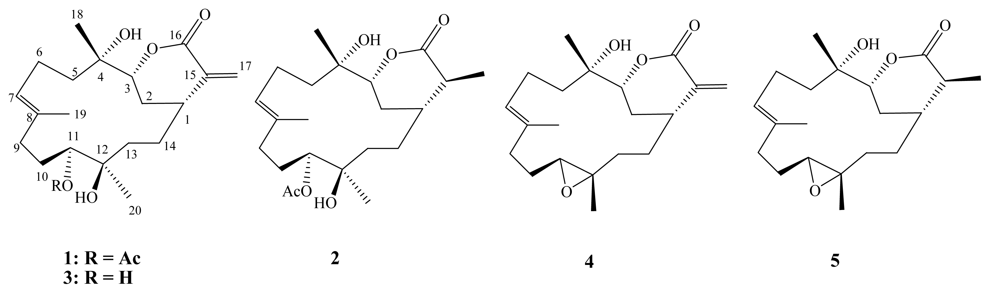
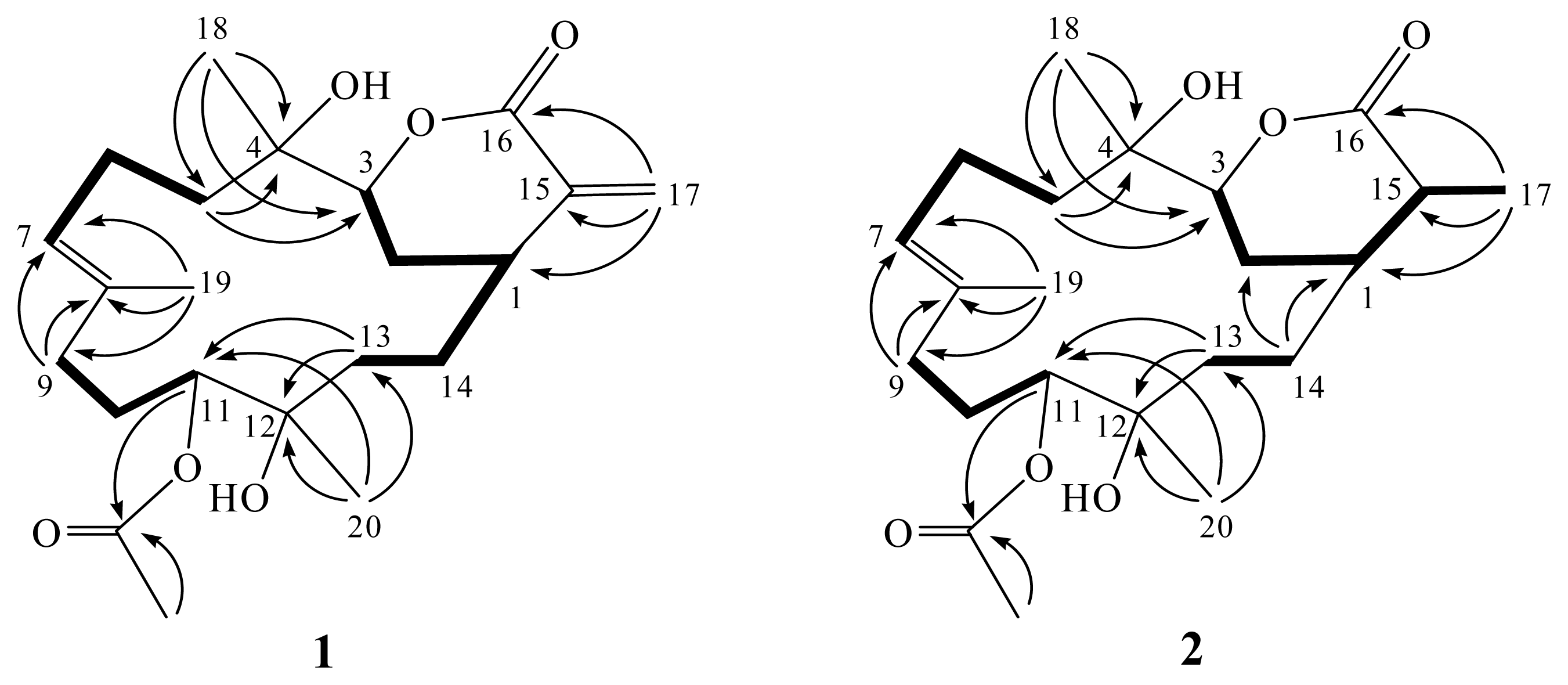
2. Results and Discussion

Frozen samples of Sinularia flexibilis were extracted with EtOAc. The dry EtOAc extracts were fractionated by silica gel gravity column chromatography, and the eluted fractions were further purified by HPLC to yield cembranoids 15.
The HR-ESI-MS (m/z 417.2250 [M + Na]+) of 11-acetylsinuflexolide (1) established the molecular formula C22H34O6, appropriate for six degrees of unsaturation. Inspection of the 13C-NMR and DEPT spectroscopic data (Table 1) (Figures S1 and S2) showed signals of four methyls (including one acetate methyl), seven sp3 methylenes, one sp2 methylene, three sp3 methines (including two oxymethines), one sp2 methine, two sp3 and four sp2 quaternary carbons (including two ester carbonyls). The 13C NMR signals appearing at δC 166.6 (C), 140.4 (C), 125.5 (CH2), 84.5 (CH), 36.7 (CH), and 29.3 (CH2) were assigned to an α-exomethylenic–δ-lactone ring functionality by comparing the very similar NMR data of the cembranoids with the same six-membered lactone ring [23,24]. Resonances in the 13C NMR spectrum of 1 at δC 170.6 (C) supported the presence of one additional ester group (Table 1). The ester was identified as acetate by the presence of one methyl resonance in the 1H NMR spectrum at δH 2.11 (3H, s) (Table 1). Furthermore, carbon signals of three methyls (δC 16.1, 25.4 and 25.5), one trisubstituted double bond (δC 135.1, C; 127.2, CH), two oxygen-bearing methines (δC 84.5 and 77.5), and two oxygenated quaternary carbons (δC 74.8 and 73.7) were also determined. The 1H-NMR spectral data revealed the presence of two olefinic methylene protons (δ 6.43, d, J = 2.0 Hz and 5.63, d, J = 2.0 Hz) and one olefinic methine proton (δ 5.26, dd, J = 7.5, 7.5 Hz). Furthermore, two oxygenated methines (δ 4.79, dd, J = 6.5, 2.5 Hz and 4.05, d, J = 11.5 Hz) were also designated from the 1H NMR signals. By interpretation of 1H-1H COSY correlations (Figure S5), it was possible to establish three partial structures from H-1 to H-3, from H2-5 to H-7, from H2-9 to H-11, and from H2-13 to H-1 through H2-14 (Figure 3). These data, together with the HMBC correlations (Figure 3) (Figure S4) from H2-5 to C-3 and C-4, H2-9 to C-7 and C-8, and H2-13 to C-11 and C-12 established the connectivity within the 14-membered ring. Three methyl groups attached at C-4, C-8 and C-12 were confirmed by the HMBC correlations from H3-18 to C-3, C-4 and C-5, H3-19 to C-7, C-8 and C-9, H3-20 to C-11, C-12 and C-13. A 1,1-disubstituted double bond attached at C-15 was confirmed by the HMBC correlations from H2-17 to C-1, C-15 and C-16. Moreover, one acetoxy group positioned at C-11 was confirmed from the HMBC correlations of H-11 (δ 4.79) and protons of an acetate methyl (δ 2.11) to the ester carbonyl carbon at δ 170.6 (C). The E–configuration of one double bond at C-7/C-8 was assigned based on the 13C NMR chemical shifts at C-19 (δC 16.1). Thus, 1 was revealed as a cembranoid possessing an α-exomethylenic–δ-lactone ring, based on the above analysis. Furthermore, the relative stereochemistry of 1 was mostly confirmed to be the same as that of the known metabolite sinuflexolide (3) by comparison of the proton chemical shifts and coupling constants [24]. Further comparison of the 1H and 13C NMR data of 1 with those of 3, showed that 1 contains an extra acetyl group relative to 3. The chemical shift of H-11 in 3H 3.47, dd, J = 6.4, 2.4 Hz) was shifted downfield (δH 4.79, dd, J = 6.5, 2.5 Hz) in 1, suggesting that 1 is the 11-acetyl derivative of 3. This was further supported by acetylation of 3 with acetic anhydride in pyridine to yield 1. Thus, compound 1 was established as the 11-acetyl derivative of 3.
11-acetyldihydrosinuflexolide (2) obtained as a white powder. The HRESIMS (m/z 419.2411, [M + Na]+) and NMR data of 2 indicated the molecular formula, C22H36O6. Both the 1H and 13C NMR signals of 2 were found to be very closely related to those of compound 1, suggesting the same skeleton. Further comparison of NMR data of 2 with those of 1 (Table 1) (Figures S1–S10), revealed that the two exomethylene proton signals (δH 6.43 and 5.63) in 1 was replaced by a methyl proton signal (δH 1.35 d, J = 7.0 Hz) in 2. This was confirmed by the HMBC correlations (Figure 3) from H3-17 to C-1, C-15 and C-16. The relative stereochemistry of all stereocenters except C-15 of 2 was determined to be the same as that of 1 by comparison of the proton shifts and coupling constants. The methyl group at C-15 was assigned the β-configuration primarily due to the NOE correlation between H3-17 and H-1. Furthermore, comparison of the NMR data between 2 and 5 confirmed both compounds have the same relative stereochemistry at C-15 [22]. Thus, the structure of 2 was established.
Finally, a 3-(4,5-dimethylthiazol-2-yl)-2,5-diphenyl tetrazolium bromide (MTT) assay was used to examine the cytotoxic activities of compounds 1–5 against four cancer cell lines, including human cervical epitheloid carcinoma (HeLa), laryngeal carcinoma (HEp-2) and breast carcinoma (MCF-7 and MDA-MB-231) cancer cells. Cells were treated with different concentrations of 1–5 for 72 h. The results show that compound 3, the most potent of compounds 1–5, exhibited cytotoxicity against the HeLa, HEp-2, MCF-7 and MDA-MB-231 cancer cell lines with IC50 values of 8.6, 8.2, 16.0 and 11.3 μg/mL, respectively. Furthermore, compounds 1 and 4 were found to exhibit weak cytotoxicity towards some of the cell lines (Table 2).

3. Experimental Section

3.1. General Procedures

Optical rotation values were measured using a Jasco P-1010 digital polarimeter. IR spectra were recorded on a Varian Digilab FTS 1000 Fourier transform infrared spectrophotometer. NMR spectra were recorded on a Varian Mercury Plus 400 FT-NMR (or Varian Unity INOVA 500 FT-NMR) instrument at 400 MHz (or 500 MHz) for 1H-NMR and 100 MHz (or 125 MHz) for 13C-NMR, respectively, in CDCl3. ESIMS and HRESIMS data were recorded with a Bruker APEX II mass spectrometer. Gravity column chomatography was performed on silica gel (230–400 mesh, Merck, Darmstadt, Germany). Thin layer chomatography (TLC) was carried out on precoated Kieselgel 60 F254 (0.2 mm, Merck) and spots were visualized by spraying with 10% H2SO4 solution followed by heating. HPLC was performed using a system comprised of a Hitachi L-7100 pump (Tokyo, Japan) and a Rheodyne 7725 injection (Cotati, CA, USA) port. A preparative normal phase column (Hibar 250 × 21.2 mm, Supelco, silica gel 60, 5 μm, Bellefonte, PA, USA) was used for HPLC.

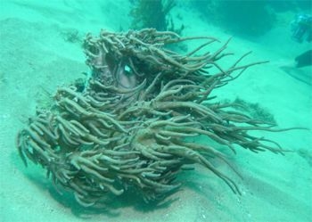
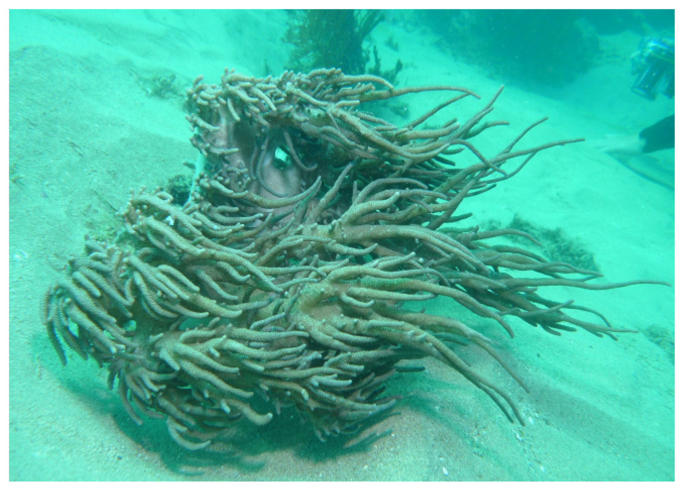
3.2. Animal Material

The marine soft coral Sinularia flexibilis (Quoy and Gaimard, 1833) was collected by scuba divers at a depth of around 10 m off the coast of Pingtung County, Taiwan, in July 2012, and the sample was frozen immediately after collection. A voucher sample was deposited at the National Museum of Marine Biology and Aquarium, Taiwan (specimen No. 2012-0709-10).

3.3. Extraction and Separation

The soft coral (2.0 kg, wet wt.) was stored frozen and then freeze dried. The freeze-dried material (450 g) was minced and extracted five times with EtOAc (2 L) for 24 h each time at room temperature. The organic extract was evaporated to yield a residue (60.5 g), which was subjected to open column chomatography on silica gel eluting with gradients of n-hexane (H)–EtOAc (E), to give 14 fractions: Fr-1 (eluted by n-hexane), Fr-2 (eluted by H–E 100:1), Fr-3 (eluted by H–E 50:1), Fr-4 (eluted by H–E 30:1), Fr-5 (eluted by H–E 20:1), Fr-6 (eluted by H–E 15:1), Fr-7 (eluted by H–E 10:1), Fr-8 (eluted by H–E 8:1), Fr-9 (eluted by H–E 5:1), Fr-10 (eluted by H–E 3:1), Fr-11 (eluted by H–E 2:1), Fr-12 (eluted by H–E 1:1), Fr-13 (eluted by H–E 1:2), and Fr-14 (eluted by EtOAc). Fraction 10 was further separated by silica gel column chomatography with gradient elution (n-hexane–EtOAc, 5:1 to 1:1) to yield five subfractions (10A–E). Subfraction 10C was subjected to normal phase HPLC with n-hexane–EtOAc (4:1) as the eluent (flow rate 2 mL/min) to obtain compounds 4 (250 mg, 0.41% dry wt. of extract) and 5 (330 mg, 0.55% dry wt. of extract). Fraction 12 was further separated by silica gel column chomatography with gradient elution (n-hexane–EtOAc, 1:1 to 1:3) to yield seven subfractions (12A–G). Subfraction 12C was subjected to normal phase HPLC with n-hexane–EtOAc (1:1) as the eluent (flow rate 2 mL/min) to obtain compounds 1 (8.0 mg, 0.013% dry wt. of extract) and 2 (6.5 mg, 0.011% dry wt. of extract). Subfraction 12F was subjected to normal phase HPLC with n-hexane–acetone (1:1) as the eluent (flow rate 2 mL/min) to obtain compound 3 (6.5 mg, 0.011% dry wt. of extract).
11-Acetylsinuflexolide (1): white solid; mp 82.0–85.0 °C; [α]D25 −12 (c 0.7, CHCl3); IR (neat) νmax 3434, 2974, 2937, 1712, 1622, 1452, 1376 and 1256 cm−1; UV (MeOH) λmax (log ɛ) 212 (3.9) nm; 13C and 1H NMR data, see Table 1; ESIMS m/z 417 [M + Na]+; HRESIMS m/z 417.2250 [M + Na]+ (calcd for C22H34O6Na, 417.2253).
11-Acetyldihydrosinuflexolide (2): white solid; mp 75.0–78.0 °C; [α]D25−15 (c 0.6, CHCl3); IR (neat) νmax 3434, 2975, 2938, 1714, 1639, 1458, 1377 and 1245 cm−1; 13C and 1H NMR data, see Table 1; ESIMS m/z 419 [M + Na]+; HRESIMS m/z 419.2411 [M + Na]+ (calcd for C22H36O6Na, 419.2409).
Sinuflexolide (3): white solid; [α]D24 −7.0 (c 0.2, CHCl3); IR (neat) νmax 3400, 2972, 1714, 1458, 1381, and 1235 cm−1; UV (MeOH) max 215 (log ɛ =3.8); [lit. [α]D25 −8.6 (c 0.17, CHCl3)] [21].
Sinularin (4): white solid; [α]D25 –105 (c 1.0, CHCl3); IR (neat) νmax 3404, 2965, 1710, 1455, 1381, and 1237 cm−1; UV (MeOH) λmax 212 (log ɛ = 3.8); [lit. [α]D20 −127] [22].
Dihydrosinularin (5): white solid; [α]D25 –30 (c 0.3, CHCl3); IR (neat) νmax 3400, 2960, 1714, 1459, 1385, and 1231 cm−1; [lit. [α]D20 −45] [22].
Acetylation of 3: A solution of 3 (5.0 mg) in pyridine (0.5 mL) was mixed with Ac2O (0.1 mL), and stirred at room temperature for 24 h. After evaporation of excess reagent, the residue was subjected to column chromatograph over silica gel using n-hexane–EtOAc (1:2) to give the acetyl derivative 1 (4.8 mg, 81%).

3.4. Cytotoxicity Testing

Cell lines were purchased from the American Type Culture Collection (ATCC). Cytotoxicity assays of compounds 15 were performed using the 3-(4,5-dimethylthiazol-2-yl)-2,5-diphenyltetrazolium bromide (MTT) colorimetric method [25,26].

4. Conclusions

In the previous reports, cembranoids possessing a δ-lactone have been mostly isolated from soft coral (Alcyonaceae) belonging to the genera Sinularia [2,2123,27,28] and Lobophytum [29]. Some of these metabolites have been found to possess several kinds of biological activities, such as cytotoxic [2123] and anti-inflammatory activity [2,29]. In the present study, compounds 1, 3 and 4 exhibited moderate or weak cytotoxicity against the growth of HeLa, HEp-2, MCF-7 and MDA-MB-231 cancer cell lines. According to the structures of 15, it seems that the α-exomethylenic–ä-lactone ring group in compounds 1, 3 and 4 is critical for the cytotoxic activity of metabolites 15.

Acknowledgements

This study was supported in part by a grant from National Science Council of Taiwan (NSC 98-2313-B-276-001-MY3 and NSC 101-2313-B-276-002) and part by grant from Antai Medical Care Cooperation Antai Tian-Sheng Memorial Hospital Research Fund. (Project No. 101-FI-DBS-IAC-R-003).

References and Notes

  1. Rocha, J.; Peixe, L.; Gomes, N.C.; Calado, R. Cnidarians as a source of new marine bioactive compounds—An overview of the last decade and future steps for bioprospecting. Mar. Drugs 2011, 9, 1860–1886. [Google Scholar]
  2. Su, J.-H.; Wen, Z.-H. Bioactive cembrane-based diterpenoids from the soft coral Sinularia triangular. Mar. Drugs 2011, 9, 944–951. [Google Scholar]
  3. Chao, C.-H.; Chou, K.-J.; Huang, C.-Y.; Wen, Z.-H.; Hsu, C.-H.; Wu, Y.-C.; Dai, C.-F.; Sheu, J.-H. Bioactive cembranoids from the soft coral Sinularia crassa. Mar. Drugs 2011, 9, 1955–1968. [Google Scholar]
  4. Shih, H.-J.; Tseng, Y.-J.; Huang, C.-Y.; Wen, Z.-H.; Dai, C.-F.; Sheu, J.-H. Cytotoxic and anti-inflammatory diterpenoids from the Dongsha Atoll soft coral Sinularia flexibilis. Tetrahedron 2012, 68, 244–249. [Google Scholar]
  5. Lu, Y.; Huang, C.-Y.; Lin, Y.-F.; Wen, Z.-H.; Su, J.-H.; Kuo, Y.-H.; Chiang, M.Y.; Sheu, J.-H. Anti-inflammatory cembranoids from the soft corals Sinularia querciformis and Sinularia granosa. J. Nat. Prod 2008, 71, 1754–1759. [Google Scholar]
  6. Chen, B.-W.; Chao, C.-H.; Su, J.-H.; Huang, C.-Y.; Dai, C.-F.; Wen, Z.-H.; Sheu, J.-H. A novel symmetric sulfur-containing biscembranoid from the Formosan soft coral Sinularia flexibilis. Tetrahedron Lett 2010, 44, 5764–5766. [Google Scholar]
  7. Lin, Y.-S.; Lee, N.-L.; Lu, M.-C.; Su, J.-H. Two new cembrane-based diterpenoids from the marine soft coral Sinularia crassa. Molecules 2012, 17, 5422–5429. [Google Scholar]
  8. Huang, S.-Y.; Chen, N.-F.; Chen, W.-F.; Hung, H.-C.; Lee, H.-P.; Lin, Y.-Y.; Wang, H.-M.; Sung, P.-J.; Sheu, J.-H.; Wen, Z.-H. Sinularin from indigenous soft coral attenuates nociceptive responses and spinal neuroinflammation in carrageenan-induced inflammatory rat model. Mar. Drugs 2012, 10, 1899–1919. [Google Scholar]
  9. Liu, C.-I.; Chen, C.-C.; Chen, J.-C.; Su, J.-H.; Huang, H.H.; Chen, J.-F.; Wu, Y.-J. Proteomic analysis of anti-tumor effects of 11-dehydrosinulariolide on CAL-27 cells. Mar. Drugs 2011, 9, 1254–1272. [Google Scholar]
  10. Neoh, C.-A.; Wang, R.Y.-L.; Din, Z.-H.; Su, J.-H.; Chen, Y.-K.; Tsai, F.-J.; Weng, S.-H.; Wu, Y.-J. Induction of apoptosis by sinulariolide from soft coral through mitochondrial-related and p38MAPK pathways on human bladder carcinoma cells. Mar. Drugs 2012, 10, 2893–2911. [Google Scholar]
  11. Su, T.-R.; Tsai, F.-J.; Lin, J.-J.; Huang, H.H.; Chiu, C.-C.; Su, J.-H.; Yang, Y.-T.; Chen, J.-F.; Wong, B.-S.; Wu, Y.-J. Induction of apoptosis by 11-dehydrosinulariolide via mitochondrial dysregulation and ER stress pathways in human melanoma cells. Mar. Drugs 2012, 10, 1883–1898. [Google Scholar]
  12. Lin, W.-Y.; Lu, Y.; Su, J.-H.; Wen, Z.-H.; Dai, C.-F.; Kuo, Y.-H.; Sheu, J.-H. Bioactive cembranoids from the Dongsha Atoll soft coral Sarcophyton crassocaule. Mar. Drugs 2011, 9, 994–1006. [Google Scholar]
  13. Lin, W.-Y.; Su, J.-H.; Lu, Y.; Wen, Z.-H.; Dai, C.-F.; Kuo, Y.-H.; Sheu, J.-H. Cytotoxic and Anti-inflammatory cembranoids from the Dongsha Atoll soft coral Sarcophyton crassocaule. Bioorg. Med. Chem 2010, 18, 1936–1941. [Google Scholar]
  14. Lin, W.-Y.; Lu, Y.; Chen, B.-W.; Huang, C.-Y.; Su, J.-H.; Wen, Z.-H.; Dai, C.-F.; Kuo, Y.-H.; Sheu, J.-H. Sarcocrassocolides M–O, bioactive cembranoids from the Dongsha Atoll soft coral Sarcophyton crassocaule. Mar. Drugs 2012, 10, 617–626. [Google Scholar]
  15. Su, C.-C.; Su, J.-H.; Lin, J.-J.; Chen, C.-C.; Hwang, W.-I.; Huang, H.H.; Wu, Y.-J. An investigation into the cytotoxic effects of 13-acetoxysarcocrassolide from the soft coral Sarcophyton crassocaule on bladder cancer Cells. Mar. Drugs 2011, 9, 2622–2642. [Google Scholar]
  16. Cheng, S.-Y.; Wang, S.-K.; Chiou, S.-F.; Hsu, C.-H.; Dai, C.-F.; Chiang, M.Y.; Duh, C.-Y. Cembranoids from the octocoral Sarcophyton ehrenbergi. J. Nat. Prod 2010, 73, 197–203. [Google Scholar]
  17. Wang, S.-K.; Duh, C.-Y. New cytotoxic cembranolides from the soft coral Lobophytum michaelae. Mar. Drugs 2012, 10, 306–318. [Google Scholar]
  18. Chao, C.-H.; Wen, Z.-H.; Wu, Y.-C.; Yeh, H.-C.; Sheu, J.-H. Cytotoxic and anti-inflammatory cembranoids from the soft coral Lobophytum crassum. J. Nat. Prod 2008, 71, 1819–1824. [Google Scholar]
  19. Cheng, S.-Y.; Wen, Z.-H.; Wang, S.-K.; Chiou, S.-F.; Hsu, C.-H.; Dai, C.-F.; Chiang, M.Y.; Duh, C.-Y. Unprecedented hemiketal cembranolides with anti-inflammatory activity from the soft coral Lobophytum durum. J. Nat. Prod 2009, 72, 152–155. [Google Scholar]
  20. Su, J.-H.; Lin, Y.-F.; Lu, Y.; Huang, C.-Y.; Wang, W.-H.; Fang, T.-Y.; Sheu, J.-H. Oxygenated cembranoids from the cultured and wild-type soft corals Sinularia flexibilis. Chem. Pharm. Bull 2009, 57, 1189–1192. [Google Scholar]
  21. Duh, C.-Y.; Wang, S.-K.; Tseng, H.-K.; Sheu, J.-H.; Chiang, M.Y. Novel cytotoxic cembranoids from the soft coral Sinularia flexibilis. J. Nat. Prod 1998, 61, 844–847. [Google Scholar]
  22. Weinheimer, A.J.; Matson, J.A.; Hossain, M.B.; van der Helm, D. Marine anticancer agents: Sinularin and dihydrosinularin, new cembranolides from the soft coral, Sinularia flexibilis. Tetrahedron Lett 1977, 34, 2923–2926. [Google Scholar]
  23. Su, J.-H.; Ahmed, A.F.; Sung, P.-J.; Chao, C.-H.; Kuo, Y.-H.; Sheu, J.-H. Manaarenolides A–I, new diterpenoids from the soft coral Sinularia manaarensis. J. Nat. Prod 2006, 69, 1134–1139. [Google Scholar]
  24. Sinuflexolide (3): Selected 1H NMR (CDCl3, 400 MHz) data: δ 6.43 (1H, d, J = 2.0 Hz, H-17a), 5.64 (1H, d, J = 2.0 Hz, H-17b), 5.24 (1H, dd, J = 7.2, 6.4 Hz, H-7), 4.06 (1H, dd, J = 10.8, 2.0 Hz, H-3), 3.47 (1H, dd, J = 6.4, 2.4 Hz, H-11), 2.82 (1H, m, H-1), 1.63 (3H, s, H3–19), 1.37 (1H, s, H3–18), 1.22 (1H, s, H3–20); 13C NMR (CDCl3, 100 MHz) δ 166.6 (C, C-16), 140.4 (C, C-15), 135.9 (C, C-8), 126.7 (CH, C-7), 125.7 (CH2, C-17), 84.4 (CH, C-3), 75.0 (CH, C-12), 74.7 (C, C-11), 73.8 (C, C-4), 37.9 (CH2, C-5), 36.3 (CH, C-1), 35.2 (CH2, C-9), 35.0 (CH2, C-13), 29.4 (CH2, C-2), 29.3 (CH2, C-14), 25.3 (CH3, C-18),25.1 (CH3, C-20), 22.2 (CH2, C-6), 16.0 (CH3, C-19).
  25. Alley, M.C.; Scudiero, D.A.; Monks, A.; Hursey, M.L.; Czerwinski, M.J.; Fine, D.L.; Abbott, B.J.; Mayo, J.G.; Shoemaker, R.H.; Boyd, M.R. Feasibility of drug screening with panels of human tumor cell lines using a microculture tetrazolium assay. Cancer Res 1988, 48, 589–601. [Google Scholar]
  26. Scudiero, D.A.; Shoemaker, R.H.; Paull, K.D.; Monks, A.; Tierney, S.; Nofziger, T.H.; Currens, M.J.; Seniff, D.; Boyd, M.R. Evaluation of a soluble tetrazolium/formazan assay for cell growth and drug sensitivity in culture using human and other tumor cell lines. Cancer Res 1988, 48, 4827–4833. [Google Scholar]
  27. Wen, T.; Ding, Y.; Deng, Z.; van Ofwegen, L.; Proksch, P.; Lin, W. Sinulaflexiolides A–K, cembrane-type diterpenoids from the Chinese soft coral Sinularia flexibilis. J. Nat. Prod 2008, 71, 1133–1140. [Google Scholar]
  28. Lin, Y.-S.; Chen, C.-H.; Liaw, C.-C.; Chen, Y.-C.; Kuo, Y.-H.; Shen, Y.-C. Cembrane diterpenoids from the Taiwanese soft coral Sinularia flexibilis. Tetrahedron 2009, 65, 9157–9164. [Google Scholar]
  29. Kao, C.-Y.; Su, J.-H.; Lu, M.-C.; Hwang, T.-L.; Wang, W.-H.; Chen, J.-J.; Sheu, J.-H.; Kuo, Y.-H.; Weng, C.-F.; Fang, L.-S.; et al. Lobocrassins A–E: New cembrane-type diterpenoids from the soft coral Lobophytum crassum. Mar. Drugs 2011, 9, 1319–1331. [Google Scholar]
Figure 1. The soft coral Sinularia flexibilis.
Ijms 14 04317f1
Figure 2. Structures of metabolites 15.
Ijms 14 04317f2
Figure 3. The structures of metabolites 1 and 2 and selected 1H-1H COSY (−) and HMBC (→) correlations.
Ijms 14 04317f3
Table 1. 1H and 13C NMR data for 1 and 2.
C/H12


δH (J in Hz) aδC (mult.) bδH (J in Hz) aδC (mult.) b
12.75 m36.7 (CH)1.71 m38.4 (CH)
22.25 m; 1.57 m29.3 (CH2)2.24 m; 1.44 m29.8 (CH2)
34.05 d (11.5)84.5 (CH)4.05 d (11.5, 2.5)85.0 (CH)
473.7 (C)73.7 (C)
51.83 m; 1.77 m37.8 (CH2)1.77 m37.5 (CH2)
62.28 m; 2.02 m22.1 (CH2)2.28 m; 1.98 m22.1 (CH2)
75.26 dd (7.5, 7.5)127.2 (CH)5.23 dd (7.0, 7.0)127.2 (CH)
8135.1 (C)135.0 (C)
92.31 m; 1.93 m35.3 (CH2)2.27 m; 1.92 m35.1 (CH2)
101.92 m; 1.72 m27.9 (CH2)1.90 m; 1.72 m28.1 (CH2)
114.79 dd (6.5, 2.5)77.5 (CH)4.80 dd (7.0, 2.0)77.2 (CH)
1274.8 (C)74.8 (C)
131.74 m; 1.53 m35.2 (CH2)1.68 m; 1.48 m36.1 (CH2)
141.92 m; 1.36 m28.6 (CH2)1.68 m; 1.12 m28.7 (CH2)
15140.4 (C)2.09 m43.5 (CH)
16166.6 (C)174.8 (C)
176.43 d (2.0); 5.63 d (2.0)125.5 (CH2)1.35 d (7.0)15.3 (CH3)
181.38 s25.5 (CH3)1.39 s25.6 (CH3)
191.62 s16.1 (CH3)1.62 s16.4 (CH3)
201.19 s25.4 (CH3)1.17 s25.4 (CH3)
OAC170.6 (C)170.6 (C)
2.11 s21.1 (CH3)2.11 s21.1 (CH3)
a500 MHz in CDCl3;
b125 MHz in CDCl3.
Table 2. Cytotoxicity (IC50 μg/mL) of compounds 15.
CompoundCell Lines

HeLaHEp-2MCF-7MDA-MB-231
19.511.317.815.7
2NA bNA bNA bNA b
38.68.216.011.3
4NA b12.617.513.5
5NA bNA bNA bNA b
Doxorubicina0.050.10.070.5
aClinical anticancer drug as positive control;
bNA, not active at 20 μg/mL.

Supplementary Files

  • Supplementary File 1:

    Supporting Information (PDF, 230 KB)

  • Share and Cite

    MDPI and ACS Style

    Su, C.-C.; Wong, B.-S.; Chin, C.; Wu, Y.-J.; Su, J.-H. Oxygenated Cembranoids from the Soft Coral Sinularia flexibilis. Int. J. Mol. Sci. 2013, 14, 4317-4325. https://doi.org/10.3390/ijms14024317

    AMA Style

    Su C-C, Wong B-S, Chin C, Wu Y-J, Su J-H. Oxygenated Cembranoids from the Soft Coral Sinularia flexibilis. International Journal of Molecular Sciences. 2013; 14(2):4317-4325. https://doi.org/10.3390/ijms14024317

    Chicago/Turabian Style

    Su, Ching-Chyuan, Bing-Sang Wong, Chuen Chin, Yu-Jen Wu, and Jui-Hsin Su. 2013. "Oxygenated Cembranoids from the Soft Coral Sinularia flexibilis" International Journal of Molecular Sciences 14, no. 2: 4317-4325. https://doi.org/10.3390/ijms14024317

    Article Metrics

    Back to TopTop