Article

Isolation and Identification of Cellulolytic Bacteria from the Gut of Holotrichia parallela Larvae (Coleoptera: Scarabaeidae)

1
State Key Laboratory of Agricultural Microbiology, Huazhong Agricultural University, Wuhan 430070, China
2
Institute of Urban and Horticultural Pests, College of Plant Science and Technology, Huazhong Agricultural University, Wuhan 430070, China
3
Hubei Insect Resources Utilization and Sustainable Pest Management Key Laboratory, College of Plant Science and Technology, Huazhong Agricultural University, Wuhan 430070, China
*
Author to whom correspondence should be addressed.
Int. J. Mol. Sci. 2012, 13(3), 2563-2577; https://doi.org/10.3390/ijms13032563
Submission received: 31 January 2012 / Revised: 17 February 2012 / Accepted: 20 February 2012 / Published: 23 February 2012

Abstract

:
In this study, 207 strains of aerobic and facultatively anaerobic cellulolytic bacteria were isolated from the gut of Holotrichia parallela larvae. These bacterial isolates were assigned to 21 genotypes by amplified ribosomal DNA restriction analysis (ARDRA). A partial 16S rDNA sequence analysis and standard biochemical and physiological tests were used for the assignment of the 21 representative isolates. Our results show that the cellulolytic bacterial community is dominated by the Proteobacteria (70.05%), followed by the Actinobacteria (24.15%), the Firmicutes (4.35%), and the Bacteroidetes (1.45%). At the genus level, Gram-negative bacteria including Pseudomonas, Ochrobactrum, Rhizobium, Cellulosimicrobium, and Microbacterium were the predominant groups, but members of Bacillus, Dyadobacter, Siphonobacter, Paracoccus, Kaistia, Devosia, Labrys, Ensifer, Variovorax, Shinella, Citrobacter, and Stenotrophomonas were also found. Furthermore, our results suggest that a significant amount of bacterial diversity exists among the cellulolytic bacteria, and that Siphonobacter aquaeclarae, Cellulosimicrobium funkei, Paracoccus sulfuroxidans, Ochrobactrum cytisi, Ochrobactrum haematophilum, Kaistia adipata, Devosia riboflavina, Labrys neptuniae, Ensifer adhaerens, Shinella zoogloeoides, Citrobacter freundii, and Pseudomonas nitroreducens are reported to be cellulolytic for the first time in this study. Our results indicate that the scarab gut is an attractive source for the study of novel cellulolytic microorganisms and enzymes useful for cellulose degradation.

1. Introduction

Interest in bioenergy has been sharply increasing in recent years due to the necessity of sustainable economies and clean environments [13]. Cellulose and hemicellulose are the most abundant biomasses on earth, and therefore have the greatest potential to resolve both the energetic and environmental demands of bioenergy [4,5]. The production of ethanol and other biofuels from lignocellulosic biomass has recently received tremendous attention both in industry and in academic communities worldwide, and great progress has been made in the production and use of biofuels [1,3,6,7]. Lignocellulosic raw materials can be converted to ethanol by hydrolysis and subsequent fermentation [8]. In the hydrolysis step, acid-reliant hydrolytic processes have been used for many decades, but have also been blamed for negative effects on the environment, including the formation of large quantities of calcium sulfate that require disposal [3,7,8]. Thermochemical processes are another significant method of bioethanol production [9,10], but developing a cost-effective, all-thermochemical process has been difficult [11,12]. Consequently, enzymatic hydrolysis is a more environmentally sound approach [8], and the costs of this approach can be reduced with recent breakthroughs in molecular genetics, enzyme engineering and metabolic engineering, which has drawn greater attention from researchers [3,1214]. In the fermentation step, the conversion of cellulose, the recovery efficiency and the cost depends strongly on the fermentation efficiency of the microorganisms and enzymes [15,16]. Currently, the mainstream process of bioethanol production makes use of the basic yeast Saccharomyces cerevisiae due to its unique advantages (e.g., genetically tractable, superior conversion yields of ethanol from glucose, high alcohol tolerance) [17,18]. However, the rising concentration of sugars and aromatic components in the industrial conversion of lignocellulose to ethanol as well as other adverse conditions can cause harm to the S. cerevisiae and restrict its applications [18]. Therefore, isolating ideal biofuel-producing microorganism with ability to degrade different lignocellulosic materials, resist different inhibitors and biosynthesize specific biofuels with high yield has an important role in developing biofuel production systems [3,18]. Some insects, such as termites, wood-feeding roaches, beetles, and leaf-cutting ants, can use lignocellulosic substrates as their main food source and are highly efficient at degrading cellulose to glucose as an energy source [1]. There have been numerous reports on the cellulolytic activity of these insects, which include Reticulitermes flavipes [19], Anoplophora glabripennis [20], Tenebrio molitor [21], and Pachnoda marginata [22]. The gut systems of these insects are diverse and highly adapted and are considered to be highly efficient natural bioreactors [1,4,23]. Furthermore, the intestinal microorganisms of these lignocellulose-degrading insects are considered to be essential for cellulose digestion. Gut microbiota symbiotically associated with lower termites are responsible for the decomposition of various kinds of organic matter and for biomass recycling [2426]. Additionally, a number of protozoa and bacteria with cellulolytic activity have been isolated from Zootermopsis angusticollis [25], silver cricket Lepisma sp. [27], Tipula abdominalis [28], Saperda vestita [29], Dendroctonus frontalis [29], and Pachnoda marginata [30]. These studies suggest that lignocellulose-degrading insects are an attractive potential source of novel cellulolytic microorganisms and enzymes and suggest that these enzymes may be useful in biofuel production [28,31].
The family Scarabaeidae, as currently defined, comprises over 30,000 species of beetles, which are almost exclusively herbivorous or saprophagous [31]. Many scarab larvae live in the soil and feed on roots or other organic matter [32]. The hindgut of scarab larvae (also referred to as the fermentation sac) is enlarged and houses dense microbial communities [33,34]. Previous studies have shown that 25–65% of the ingested pure cellulose or neutral detergent fibers in their diet are degraded by scarab larvae and that the intestinal bacteria in the hindgut of these larvae are responsible for cellulose degradation [22,35]. Furthermore, several cellulolytic bacterial species have been successfully isolated from the gut contents of some scarab species [30,36]. These studies demonstrated that the hindgut of scarab larvae represent an ideal prospecting resource for identifying microorganisms and enzymes that can be used for biofuel production and to improve biofuel production technology [31].
The root-feeding larvae of Holotrichia parallela live in the soil in China and feed on a variety of plants, resulting in significant economic damage [37]. In this study, we isolated and identified cellulose-degrading bacteria from the gut of H. parallela to obtain a more precise estimation of their occurrence in scarabs, and we specifically evaluated the nutritional contributions of gut microbiota to scarabs, and also assessed their potential to future biofuel production.

2. Results and Discussion

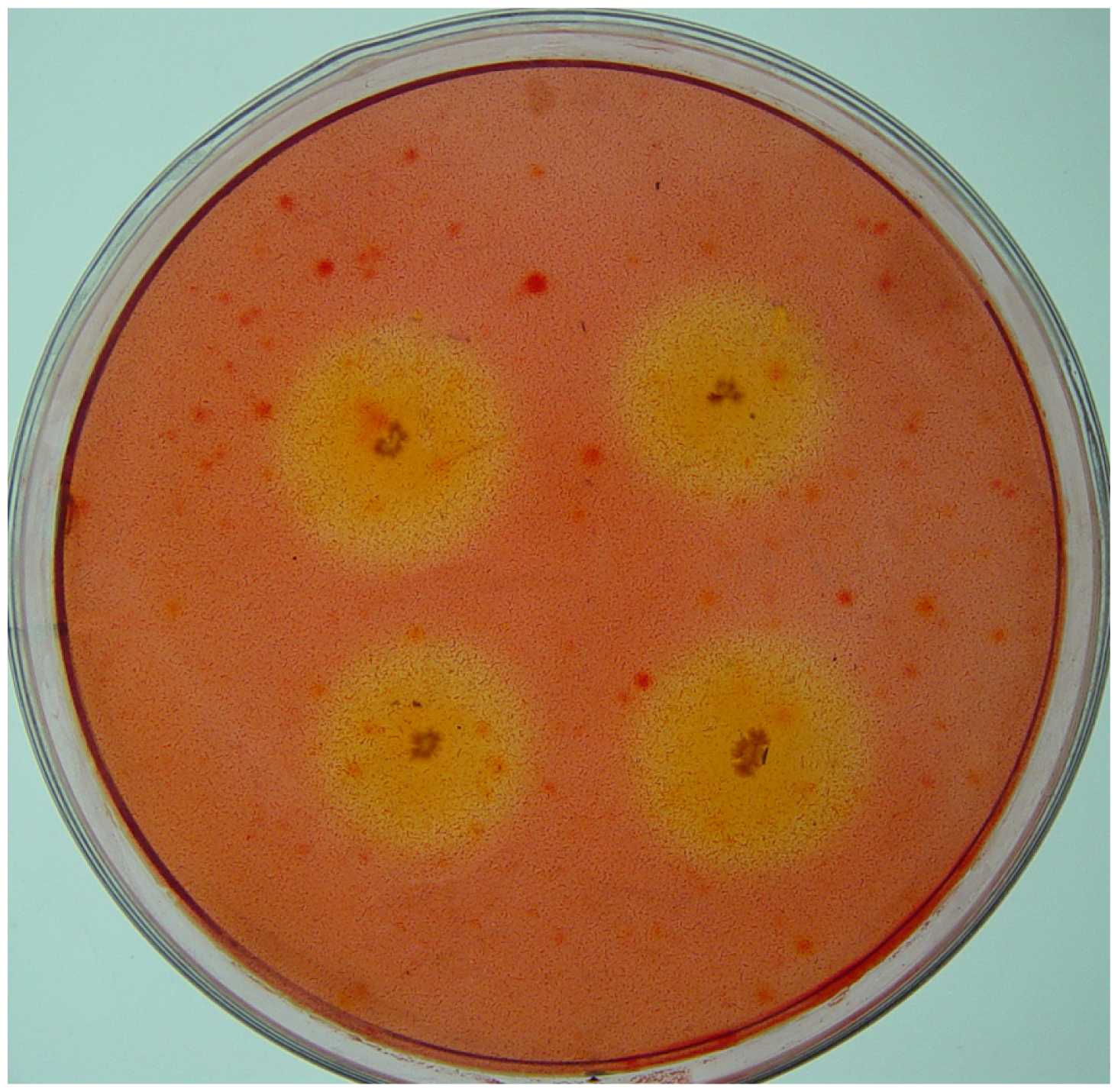
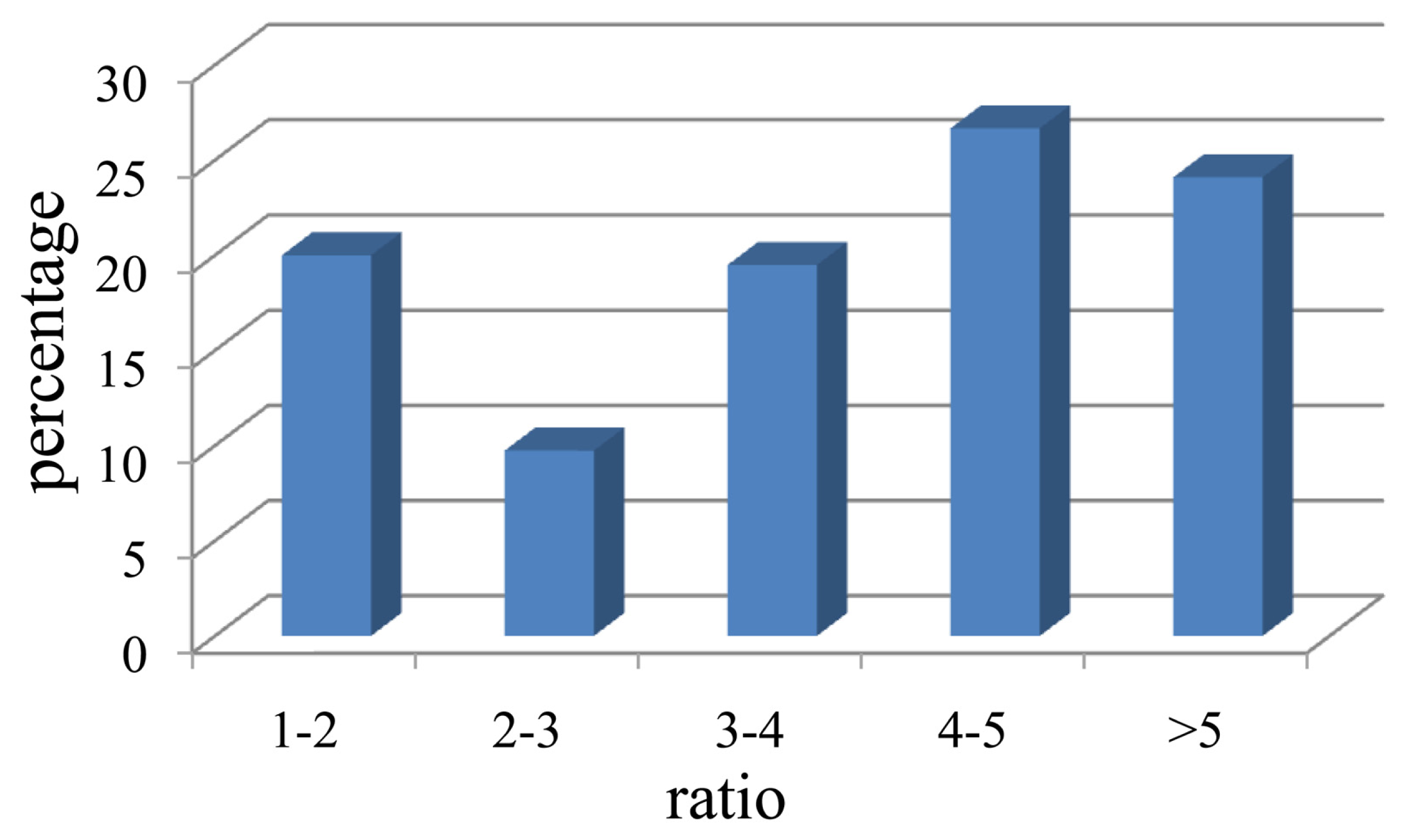
2.1. Isolation of Cellulolytic Bacteria

Bacteria with CMCase activity were abundant (1.14 ± 0.13 × 10⁸ colony forming units (CFU)/gut) in the hindgut of H. parallela. However, bacteria with CMCase activity were seldom found in the midgut (only 20 ± 1.45 CFU/gut). These results are similar to those from another scarab beetle, P. marginata [30]. Cazemier et al. [30] observed that a large number of bacteria with CMCase and xylanase activities were present in the hindgut of P. marginata (2.5 ± 1.1 × 10⁸ CFU (mLgut) −1), but that these bacteria were not detected in the midgut. Studies of the gut microbiota of other scarab beetles showed that the hindgut microbiota was dominated by groups of fermentative bacteria such as Clostridiales, Actinobacteria, and Cytophaga-Flavobacterium-Bacteroides, which contain a wide range of species able to ferment cellulose, hemicellulose, pectin and other polysaccharides [34,3840]. These results indicate that the bacteria in the scarab hindgut play an important role in the degradation of roots and other organic matter consumed by scarab larvae, as suggested by Cazemier et al. [30] and Huang et al. [31]. As bacteria with cellulolytic activity appear to be absent in the midgut, it seems likely that the midgut of scarabs serves a predigestive function for lignocellulose rather than for the microbial degradation of cellulose and hemicellulose [30].
A total of 207 isolates with CMCase activity were obtained from the gut contents of H. parallela either by plating on CMC medium or by enrichment on filter paper. Among these cellulolytic bacteria, 81 isolates were obtained using the filter paper inoculation method, and 126 isolates were obtained from direct plating. These isolates produced variable zones of CMC clearance (Figure 1). Based on the calculation of the ratio of the diameter (mm) of the zone of clearance to the diameter of the colony, it was determined that these bacterial isolates demonstrated large differences in their ability to degrade CMC (Figure 2). This ratio ranged from 1.1 to 9.0 among all the isolates, with 24.1% of the isolates showing high CMC-degrading activity (ratio > 5), demonstrating that multiple bacterial isolates from the scarab gut possess the ability to produce CMCase (Figure 2).

2.2. Assignment and Identification of Cellulolytic Bacteria

The 207 cellulolytic bacterial isolates obtained in this study were grouped into 21 clusters or genotypic groups (Table 1). Each group displayed a specific ARDRA banding pattern, and the number of isolates belonging to each group was different (Table 1). A total of 21 isolates were chosen to represent each ARDRA group, and these isolates were investigated both by 16S rDNA sequencing and by physical and biochemical characterization (Table 2). Overall, the 16S rDNA sequences from the 21 isolates showed a high degree of similarity (99–100%) to a number of annotated sequences found in the databases (data not shown), and their identification was in agreement with the biochemical and physiological tests. The 21 isolates clustered into four phyla (Proteobacteria, Actinobacteria, Bacteroidetes, and Firmicutes), and represented 17 different genera (Table 1). The cellulolytic bacterial community was represented by members of the phylum Proteobacteria (67.13%), followed by Actinobacteria (23.15%), Firmicutes (4.35%), and Bacteroidetes (1.45%). At genus level, Pseudomonas (31.4%), Cellulosimicrobium (13.53%), Ochrobactrum (12.08%), Rhizobium (11.59%), and Microbacterium (10.63%) were the dominant genera identified, with 65, 28, 25, 24 and 22 isolates, respectively, while Siphonobacter (group 3), Devosia (group 14), Variovorax (group17), Shinella (group 18) each consisted of a single bacteria isolate. Furthermore, the ARDRA grouping results also revealed that bacterial isolates belonging to Bacillus licheniformis, Microbacterium oxydans, Microbacterium binotii, Microbacterium aurum., Cellulosimicrobium funkei, Ochrobactrum cytisi, Rhizobium radiobacter, Labrys neptuniae, Pseudomonas nitroreducens, Stenotrophomonas maltophilia can obtained both by the direct plating method and by the filter papers enrichment method. The fact that these bacterial isolates can be obtained using both methods demonstrates that bacteria with cellulolytic ability are commonly present in the hindgut of H. parallela.
Our results showed that Pseudomonas was the most dominant group in the cellulolytic bacterial community in the gut of soil-dwelling scarab larvae. The dominance of Pseudomonas in the present study is similar to the results of previous studies on cellulolytic bacteria present on native Chaco soil, which showed that the Pseudomonas was the only genus that exists stably in three samples (native forest soil, CMC- and filter paper-enriched samples) [41]. Bacteria of the genus Pseudomonas can be found in many different environments including soil, water, plant and animal tissue, and these bacteria have the ability to metabolize a variety of diverse nutrients [42]. Many Pseudomonas species are opportunistic pathogens that infect humans, animals, and plants [4345], but other Pseudomonas species also have been reported to degrade cellulose [4648]. There have been no reports, however, describing the cellulolytic activity of P. nitroreducens, which we observed in this study.
The cellulolytic activity of some of the bacteria found in this study has been reported previously. B. licheniformis is characterized by strong xylanase activity, and also possesses CMCase, mannanase, and pectinase activities [49]. Though Dyadobacter fermentans NS114T does not hydrolyze cellulose or starch [50], whole genome sequencing of D. fermentans DSM 18053 has revealed several genes encoding for 1,4-β-cellobiosidase, β-glucosidase, and endo-1,4-β-xylanase enzymes [51]. The Microbacterium genus contains many species with cellulolytic or xylanolytic activities. A cellulolytic bacterium that showed 99% 16S rDNA sequence similarity to M. oxydans has been found to produce an array of cellulolytic-xylanolytic enzymes (filter paper cellulase, β-glucosidase, xylanase, and β-xylosidase) [52]. M. binotii have also been reported to produce an enzyme with β-glucosidase activity [53]. Rhizobium species are known to produce cellulolytic and pectinolytic enzymes that can break the glycosidic bonds present in the plant cell wall, and these enzymes are essential for the primary symbiotic infection of legume host roots [5456]. However, little attention has been paid to their potential ability to degrade organic compounds during their growth as free-living saprophytes [41,57]. An analysis of the genome sequence of R. radiobacter (formerly Agrobacterium tumefaciens) has identified several genes encoding pectinase, ligninase, and xylanase as well as genes encoding regulators of pectinase and cellulase production [58]. Variovorax paradoxus is a microorganism of special interest due to its diverse metabolic capabilities. Whole genome sequencing of V. paradoxus revealed a single gene encoding β-glucosidase, but genes involved in the production of pectinases and other cellulases remained unidentified [59]. The Stenotrophomonas genus contains species ranging from common soil organisms (Stenotrophomonas nitritireducens) to opportunistic human pathogens (S. maltophilia) [41]; one S. maltophilia strain from the mesophilic microbial community BYND-8 has also been reported to be cellulolytic [60].
In addition to those bacterial isolates for which cellulolytic activity has been well described, our results demonstrate cellulolytic activity for several bacterial strains that have not been previously reported to be cellulolytic. To the best of our knowledge, this is the first report describing Siphonobacter aquaeclarae, C. funkei, Paracoccus sulfuroxidans, O. cytisi, Ochrobactrum haematophilum, Kaistia adipata, Devosia riboflavina, L. neptuniae, Ensifer adhaerens, Shinella zoogloeoides, Citrobacter freundii, and P. nitroreducens as being cellulolytic, with some isolates displaying high cellulolytic activity. In the case of S. aquaeclarae, the ratio of the CMC clearance zone to the colony diameter was greater than 7, and for C. funkei, the ratio ranged from 3.3 to 5.3, indicating robust CMC-ase production. These cellulolytic bacterial isolates demonstrate great potential for the study of novel enzymes in cellulose degradation and for improving the bioconversion of lignocellulosic biomass.

3. Experimental Section

3.1. Insect and Dissection

Third-instar larvae of H. parallela were collected from a peanut field and were maintained individually in containers with sterile soil. All the larvae were fed with peanuts surface sterilized with 70% ethanol, and the diets were replaced every 3 days. After 3 weeks, 9 healthy larvae were surface sterilized with 70% ethanol to remove contamination, washed twice in sterile distilled water, and allowed to air dry for 1 min. The preparation of the intestinal tract (mid- and hindgut) was performed as described previously by Zhang and Jackson [34].

3.2. Media

Medium I and Medium II were prepared as described by Cazemier, et al. [30], with some modifications, as follows:
Medium I: peptone, 5 g/L; yeast extract, 0.1 g/L; K2HPO4, 1 g/L; MgSO4·7H2O, 0.2 g/L; carboxymethylcellulose (CMC), 10 g/L (sodium salt, low viscosity, Sigma); Na2CO3, 10 g/L (sterilized separately); pH 10.3.
Medium II: K2HPO4, 1.9 g/L; KH2PO4, 0.94 g/L; KCl, 1.6 g/L; NaCl, 1.43 g/L; NH4Cl, 0.15 g/L; MgSO4·7H2O, 0.037 g/L; CaCl2·2H2O, 0.017 g/L; yeast extract, 0.1 g/L; CMC, 10 g/L; pH 7.2.
Medium III was prepared as described by Wenzel et al. [25], with the following modifications: yeast extract, 0.04 g/L; malt extract, 0.1 g/L; CaCO3, 0.5 g/L; filter paper strips, 5 g/L (Whatman Filter Paper No.1); pH 10.3.
The media were sterilized (121 °C, 20 min) and solidified with agar (17 g/L) when necessary.

3.3. Counting and Isolation of Cellulolytic Bacteria

For viable counts, an individual gut was homogenized and suspended in 10 mL of medium I and serially diluted ten-fold (to 10−9). From each dilution, 100 μL was spread on plates with solid medium I (midgut) or medium II (hindgut). A triplicate series of dilutions from the midguts and hindguts of three different larvae were incubated at 28 °C. Colonies were counted following 4 weeks of incubation. Only the colonies that were encircled by a clear zone after staining with a solution of congo red (1 mg/mL) were counted.
To isolate cellulolytic bacteria, the midgut or hindgut sections from six individual larvae were pooled and homogenized. Serial dilutions and plating were performed as described above, and the plates were incubated aerobically at 28 °C for up to 4 weeks. In addition to directly plating the gut samples on solid media, 0.5 mL of the homogenized midgut or hindgut suspension was inoculated into 100 mL of medium III and incubated at 28 °C. After 3 weeks of enrichment, 100 μL of the growing cultures were cultivated on solid medium I (midgut) or medium II (hindgut). Bacteria from single colonies were repeatedly grown on solid agar plates until a pure culture was obtained.

3.4. CMCase Activity Assay

CMC degradation by the isolates was tested on solid medium II by covering the Petri dishes with congo red dye, as described by Teather and Wood [61]. Carboxymethylcellulose degradation was indicated by a clear zone around the colonies. Enzyme activity was indexed as the diameter of the colony plus the surrounding clear zone divided by the diameter of the colony [29]. Three measurements were taken from each isolate, and only the isolates that produced a clear zone around the colony were chosen for further study.

3.5. DNA Extraction and PCR Amplification of 16S rDNA

Bacterial isolates were grown in LB medium (Tryptone, 10 g/L; yeast extract, 5 g/L; NaCl, 10 g/L; pH 7.0) at 30 °C for 48 h. The cultures were centrifuged at 10,000× g for 1 min, and the supernatant was removed. DNA extraction was performed using a Cell/Tissue Genomic DNA Extraction Kit (BioTeke Corporation, Beijing, China) according to the manufacturer’s instructions, and the genomic DNA was stored at −80 °C until further analysis. Bacterial universal primers 27F (5′-AGAGTTT GATCMTGGCTCAG-3′) and 1492R (5′-TACGGYTACCTTGTTACGACTT-3′) were used to amplify the 16S rDNA from genomic DNA [62]. Polymerase chain reaction (PCR) was performed in a thermocycler (MyCycler, Bio-Rad, USA). Each reaction mixture (50 μL) contained 5 μL of 10× reaction buffer without MgCl2, 1.5 mM MgCl2, 0.2 μM of each primer, 0.2 mM of each dNTP, 2.5 U of Taq DNA polymerase (TaKaRa Biotechnology (Dalian) Co., Ltd., China), and 25 ng of template DNA. The amplification was performed as follows: initial denaturation for 5 min at 94 °C, 35 cycles each of denaturation for 30 s at 94 °C, annealing for 30 s at 55 °C, and primer extension for 1.5 min at 72 °C, and a final extension for 10 min at 72 °C. The PCR products were checked by gel electrophoresis in 1.2% (w/v) agarose gels stained with ethidium bromide (10 mg/mL) and cleaned using an EasyPure Quick Gel Extraction Kit (Transgen Biotech, China) according to the manufacturer’s instructions.

3.6. Genotyping of Bacterial Isolates by ARDRA

Amplified ribosomal DNA restriction analysis (ARDRA) was performed on the PCR-amplified 16S rDNA products from each of the isolates using three specific restriction enzymes: HhaI, AfaI, and MspI (TaKaRa Biotechnology (Dalian) Co., Ltd., China). Five microliters of each PCR product was digested for 2 h at 37 °C with 1.5 U of each restriction endonuclease. Aliquots (5 μL) of each digested product were analyzed by gel electrophoresis in an 8% nondenaturing acrylamide gel (acrylamide: N,N′-Methylenebisacrylamide, 29:1) [63] and by silver nitrate staining, as described previously [64]. Fragment sizes were estimated using a low range, 50 bp DNA ladder (Dongsheng Biotech Co., Ltd., China), and a final grouping of isolates was performed by a visual comparison of the restriction patterns. For each distinct ARDRA group, one bacterial isolate was selected for sequencing and standard physical and biochemical characterization.

3.7. 16S rDNA Sequencing Analysis

Nearly full-length bacterial 16S rDNA fragments were amplified by PCR from each representative isolate using the universal primers 27F and 1492R, as described above. The PCR products were cleaned and cloned using the pEASY-T1 cloning kit (Transgen Biotech, China) with blue-white screening. The clones containing inserts of the correct size were sequenced, and the sequences were aligned against those found in the NCBI database [65], in the RDP II database [66], and on the EzTaxon server [67] using the BLAST (Basic Local Alignment and Search Tool) algorithm [68]. All the sequences have been submitted to the GenBank database under the accession numbers JQ291585-JQ291605.

3.8. Identification of Cellulolytic Isolates

For each ARDRA group, one representative isolate was identified based on standard physical and biochemical tests [69], including motility, Gram staining, the methyl red (MR) test, the Voges-Proskauer (VP) test, the activities of catalase, oxidase, urease, and arginine dihydrolase, tests for nitrate reduction, the production of indole, the utilization of citrate, and acid and gas production from glucose. Different carbon sources (d-Lactose, d-Glucose, d-Fructose, d-Maltose, Mannose, Xylose, d-Rhamnose, d-Mannitol, and d-Sorbitol) were used to evaluate carbon utilization. Except for the gelatinase activity test (which was performed at 20 °C), all of the tests were performed at 28 °C in the appropriate medium and were conducted according to standard methods [69].

4. Conclusions

This study demonstrates that the larvae of H. parallela harbor a dense and diverse community of cellulolytic bacteria in their hindgut and that the bacteria in the hindgut have an important role in the degradation of the roots and other organic matter consumed by scarab larvae. The 21 species of cellulolytic bacteria represent 17 genera, with the cellulolytic activity varying among the different strains, indicating that cellulolytic bacteria possess a significant amount of genetic diversity. Moreover, many bacterial species were reported to be cellulolytic for the first time in this study, which demonstrates that the scarab gut has a great potential to be a source of novel cellulolytic microorganisms and enzymes useful for future biofuel production.

Acknowledgments

This research was supported by National Natural Science Foundation of China (No. 30671404), the Special Fund for Agro-Scientific Research of the Public Interest (Grant No. 201003025), the earmarked fund for Modern Agro-industry Technology Research System of China (No. CARS-27), and the Specialized Research Fund for the Doctoral Program of Higher Education of China (No. 200805040010). The authors would also like to thank Qi Wang and Ailing Wang for their contributions to the accomplishment of this task.

References

  1. Sun, J.Z.; Scharf, M.E. Exploring and integrating cellulolytic systems of insects to advance biofuel technology. Insect Sci 2010, 17, 163–165. [Google Scholar]
  2. Lynd, L.R.; Cushman, J.H.; Nichols, R.J.; Wyman, C.E. Fuel ethanol from cellulosic biomass. Science 1991, 251, 1318–1323. [Google Scholar]
  3. Lynd, L.R.; Laser, M.S.; Bransby, D.; Dale, B.E.; Davison, B.; Hamilton, R.; Himmel, M.; Keller, M.; McMillan, J.D.; Sheehan, J.; et al. How biotech can transform biofuels. Nat. Biotechnol 2008, 26, 169–172. [Google Scholar]
  4. Kim, N.; Choo, Y.M.; Lee, K.S.; Hong, S.J.; Seol, K.Y.; Je, Y.H.; Sohn, H.D.; Jin, B.R. Molecular cloning and characterization of a glycosyl hydrolase family 9 cellulase distributed throughout the digestive tract of the cricket Teleogryllus emma. Comp. Biochem. Physiol. B Biochem. Mol. Biol 2008, 150, 368–376. [Google Scholar]
  5. Sánchez, Ó.J.; Cardona, C.A. Trends in biotechnological production of fuel ethanol from different feedstocks. Bioresour. Technol. 2008, 99, 5270–5295. [Google Scholar]
  6. Sanderson, K. Us biofuels: A field in ferment. Nature 2006, 444, 673–676. [Google Scholar]
  7. Badger, P.C. Ethanol from Cellulose: A General Review. In Trends in New Crops and New Uses; Janick, J., Whipkey, A., Eds.; American Society for Horticultural Science (ASHS) Press: Alexandria, VA, USA, 2002; pp. 17–21. [Google Scholar]
  8. Hamelinck, C.N.; van Hooijdonk, G.; Faaij, A.P.C. Ethanol from lignocellulosic biomass: Techno-economic performance in short-, middle- and long-term. Biomass Bioenergy 2005, 28, 384–410. [Google Scholar]
  9. Martin, M.; Ahmetovic, E.; Grossmann, I.E. Optimization of water consumption in second generation bioethanol plants. Ind. Eng. Chem. Res 2010, 50, 3705–3721. [Google Scholar]
  10. Mabee, W.E.; Saddler, J.N. Bioethanol from lignocellulosics: Status and perspectives in Canada. Bioresour. Technol 2010, 101, 4806–4813. [Google Scholar]
  11. Demirbas, A. Options and trends of thorium fuel utilization in turkey. Energy Sources 2005, 27, 597–603. [Google Scholar]
  12. Balat, M.; Balat, H.; Öz, C. Progress in bioethanol processing. Prog. Energy Combust. Sci 2008, 34, 551–573. [Google Scholar]
  13. Eriksson, T.; Börjesson, J.; Tjerneld, F. Mechanism of surfactant effect in enzymatic hydrolysis of lignocellulose. Enzyme Microb. Technol 2002, 31, 353–364. [Google Scholar]
  14. Ferreira, S.; Duarte, A.P.; Ribeiro, M.H.L.; Queiroz, J.A.; Domingues, F.C. Response surface optimization of enzymatic hydrolysis of cistus ladanifer and cytisus striatus for bioethanol production. Biochem. Eng. J 2009, 45, 192–200. [Google Scholar]
  15. Eijsink, V.G.H.; Vaaje-Kolstad, G.; Vårum, K.M.; Horn, S.J. Towards new enzymes for biofuels: Lessons from chitinase research. Trends Biotechnol 2008, 26, 228–235. [Google Scholar]
  16. Mojović, L.; Nikolić, S.; Rakin, M.; Vukasinović, M. Production of bioethanol from corn meal hydrolyzates. Fuel 2006, 85, 1750–1755. [Google Scholar]
  17. Piskur, J.; Rozpedowska, E.; Polakova, S.; Merico, A.; Compagno, C. How did Saccharomyces evolve to become a good brewer? Trends Genet 2006, 22, 183–186. [Google Scholar]
  18. Alper1, H.; Stephanopoulos, G. Engineering for biofuels: Exploiting innate microbial capacity or importing biosynthetic potential? Nat. Rev. Microbiol 2009, 7, 715–723. [Google Scholar]
  19. Zhou, X.; Smith, J.A.; Oi, F.M.; Koehler, P.G.; Bennett, G.W.; Scharf, M.E. Correlation of cellulase gene expression and cellulolytic activity throughout the gut of the termite Reticulitermes flavipes. Gene 2007, 395, 29–39. [Google Scholar]
  20. Geib, S.M.; Tien, M.; Hoover, K. Identification of proteins involved in lignocellulose degradation using in gel zymogram analysis combined with mass spectroscopy-based peptide analysis of gut proteins from larval asian longhorned beetles, Anoplophora glabripennis. Insect Sci 2010, 17, 253–264. [Google Scholar]
  21. Ferreira, A.H.; Marana, S.R.; Terra, W.R.; Ferreira, C. Purification, molecular cloning, and properties of a beta-glycosidase isolated from midgut lumen of Tenebrio molitor (Coleoptera) larvae. Insect Biochem. Mol. Biol 2001, 31, 1065–1076. [Google Scholar]
  22. Cazemier, A.E.; Op den Camp, H.J.M.; Hackstein, J.H.P.; Vogels, G.D. Fibre digestion in arthropods. Comp. Biochem. Physiol. A Physiol 1997, 118, 101–109. [Google Scholar]
  23. Brune, A. Termite guts: The world’s smallest bioreactors. Trends Biotechnol 1998, 16, 16–21. [Google Scholar]
  24. Breznak, J.A.; Brune, A. Role of microorganisms in the digestion of lignocellulose by termites. Annu. Rev. Entomol 1994, 39, 453–487. [Google Scholar]
  25. Wenzel, M.; Schonig, I.; Berchtold, M.; Kampfer, P.; Konig, H. Aerobic and facultatively anaerobic cellulolytic bacteria from the gut of the termite Zootermopsis angusticollis. J. Appl. Microbiol 2002, 92, 32–40. [Google Scholar]
  26. Watanabe, H.; Tokuda, G. Cellulolytic systems in insects. Annu. Rev. Entomol 2010, 55, 609–632. [Google Scholar]
  27. Chakraborty, N.; Sarkar, G.M.; Lahiri, S.C. Cellulose degrading capabilities of cellulolytic bacteria isolated from the intestinal fluids of the silver cricket. Environmentalist 2000, 20, 9–11. [Google Scholar]
  28. Cook, D.M.; Doran-Peterson, J. Mining diversity of the natural biorefinery housed within Tipula abdominalis larvae for use in an industrial biorefinery for production of lignocellulosic ethanol. Insect Sci 2010, 17, 303–312. [Google Scholar]
  29. Delalibera, I.; Handelsman, J.; Raffa, K.F. Contrasts in cellulolytic activities of gut microorganisms between the wood borer, Saperda vestita (Coleoptera: Cerambycidae), and the bark beetles, Ips pini and Dendroctonus frontalis (Coleoptera: Curculionidae). Environ. Entomol 2005, 34, 541–547. [Google Scholar]
  30. Cazemier, A.E.; Verdoes, J.C.; Reubsaet, F.A.; Hackstein, J.H.; van der Drift, C.; Op den Camp, H.J. Promicromonospora pachnodae sp. nov., a member of the (hemi)cellulolytic hindgut flora of larvae of the scarab beetle Pachnoda marginata. Antonie Van Leeuwenhoek 2003, 83, 135–148. [Google Scholar]
  31. Huang, S.W.; Zhang, H.Y.; Marshall, S.; Jackson, T.A. The scarab gut: A potential bioreactor for bio-fuel production. Insect Sci 2010, 17, 175–183. [Google Scholar]
  32. Lavelle, P.; Bignell, D.; Lepage, M.; Wolters, V.; Roger, P.; Ineson, P.; Heal, O.W.; Dhillion, S. Soil function in a changing world: The role of invertebrate ecosystem engineers. Eur. J. Soil Biol 1997, 33, 159–193. [Google Scholar]
  33. Cazemier, A.E.; Hackstein, J.H.P.; Op den Camp, H.J.M.; Rosenberg, J.; van der Drift, C. Bacteria in the intestinal tract of different species of arthropods. Microb. Ecol 1997, 33, 189–197. [Google Scholar]
  34. Zhang, H.Y.; Jackson, T.A. Autochthonous bacterial flora indicated by PCR-DGGE of 16S rRNA gene fragments from the alimentary tract of Costelytra zealandica (Coleoptera: Scarabaeidae). J. Appl. Microbiol 2008, 105, 1277–1285. [Google Scholar]
  35. Bayon, C.; Mathelin, J. Carbohydrate fermentation and by-product absorption studied with labeled cellulose in Oryctes nasicornis larvae (Coleoptera: Scarabaeidae). J. Insect Physiol 1980, 26, 833–840. [Google Scholar]
  36. Geissinger, O.; Herlemann, D.P.R.; Mörschel, E.; Maier, U.G.; Brune, A. The ultramicrobacterium “Elusimicrobium minutum” gen. nov., sp. nov., the first cultivated representative of the termite group 1 phylum. Appl. Environ. Microbiol 2009, 75, 2831–2840. [Google Scholar]
  37. Zhou, L.M.; Ju, Q.; Qu, M.J.; Zhao, Z.Q.; Dong, S.L.; Han, Z.J.; Yu, S.L. EAG and behavioral responses of the large black chafer, Holotrichia parallela (Coleoptera: Scarabaeidae) to its sex pheromone. Acta Entomol. Sin 2009, 52, 121–125. [Google Scholar]
  38. Egert, M.; Wagner, B.; Lemke, T.; Brune, A.; Friedrich, M.W. Microbial community structure in midgut and hindgut of the humus-feeding larva of Pachnoda ephippiata (Coleoptera: Scarabaeidae). Appl. Environ. Microbiol 2003, 69, 6659–6668. [Google Scholar]
  39. Egert, M.; Stingl, U.; Bruun, D.L.; Wagner, B.; Brune, A.; Friedrich, M.W. Structure and topology of microbial communities in the major gut compartments of Melolontha melolontha larvae (Coleoptera: Scarabaeidae). Appl. Environ. Microbiol 2005, 71, 4556–4566. [Google Scholar]
  40. Lemke, T.; Stingl, U.; Egert, M.; Friedrich, M.W.; Brune, A. Physicochemical conditions and microbial activities in the highly alkaline gut of the humus-feeding larva of Pachnoda ephippiata (Coleoptera: Scarabaeidae). Appl. Environ. Microbiol 2003, 69, 6650–6658. [Google Scholar]
  41. Talia, P.; Sede, S.M.; Campos, E.; Rorig, M.; Principi, D.; Tosto, D.; Hopp, H.E.; Grasso, D.; Cataldi, A. Biodiversity characterization of cellulolytic bacteria present on native Chaco soil by comparison of ribosomal RNA genes. Res. Microbiol 2011. [Google Scholar] [CrossRef]
  42. Palleroni, N.J. The Pseudomonas story. Environ. Microbiol 2010, 12, 1377–1383. [Google Scholar]
  43. Brodey, C.L.; Rainey, P.B.; Tester, M.; Johnstone, K. Bacterial blotch disease of the cultivated mushroom is caused by an ion channel forming lipodepsipeptide toxin. Mol. Plant Microbe Interact 1991, 4, 407–411. [Google Scholar]
  44. Young, J.M. Drippy gill: A bacterial disease of cultivated mushrooms caused by Pseudomonas agarici n. sp. N. Z. J. Agric. Res 1970, 13, 977–990. [Google Scholar]
  45. Kodama, K.; Kimura, K.; Komagata, K. Two new species of Pseudomonas: P. oryzihabitans isolated from rice paddy and clinical specimens and P. Luteola isolated from clinical specimens. Int. J. Syst. Evol. Microbiol 1985, 35, 467–474. [Google Scholar]
  46. Meyers, M.; Poffe, R.; Verachtert, H. Properties of a cellulolytic Pseudomonas. Antonie Van Leeuwenhoek 1984, 50, 301. [Google Scholar]
  47. Sindhu, S.S.; Dadarwal, K.R. Chitinolytic and cellulolytic Pseudomonas sp. Antagonistic to fungal pathogens enhances nodulation by Mesorhizobium sp. Cicer in chickpea. Microbiol. Res 2001, 156, 353–358. [Google Scholar]
  48. Millward-Sadler, S.J.; Davidson, K.; Hazlewood, G.P.; Black, G.W.; Gilbert, H.J.; Clarke, J.H. Novel cellulose-binding domains, NodB homologues and conserved modular architecture in xylanases from the aerobic soil bacteria Pseudomonas fluorescens subsp. cellulosa and Cellvibrio mixtus. Biochem. J 1995, 312, 39–48. [Google Scholar]
  49. van Dyk, J.S.; Sakka, M.; Sakka, K.; Pletschke, B.I. The cellulolytic and hemi-cellulolytic system of Bacillus licheniformis SVD1 and the evidence for production of a large multi-enzyme complex. Enzyme Microb. Technol 2009, 45, 372–378. [Google Scholar]
  50. Chelius, M.K.; Triplett, E.W. Dyadobacter fermentans gen. nov., sp. nov., a novel gram-negative bacterium isolated from surface-sterilized Zea mays stems. Int. J. Syst. Evol. Microbiol 2000, 50, 751–758. [Google Scholar]
  51. Lang, E.; Lapidus, A.; Chertkov, O.; Brettin, T.; Detter, J.C.; Han, C.; Copeland, A.; Glavina Del Rio, T.; Nolan, M.; Chen, F.; et al. Complete genome sequence of Dyadobacter fermentans type strain (NS114T). Stand. Genomic Sci 2009, 1, 133–140. [Google Scholar]
  52. Benedict, C.; Okeke, B.C.; Lu, J. Characterization of a defined cellulolytic and xylanolytic bacterial consortium for bioprocessing of cellulose and hemicelluloses. Appl. Biochem. Biotechnol 2011, 163, 869–881. [Google Scholar]
  53. Clermont, D.; Diard, S.; Bouchier, C.; Vivier, C.; Bimet, F.; Motreff, L.; Welker, M.; Kallow, W.; Bizet, C. Microbacterium binotii sp. nov., isolated from human blood. Int. J. Syst. Evol. Microbiol 2009, 59, 1016–1022. [Google Scholar]
  54. Robledo, M.; Jiménez-Zurdo, J.I.; Velázquez, E.; Trujillo, M.E.; Zurdo-Piñeiro, J.L.; Ramírez-Bahena, M.H.; Ramos, B.; Díaz-Mínguez, J.M.; Dazzo, F.; Martínez-Molina, E.; et al. Rhizobium cellulase CelC2 is essential for primary symbiotic infection of legume host roots. Proc. Natl. Acad. Sci. USA 2008, 105, 7064–7069. [Google Scholar]
  55. Mateos, P.F.; Jimenez-Zurdo, J.I.; Chen, J.; Squartini, A.S.; Haack, S.K.; Martinez-Molina, E.; Hubbell, D.H.; Dazzo, F.B. Cell-associated pectinolytic and cellulolytic enzymes in Rhizobium leguminosarum biovar trifolii. Appl. Environ. Microbiol 1992, 58, 1816–1822. [Google Scholar]
  56. Berge, O.; Lodhi, A.; Brandelet, G.; Santaella, C.; Roncato, M-A.; Christen, R.; Heulin, T.; Achouak, W. Rhizobium alamii sp. nov., an exopolysaccharide-producing species isolated from legume and non-legume rhizospheres. Int. J. Syst. Evol. Microbiol 2009, 59, 367–372. [Google Scholar]
  57. Germida, J.J. Growth of indigenous Rhizobium leguminosarum and Rhizobium meliloti in soils amended with organic nutrients. Appl. Environ. Microbiol 1988, 54, 257–263. [Google Scholar]
  58. Wood, D.W.; Setubal, J.C.; Kaul, R.; Monks, D.E.; Kitajima, J.P.; Okura, V.K.; Zhou, Y.; Chen, L.; Wood, G.E.; Almeida, N.F., Jr; et al. Science 2001, 294, 2317–2323.
  59. Han, J-I.; Choi, H-K.; Lee, S-W.; Orwin, P.M.; Kim, J.; LaRoe, S.L.; Kim, T-G.; O’Neil, J.; Leadbetter, J.R.; Lee, S.Y.; et al. Complete genome sequence of the metabolically versatile plant growth-promoting endophyte Variovorax paradoxus S110. J. Bacteriol 2011, 193, 1183–1190. [Google Scholar]
  60. Wang, W.D.; Song, Y.B.; Wang, Y.J.; Gao, Y.M.; Jing, R.Y.; Cui, Z.J. Biodiversity of mesophilic microbial community BYND-8 capability of lignocellulose degradation and its effect on biogas production. Huan Jing Ke Xue 2011, 32, 253–258. [Google Scholar]
  61. Teather, R.M.; Wood, P.J. Use of Congo red-polysaccharide interactions in enumeration and characterization of cellulolytic bacteria from the bovine rumen. Appl. Environ. Microbiol 1982, 43, 777–780. [Google Scholar]
  62. Heuer, H.; Krsek, M.; Baker, P.; Smalla, K.; Wellington, E.M. Analysis of actinomycete communities by specific amplification of genes encoding 16S rRNA and gel-electrophoretic separation in denaturing gradients. Appl. Environ. Microbiol 1997, 63, 3233–3241. [Google Scholar]
  63. Martínez-Murcia, A.J.; Acinas, S.G.; Rodriguez-Valera, F. Evaluation of prokaryotic diversity by restrictase digestion of 16S rDNA directly amplified from hypersaline environments. FEMS Microbiol. Ecol 1995, 17, 247–255. [Google Scholar]
  64. Sanguinetti, C.J.; Neto, E.D.; Simpson, A.J. Rapid silver staining and recovery of PCR products separated on polyacrylamide gels. Biotechniques 1994, 17, 914–921. [Google Scholar]
  65. National Center for Biotechnology Information. Available online: http://www.ncbi.nlm.nih.gov accessed on 2 December 2011.
  66. Cole, J.R.; Wang, Q.; Cardenas, E.; Fish, J.; Chai, B.; Farris, R.J.; Kulam-Syed-Mohideen, A.S.; McGarrell, D.M.; Marsh, T.; Garrity, G.M.; et al. The ribosomal database project: Improved alignments and new tools for rRNA analysis. Nucleic Acids Res 2009, 37, D141–D145. [Google Scholar]
  67. Chun, J.; Lee, J-H.; Jung, Y.; Kim, M.; Kim, S.; Kim, B.K.; Lim, Y-W. Eztaxon: A web-based tool for the identification of prokaryotes based on 16S ribosomal RNA gene sequences. Int. J. Syst. Evol. Microbiol 2007, 57, 2259–2261. [Google Scholar]
  68. Altschul, S.F.; Gish, W.; Miller, W.; Myers, E.W.; Lipman, D.J. Basic local alignment search tool. J. Mol. Biol 1990, 215, 403–410. [Google Scholar]
  69. Smibert, R.M.; Krieg, N.R. Phenotypic Characterization. In Methods for General and Molecular Bacteriology; Gerhardt, P., Murray, R.G.E., Wood, W.A., Krieg, N.R., Eds.; American Society for Microbiology Press: Washington, DC, USA, 1994; pp. 607–654. [Google Scholar]
Figure 1. Screening of cellulolytic bacteria by covering the petri dishes with congo red dye. A zone of clearance surrounding a colony is indicative of carboxymethylcellulose (CMC) hydrolysis by secreted CMCase.
Ijms 13 02563f1
Figure 2. The ratio of the diameter of the zone of clearance to the diameter of the colony and the percentage of the bacterial isolates in each range of ratios.
Ijms 13 02563f2
Table 1. The group identities and number of isolates.
GroupRepresentative strainsPhylum/classIdentities of isolatesNumbers of Strains

Medium IIMedium III
1H16FirmicutesBacillus licheniformis63
2H212BacteroidetesDyadobacter fermentans20
3H59Siphonobacter aquaeclarae01
4H99ActinobacteriaCellulosimicrobium funkei1612
5H97Microbacterium oxydans11
6H63Microbacterium binotii54
7H1Microbacterium pumilum29
8H122α-ProteobacteriaParacoccus sulfuroxidans30
9H108Ochrobactrum lupini10
10H191Ochrobactrum cytisi1012
11H70Ochrobactrum haematophilum20
12H87Rhizobium radiobacter186
13H6Kaistia adipata02
14H162Devosia riboflavina01
15H37Labrys neptuniae11
16H75Ensifer adhaerens20
17H173β-ProteobacteriaVariovorax paradoxus10
18H19Shinella zoogloeoides01
19H143γ-ProteobacteriaCitrobacter freundii50
20H45Pseudomonas nitroreducens4025
21H72Stenotrophomonas maltophilia113
Table 2. Physiological and biochemical characteristic of isolated strains.
CharacteristicRepresentative Strains

H16H212H59H97H99H63H1H122H108H191H87H6H162H37H70H75H173H19H143H45H72
Gram strain+++++
Motility++++++++++++
Catalase+++++++++++++++++++
Oxidase+++++++++++++
MR test++++++++
V-P test++++
Indole test++++
Nitrate reduction+++++++++++++
Urease++++++++++
Hydrolysis of
Starch++++++
Gelatin++++++++
Acid produced from glucose+++++++++++++
Gas produced from glucose+++++
Arginine dihydrolase+++++
Assimilation of
Citrate++++++++++++++
Fructose+++++++++++++++++
Glucose+++++++++++++++++++++
Lactose++++++++++++++++
Maltose++++++++++++++++++
Mannose++++++++++++++++++
Mannitol++++++++++++++++++
Rhamnose++++++++++++++++++
Xylose++++++++++++++++
Sorbitol++++++++++++
Symbols: + positive; − negative.

