<?xml version="1.0" encoding="UTF-8"?>
<!DOCTYPE article PUBLIC "-//NLM//DTD Journal Publishing DTD v2.3 20070202//EN" "journalpublishing.dtd">
<article xmlns:xlink="http://www.w3.org/1999/xlink" xml:lang="en" article-type="research-article">
<front>
<journal-meta>
<journal-id journal-id-type="publisher-id">ijms</journal-id>
<journal-title>International Journal of Molecular Sciences</journal-title>
<abbrev-journal-title>Int. J. Mol. Sci.</abbrev-journal-title>
<issn pub-type="epub">1422-0067</issn>
<publisher>
<publisher-name>Molecular Diversity Preservation International (MDPI)</publisher-name></publisher></journal-meta>
<article-meta>
<article-id pub-id-type="doi">10.3390/ijms13022314</article-id>
<article-id pub-id-type="publisher-id">ijms-13-02314</article-id>
<article-categories>
<subj-group>
<subject>Article</subject></subj-group></article-categories>
<title-group>
<article-title>Antioxidant and Hepatoprotective Effects of the Red Ginseng Essential Oil in H<sub>2</sub>O<sub>2</sub>-Treated HepG2 Cells and CCl<sub>4</sub>-Treated Mice</article-title></title-group>
<contrib-group>
<contrib contrib-type="author">
<name><surname>Bak</surname><given-names>Min-Ji</given-names></name><xref ref-type="aff" rid="af1-ijms-13-02314">1</xref><xref ref-type="author-notes" rid="fn1-ijms-13-02314">†</xref></contrib>
<contrib contrib-type="author">
<name><surname>Jun</surname><given-names>Mira</given-names></name><xref ref-type="aff" rid="af2-ijms-13-02314">2</xref><xref ref-type="author-notes" rid="fn1-ijms-13-02314">†</xref></contrib>
<contrib contrib-type="author">
<name><surname>Jeong</surname><given-names>Woo-Sik</given-names></name><xref ref-type="aff" rid="af1-ijms-13-02314">1</xref><xref ref-type="corresp" rid="c1-ijms-13-02314">*</xref></contrib></contrib-group>
<aff id="af1-ijms-13-02314">
<label>1</label>Institute for Phytochemical-Drug Interactions, Department of Food &amp; Life Sciences, College of Biomedical Science &amp; Engineering, Inje University, Gimhae 621-749, Korea; E-Mail: <email>redapplemj@hanmail.net</email></aff>
<aff id="af2-ijms-13-02314">
<label>2</label>Department of Food Science &amp; Nutrition, Dong-A University, Busan 604-714, Korea; E-Mail: <email>mjun@dau.ac.kr</email></aff>
<author-notes>
<corresp id="c1-ijms-13-02314">
<label>*</label>Author to whom correspondence should be addressed; E-Mail: <email>jeongws@inje.ac.kr</email>; Tel.: +82-55-320-3238; Fax: +82-55-320-0691.</corresp><fn id="fn1-ijms-13-02314">
<label>†</label>
<p>These authors contributed equally to this work.</p></fn></author-notes>
<pub-date pub-type="collection">
<year>2012</year></pub-date>
<pub-date pub-type="epub">
<day>21</day>
<month>2</month>
<year>2012</year></pub-date>
<volume>13</volume>
<issue>2</issue>
<fpage>2314</fpage>
<lpage>2330</lpage>
<history>
<date date-type="received">
<day>19</day>
<month>12</month>
<year>2011</year></date>
<date date-type="rev-recd">
<day>10</day>
<month>2</month>
<year>2012</year></date>
<date date-type="accepted">
<day>14</day>
<month>2</month>
<year>2012</year></date></history>
<permissions>
<copyright-statement>© 2012 by the authors; licensee Molecular Diversity Preservation International, Basel, Switzerland.</copyright-statement>
<copyright-year>2012</copyright-year>
<license license-type="open-access" xlink:href="http://creativecommons.org/licenses/by/3.0">
<p>This article is an open-access article distributed under the terms and conditions of the Creative Commons Attribution license (http://creativecommons.org/licenses/by/3.0/).</p></license></permissions>
<abstract>
<p>The aim of this study was to evaluate the antioxidant mechanisms of red ginseng essential oil (REO) in cells as well as in an animal model. REO was prepared by a supercritical CO<sub>2</sub> extraction of waste-products generated after hot water extraction of red ginseng. In HepG2 cells, REO diminished the H<sub>2</sub>O<sub>2</sub>-mediated oxidative stress and also restored both the activity and expression of antioxidant enzymes such as superoxide dismutase, catalase and glutathione peroxidase. Administration of REO inhibited the phosphorylation of upstream mitogen-activated protein kinases (MAPKs) such as c-Jun <italic>N</italic>-terminal kinase, extracellular signal-regulated kinase, and p38. In mice, the CCl<sub>4</sub>-mediated elevation of serum aspartate transaminase and alanine transaminase as well as the induction of hepatic lipid peroxidation were decreased by REO administration. REO treatments also resulted in up-regulation of the antioxidant enzyme expression in the liver. Moreover, increased phosphorylations of MAPKs were inhibited after REO administration. Overall, REO seems to protect the liver from oxidative stress through the activation and induction of antioxidant enzymes via inhibition of MAPKs pathways.</p></abstract>
<kwd-group>
<kwd>antioxidant enzymes</kwd>
<kwd>SOD</kwd>
<kwd>GPx</kwd>
<kwd>Catalase</kwd>
<kwd>MAPK</kwd>
<kwd>red ginseng essential oil</kwd>
<kwd><italic>Panax ginseng</italic></kwd></kwd-group></article-meta></front>
<body>
<sec sec-type="intro">
<title>1. Introduction</title>
<p>Oxidative stress is an important contributory factor to the pathophysiology of a variety of pathological conditions including cardiovascular dysfunctions, atherosclerosis, inflammation, carcinogenesis, reperfusion injury and neurodegenerative diseases [<xref ref-type="bibr" rid="b1-ijms-13-02314">1</xref>]. Oxidative stress is defined as an increase in reactive oxygen species (ROS) and/or a decrease in the antioxidant defense mechanisms [<xref ref-type="bibr" rid="b2-ijms-13-02314">2</xref>]. ROS include free radicals such as superoxide (O<sub>2</sub><sup>•−</sup>), hydroxyl radical (•OH), peroxyl radical (RO<sub>2</sub><sup>•</sup>) as well as non radical species such as hydrogen peroxide (H<sub>2</sub>O<sub>2</sub>). These ROS are created as part of the body’s normal metabolic process as well as by exogenous stimuli such as heavy metals and other toxicants. Increased generation of ROS and free radicals has been shown to occur in various disease conditions [<xref ref-type="bibr" rid="b3-ijms-13-02314">3</xref>].</p>
<p>The effects of ROS/free radicals can be balanced by endogenous antioxidant enzymes including superoxide dismutase (SOD), catalase (CAT), glutathione dependent enzymes such as glutathione peroxidase (GPx), glutathione reductase (GR) as well as by antioxidant compounds such as ascorbic acid, α-tocopherol, glutathione and other dietary antioxidants, which scavenge radicals or neutralize ROS, thus maintaining redox balance [<xref ref-type="bibr" rid="b4-ijms-13-02314">4</xref>]. Therefore, the antioxidant enzymes and antioxidant molecules are believed to play an important role in the prevention of oxidative stress-related diseases such as cancer, cardiovascular disease, Alzheimer’s disease and muscular degeneration [<xref ref-type="bibr" rid="b5-ijms-13-02314">5</xref>]. The activity and expression of the antioxidant enzymes can be modulated by cellular proteins like mitogen-activated protein kinases (MAPKs) such as c-Jun <italic>N</italic>-terminal kinase (JNK), extracellular-regulated kinase (ERK), and p38, as well as the upstream kinases including phosphoinositide kinase-3 (PI3K)/Akt [<xref ref-type="bibr" rid="b6-ijms-13-02314">6</xref>]. A variety of phytochemicals from fruits, vegetables and herbs have been studied for their ability to protect cells against oxidative stress via the induction or activation of antioxidant enzymes [<xref ref-type="bibr" rid="b7-ijms-13-02314">7</xref>–<xref ref-type="bibr" rid="b10-ijms-13-02314">10</xref>].</p>
<p>Ginseng (<italic>Panax ginseng</italic>) in Korea is classified into three categories comprising fresh, white and red ginseng [<xref ref-type="bibr" rid="b11-ijms-13-02314">11</xref>]. Red ginseng is made by steaming fresh ginseng followed by a drying process and is the most widely consumed ginseng product in Korea. Red ginseng has been shown to have various pharmacological properties such as anti-diabetic [<xref ref-type="bibr" rid="b12-ijms-13-02314">12</xref>], antioxidant [<xref ref-type="bibr" rid="b13-ijms-13-02314">13</xref>,<xref ref-type="bibr" rid="b14-ijms-13-02314">14</xref>], anticarcinogenic [<xref ref-type="bibr" rid="b14-ijms-13-02314">14</xref>], anti-aging [<xref ref-type="bibr" rid="b15-ijms-13-02314">15</xref>], and anti-obesity effects [<xref ref-type="bibr" rid="b12-ijms-13-02314">12</xref>,<xref ref-type="bibr" rid="b16-ijms-13-02314">16</xref>]. However, these beneficial effects of red ginseng have been studied mostly with its water-soluble fractions, probably because red ginseng is widely consumed in the form of hot-water extract or its concentrates.</p>
<p>Plant-derived essential oils and lipid-soluble bioactive compounds have gained attention for biological roles due to their higher bioavailability compared to water-soluble bioactive compounds. Recently, some essential oils extracted from plants have been reported to have antioxidant effects through scavenging radicals and inducing antioxidant enzymes [<xref ref-type="bibr" rid="b17-ijms-13-02314">17</xref>–<xref ref-type="bibr" rid="b20-ijms-13-02314">20</xref>]. Essential oil isolated from fresh ginseng has been shown to exert anti-obesity effects in a mouse model fed on a high-fat diet through the suppression of dietary TG absorption and/or regulation of PPAR-gamma expression [<xref ref-type="bibr" rid="b21-ijms-13-02314">21</xref>]. Moreover, a recent study using hexane extract of red ginseng has shown a growth inhibition of human lung tumor xenografts in nude mice [<xref ref-type="bibr" rid="b22-ijms-13-02314">22</xref>].</p>
<p>To elucidate the antioxidant activity and the underlying molecular mechanisms of lipophilic moiety of red ginseng, we prepared a red ginseng essential oil (REO) using a supercritical CO<sub>2</sub> extraction and evaluated its effects on ROS formation, both on activity and expression of antioxidant enzymes as well as on the upstream kinases including MAPKs/Akt pathways, which were studied in cells and a mouse model.</p></sec>
<sec sec-type="results|discussion">
<title>2. Results and Discussion</title>
<sec>
<title>
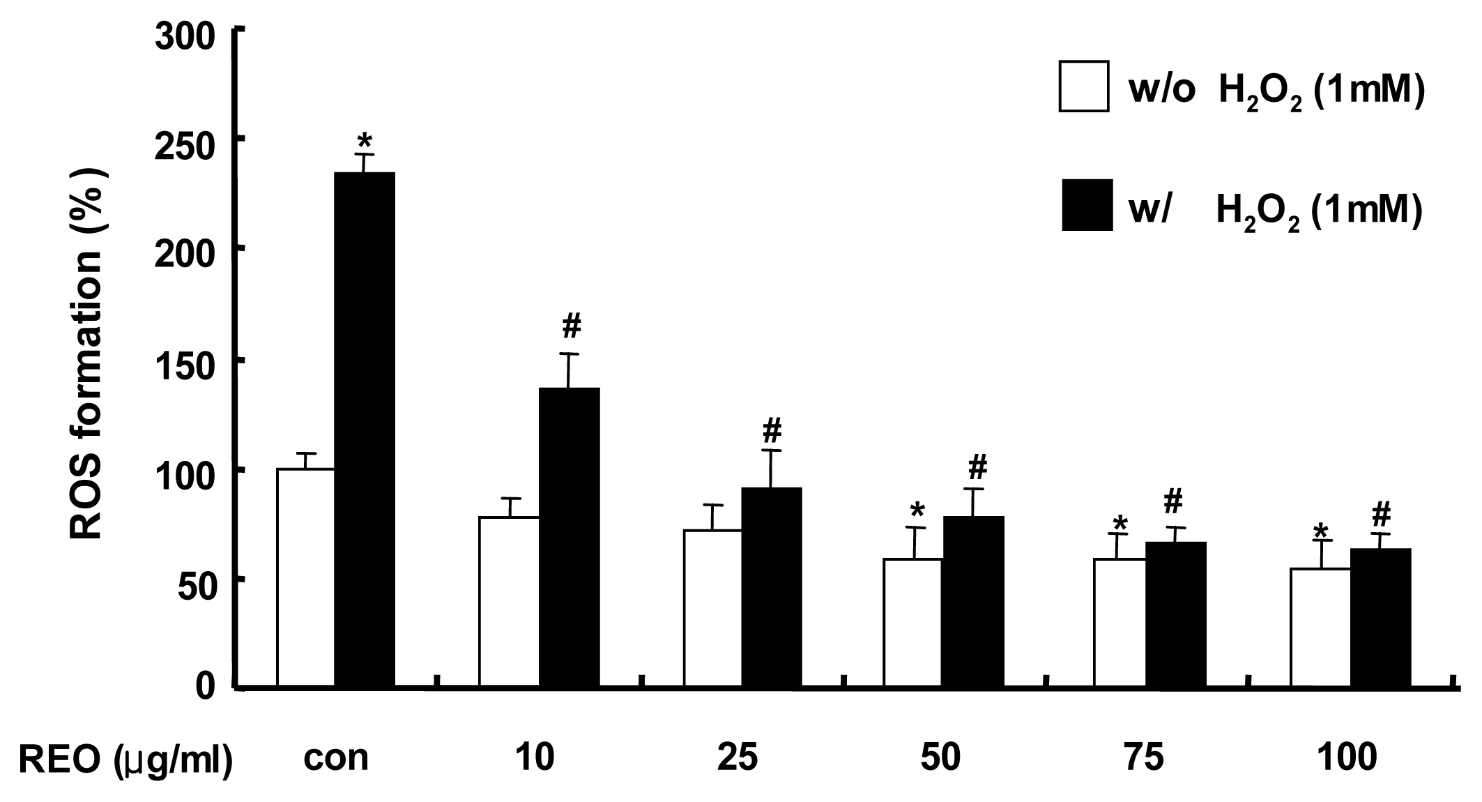
2.1. Inhibition of ROS Production in HepG2 Cells</title>
<p>The oxidative damage caused by ROS may generate various diseases in humans such as aging, arthritis, cancer, inflammation, and heart diseases [<xref ref-type="bibr" rid="b23-ijms-13-02314">23</xref>,<xref ref-type="bibr" rid="b24-ijms-13-02314">24</xref>]. In the present study, 2′,7′-dichlorofluorescin diacetate (DCF-DA) staining was applied to examine whether REO was able to inhibit ROS production in H<sub>2</sub>O<sub>2</sub>-treated HepG2 cells. Hydrogen peroxide (H<sub>2</sub>O<sub>2</sub>) is produced endogenously from microsomes and peroxisomes and also by several physiological processes such as inflammatory respiratory burst and oxidative phosphorylation [<xref ref-type="bibr" rid="b6-ijms-13-02314">6</xref>,<xref ref-type="bibr" rid="b25-ijms-13-02314">25</xref>]. H<sub>2</sub>O<sub>2</sub> has often been used as a model to investigate the mechanism of cell injury by oxidative stress [<xref ref-type="bibr" rid="b26-ijms-13-02314">26</xref>,<xref ref-type="bibr" rid="b27-ijms-13-02314">27</xref>]. When HepG2 cells were challenged with 1 mM H<sub>2</sub>O<sub>2</sub>, ROS were generated over 2-fold compared to the unchallenged control and the pretreatment with REO dose-dependently decreased the H<sub>2</sub>O<sub>2</sub>-mediated ROS formation (<xref ref-type="fig" rid="f1-ijms-13-02314">Figure 1</xref>). This dose-dependent antioxidant activity of REO was also observed even in the absence of H<sub>2</sub>O<sub>2</sub>. These results imply that REO may have an ability to directly scavenge ROS and/or free radicals.</p></sec>
<sec>
<title>
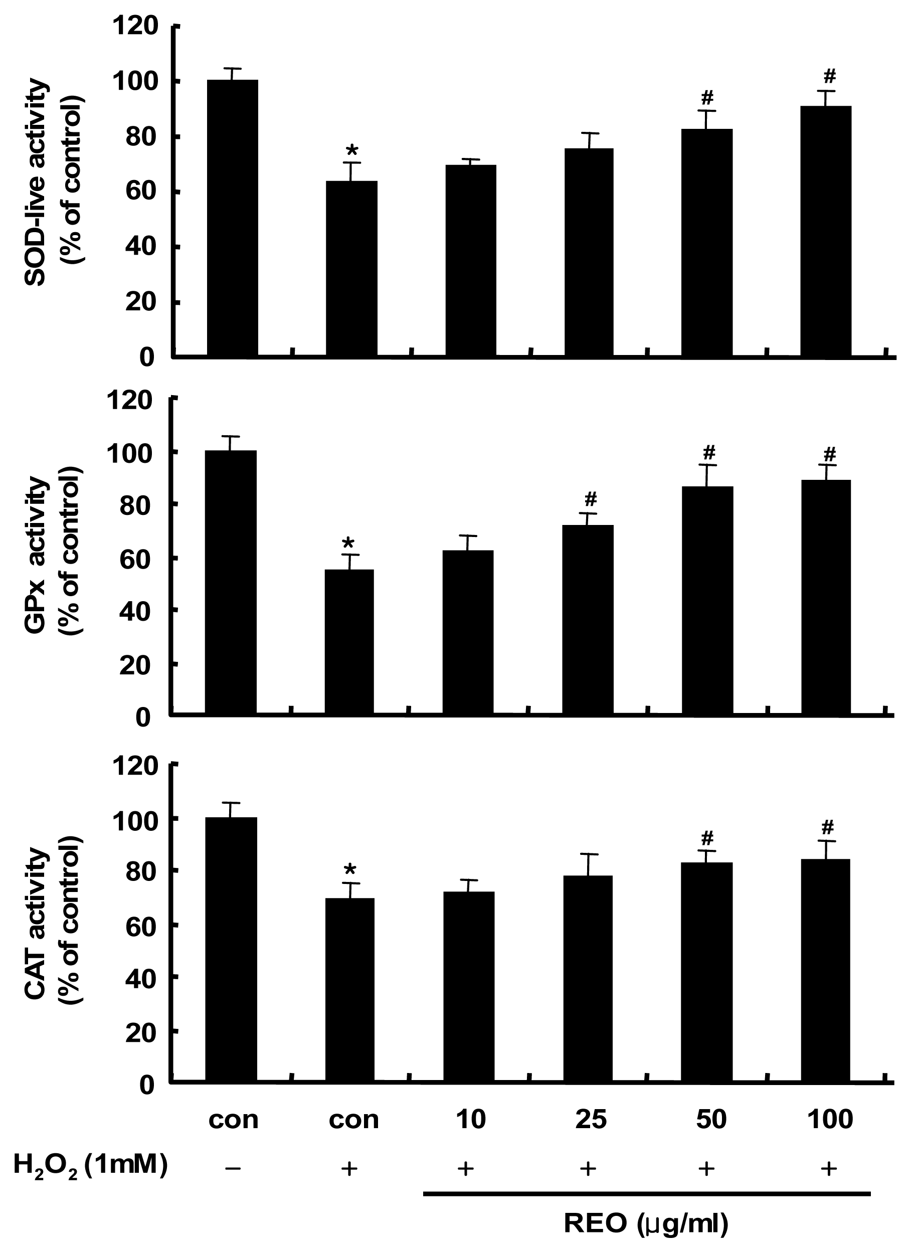
2.2. Induction of Antioxidant Enzyme Activities in HepG2 Cells</title>
<p>In order to investigate whether these antioxidant properties of REO are related to the activity induction of antioxidant enzymes, HepG2 cells were treated with REO and the activities of antioxidant enzymes including SOD, GPx and CAT were measured. These enzymes are regarded as the first line of the antioxidant defense system against ROS generated during oxidative stress [<xref ref-type="bibr" rid="b28-ijms-13-02314">28</xref>]. As shown in <xref ref-type="fig" rid="f2-ijms-13-02314">Figure 2</xref>, the activities of the three antioxidant enzymes were dramatically down-regulated by H<sub>2</sub>O<sub>2</sub> (1 mM) treatment, while treatment with REO restored the enzymes activities in a dose dependent manner. Therefore, the REO appears to have antioxidant properties by stimulating the activity of endogenous antioxidant enzymes as well as by directly scavenging ROS/free radicals. Recently, a water-soluble ginsenoside Rb1 was reported to prevent H<sub>2</sub>O<sub>2</sub>-induced HUVECs senescence through upregulating endogenous antioxidants like SOD and decreasing vascular lipid peroxidation [<xref ref-type="bibr" rid="b29-ijms-13-02314">29</xref>]. This study affirmed for the first time the antioxidant properties of the essential oil of red ginseng byproducts.</p></sec>
<sec>
<title>
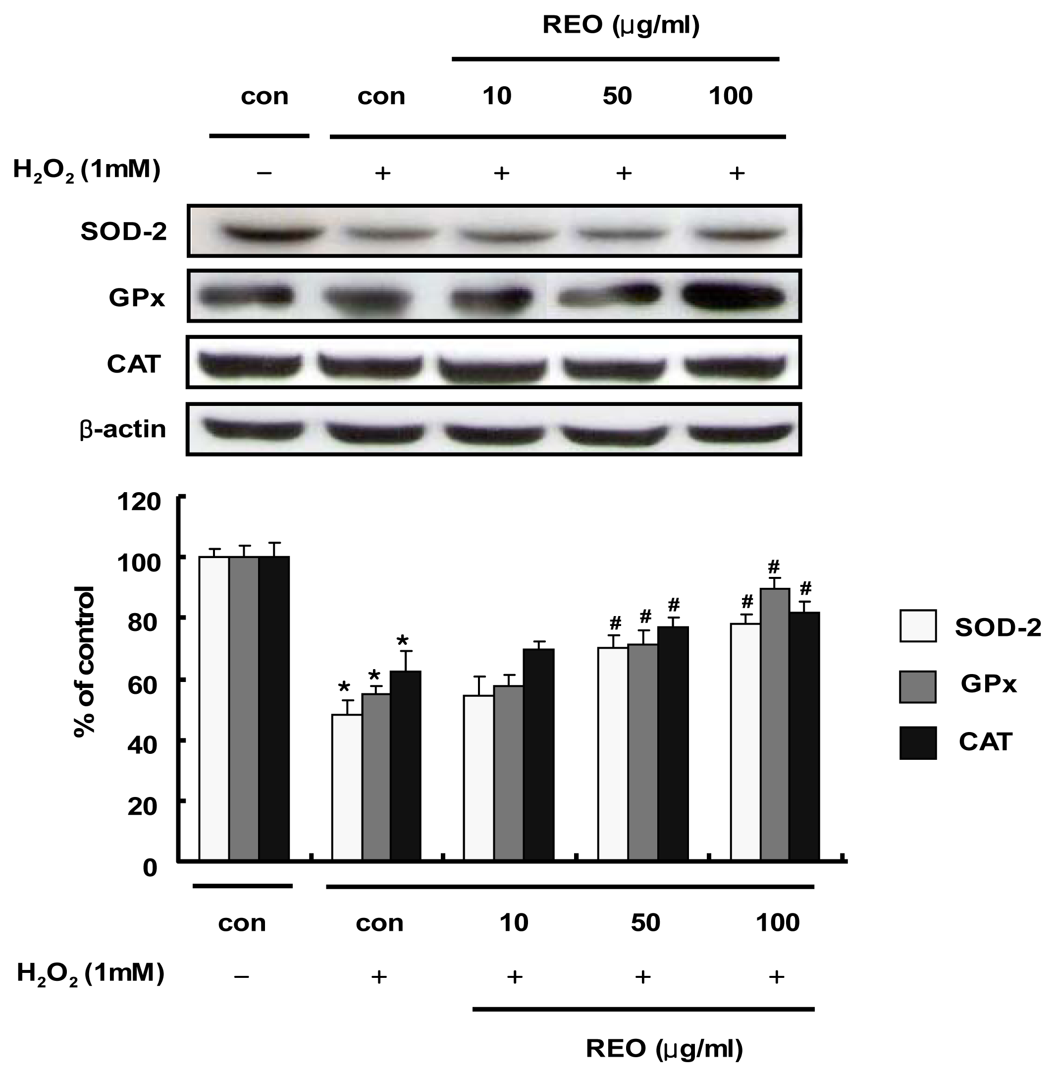
2.3. Induction of Antioxidant Enzyme Expressions in HepG2 Cells</title>
<p>In addition to the enzyme activity stimulation by REO, the effects of REO on the protein expression of these antioxidant enzymes were evaluated in HepG2 cells. When cells were treated with H<sub>2</sub>O<sub>2</sub> (1 mM) alone, the protein expressions of SOD, GPx and CAT were significantly diminished compared to the vehicle-treated control (<xref ref-type="fig" rid="f3-ijms-13-02314">Figure 3</xref>). However, the treatment with REO potently and dose-dependently re-induced the expression of the antioxidant enzymes. Accordingly, the REO seems to induce both activity and expression of the antioxidant enzymes. An aqueous extract of ginseng has been reported to restore the expression of these antioxidant enzymes in H<sub>2</sub>O<sub>2</sub>-injured primary cultures of rat astrocytes [<xref ref-type="bibr" rid="b30-ijms-13-02314">30</xref>]. Ginseng saponins are also protective against alcohol-induced hepatic injury in mice by up-regulating the expression of these antioxidant enzymes [<xref ref-type="bibr" rid="b31-ijms-13-02314">31</xref>]. Our data strongly imply that essential oil from red ginseng byproducts, but neither the water-soluble extract nor saponins, may participate in cellular protection not only directly as an antioxidant molecule (as shown in <xref ref-type="fig" rid="f1-ijms-13-02314">Figure 1</xref>) but also indirectly as a stimulator of antioxidant enzymes.</p></sec>
<sec>
<title>
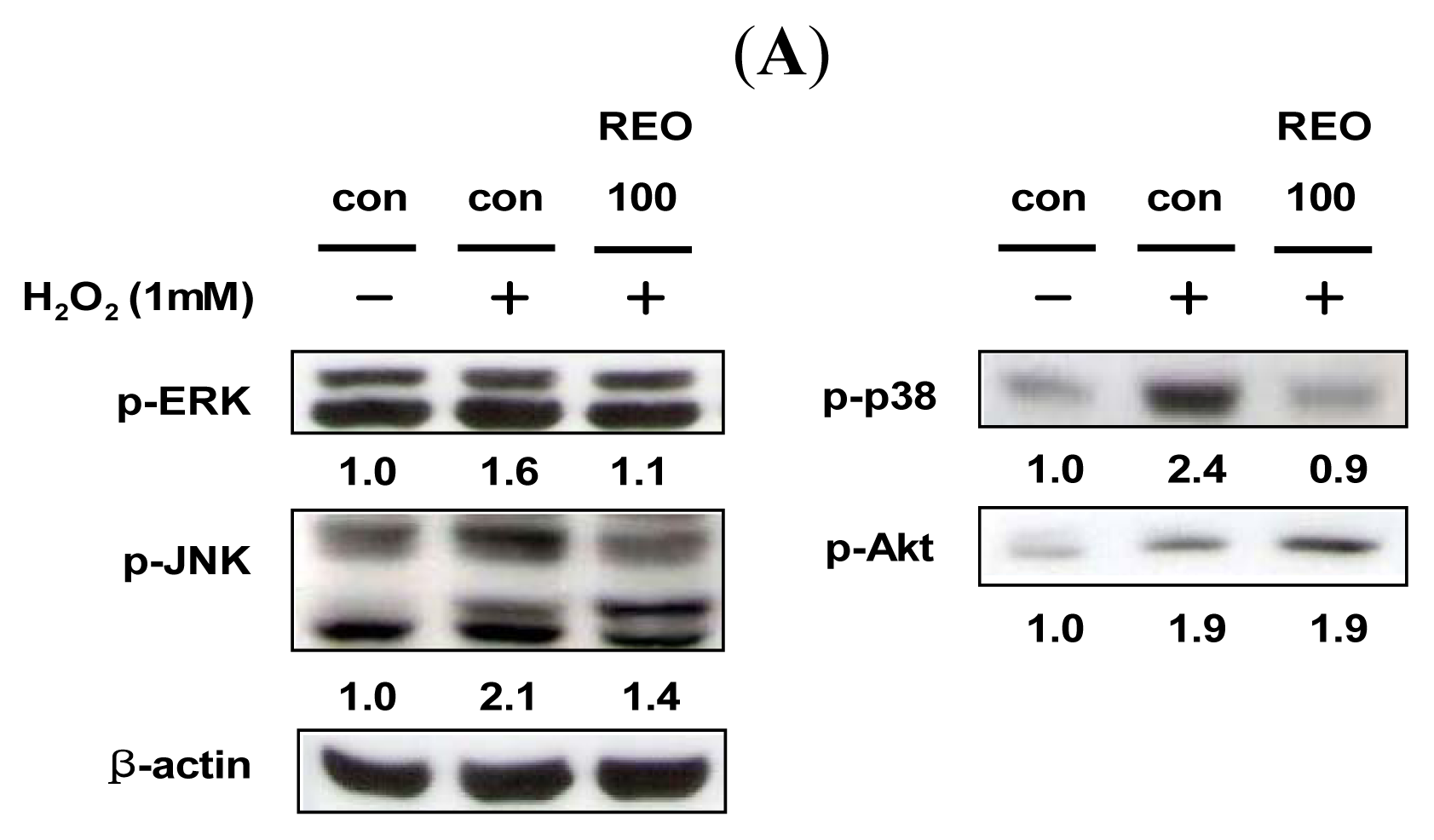
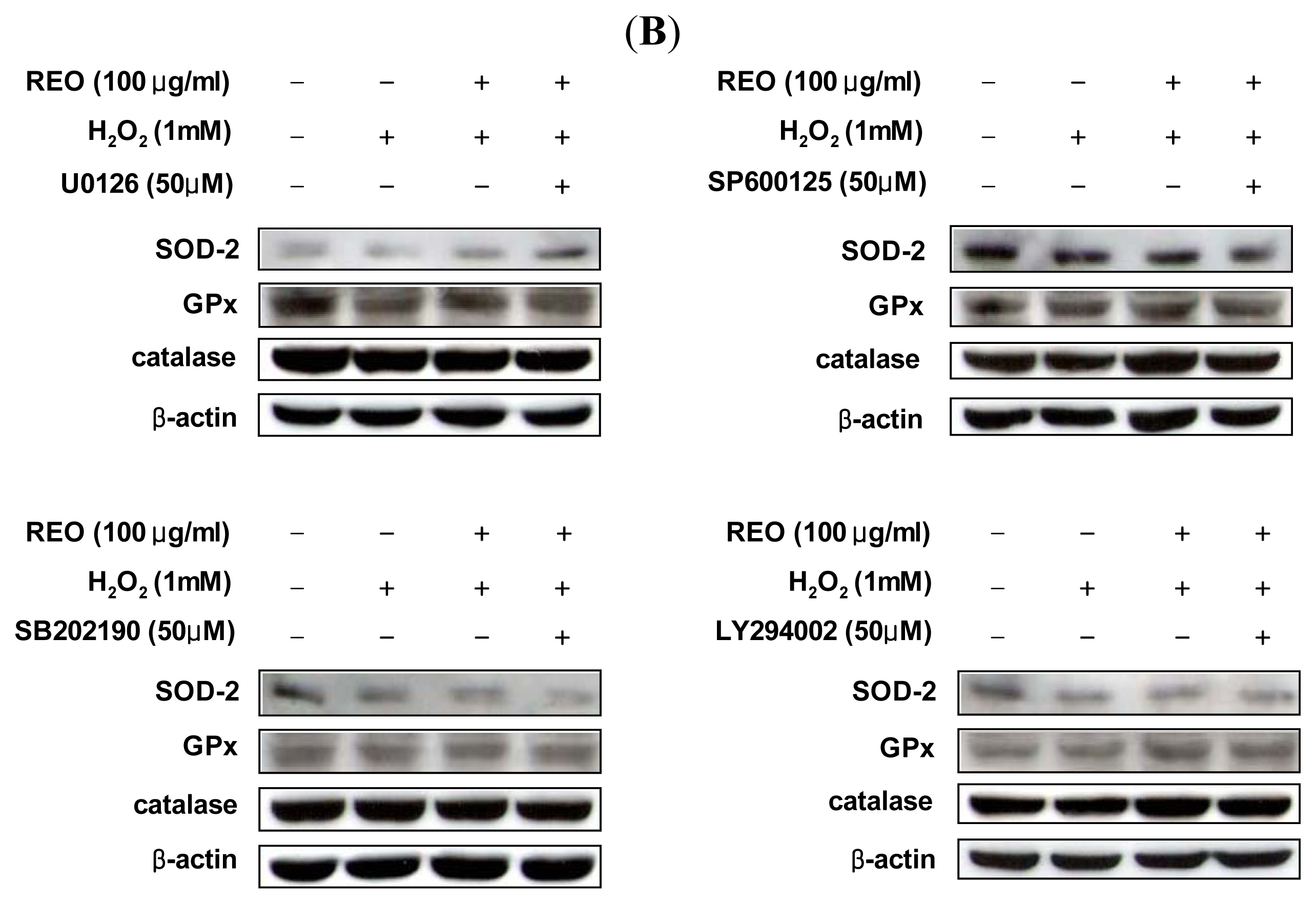
2.4. Effects of REO on Phosphorylations of Upstream Kinases in HepG2 Cells</title>
<p>The MAPK family plays important roles in regulation of cell proliferation and cell death in response to various cellular stresses [<xref ref-type="bibr" rid="b32-ijms-13-02314">32</xref>]. To determine whether REO can modulate the upstream signaling pathways, HepG2 cells were treated with REO and the phosphorylations of ERK, JNK, p38 and Akt were analyzed. The results indicated that H<sub>2</sub>O<sub>2</sub> stimulated phosphorylations of all MAPKs and Akt (<xref ref-type="fig" rid="f4-ijms-13-02314">Figures 4A</xref>). The induced expression of phosphorylated ERK, JNK and p38 by H<sub>2</sub>O<sub>2</sub> was down-regulated by REO, whereas phosphorylation of Akt was up-regulated (<xref ref-type="fig" rid="f4-ijms-13-02314">Figure 4A</xref>). To further elucidate the modulatory effects of REO on MAPK and Akt signaling pathways and their involvement in antioxidant enzyme expression, cells were treated with specific inhibitors of each MAPKs and Akt (U0126, an inhibitor of MEK1/2; SP600125, an inhibitor of JNK; SB202190, an inhibitor of p38; and LY294002, an inhibitor of phosphoinositide 3-kinase (PI3K)/Akt). The stimulated expression of SOD by REO was down-regulated by treatment with the p38-specific inhibitor, and those of GPx and CAT by REO were decreased by treatments with JNK specific inhibitor (<xref ref-type="fig" rid="f4-ijms-13-02314">Figure 4B</xref>). Accordingly, REO seems to induce the expression of SOD through inhibition of p38 phosphorylation, while it induces the expression of GPx and CAT via blockade of JNK phosphorylation. Zhang and Wang [<xref ref-type="bibr" rid="b33-ijms-13-02314">33</xref>] have reported that a notoginseng saponin notoginsenoside R1 inhibits TNF-alpha-induced ERK activation in human arterial smooth muscle cells by suppressing NADPH oxidase-mediated ROS generation and directly scavenging ROS. Notoginseng n-butanol extracts have been shown to inhibit the LPS-stimulated activation of JNK and ERK signaling in RAW264.7 cells [<xref ref-type="bibr" rid="b34-ijms-13-02314">34</xref>]. So far, there have been few reports on the effect of ginseng ingredients on the relationship between antioxidant enzymes and MAPKs pathways. Our previous study demonstrated that a flavonoid quercitrin from <italic>Cedrela sinensis</italic> down-regulates H<sub>2</sub>O<sub>2</sub>-induced phosphorylations of Akt and three MAPKs (ERK, JNK, and p38) in HepG2 cells [<xref ref-type="bibr" rid="b7-ijms-13-02314">7</xref>]. In the study, we found that quercitrin-induced expression of GPx was ameliorated by the p38-specific inhibitor and the Akt/PI3K inhibitor, whereas the expression of SOD by quercitrin was diminished only by the p38 inhibitor. Upstream molecules such as MAPKs and Akt are certainly involved in modulation of the expression and activity of the antioxidant enzymes. However, the mode of regulation seems to be different by cell types as well as by compounds treated.</p></sec>
<sec>
<title>
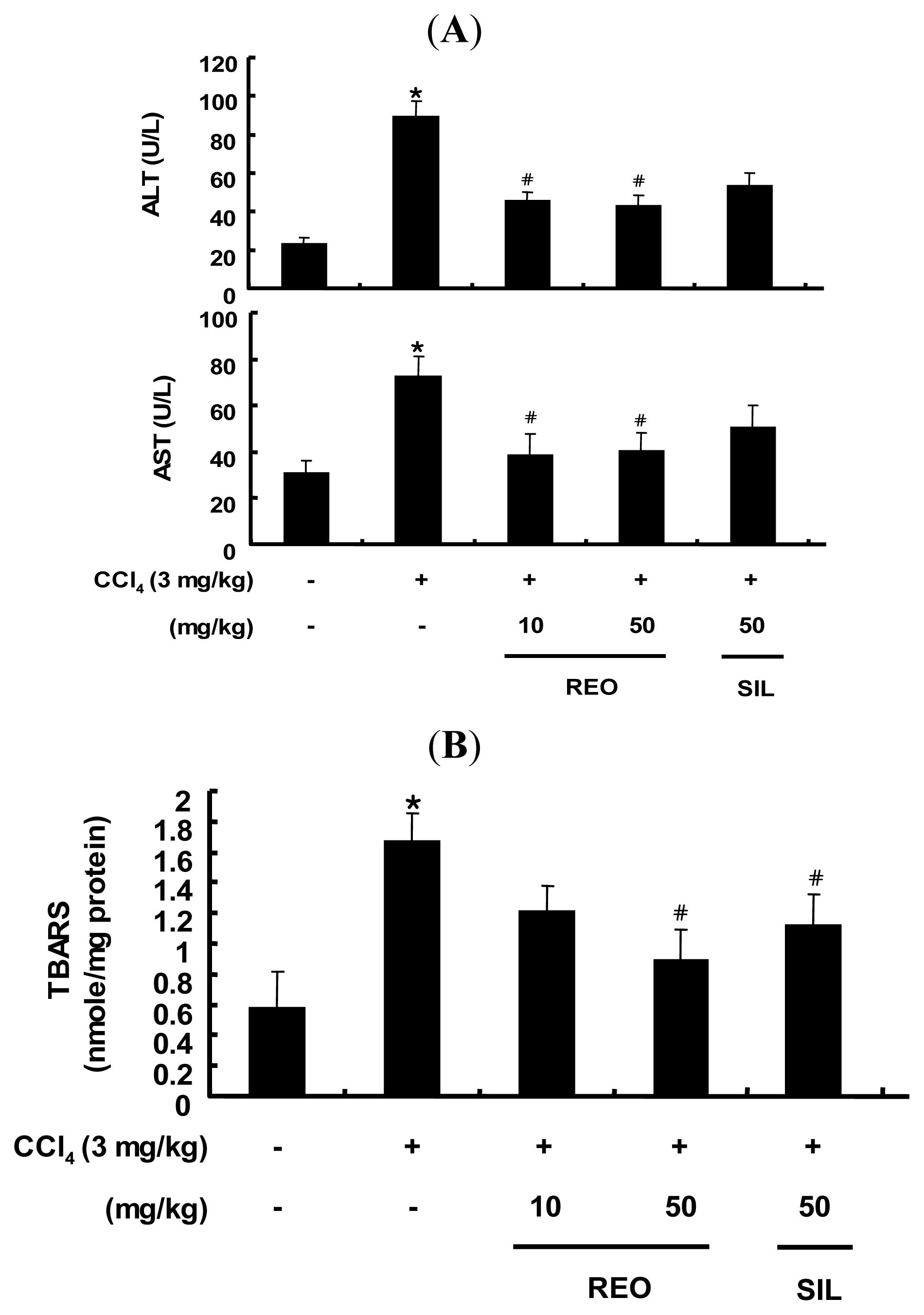
2.5. Effects of REO on Serum ALT and AST Activity in Mice</title>
<p>Based on the antioxidant activity of REO at cellular level, the effect of REO was further examined in an animal model. Male Balb/c mice were treated with either REO (10 and 50 mg/kg b.w.) or silymarin (50 mg/kg b.w.), a known hepatoprotective phytochemical as a positive control, and challenged with CCl<sub>4</sub> (carbon tetrachloride, dissolved in corn oil (20% v/v, 3 mL/kg b.w.)) for three days per week for four weeks. CCl<sub>4</sub> is often used in experimental animal models of severe hepatic damage by generation of oxidative stress and activation of immune cells, which can lead to architectural and functional alteration [<xref ref-type="bibr" rid="b35-ijms-13-02314">35</xref>].</p>
<p>As shown in <xref ref-type="fig" rid="f5-ijms-13-02314">Figure 5A</xref>, the administration of CCl<sub>4</sub> for 4 weeks dramatically elevated the serum levels of routine liver markers of aspartate transaminase (AST) and alanine transaminase (ALT) compared to the vehicle-treated normal control group, indicating significant hepatotoxicity of CCl<sub>4</sub> treatment. In contrast, administration of REO at both 10 and 50 mg/kg b.w. significantly restored ALT and AST levels in CCl<sub>4</sub>-treated mice. Silymarin at 50 mg/kg b.w. also decreased the levels of ALT and AST but not as effectively as REO in attenuation of hepatotoxicity caused by CCl<sub>4</sub>. There have been several animal studies on the hepatoprotective effects of ginseng extracts and saponins against exposure to toxicants such as CCl<sub>4</sub> [<xref ref-type="bibr" rid="b36-ijms-13-02314">36</xref>], benzo[alpha]pyrene [<xref ref-type="bibr" rid="b37-ijms-13-02314">37</xref>], ethanol [<xref ref-type="bibr" rid="b38-ijms-13-02314">38</xref>] and tert-butyl hdroperoxide [<xref ref-type="bibr" rid="b39-ijms-13-02314">39</xref>]. Yet, the current study affirmed for the first time the hepatoprotective activity of the essential oil of red ginseng byproducts.</p></sec>
<sec>
<title>2.6. Effect of REO on Hepatic TBARS Content in Mice</title>
<p>Lipid peroxidation is considered to be one of the principal results of CCl<sub>4</sub>-induced liver injury [<xref ref-type="bibr" rid="b40-ijms-13-02314">40</xref>]. As a biomarker for lipid peroxidation, hepatic lipid peroxidation was determined by measuring the TBARS concentration in liver tissues in CCl<sub>4</sub>-treated mice. The hepatic TBARS concentrations were increased by CCl<sub>4</sub> treatment, but these increases of lipid peroxidation were dose-dependently inhibited in both REO groups and silymarin group and the inhibition by REO was more potent than that by silymarin at the same dose of 50 mg/kg b.w. (<xref ref-type="fig" rid="f5-ijms-13-02314">Figure 5B</xref>). These data suggest that REO may exert its antioxidant effects through the inhibition of hepatic lipid peroxidation.</p></sec>
<sec>
<title>2.7. Effects of REO on the Activity and Expression of Hepatic Antioxidant Enzymes in Mice</title>
<p>The administration of CCl<sub>4</sub> is known to increase the production of ROS/free radicals, resulting in a significant reduction of the endogenous antioxidant enzymes activity [<xref ref-type="bibr" rid="b41-ijms-13-02314">41</xref>]. The activities of the three hepatic antioxidant enzymes including SOD, GPx and CAT were evaluated in mice treated with CCl<sub>4</sub> alone (<xref ref-type="fig" rid="f6-ijms-13-02314">Figure 6</xref>). However, administration of REO at both doses of 10 and 50 mg/kg b.w. resulted in significant increase in the SOD activity and similar results were observed in the silymarin-treated group (50 mg/kg b.w.). In GPx activity, only REO administration at 50 mg/kg b.w. resulted in a significant recovery. CAT activity increased significantly in both REO and silymarin groups at 50 mg/kg b.w. In addition to the activity of the hepatic antioxidant enzymes, the protein expression levels after exposure of mice to REO and CCl<sub>4</sub> were evaluated by Western blotting analysis of liver tissue. Injection with CCl<sub>4</sub> resulted in significant down-regulations of the protein expression levels of SOD, GPx and CAT compared with the normal mice control (<xref ref-type="fig" rid="f7-ijms-13-02314">Figure 7</xref>). However, administration of REO at 50 mg/kg b.w. significantly improved protein expressions of all three antioxidant enzymes in the liver of mice treated with CCl<sub>4</sub>. Therefore, REO seems to enhance both the activity and expression of hepatic antioxidant enzymes <italic>in vivo</italic> as well as <italic>in vitro</italic> cell lines. Earlier studies have reported that administration of ginsenosides increases the activities SOD, GPx and CAT in mice [<xref ref-type="bibr" rid="b42-ijms-13-02314">42</xref>] Our results suggest that REO may attenuate oxidative stress by increasing both the activity and expression of the antioxidant enzymes <italic>in vitro</italic> as well as <italic>in vivo</italic>.</p></sec>
<sec>
<title>
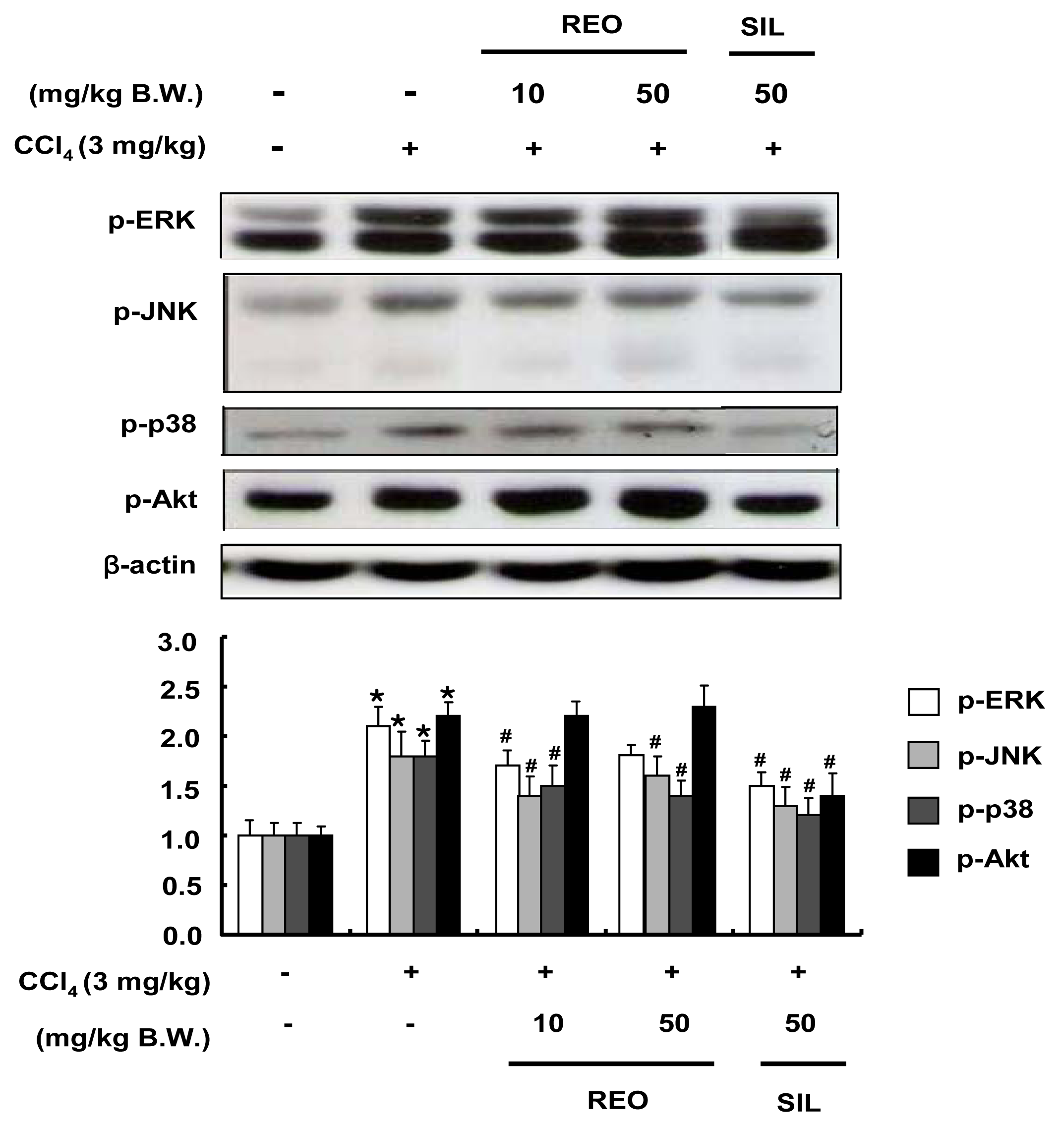
2.8. Effects of REO on Phosphorylations of Upstream Kinases in Mice</title>
<p>During CCl<sub>4</sub> challenge, oxidative stress activates MAP kinase, leading to phosphorylation of JNK and p38 [<xref ref-type="bibr" rid="b43-ijms-13-02314">43</xref>]. On the other hand, ERK 1/2 is involved in survival signals by regulating cell proliferation after partial hepatectomy or CCl<sub>4</sub> intoxication [<xref ref-type="bibr" rid="b44-ijms-13-02314">44</xref>]. The effects of REO on the phosphorylation of hepatic upstream kinases such as MAPKs and Akt were analyzed to better understand the underlying mechanisms association between REO and the hepatic defense system. As shown in <xref ref-type="fig" rid="f8-ijms-13-02314">Figure 8</xref>, the phosphorylation levels of all three MAPKs and Akt were up-regulated (1.8–2.2 fold) by CCl<sub>4</sub> treatment. Administration of REO resulted in inhibition of CCl<sub>4</sub>-mediated phosphorylations of hepatic MAPKs while it did not affect the phosphorylation of Akt. These results correspond with those observed in the cell line model. This study affirmed for the first time the involvement of upstream signaling pathways in hepatoprotective activity of the essential oil of red ginseng byproducts.</p></sec></sec>
<sec>
<title>3. Experimental Section</title>
<sec>
<title>3.1. Chemicals and Reagents</title>
<p>H<sub>2</sub>O<sub>2</sub>, nitro blue tetrazolium salt, xanthine, copper chloride, glutathione, xanthine oxidase from bovine milk (0.1–0.4 U/mg of protein), polyethylene glycol oil, silymarin, carbon tetrachloride and glutathione reductase from baker’s yeast (<italic>Saccharimyces cerevisiae</italic>) (100–300 U/mg of protein) were purchased from Sigma Chemical Co. (St. Louis, MO, USA). 2′,7′-dichlorodihydrofluorescein (DCF-DA) was purchased from Molecular Probe Inc. (Eugene, OR, USA). F-12 medium, fetal bovine serum and penicillin/streptomycin were obtained from Hyclone (Logan, UT, USA). Antibodies against SOD, CAT, GPx, phosphorylated ERK, phosphorylated JNK, phosphorylated p38, and phosphorylated Akt were purchased from Cell Signaling Technology Inc. (Beverly, MA, USA). Anti β-actin and horseradish peroxidase-conjugated anti-goat and anti-rabbit immunoglobulin IgG were purchased from Santa Cruz Biotechnology (Santa Cruz, CA, USA). Specific inhibitors against upstream targets, including U0126, SP600125, SB202190 and LY294002 were obtained from Cell Signaling Technology. All other reagents were of the highest quality generally available.</p></sec>
<sec>
<title>3.2. Preparation of REO</title>
<p>The supercritical CO<sub>2</sub> extraction was performed using a pilot-scale automated equipment (Ilshin Autoclave Co., LTD, Daejeon, Korea). One hundred kilograms of red ginseng byproduct powder were placed into a high-pressure extraction tank and the supercritical CO<sub>2</sub> extraction was conducted with a pressure of 450 bar at 65 °C. The CO<sub>2</sub> was liquefied by a cooler and pressurized to operating pressure using a high pressure pump. The pressurized carbon dioxide was then heated to operating temperature and pumped into the extraction tank (cell) during the supercritical fluid extraction process. The essential oil and other lipophilic substances in the separator were collected via supercritical CO<sub>2</sub> through a needle valve with heater for 3–4 h extraction times.</p></sec>
<sec>
<title>3.3. Cell Culture</title>
<p>HepG2 human hepatoma cells were obtained from American Type Culture Collections (Rockville, MD, USA). Cells were maintained in F-12 medium supplemented with 10% fetal bovine serum, 100 units/mL penicillin, 100 μg/mL streptomycin, 1% essential amino acids, 1% glutamax and 0.1% insulin, in a humidified atmosphere of 95%, 5% CO<sub>2</sub> at 37 °C.</p></sec>
<sec>
<title>3.4. Animals</title>
<p>Male Balb/c mice, 6 weeks old (weighing 18–20 g), were purchased from Hyochang Science (Daegu, Korea) and were given a standard laboratory diet and distilled water <italic>ad libitum</italic> under a 12/12 h light/dark cycle in a temperature controlled room (22 ± 2 °C). The protocol for all animal experiments was approved by the Animal Care and Used Committee of Inje University. The animals were acclimatized for seven days before experiments were performed. The mice were divided into five groups (<italic>n</italic> = 8 per group) including: the normal control group and received polyethylene glycol oil (PEG) as vehicle for 5 weeks; the CCl<sub>4</sub> group as a negative control received PEG for 1 week and then injected i.p. with CCl<sub>4</sub> dissolved in corn oil (20% v/v, 3 mL/kg) three times per week for 4 weeks. REO groups were administered with 10 or 50 mg/kg of REO, <italic>p.o.</italic> three times per week and then treated with REO and CCl<sub>4</sub> for another 4 weeks. The Silymarin group served as a positive control and was administered with 50 mg/kg of silymarin, <italic>p.o.</italic> three times per week and then treated with REO and CCl<sub>4</sub> for another 4 weeks.</p></sec>
<sec>
<title>3.5. Assessment of ROS Production in Cells</title>
<p>The effect of REO on ROS production was evaluated in HepG2 cells using a cell permeable probe, DCF-DA [<xref ref-type="bibr" rid="b45-ijms-13-02314">45</xref>]. The cells were seeded onto 96 well plates, grown until confluency and pre-incubated with DCF-DA for 1 h at 37 °C in darkness. After washing out the excess probe, the cells were treated with vehicle (0.1% DMSO) or REO in the presence or absence of 1 mM H<sub>2</sub>O<sub>2</sub> for 12 h and then washed twice with 1× ice-cold PBS. The fluorescence was measured at 485/20 nm excitation and 528/20 nm emission in a fluorescence multi-detection reader (Synergy HT™ Multi-detecction microplate reader; BioTek Instruments Inc, VT, USA).</p></sec>
<sec>
<title>3.6. Assessment of CAT and GPx Activities in HepG2 Cells and Liver Tissues</title>
<p>HepG2 cells were seeded in a six-well plate until 80–90% confluency. After starvation for 12 h, the cells were treated with samples for 24 h and harvested in an ice-cold cell lysis buffer (pH 7.4). The cell debris was removed by centrifugation at 13,000 <italic>g</italic> for 10 min and the supernatant was used for further experiments. For the mouse model, liver tissue was homogenized in cell lysis buffer. The protein concentration for both cells and tissues was determined using the BCA protein assay (Pierce Biotechnology, Inc., Rockford, IL, USA) according to the manufacturer’s instructions. Each supernatant containing an equal amount of protein (10 μg) was used for the enzyme activity assay.</p>
<p>CAT activity was measured according to the method of Carrillo <italic>et al</italic>. [<xref ref-type="bibr" rid="b46-ijms-13-02314">46</xref>]. One unit of CAT was defined as the amount of enzyme required to decompose 1.0 μM of H<sub>2</sub>O<sub>2</sub> in 1 min. The reaction was initiated by the addition of freshly prepared 20 mM H<sub>2</sub>O<sub>2</sub> (1 mL). The rate of decomposition of H<sub>2</sub>O<sub>2</sub> was measured at 240 nm for 1 min. The enzyme activity was expressed as U/mg protein.</p>
<p>GPx activity was evaluated by the method of Bogdanska <italic>et al</italic>. [<xref ref-type="bibr" rid="b47-ijms-13-02314">47</xref>]. The reaction mixture consisted of 0.1 M phosphate buffer (pH 7.0), 1 mM EDTA, 10 mM glutathione (GSH), 1 mM NaN<sub>3</sub>, 1 unit of glutathione reductase, 1.5 mM NADPH and cell lysates. The activity was calculated using the molar extinction coefficient for NADPH of 6.22 μmol/cm at 340 nm.</p></sec>
<sec>
<title>3.7. Assessment of SOD-Like Activity in HepG2 Cells and Liver Tissues</title>
<p>SOD-like activity was measured by the inhibition of nitro blue tetrazolium reduction due to the superoxide anion generated by the combination xanthine and xanthine oxidase [<xref ref-type="bibr" rid="b48-ijms-13-02314">48</xref>]. The reaction mixture was buffered with 50 mM sodium carbonate buffer (pH 10.2) containing 3 mM xanthine, 0.75 mM nitro blue tetrazolium, 3 mM EDTA and 50 μL cell lysates. After pre-incubation for 30 min at room temperature, the reaction was initiated by adding 50 μL xanthine oxidase (0.1 mg/mL) and stopped by addition of 6 mM copper (II) chloride and centrifuged at 1500 <italic>g</italic> for 15 min. The absorbance of the reaction mixture was measured at 560 nm.</p></sec>
<sec sec-type="methods">
<title>3.8. Western Blotting Analysis</title>
<p>The whole cell extracts for immunoblotting SOD, CAT and GPx were isolated from either H<sub>2</sub>O<sub>2</sub>-stimulated HepG2 cells or liver tissue from the CCl<sub>4</sub>-treated mice. These sample proteins were separated in a 10% polyacrylamide gel at 100 V and transferred onto polyvinylidene difluoride (PVDF) membranes (Millipore, Bedford, MA, USA) using a semi-dry transfer system. The membranes were blocked with 5% non-fat dry milk in 1× PBST buffer (0.1% Tween 20 in PBS) overnight at 4 °C and incubated with rabbit polyclonal antibody in PBST solution containing 5% non-fat dry milk. After washing 3 times with PBST, the membranes were incubated for 1 h at room temperature with anti-rabbit and anti-goat antibodies with horseradish peroxidase for 1 h at room temperature, and washed with PBST 3 times. Final detection was performed with enhanced chemiluminescence (ECL™) Western blotting reagents (Santa Cruz).</p></sec>
<sec>
<title>3.9. Assessment of Serum Marker Enzymes</title>
<p>The enzymatic activities of serum ALT and AST were used as biochemical markers for hepatotoxicity. Blood samples from mice were centrifuged at 3000 <italic>g</italic> at 4 °C for 10 min and the ALT and AST activities in serum supernatants were determined using commercially available kits (Youngdong Pharm, Seoul, Korea).</p></sec>
<sec>
<title>3.10. Assessment of Thiobarbituric Acid Reactive Substance (TBARS) in Tissue</title>
<p>Liver tissues were homogenized in an ice-cold lysis buffer and a portion of the homogenate was measured immediately for TBARS value using the method of Ohkawa <italic>et al</italic>. [<xref ref-type="bibr" rid="b49-ijms-13-02314">49</xref>]. Malondialdehyde, an end product of lipid peroxidation, reacted with thiobarbituric acid. The absorbance of the resulting chromophore was measured at 535 nm.</p></sec>
<sec sec-type="methods">
<title>3.11. Statistical Analysis</title>
<p>The data were expressed as mean ± SD values. The values were compared with the control using analysis of variance followed by unpaired Student’s t tests. A <italic>P</italic> value of &lt;0.05 was considered significant.</p></sec></sec>
<sec sec-type="conclusions">
<title>4. Conclusions</title>
<p>Red ginseng has been extensively studied during recent decades and most of the studies have focused on the water soluble parts with ginsenoside saponins due to its normal consumption pattern. Results of this study clearly suggest that red ginseng oil has the ability to protect cells/tissues against oxidative damage by directly scavenging ROS, by inhibiting lipid peroxidation, as well as by inducing both activity and expression of cellular antioxidant enzymes activity and expression through inhibiting MAPK signaling pathways. Therefore, REO might be considered as a useful source of cellular defense agents at least in liver cells and/or tissues. However, studies on the active ingredients in REO should be followed up and will be reported later.</p></sec></body>
<back>
<ack>
<title>Acknowledgments</title>
<p>We thank Korea Ginseng Cooperation for the preparation of red ginseng essential oil. This work was supported by the National Research Foundation of Korea (NRF) grant funded by the Korea government (MEST) (No. 20100014447)</p></ack>
<ref-list>
<title>References</title>
<ref id="b1-ijms-13-02314"><label>1</label><citation citation-type="journal"><person-group person-group-type="author"><name><surname>Aruoma</surname><given-names>O.I.</given-names></name><name><surname>Grootveld</surname><given-names>M.</given-names></name><name><surname>Bahorun</surname><given-names>T.</given-names></name></person-group><article-title>Free radicals in biology and medicine: from inflammation to biotechnology</article-title><source>Biofactors</source><year>2006</year><volume>27</volume><fpage>1</fpage><lpage>3</lpage><pub-id pub-id-type="doi">10.1002/biof.5520270101</pub-id><pub-id pub-id-type="pmid">17012759</pub-id></citation></ref>
<ref id="b2-ijms-13-02314"><label>2</label><citation citation-type="journal"><person-group person-group-type="author"><name><surname>Heyman</surname><given-names>S.N.</given-names></name><name><surname>Rosen</surname><given-names>S.</given-names></name><name><surname>Rosenberger</surname><given-names>C.</given-names></name></person-group><article-title>A role for oxidative stress</article-title><source>Contrib. Nephrol</source><year>2011</year><volume>174</volume><fpage>138</fpage><lpage>148</lpage><pub-id pub-id-type="pmid">21921618</pub-id></citation></ref>
<ref id="b3-ijms-13-02314"><label>3</label><citation citation-type="journal"><person-group person-group-type="author"><name><surname>Whaley-Connell</surname><given-names>A.</given-names></name><name><surname>McCullough</surname><given-names>P.A.</given-names></name><name><surname>Sowers</surname><given-names>J.R.</given-names></name></person-group><article-title>The role of oxidative stress in the metabolic syndrome</article-title><source>Rev. Cardiovasc. Med</source><year>2011</year><volume>12</volume><fpage>21</fpage><lpage>29</lpage><pub-id pub-id-type="pmid">21546885</pub-id></citation></ref>
<ref id="b4-ijms-13-02314"><label>4</label><citation citation-type="journal"><person-group person-group-type="author"><name><surname>Datta</surname><given-names>K.</given-names></name><name><surname>Sinha</surname><given-names>S.</given-names></name><name><surname>Chattopadhyay</surname><given-names>P.</given-names></name></person-group><article-title>Reactive oxygen species in health and disease</article-title><source>Natl. Med. J. India</source><year>2000</year><volume>13</volume><fpage>304</fpage><lpage>310</lpage><pub-id pub-id-type="pmid">11209486</pub-id></citation></ref>
<ref id="b5-ijms-13-02314"><label>5</label><citation citation-type="journal"><person-group person-group-type="author"><name><surname>Winterbourn</surname><given-names>C.C.</given-names></name></person-group><article-title>Nutritional antioxidants: their role in disease prevention</article-title><source>N. Z. Med. J</source><year>1995</year><volume>108</volume><fpage>447</fpage><lpage>449</lpage><pub-id pub-id-type="pmid">8538960</pub-id></citation></ref>
<ref id="b6-ijms-13-02314"><label>6</label><citation citation-type="journal"><person-group person-group-type="author"><name><surname>Valko</surname><given-names>M.</given-names></name><name><surname>Rhodes</surname><given-names>C.J.</given-names></name><name><surname>Moncol</surname><given-names>J.</given-names></name><name><surname>Izakovic</surname><given-names>M.</given-names></name><name><surname>Mazur</surname><given-names>M.</given-names></name></person-group><article-title>Free radicals, metals and antioxidants in oxidative stress-induced cancer</article-title><source>Chem. Biol. Interact</source><year>2006</year><volume>160</volume><fpage>1</fpage><lpage>40</lpage><pub-id pub-id-type="doi">10.1016/j.cbi.2005.12.009</pub-id><pub-id pub-id-type="pmid">16430879</pub-id></citation></ref>
<ref id="b7-ijms-13-02314"><label>7</label><citation citation-type="journal"><person-group person-group-type="author"><name><surname>Bak</surname><given-names>M.J.</given-names></name><name><surname>Jeong</surname><given-names>J.H.</given-names></name><name><surname>Kang</surname><given-names>H.S.</given-names></name><name><surname>Jin</surname><given-names>K.S.</given-names></name><name><surname>Jun</surname><given-names>M.</given-names></name><name><surname>Jeong</surname><given-names>W.S.</given-names></name></person-group><article-title>Stimulation of Activity and Expression of Antioxidant Enzymes by Solvent Fractions and Isolated Compound from <italic>Cedrela sinensis</italic> Leaves in HepG2 Cells</article-title><source>J. Med. Food</source><year>2011</year><volume>14</volume><fpage>405</fpage><lpage>412</lpage><pub-id pub-id-type="doi">10.1089/jmf.2010.1213</pub-id><pub-id pub-id-type="pmid">21303261</pub-id></citation></ref>
<ref id="b8-ijms-13-02314"><label>8</label><citation citation-type="journal"><person-group person-group-type="author"><name><surname>El-Mowafy</surname><given-names>A.M.</given-names></name><name><surname>Salem</surname><given-names>H.A.</given-names></name><name><surname>Al-Gayyar</surname><given-names>M.M.</given-names></name><name><surname>El-Mesery</surname><given-names>M.E.</given-names></name><name><surname>El-Azab</surname><given-names>M.F.</given-names></name></person-group><article-title>Evaluation of renal protective effects of the green-tea (EGCG) and red grape resveratrol: Role of oxidative stress and inflammatory cytokines</article-title><source>Nat. Prod. Res</source><year>2011</year><volume>25</volume><fpage>850</fpage><lpage>856</lpage><pub-id pub-id-type="doi">10.1080/14786419.2010.533669</pub-id><pub-id pub-id-type="pmid">21462079</pub-id></citation></ref>
<ref id="b9-ijms-13-02314"><label>9</label><citation citation-type="journal"><person-group person-group-type="author"><name><surname>Yurt</surname><given-names>B.</given-names></name><name><surname>Celik</surname><given-names>I.</given-names></name></person-group><article-title>Hepatoprotective effect and antioxidant role of sun, sulphited-dried apricot (<italic>Prunus armeniaca</italic> L.) and its kernel against ethanol-induced oxidative stress in rats</article-title><source>Food Chem. Toxicol</source><year>2011</year><volume>49</volume><fpage>508</fpage><lpage>513</lpage><pub-id pub-id-type="doi">10.1016/j.fct.2010.11.035</pub-id><pub-id pub-id-type="pmid">21115094</pub-id></citation></ref>
<ref id="b10-ijms-13-02314"><label>10</label><citation citation-type="journal"><person-group person-group-type="author"><name><surname>Aruoma</surname><given-names>O.I.</given-names></name><name><surname>Sun</surname><given-names>B.</given-names></name><name><surname>Fujii</surname><given-names>H.</given-names></name><name><surname>Neergheen</surname><given-names>V.S.</given-names></name><name><surname>Bahorun</surname><given-names>T.</given-names></name><name><surname>Kang</surname><given-names>K.S.</given-names></name><name><surname>Sung</surname><given-names>M.K.</given-names></name></person-group><article-title>Low molecular proanthocyanidin dietary biofactor Oligonol: Its modulation of oxidative stress, bioefficacy, neuroprotection, food application and chemoprevention potentials</article-title><source>Biofactors</source><year>2006</year><volume>27</volume><fpage>245</fpage><lpage>265</lpage><pub-id pub-id-type="doi">10.1002/biof.5520270121</pub-id><pub-id pub-id-type="pmid">17012779</pub-id></citation></ref>
<ref id="b11-ijms-13-02314"><label>11</label><citation citation-type="journal"><person-group person-group-type="author"><name><surname>Jang</surname><given-names>D.J.</given-names></name><name><surname>Lee</surname><given-names>M.S.</given-names></name><name><surname>Shin</surname><given-names>B.C.</given-names></name><name><surname>Lee</surname><given-names>Y.C.</given-names></name><name><surname>Ernst</surname><given-names>E.</given-names></name></person-group><article-title>Red ginseng for treating erectile dysfunction: a systematic review</article-title><source>Br. J. Clin. Pharmacol</source><year>2008</year><volume>66</volume><fpage>444</fpage><lpage>450</lpage><pub-id pub-id-type="doi">10.1111/j.1365-2125.2008.03236.x</pub-id><pub-id pub-id-type="pmid">18754850</pub-id></citation></ref>
<ref id="b12-ijms-13-02314"><label>12</label><citation citation-type="journal"><person-group person-group-type="author"><name><surname>Hwang</surname><given-names>J.T.</given-names></name><name><surname>Lee</surname><given-names>M.S.</given-names></name><name><surname>Kim</surname><given-names>H.J.</given-names></name><name><surname>Sung</surname><given-names>M.J.</given-names></name><name><surname>Kim</surname><given-names>H.Y.</given-names></name><name><surname>Kim</surname><given-names>M.S.</given-names></name><name><surname>Kwon</surname><given-names>D.Y.</given-names></name></person-group><article-title>Antiobesity effect of ginsenoside Rg3 involves the AMPK and PPAR-gamma signal pathways</article-title><source>Phytother. Res</source><year>2009</year><volume>23</volume><fpage>262</fpage><lpage>266</lpage><pub-id pub-id-type="doi">10.1002/ptr.2606</pub-id><pub-id pub-id-type="pmid">18844326</pub-id></citation></ref>
<ref id="b13-ijms-13-02314"><label>13</label><citation citation-type="journal"><person-group person-group-type="author"><name><surname>Ryu</surname><given-names>J.K.</given-names></name><name><surname>Lee</surname><given-names>T.</given-names></name><name><surname>Kim</surname><given-names>D.J.</given-names></name><name><surname>Park</surname><given-names>I.S.</given-names></name><name><surname>Yoon</surname><given-names>S.M.</given-names></name><name><surname>Lee</surname><given-names>H.S.</given-names></name><name><surname>Song</surname><given-names>S.U.</given-names></name><name><surname>Suh</surname><given-names>J.K.</given-names></name></person-group><article-title>Free radical-scavenging activity of Korean red ginseng for erectile dysfunction in non-insulin-dependent diabetes mellitus rats</article-title><source>Urology</source><year>2005</year><volume>65</volume><fpage>611</fpage><lpage>615</lpage><pub-id pub-id-type="doi">10.1016/j.urology.2004.10.038</pub-id><pub-id pub-id-type="pmid">15780402</pub-id></citation></ref>
<ref id="b14-ijms-13-02314"><label>14</label><citation citation-type="journal"><person-group person-group-type="author"><name><surname>Kim</surname><given-names>J.</given-names></name></person-group><article-title>Protective effects of Asian dietary items on cancers—soy and ginseng</article-title><source>Asian Pac. J. Cancer Prev</source><year>2008</year><volume>9</volume><fpage>543</fpage><lpage>548</lpage><pub-id pub-id-type="pmid">19256735</pub-id></citation></ref>
<ref id="b15-ijms-13-02314"><label>15</label><citation citation-type="journal"><person-group person-group-type="author"><name><surname>Cho</surname><given-names>S.</given-names></name><name><surname>Won</surname><given-names>C.H.</given-names></name><name><surname>Lee</surname><given-names>D.H.</given-names></name><name><surname>Lee</surname><given-names>M.J.</given-names></name><name><surname>Lee</surname><given-names>S.</given-names></name><name><surname>So</surname><given-names>S.H.</given-names></name><name><surname>Lee</surname><given-names>S.K.</given-names></name><name><surname>Koo</surname><given-names>B.S.</given-names></name><name><surname>Kim</surname><given-names>N.M.</given-names></name><name><surname>Chung</surname><given-names>J.H.</given-names></name></person-group><article-title>Red ginseng root extract mixed with <italic>Torilus fructus</italic> and <italic>Corni fructus</italic> improves facial wrinkles and increases type I procollagen synthesis in human skin: a randomized, double-blind, placebo-controlled study</article-title><source>J. Med. Food</source><year>2009</year><volume>12</volume><fpage>1252</fpage><lpage>1259</lpage><pub-id pub-id-type="doi">10.1089/jmf.2008.1390</pub-id><pub-id pub-id-type="pmid">20041778</pub-id></citation></ref>
<ref id="b16-ijms-13-02314"><label>16</label><citation citation-type="journal"><person-group person-group-type="author"><name><surname>Kim</surname><given-names>J.H.</given-names></name><name><surname>Kang</surname><given-names>S.A.</given-names></name><name><surname>Han</surname><given-names>S.M.</given-names></name><name><surname>Shim</surname><given-names>I.</given-names></name></person-group><article-title>Comparison of the antiobesity effects of the protopanaxadiol- and protopanaxatriol-type saponins of red ginseng</article-title><source>Phytother. Res</source><year>2009</year><volume>23</volume><fpage>78</fpage><lpage>85</lpage><pub-id pub-id-type="doi">10.1002/ptr.2561</pub-id><pub-id pub-id-type="pmid">18709638</pub-id></citation></ref>
<ref id="b17-ijms-13-02314"><label>17</label><citation citation-type="journal"><person-group person-group-type="author"><name><surname>Ortet</surname><given-names>R.</given-names></name><name><surname>Regalado</surname><given-names>E.L.</given-names></name><name><surname>Thomas</surname><given-names>O.P.</given-names></name><name><surname>Pino</surname><given-names>J.A.</given-names></name><name><surname>Fernandez</surname><given-names>M.D.</given-names></name></person-group><article-title>Chemical composition and antioxidant activities of the essential oil from <italic>Tornabenea bischoffii</italic> (Apiaceae)</article-title><source>Nat. Prod. Commun</source><year>2011</year><volume>6</volume><fpage>1179</fpage><lpage>1182</lpage><pub-id pub-id-type="pmid">21922930</pub-id></citation></ref>
<ref id="b18-ijms-13-02314"><label>18</label><citation citation-type="journal"><person-group person-group-type="author"><name><surname>Sharififar</surname><given-names>F.</given-names></name><name><surname>Derakhshanfar</surname><given-names>A.</given-names></name><name><surname>Dehghan-Nudeh</surname><given-names>G.</given-names></name><name><surname>Abbasi</surname><given-names>N.</given-names></name><name><surname>Abbasi</surname><given-names>R.</given-names></name><name><surname>Gharaei</surname><given-names>R.R.</given-names></name><name><surname>Koohpayeh</surname><given-names>A.</given-names></name><name><surname>Daneshpajouh</surname><given-names>M.</given-names></name></person-group><article-title><italic>In vivo</italic> antioxidant activity of <italic>Zataria multiflora Boiss</italic> essential oil</article-title><source>Pak. J. Pharm. Sci</source><year>2011</year><volume>24</volume><fpage>221</fpage><lpage>225</lpage><pub-id pub-id-type="pmid">21454174</pub-id></citation></ref>
<ref id="b19-ijms-13-02314"><label>19</label><citation citation-type="journal"><person-group person-group-type="author"><name><surname>Kim</surname><given-names>S.S.</given-names></name><name><surname>Kim</surname><given-names>J.E.</given-names></name><name><surname>Hyun</surname><given-names>C.G.</given-names></name><name><surname>Lee</surname><given-names>N.H.</given-names></name></person-group><article-title><italic>Neolitsea aciculata</italic> essential oil inhibits drug-resistant skin pathogen growth and Propionibacterium acnes-induced inflammatory effects of human monocyte leukemia</article-title><source>Nat. Prod. Commun</source><year>2011</year><volume>6</volume><fpage>1193</fpage><lpage>1198</lpage><pub-id pub-id-type="pmid">21922933</pub-id></citation></ref>
<ref id="b20-ijms-13-02314"><label>20</label><citation citation-type="journal"><person-group person-group-type="author"><name><surname>Campelo</surname><given-names>L.M.</given-names></name><name><surname>Goncalves</surname><given-names>F.C.</given-names></name><name><surname>Feitosa</surname><given-names>C.M.</given-names></name><name><surname>de Freitas</surname><given-names>R.M.</given-names></name></person-group><article-title>Antioxidant activity of <italic>Citrus limon</italic> essential oil in mouse hippocampus</article-title><source>Pharm. Biol</source><year>2011</year><volume>49</volume><fpage>709</fpage><lpage>715</lpage><pub-id pub-id-type="doi">10.3109/13880209.2010.541924</pub-id><pub-id pub-id-type="pmid">21639684</pub-id></citation></ref>
<ref id="b21-ijms-13-02314"><label>21</label><citation citation-type="journal"><person-group person-group-type="author"><name><surname>Kim</surname><given-names>H.J.</given-names></name><name><surname>Kang</surname><given-names>H.J.</given-names></name><name><surname>Seo</surname><given-names>J.Y.</given-names></name><name><surname>Lee</surname><given-names>C.H.</given-names></name><name><surname>Kim</surname><given-names>Y.S.</given-names></name><name><surname>Kim</surname><given-names>J.S.</given-names></name></person-group><article-title>Antiobesity effect of oil extract of ginseng</article-title><source>J. Med. Food</source><year>2011</year><volume>14</volume><fpage>573</fpage><lpage>583</lpage><pub-id pub-id-type="doi">10.1089/jmf.2010.1313</pub-id><pub-id pub-id-type="pmid">21554134</pub-id></citation></ref>
<ref id="b22-ijms-13-02314"><label>22</label><citation citation-type="journal"><person-group person-group-type="author"><name><surname>Lee</surname><given-names>S.D.</given-names></name><name><surname>Park</surname><given-names>S.K.</given-names></name><name><surname>Lee</surname><given-names>E.S.</given-names></name><name><surname>Kim</surname><given-names>H.M.</given-names></name><name><surname>Lee</surname><given-names>C.W.</given-names></name><name><surname>Lee</surname><given-names>K.</given-names></name><name><surname>Lee</surname><given-names>K.H.</given-names></name><name><surname>Kang</surname><given-names>M.R.</given-names></name><name><surname>Lee</surname><given-names>K.S.</given-names></name><name><surname>Lee</surname><given-names>J.</given-names></name><name><surname>Hwang</surname><given-names>W.I.</given-names></name><name><surname>Kim</surname><given-names>D.C.</given-names></name></person-group><article-title>A lipid-soluble red ginseng extract inhibits the growth of human lung tumor xenografts in nude mice</article-title><source>J. Med. Food</source><year>2010</year><volume>13</volume><fpage>1</fpage><lpage>5</lpage><pub-id pub-id-type="doi">10.1089/jmf.2009.1142</pub-id><pub-id pub-id-type="pmid">20136429</pub-id></citation></ref>
<ref id="b23-ijms-13-02314"><label>23</label><citation citation-type="journal"><person-group person-group-type="author"><name><surname>Meerson</surname><given-names>F.Z.</given-names></name><name><surname>Kagan</surname><given-names>V.E.</given-names></name><name><surname>Kozlov Yu</surname><given-names>P.</given-names></name><name><surname>Belkina</surname><given-names>L.M.</given-names></name><name><surname>Arkhipenko Yu</surname><given-names>V.</given-names></name></person-group><article-title>The role of lipid peroxidation in pathogenesis of ischemic damage and the antioxidant protection of the heart</article-title><source>Basic Res. Cardiol</source><year>1982</year><volume>77</volume><fpage>465</fpage><lpage>485</lpage><pub-id pub-id-type="doi">10.1007/BF01907940</pub-id><pub-id pub-id-type="pmid">7181828</pub-id></citation></ref>
<ref id="b24-ijms-13-02314"><label>24</label><citation citation-type="journal"><person-group person-group-type="author"><name><surname>Abe</surname><given-names>J.</given-names></name><name><surname>Berk</surname><given-names>B.C.</given-names></name></person-group><article-title>Reactive oxygen species as mediators of signal transduction in cardiovascular disease</article-title><source>Trends Cardiovasc. Med</source><year>1998</year><volume>8</volume><fpage>59</fpage><lpage>64</lpage><pub-id pub-id-type="doi">10.1016/S1050-1738(97)00133-3</pub-id><pub-id pub-id-type="pmid">21235913</pub-id></citation></ref>
<ref id="b25-ijms-13-02314"><label>25</label><citation citation-type="journal"><person-group person-group-type="author"><name><surname>Rigoulet</surname><given-names>M.</given-names></name><name><surname>Yoboue</surname><given-names>E.D.</given-names></name><name><surname>Devin</surname><given-names>A.</given-names></name></person-group><article-title>Mitochondrial ROS generation and its regulation: mechanisms involved in H(2)O(2) signaling</article-title><source>Antioxid. Redox Signal</source><year>2011</year><volume>14</volume><fpage>459</fpage><lpage>468</lpage><pub-id pub-id-type="doi">10.1089/ars.2010.3363</pub-id><pub-id pub-id-type="pmid">20649461</pub-id></citation></ref>
<ref id="b26-ijms-13-02314"><label>26</label><citation citation-type="journal"><person-group person-group-type="author"><name><surname>Lee</surname><given-names>M.</given-names></name><name><surname>Sorn</surname><given-names>S.</given-names></name><name><surname>Baek</surname><given-names>S.</given-names></name><name><surname>Jang</surname><given-names>S.</given-names></name><name><surname>Kim</surname><given-names>S.</given-names></name></person-group><article-title>Antioxidant and apoptotic effects of korean white ginseng extracted with the same ratio of protopanaxadiol and protopanaxatriol saponins in human hepatoma HepG2 cells</article-title><source>Ann. NY Acad. Sci</source><year>2009</year><volume>1171</volume><fpage>217</fpage><lpage>227</lpage><pub-id pub-id-type="doi">10.1111/j.1749-6632.2009.04918.x</pub-id><pub-id pub-id-type="pmid">19723059</pub-id></citation></ref>
<ref id="b27-ijms-13-02314"><label>27</label><citation citation-type="journal"><person-group person-group-type="author"><name><surname>Chen</surname><given-names>X.</given-names></name><name><surname>Zhong</surname><given-names>Z.</given-names></name><name><surname>Xu</surname><given-names>Z.</given-names></name><name><surname>Chen</surname><given-names>L.</given-names></name><name><surname>Wang</surname><given-names>Y.</given-names></name></person-group><article-title>No protective effect of curcumin on hydrogen peroxide-induced cytotoxicity in HepG2 cells</article-title><source>Pharmacol. Rep</source><year>2011</year><volume>63</volume><fpage>724</fpage><lpage>732</lpage><pub-id pub-id-type="pmid">21857083</pub-id></citation></ref>
<ref id="b28-ijms-13-02314"><label>28</label><citation citation-type="journal"><person-group person-group-type="author"><name><surname>Ray</surname><given-names>G.</given-names></name><name><surname>Husain</surname><given-names>S.A.</given-names></name></person-group><article-title>Oxidants, antioxidants and carcinogenesis</article-title><source>Indian J. Exp. Biol</source><year>2002</year><volume>40</volume><fpage>1213</fpage><lpage>1232</lpage><pub-id pub-id-type="pmid">13677623</pub-id></citation></ref>
<ref id="b29-ijms-13-02314"><label>29</label><citation citation-type="journal"><person-group person-group-type="author"><name><surname>Liu</surname><given-names>D.H.</given-names></name><name><surname>Chen</surname><given-names>Y.M.</given-names></name><name><surname>Liu</surname><given-names>Y.</given-names></name><name><surname>Hao</surname><given-names>B.S.</given-names></name><name><surname>Zhou</surname><given-names>B.</given-names></name><name><surname>Wu</surname><given-names>L.</given-names></name><name><surname>Wang</surname><given-names>M.</given-names></name><name><surname>Chen</surname><given-names>L.</given-names></name><name><surname>Wu</surname><given-names>W.K.</given-names></name><name><surname>Qian</surname><given-names>X.X.</given-names></name></person-group><article-title>Rb1 protects endothelial cells from hydrogen peroxide-induced cell senescence by modulating redox status</article-title><source>Biol. Pharm. Bull</source><year>2011</year><volume>34</volume><fpage>1072</fpage><lpage>1077</lpage><pub-id pub-id-type="doi">10.1248/bpb.34.1072</pub-id><pub-id pub-id-type="pmid">21720015</pub-id></citation></ref>
<ref id="b30-ijms-13-02314"><label>30</label><citation citation-type="journal"><person-group person-group-type="author"><name><surname>Naval</surname><given-names>M.V.</given-names></name><name><surname>Gomez-Serranillos</surname><given-names>M.P.</given-names></name><name><surname>Carretero</surname><given-names>M.E.</given-names></name><name><surname>Villar</surname><given-names>A.M.</given-names></name></person-group><article-title>Neuroprotective effect of a ginseng (<italic>Panax ginseng</italic>) root extract on astrocytes primary culture</article-title><source>J. Ethnopharmacol</source><year>2007</year><volume>112</volume><fpage>262</fpage><lpage>270</lpage><pub-id pub-id-type="doi">10.1016/j.jep.2007.03.010</pub-id><pub-id pub-id-type="pmid">17467211</pub-id></citation></ref>
<ref id="b31-ijms-13-02314"><label>31</label><citation citation-type="journal"><person-group person-group-type="author"><name><surname>Li</surname><given-names>Y.G.</given-names></name><name><surname>Ji</surname><given-names>D.F.</given-names></name><name><surname>Zhong</surname><given-names>S.</given-names></name><name><surname>Shi</surname><given-names>L.G.</given-names></name><name><surname>Hu</surname><given-names>G.Y.</given-names></name><name><surname>Chen</surname><given-names>S.</given-names></name></person-group><article-title>Saponins from <italic>Panax japonicus</italic> protect against alcohol-induced hepatic injury in mice by up-regulating the expression of GPX3, SOD1 and SOD3</article-title><source>Alcohol Alcohol</source><year>2010</year><volume>45</volume><fpage>320</fpage><lpage>331</lpage><pub-id pub-id-type="doi">10.1093/alcalc/agq034</pub-id><pub-id pub-id-type="pmid">20554696</pub-id></citation></ref>
<ref id="b32-ijms-13-02314"><label>32</label><citation citation-type="journal"><person-group person-group-type="author"><name><surname>Neergheen</surname><given-names>V.S.</given-names></name><name><surname>Bahorun</surname><given-names>T.</given-names></name><name><surname>Taylor</surname><given-names>E.W.</given-names></name><name><surname>Jen</surname><given-names>L.S.</given-names></name><name><surname>Aruoma</surname><given-names>O.I.</given-names></name></person-group><article-title>Targeting specific cell signaling transduction pathways by dietary and medicinal phytochemicals in cancer chemoprevention</article-title><source>Toxicology</source><year>2011</year><volume>278</volume><fpage>229</fpage><lpage>241</lpage></citation></ref>
<ref id="b33-ijms-13-02314"><label>33</label><citation citation-type="journal"><person-group person-group-type="author"><name><surname>Zhang</surname><given-names>H.S.</given-names></name><name><surname>Wang</surname><given-names>S.Q.</given-names></name></person-group><article-title>Notoginsenoside R1 inhibits TNF-alpha-induced fibronectin production in smooth muscle cells via the ROS/ERK pathway</article-title><source>Free Radic. Biol. Med</source><year>2006</year><volume>40</volume><fpage>1664</fpage><lpage>1674</lpage><pub-id pub-id-type="doi">10.1016/j.freeradbiomed.2006.01.003</pub-id><pub-id pub-id-type="pmid">16632126</pub-id></citation></ref>
<ref id="b34-ijms-13-02314"><label>34</label><citation citation-type="journal"><person-group person-group-type="author"><name><surname>Jang</surname><given-names>Y.J.</given-names></name><name><surname>Kim</surname><given-names>M.E.</given-names></name><name><surname>Ko</surname><given-names>S.Y.</given-names></name></person-group><article-title><italic>n</italic>-Butanol extracts of Panax notoginseng suppress LPS-induced MMP-2 expression in periodontal ligament fibroblasts and inhibit osteoclastogenesis by suppressing MAPK in LPS-activated RAW264.7 cells</article-title><source>Arch. Oral Biol</source><year>2011</year><volume>56</volume><fpage>1319</fpage><lpage>1327</lpage><pub-id pub-id-type="doi">10.1016/j.archoralbio.2011.05.010</pub-id><pub-id pub-id-type="pmid">21703595</pub-id></citation></ref>
<ref id="b35-ijms-13-02314"><label>35</label><citation citation-type="journal"><person-group person-group-type="author"><name><surname>McGregor</surname><given-names>D.</given-names></name><name><surname>Lang</surname><given-names>M.</given-names></name></person-group><article-title>Carbon tetrachloride: genetic effects and other modes of action</article-title><source>Mutat. Res</source><year>1996</year><volume>366</volume><fpage>181</fpage><lpage>195</lpage><pub-id pub-id-type="pmid">9033667</pub-id></citation></ref>
<ref id="b36-ijms-13-02314"><label>36</label><citation citation-type="journal"><person-group person-group-type="author"><name><surname>Shim</surname><given-names>J.Y.</given-names></name><name><surname>Kim</surname><given-names>M.H.</given-names></name><name><surname>Kim</surname><given-names>H.D.</given-names></name><name><surname>Ahn</surname><given-names>J.Y.</given-names></name><name><surname>Yun</surname><given-names>Y.S.</given-names></name><name><surname>Song</surname><given-names>J.Y.</given-names></name></person-group><article-title>Protective action of the immunomodulator ginsan against carbon tetrachloride-induced liver injury via control of oxidative stress and the inflammatory response</article-title><source>Toxicol. Appl. Pharmacol</source><year>2010</year><volume>242</volume><fpage>318</fpage><lpage>325</lpage><pub-id pub-id-type="doi">10.1016/j.taap.2009.11.005</pub-id><pub-id pub-id-type="pmid">19913046</pub-id></citation></ref>
<ref id="b37-ijms-13-02314"><label>37</label><citation citation-type="journal"><person-group person-group-type="author"><name><surname>Gum</surname><given-names>S.I.</given-names></name><name><surname>Jo</surname><given-names>S.J.</given-names></name><name><surname>Ahn</surname><given-names>S.H.</given-names></name><name><surname>Kim</surname><given-names>S.G.</given-names></name><name><surname>Kim</surname><given-names>J.T.</given-names></name><name><surname>Shin</surname><given-names>H.M.</given-names></name><name><surname>Cho</surname><given-names>M.K.</given-names></name></person-group><article-title>The potent protective effect of wild ginseng (<italic>Panax ginseng</italic> C.A. Meyer) against benzo[alpha]pyrene-induced toxicity through metabolic regulation of CYP1A1 and GSTs</article-title><source>J. Ethnopharmacol</source><year>2007</year><volume>112</volume><fpage>568</fpage><lpage>576</lpage><pub-id pub-id-type="doi">10.1016/j.jep.2007.05.014</pub-id><pub-id pub-id-type="pmid">17590295</pub-id></citation></ref>
<ref id="b38-ijms-13-02314"><label>38</label><citation citation-type="journal"><person-group person-group-type="author"><name><surname>Horie</surname><given-names>Y.</given-names></name><name><surname>Kikuchi</surname><given-names>M.</given-names></name><name><surname>Yamagishi</surname><given-names>Y.</given-names></name><name><surname>Umeda</surname><given-names>R.</given-names></name><name><surname>Ebinuma</surname><given-names>H.</given-names></name><name><surname>Saito</surname><given-names>H.</given-names></name><name><surname>Kato</surname><given-names>S.</given-names></name><name><surname>Ishii</surname><given-names>H.</given-names></name><name><surname>Hibi</surname><given-names>T.</given-names></name><name><surname>Han</surname><given-names>J.Y.</given-names></name></person-group><article-title>Effect of a herbal medicine on fatty liver in rats fed ethanol chronically</article-title><source>Nihon Arukoru Yakubutsu Igakkai Zasshi</source><year>2009</year><volume>44</volume><fpage>636</fpage><lpage>648</lpage><pub-id pub-id-type="pmid">20077758</pub-id></citation></ref>
<ref id="b39-ijms-13-02314"><label>39</label><citation citation-type="journal"><person-group person-group-type="author"><name><surname>Lee</surname><given-names>H.U.</given-names></name><name><surname>Bae</surname><given-names>E.A.</given-names></name><name><surname>Han</surname><given-names>M.J.</given-names></name><name><surname>Kim</surname><given-names>D.H.</given-names></name></person-group><article-title>Hepatoprotective effect of 20(<italic>S</italic>)-ginsenosides Rg3 and its metabolite 20(<italic>S</italic>)-ginsenoside Rh2 on tert-butyl hydroperoxide-induced liver injury</article-title><source>Biol. Pharm. Bull</source><year>2005</year><volume>28</volume><fpage>1992</fpage><lpage>1994</lpage><pub-id pub-id-type="doi">10.1248/bpb.28.1992</pub-id><pub-id pub-id-type="pmid">16204963</pub-id></citation></ref>
<ref id="b40-ijms-13-02314"><label>40</label><citation citation-type="journal"><person-group person-group-type="author"><name><surname>Brattin</surname><given-names>W.J.</given-names></name><name><surname>Glende</surname><given-names>E.A.</given-names><suffix>Jr</suffix></name><name><surname>Recknagel</surname><given-names>R.O.</given-names></name></person-group><article-title>Pathological mechanisms in carbon tetrachloride hepatotoxicity</article-title><source>J. Free Radic. Biol. Med.</source><year>1985</year><volume>1</volume><fpage>27</fpage><lpage>38</lpage><pub-id pub-id-type="doi">10.1016/0748-5514(85)90026-1</pub-id><pub-id pub-id-type="pmid">3915301</pub-id></citation></ref>
<ref id="b41-ijms-13-02314"><label>41</label><citation citation-type="journal"><person-group person-group-type="author"><name><surname>Thomas</surname><given-names>C.E.</given-names></name><name><surname>Aust</surname><given-names>S.D.</given-names></name></person-group><article-title>Free radicals and environmental toxins</article-title><source>Ann. Emerg. Med</source><year>1986</year><volume>15</volume><fpage>1075</fpage><lpage>1083</lpage><pub-id pub-id-type="doi">10.1016/S0196-0644(86)80132-9</pub-id><pub-id pub-id-type="pmid">3526996</pub-id></citation></ref>
<ref id="b42-ijms-13-02314"><label>42</label><citation citation-type="journal"><person-group person-group-type="author"><name><surname>Zhang</surname><given-names>Q.H.</given-names></name><name><surname>Wu</surname><given-names>C.F.</given-names></name><name><surname>Duan</surname><given-names>L.</given-names></name><name><surname>Yang</surname><given-names>J.Y.</given-names></name></person-group><article-title>Protective effects of ginsenoside Rg(3) against cyclophosphamide-induced DNA damage and cell apoptosis in mice</article-title><source>Arch. Toxicol</source><year>2008</year><volume>82</volume><fpage>117</fpage><lpage>123</lpage><pub-id pub-id-type="doi">10.1007/s00204-007-0224-3</pub-id><pub-id pub-id-type="pmid">17598087</pub-id></citation></ref>
<ref id="b43-ijms-13-02314"><label>43</label><citation citation-type="journal"><person-group person-group-type="author"><name><surname>Iida</surname><given-names>C.</given-names></name><name><surname>Fujii</surname><given-names>K.</given-names></name><name><surname>Kishioka</surname><given-names>T.</given-names></name><name><surname>Nagae</surname><given-names>R.</given-names></name><name><surname>Onishi</surname><given-names>Y.</given-names></name><name><surname>Ichi</surname><given-names>I.</given-names></name><name><surname>Kojo</surname><given-names>S.</given-names></name></person-group><article-title>Activation of mitogen activated protein kinase (MAPK) during carbon tetrachloride intoxication in the rat liver</article-title><source>Arch. Toxicol</source><year>2007</year><volume>81</volume><fpage>489</fpage><lpage>493</lpage><pub-id pub-id-type="doi">10.1007/s00204-007-0181-x</pub-id><pub-id pub-id-type="pmid">17285312</pub-id></citation></ref>
<ref id="b44-ijms-13-02314"><label>44</label><citation citation-type="journal"><person-group person-group-type="author"><name><surname>Taniguchi</surname><given-names>M.</given-names></name><name><surname>Takeuchi</surname><given-names>T.</given-names></name><name><surname>Nakatsuka</surname><given-names>R.</given-names></name><name><surname>Watanabe</surname><given-names>T.</given-names></name><name><surname>Sato</surname><given-names>K.</given-names></name></person-group><article-title>Molecular process in acute liver injury and regeneration induced by carbon tetrachloride</article-title><source>Life Sci</source><year>2004</year><volume>75</volume><fpage>1539</fpage><lpage>1549</lpage><pub-id pub-id-type="doi">10.1016/j.lfs.2004.02.030</pub-id><pub-id pub-id-type="pmid">15261760</pub-id></citation></ref>
<ref id="b45-ijms-13-02314"><label>45</label><citation citation-type="journal"><person-group person-group-type="author"><name><surname>Jiao</surname><given-names>H.L.</given-names></name><name><surname>Zhao</surname><given-names>B.L.</given-names></name></person-group><article-title>Cytotoxic effect of peroxisome proliferator fenofibrate on human HepG2 hepatoma cell line and relevant mechanisms</article-title><source>Toxicol. Appl. Pharmacol</source><year>2002</year><volume>185</volume><fpage>172</fpage><lpage>179</lpage><pub-id pub-id-type="doi">10.1006/taap.2002.9538</pub-id><pub-id pub-id-type="pmid">12498734</pub-id></citation></ref>
<ref id="b46-ijms-13-02314"><label>46</label><citation citation-type="journal"><person-group person-group-type="author"><name><surname>Carrillo</surname><given-names>M.C.</given-names></name><name><surname>Kanai</surname><given-names>S.</given-names></name><name><surname>Nokubo</surname><given-names>M.</given-names></name><name><surname>Kitani</surname><given-names>K.</given-names></name></person-group><article-title>(−) deprenyl induces activities of both superoxide dismutase and catalase but not of glutathione peroxidase in the striatum of young male rats</article-title><source>Life Sci.</source><year>1991</year><volume>48</volume><fpage>517</fpage><lpage>521</lpage><pub-id pub-id-type="doi">10.1016/0024-3205(91)90466-O</pub-id><pub-id pub-id-type="pmid">1899460</pub-id></citation></ref>
<ref id="b47-ijms-13-02314"><label>47</label><citation citation-type="journal"><person-group person-group-type="author"><name><surname>Bogdanska</surname><given-names>J.J.</given-names></name><name><surname>Korneti</surname><given-names>P.</given-names></name><name><surname>Todorova</surname><given-names>B.</given-names></name></person-group><article-title>Erythrocyte superoxide dismutase, glutathione peroxidase and catalase activities in healthy male subjects in Republic of Macedonia</article-title><source>Bratisl. Lek. Listy</source><year>2003</year><volume>104</volume><fpage>108</fpage><lpage>114</lpage><pub-id pub-id-type="pmid">12940695</pub-id></citation></ref>
<ref id="b48-ijms-13-02314"><label>48</label><citation citation-type="journal"><person-group person-group-type="author"><name><surname>Liu</surname><given-names>F.</given-names></name><name><surname>Ooi</surname><given-names>V.E.</given-names></name><name><surname>Chang</surname><given-names>S.T.</given-names></name></person-group><article-title>Free radical scavenging activities of mushroom polysaccharide extracts</article-title><source>Life Sci</source><year>1997</year><volume>60</volume><fpage>763</fpage><lpage>771</lpage><pub-id pub-id-type="doi">10.1016/S0024-3205(97)00004-0</pub-id><pub-id pub-id-type="pmid">9064481</pub-id></citation></ref>
<ref id="b49-ijms-13-02314"><label>49</label><citation citation-type="journal"><person-group person-group-type="author"><name><surname>Ohkawa</surname><given-names>H.</given-names></name><name><surname>Ohishi</surname><given-names>N.</given-names></name><name><surname>Yagi</surname><given-names>K.</given-names></name></person-group><article-title>Assay for lipid peroxides in animal tissues by thiobarbituric acid reaction</article-title><source>Anal. Biochem</source><year>1979</year><volume>95</volume><fpage>351</fpage><lpage>358</lpage><pub-id pub-id-type="doi">10.1016/0003-2697(79)90738-3</pub-id><pub-id pub-id-type="pmid">36810</pub-id></citation></ref></ref-list>
<sec sec-type="display-objects">
<title>Figures</title>
<fig id="f1-ijms-13-02314" position="float">
<label>Figure 1</label>
<caption>
<p>Effect of red ginseng essential oil (REO) on H<sub>2</sub>O<sub>2</sub>-induced reactive oxygen species (ROS) formation in HepG2 cells pre-treated with 2′,7′-dichlorofluorescin diacetate (DCF-DA) for 1 h and exposed to REO for another 1 h with or without H<sub>2</sub>O<sub>2</sub>. Values are means of three independent experiments ± S.D. (<italic>n</italic> = 3). <bold>*</bold> <italic>p</italic> &lt; 0.05, significantly different from the vehicle control in the absence of H<sub>2</sub>O<sub>2</sub>. <sup>#</sup> <italic>p</italic> &lt; 0.05, significantly different from the vehicle control in the presence of H<sub>2</sub>O<sub>2</sub>.</p></caption>
<graphic xlink:href="ijms-13-02314f1.gif"/></fig>
<fig id="f2-ijms-13-02314" position="float">
<label>Figure 2</label>
<caption>
<p>Effects of REO on superoxide dismutase (SOD)-like, glutathione peroxidase (GPx) and catalase (CAT) activities in H<sub>2</sub>O<sub>2</sub>-treated HepG2 cells. Data are mean ± SD values of three individual experiments. The values were compared with the control using analysis of variance followed by unpaired student’s <italic>t</italic> tests. <bold>*</bold> <italic>p</italic> &lt; 0.05, significant differences from the unstimulated control group. # <italic>p</italic> &lt; 0.05, significant differences from the H<sub>2</sub>O<sub>2</sub>-treated group.</p></caption>
<graphic xlink:href="ijms-13-02314f2.gif"/></fig>
<fig id="f3-ijms-13-02314" position="float">
<label>Figure 3</label>
<caption>
<p>Effects of REO on SOD, GPx and CAT protein expressions in HepG2 cells. The bands shown are representative of three independent experiments with similar results. Values are means ± SD; <italic>n</italic> = 3. <bold>*</bold> <italic>p</italic> &lt; 0.05, significantly different from the unstimulated control group. <sup>#</sup> <italic>p</italic> &lt; 0.05, significantly different from the H<sub>2</sub>O<sub>2</sub>-treated group.</p></caption>
<graphic xlink:href="ijms-13-02314f3.gif"/></fig>
<fig id="f4-ijms-13-02314" position="float">
<label>Figure 4</label>
<caption>
<p>Effects of REO on mitogen-activated protein kinase (MAPK) and Akt phosphorylations. (<bold>A</bold>) HepG2 cells pretreated with vehicle or REO (100 μg/mL) before being incubated with H<sub>2</sub>O<sub>2</sub> (1 mM) for 1 h. Then, the phosphorylations of target proteins were detected by Western blotting; (<bold>B</bold>) HepG2 cells pretreated with vehicle or 50 μM inhibitors against MAPK/ERK kinase (MEK) 1/2 (U0126), c-Jun <italic>N</italic>-terminal kinase (JNK) (SP600125), p38 (SB202190), and serine/threonine protein kinase Akt (LY294002) for 1 h prior to treatment with 100 μg/mL REO for 1 h. The bands shown are representatives of three independent experiments with similar results.</p></caption>
<graphic xlink:href="ijms-13-02314f4a.gif"/>
<graphic xlink:href="ijms-13-02314f4b.gif"/></fig>
<fig id="f5-ijms-13-02314" position="float">
<label>Figure 5</label>
<caption>
<p>Effects of REO administration on the activities of (<bold>A</bold>) serum alanine transaminase, serum aspartate transaminase and (<bold>B</bold>) hepatic thiobarbituric acid reactive Substance (TBARS) content in CCl<sub>4</sub>-treated mice. Each value represents the mean ± SD for 8 mice. <bold>*</bold> <italic>p</italic> &lt; 0.05, significantly different from the unstimulated control group. <sup>#</sup> <italic>p</italic> &lt; 0.05, significant differences from the CCl<sub>4</sub>-treated group.</p></caption>
<graphic xlink:href="ijms-13-02314f5.gif"/></fig>
<fig id="f6-ijms-13-02314" position="float">
<label>Figure 6</label>
<caption>
<p>Effects of REO on hepatic SOD, GPx and CAT activities in CCl<sub>4</sub>-treated mice. Each value represents the mean ± SD for 8 mice. <bold>*</bold> <italic>p</italic> &lt; 0.05, significantly different from the unstimulated control group. <sup>#</sup> <italic>p</italic> &lt; 0.05, significant differences from the CCl<sub>4</sub>-treated group.</p></caption>
<graphic xlink:href="ijms-13-02314f6.gif"/></fig>
<fig id="f7-ijms-13-02314" position="float">
<label>Figure 7</label>
<caption>
<p>Effects of REO on SOD, GPx and CAT protein expression in CCl<sub>4</sub>-treated mice. Each value represents the mean ± SD for 8 mice. <bold>*</bold> <italic>p</italic> &lt; 0.05, significant differences from the unstimulated control group. <sup>#</sup> <italic>p</italic> &lt; 0.05, significant differences from the CCl<sub>4</sub>-treated group.</p></caption>
<graphic xlink:href="ijms-13-02314f7.gif"/></fig>
<fig id="f8-ijms-13-02314" position="float">
<label>Figure 8</label>
<caption>
<p>Effects of REO on MAP kinase and Akt protein expressions in liver of CCl<sub>4</sub>-treated mice. Each value represents the mean ± SD for 8 mice. <bold>*</bold> <italic>p</italic> &lt; 0.05, significant differences from the unstimulated control group. <sup>#</sup> <italic>p</italic> &lt; 0.05, significant differences from the CCl<sub>4</sub>-treated group.</p></caption>
<graphic xlink:href="ijms-13-02314f8.gif"/></fig></sec></back></article>
