<?xml version="1.0" encoding="UTF-8"?>
<!DOCTYPE article PUBLIC "-//NLM//DTD Journal Publishing DTD v2.3 20070202//EN" "journalpublishing.dtd">
<article xmlns:mml="http://www.w3.org/1998/Math/MathML" xmlns:xlink="http://www.w3.org/1999/xlink" xml:lang="en" article-type="research-article">
<front>
<journal-meta>
<journal-id journal-id-type="publisher-id">ijms</journal-id>
<journal-title>International Journal of Molecular Sciences</journal-title>
<abbrev-journal-title>Int. J. Mol. Sci.</abbrev-journal-title>
<issn pub-type="epub">1422-0067</issn>
<publisher>
<publisher-name>Molecular Diversity Preservation International (MDPI)</publisher-name></publisher></journal-meta>
<article-meta>
<article-id pub-id-type="doi">10.3390/ijms13010758</article-id>
<article-id pub-id-type="publisher-id">ijms-13-00758</article-id>
<article-categories>
<subj-group>
<subject>Article</subject></subj-group></article-categories>
<title-group>
<article-title>Mechanisms of Action of (Meth)acrylates in Hemolytic Activity, <italic>in Vivo</italic> Toxicity and Dipalmitoylphosphatidylcholine (DPPC) Liposomes Determined Using NMR Spectroscopy</article-title></title-group>
<contrib-group>
<contrib contrib-type="author">
<name><surname>Fujisawa</surname><given-names>Seiichiro</given-names></name><xref ref-type="aff" rid="af1-ijms-13-00758">1</xref></contrib>
<contrib contrib-type="author">
<name><surname>Kadoma</surname><given-names>Yoshinori</given-names></name><xref ref-type="aff" rid="af2-ijms-13-00758">2</xref><xref ref-type="corresp" rid="c1-ijms-13-00758">*</xref></contrib></contrib-group>
<aff id="af1-ijms-13-00758">
<label>1</label>Meikai University School of Dentistry, Sakado, Saitama 350-0283, Japan; E-Mail: <email>fujisawa33@nifty.com</email></aff>
<aff id="af2-ijms-13-00758">
<label>2</label>Institute of Biomaterials and Bioengineering, Tokyo Medical and Dental University, Kanda-surugadai, Chiyoda-ku, Tokyo 101-0062, Japan</aff>
<author-notes>
<corresp id="c1-ijms-13-00758">
<label>*</label>Author to whom correspondence should be addressed; E-Mail: <email>y-kadoma.fm@tmd.ac.jp</email>; Tel.: +81-3-5280-8030; Fax: +81-3-5280-8005.</corresp></author-notes>
<pub-date pub-type="collection">
<year>2012</year></pub-date>
<pub-date pub-type="epub">
<day>12</day>
<month>1</month>
<year>2012</year></pub-date>
<volume>13</volume>
<issue>1</issue>
<fpage>758</fpage>
<lpage>773</lpage>
<history>
<date date-type="received">
<day>13</day>
<month>12</month>
<year>2011</year></date>
<date date-type="rev-recd">
<day>30</day>
<month>12</month>
<year>2011</year></date>
<date date-type="accepted">
<day>04</day>
<month>1</month>
<year>2012</year></date></history>
<permissions>
<copyright-statement>© 2012 by the authors; licensee Molecular Diversity Preservation International, Basel, Switzerland.</copyright-statement>
<copyright-year>2012</copyright-year>
<license license-type="open-access" xlink:href="http://creativecommons.org/licenses/by/3.0">
<p>This article is an open-access article distributed under the terms and conditions of the Creative Commons Attribution license (http://creativecommons.org/licenses/by/3.0/).</p></license></permissions>
<abstract>
<p>We investigated the quantitative structure-activity relationships between hemolytic activity (log 1/H<sub>50</sub>) or <italic>in vivo</italic> mouse intraperitoneal (ip) LD<sub>50</sub> using reported data for α,β-unsaturated carbonyl compounds such as (meth)acrylate monomers and their <sup>13</sup>C-NMR β-carbon chemical shift (δ). The log 1/H<sub>50</sub> value for methacrylates was linearly correlated with the δC<sub>β</sub> value. That for (meth)acrylates was linearly correlated with log <italic>P</italic>, an index of lipophilicity. The ipLD<sub>50</sub> for (meth)acrylates was linearly correlated with δC<sub>β</sub> but not with log <italic>P</italic>. For (meth)acrylates, the δC<sub>β</sub> value, which is dependent on the π-electron density on the β-carbon, was linearly correlated with PM3-based theoretical parameters (chemical hardness, <italic>η</italic>; electronegativity, <italic>χ</italic>; electrophilicity, <italic>ω</italic>), whereas log <italic>P</italic> was linearly correlated with heat of formation (HF). Also, the interaction between (meth)acrylates and DPPC liposomes in cell membrane molecular models was investigated using <sup>1</sup>H-NMR spectroscopy and differential scanning calorimetry (DSC). The log 1/H<sub>50</sub> value was related to the difference in chemical shift (ΔδHa) (Ha: H (<italic>trans</italic>) attached to the β-carbon) between the free monomer and the DPPC liposome-bound monomer. Monomer-induced DSC phase transition properties were related to HF for monomers. NMR chemical shifts may represent a valuable parameter for investigating the biological mechanisms of action of (meth)acrylates.</p></abstract>
<kwd-group>
<kwd>(meth)acrylates</kwd>
<kwd>QSA(P)R</kwd>
<kwd>biological activity</kwd>
<kwd>NMR</kwd>
<kwd>chemical shift</kwd>
<kwd>β-carbon</kwd>
<kwd>theoretical parameters</kwd>
<kwd>DSC</kwd>
<kwd>liposomes</kwd></kwd-group></article-meta></front>
<body>
<sec sec-type="intro">
<title>1. Introduction</title>
<p>Acrylates and methacrylates, (meth)acrylates (<xref ref-type="fig" rid="f1-ijms-13-00758">Figure 1</xref>), are widely used in the formulation of polymeric materials for medical, dental and industrial applications.</p>
<p>There have been many reports on the local and systemic toxicities of these monomers [<xref ref-type="bibr" rid="b1-ijms-13-00758">1</xref>–<xref ref-type="bibr" rid="b5-ijms-13-00758">5</xref>], whose volatility makes them a potential hazard in working environments [<xref ref-type="bibr" rid="b6-ijms-13-00758">6</xref>]. In particular, methacrylates are widely used in resinous dental materials for dentures and cements, and also for restorations [<xref ref-type="bibr" rid="b7-ijms-13-00758">7</xref>]. Methyl methacrylate (MMA) has been used as a bone cement for the fixation of prosthetic joints to adjacent bone [<xref ref-type="bibr" rid="b8-ijms-13-00758">8</xref>]. Acrylates and methacrylates generally do not polymerize completely in air because oxygen acts as a biradical and suppresses polymerization. Residual unpolymerized free monomers on resin surfaces are released into bio-systems through direct contact or inhalation. Therefore, the potential toxicity of these monomers is a primary concern when developing new medical and dental materials. Dillingham <italic>et al.</italic> [<xref ref-type="bibr" rid="b3-ijms-13-00758">3</xref>] and Tanii and Hashimoto [<xref ref-type="bibr" rid="b2-ijms-13-00758">2</xref>] previously reported quantitative structure-activity relationships (QSARs) between the <italic>in vivo</italic> lethal dose (LD<sub>50</sub>) and the lipophilicity factor (log <italic>P</italic>) for acrylates and methacrylates based on the Hansch model [<xref ref-type="bibr" rid="b9-ijms-13-00758">9</xref>]. The former researchers reported a good QSAR in terms of both log <italic>P</italic> and the molar refraction (MR), whereas the latter reported a good QSAR in terms of both log <italic>P</italic> and glutathione (GSH) reactivity. Dillingham <italic>et al</italic>. [<xref ref-type="bibr" rid="b3-ijms-13-00758">3</xref>] also investigated the relationships between the hemolytic activity and ipLD<sub>50</sub> of (meth)acrylates and found that the mechanism responsible for the toxicity of acrylates and methacrylates was membrane-mediated and relatively non-specific, and that <italic>in vivo</italic> biotransformation was not a significant factor for acute toxicity. However, the biotransformation of active acrylates has been reported to be causally linked to toxicity <italic>in vivo</italic> [<xref ref-type="bibr" rid="b10-ijms-13-00758">10</xref>]. Freidig <italic>et al</italic>. [<xref ref-type="bibr" rid="b11-ijms-13-00758">11</xref>] previously investigated the alkaline-catalyzed hydrolysis rate constants and GSH reactivity for (meth)acrylates and found that the hydrolysis rate constants and GSH reactivity of monomers are involved in their toxicity. Chan and O’Brien [<xref ref-type="bibr" rid="b5-ijms-13-00758">5</xref>] investigated QSARs in terms of GSH reactivity, lowest unoccupied molecular orbital (LUMO), or the partial charge on the C<sub>β</sub>, C<sub>α</sub> and carbonyl carbons for the experimentally determined hepatocyte IC<sub>50</sub>, and reported rat LD<sub>50</sub> data for (meth)acrylates, demonstrating a correlation between <italic>in vitro</italic> or <italic>in vivo</italic> toxicity and physico-chemical parameters for separation of acrylates and methacrylates.</p>
<p>According to QSAR models used by the U.S. Environmental Protection Agency (EPA) for screening information data set (SIDS) endpoints, the chemical and physical properties of methacrylates, including their melting point, boiling point, vapor pressure, log <italic>P</italic> (<italic>K</italic><sub>ow</sub>) and water solubility, are strongly independent variables, and Log <italic>P</italic> in the Hansch model has been employed successfully as an independent variable in QSAR equations [<xref ref-type="bibr" rid="b12-ijms-13-00758">12</xref>]. Eroglu <italic>et al.</italic> have employed quantum-mechanical-based descriptors in quantitative structure-toxicity relationship (QSTR) equations for organic compounds using the AM1, PM3, and DFT levels of the theory [<xref ref-type="bibr" rid="b13-ijms-13-00758">13</xref>].</p>
<p>To interpret the molecular mechanism responsible for the hemolytic activity of methacrylates, we previously investigated QSARs using the PM3-based theoretical parameters for monomers and found that the theoretical data were useful for estimating the mechanisms of hemolytic activity and toxicity [<xref ref-type="bibr" rid="b14-ijms-13-00758">14</xref>,<xref ref-type="bibr" rid="b15-ijms-13-00758">15</xref>]. Putz <italic>et al</italic>. have recently investigated QSARs for organic toxicants using chemical hardness (<italic>η</italic>) and electronegativity (<italic>χ</italic>) principles in addition to Hansch indices, and found that the mechanism responsible for the genotoxic carcinogenesis of toxicants can be interpreted using computational chemistry [<xref ref-type="bibr" rid="b16-ijms-13-00758">16</xref>]. The highest occupied molecular orbital (HOMO) and LUMO for (meth)acrylates are evident at their α,β-unsaturated carbons [<xref ref-type="bibr" rid="b17-ijms-13-00758">17</xref>]. The <sup>13</sup>C-NMR chemical shift of the β-carbon (δC<sub>β</sub>) of monomers is also quantitatively related to the π-electron density. The higher the π-electron density on the β-carbon, the higher the magnetic field where the NMR peak is observed; that is, as the π-electron density increases, the chemical shift value (δ) becomes smaller. Hence, it would be reasonable to correlate the magnitude of the chemical shift with the reactivity of acrylates and methacrylates [<xref ref-type="bibr" rid="b18-ijms-13-00758">18</xref>]. Apart from log <italic>P</italic>, we have previously reported that the NMR chemical shifts of the α,β-unsaturated carbons for (meth)acrylates may be responsible for the toxicity resulting from reaction with tissue nucleophiles via Michael addition based on QSAR studies of published LD<sub>50</sub> data in mice; there was a good correlation between the NMR chemical shifts of the β-carbon and the reactivity of reduced glutathione (GSH), and also between the GSH reactivity and LD<sub>50</sub> [<xref ref-type="bibr" rid="b15-ijms-13-00758">15</xref>,<xref ref-type="bibr" rid="b19-ijms-13-00758">19</xref>]. However, <italic>in vitro-in vivo</italic> correlation studies have indicated that the 50% cytotoxic concentration (IC<sub>50</sub>) values for (meth)acrylates cannot be used reliably to predict LD<sub>50</sub> values with a reasonable degree of precision [<xref ref-type="bibr" rid="b15-ijms-13-00758">15</xref>].</p>
<p>Here, in the light of currently available data, we investigated whether the NMR chemical shifts (δHa and δC<sub>β</sub>) for (meth)acrylates are useful as independent variables for QSAR studies of reported data for <italic>in vitro</italic> hemolytic activity (50% hemolytic concentration) and <italic>in vivo</italic> lethal toxicity (LD<sub>50</sub> in mice) [<xref ref-type="bibr" rid="b3-ijms-13-00758">3</xref>]. Also, to clarify the mechanism responsible for the hemolytic activity of (meth)acrylates, we used DPPC liposomes as cell membrane molecular models and investigated the interaction between DPPC liposomes and (meth)acrylates using <sup>1</sup>H-NMR spectroscopy and DSC.</p></sec>
<sec sec-type="results|discussion">
<title>2. Results and Discussion</title>
<sec>
<title>2.1. Hemolytic Activity</title>
<p><sup>1</sup>H-NMR chemical shifts are influenced by the π-electron density of the attached carbon. Ha represents the proton <italic>trans</italic> to the substituent, and Hb the proton <italic>cis</italic> to that (<xref ref-type="fig" rid="f1-ijms-13-00758">Figure 1</xref>). There was a good correlation between δHa and δC<sub>β</sub> for monomers [<xref ref-type="bibr" rid="b18-ijms-13-00758">18</xref>]. The <sup>1</sup>H- and <sup>13</sup>C-NMR chemical shift data for nine (meth)acrylates taken from the literature [<xref ref-type="bibr" rid="b18-ijms-13-00758">18</xref>] and their biological activity (hemolytic activity, <italic>in vivo</italic> toxicity), physicochemical parameters and theoretical parameters, also taken from the literature [<xref ref-type="bibr" rid="b1-ijms-13-00758">1</xref>,<xref ref-type="bibr" rid="b3-ijms-13-00758">3</xref>,<xref ref-type="bibr" rid="b15-ijms-13-00758">15</xref>], are shown in <xref ref-type="table" rid="t1-ijms-13-00758">Tables 1</xref>, <xref ref-type="table" rid="t2-ijms-13-00758">2</xref> and <xref ref-type="table" rid="t3-ijms-13-00758">3</xref>, respectively. We investigated QSARs between 1/H<sub>50</sub> and log <italic>P</italic> or NMR chemical shifts (δHa, δC<sub>β</sub>). The QSAR for 9 monomers (MA, EA, nPA, nBA, IBA, MMA, EMA, nPMA, and nBMA) yielded good results for log <italic>P</italic> (<italic>r</italic><sup>2</sup> = 0.95) (QSAR 1), whereas there was no QSAR for δHa. Similarly, a good linear QSAR between log 1/H<sub>50</sub> and heat of formation (HF) for the same data set was obtained (<italic>r</italic><sup>2</sup> = 0.86) (QSAR 2). Since there was a good QSPR between log <italic>P</italic> and HF for monomers (<italic>r</italic><sup>2</sup> = 0.86), the entropic and enthalpic factors could affect the overall log <italic>P</italic> value. Dillingham <italic>et al</italic>. [<xref ref-type="bibr" rid="b3-ijms-13-00758">3</xref>] previously reported a good QSAR between the hemolytic activity and log <italic>P</italic> for (meth)acrylates, and also a relationship between log <italic>P</italic> and molar refraction (MR) (<italic>r</italic><sup>2</sup> = 0.70) or molecular volume (Vm) (<italic>r</italic><sup>2</sup> = 0.92). We investigated a relation between Vm and HF (<xref ref-type="table" rid="t3-ijms-13-00758">Table 3</xref>) for 9 (meth)acrylates and it was found that the good linear relationship was obtained at <italic>r</italic><sup>2</sup> = 0.97; as HF increased, Vm declined. Putz <italic>et al</italic>. [<xref ref-type="bibr" rid="b16-ijms-13-00758">16</xref>] described Hansch physico-chemical parameters as follows: (1) hydrophobicity (log <italic>P</italic>), corresponding to trans-cellular membrane diffusion and with translation motion of the molecules; (2) polarizability, accounts for the dipole perturbation and ionic interaction; and (3) optimal total energy (Etot), which contains steric information about the molecule’s 3D structure since it is given by the equilibrium conformation. We previously investigated a relationship between log <italic>P</italic> and van der Waals (VDW) area, dipole moment (μ) or HOMO energy for seven methacrylates monomers that were calculated using the PM3 method and it was found that two molecular parameters, VDW area and HOMO energy, particularly the former, contributed significantly to the variation of log <italic>P</italic> values [<xref ref-type="bibr" rid="b15-ijms-13-00758">15</xref>]. Thus, log 1/H<sub>50</sub> could be related to HF (QSAR 2).</p>
<p>On the other hand, a linear relationship between log 1/H<sub>50</sub> and δC<sub>β</sub> was obtained for separations of acrylates and methacrylates, particularly the latter (<italic>r</italic><sup>2</sup> = 0.87). For acrylates, there was a good linear QSAR between log 1/H<sub>50</sub> and HF (<italic>r</italic><sup>2</sup> = 0.96) or log <italic>P</italic> (<italic>r</italic><sup>2</sup> = 0.95), but not δC<sub>β</sub> (<italic>r</italic><sup>2</sup> = 0.46). Thus, the hemolytic mechanism may differ between acrylates and methacrylates.</p>
<p>Acrylates and methacrylates can be classified together as a single group of unspecifically reactive chemicals [<xref ref-type="bibr" rid="b21-ijms-13-00758">21</xref>], or as two groups: the acrylates as electrophiles and the methacrylates as ester narcotics [<xref ref-type="bibr" rid="b22-ijms-13-00758">22</xref>]. In general, QSARs for acrylates and methacrylates are based on the assumption that their monomers with the same functional group (e.g., R: the alcohol moiety in <xref ref-type="fig" rid="f1-ijms-13-00758">Figure 1</xref>) have the same mode of action. However, the classes of these chemicals may not be identical to each other because the metabolic activity or conjugation with GSH differs between acrylates and methacrylates; a difference in GSH reactivity (<italic>k</italic><sub>GSH</sub>) has been observed between readily reactive acrylates and more slowly reactive methacrylates [<xref ref-type="bibr" rid="b5-ijms-13-00758">5</xref>,<xref ref-type="bibr" rid="b11-ijms-13-00758">11</xref>,<xref ref-type="bibr" rid="b20-ijms-13-00758">20</xref>]. We previously investigated the relationship between <italic>k</italic><sub>GSH</sub> values using reported data and δC<sub>β</sub> values for 12 acrylates and methacrylates and it was found that a good relationship was obtained [<xref ref-type="bibr" rid="b19-ijms-13-00758">19</xref>]. The unsaturated β-carbon atom in (meth)acrylate molecules is the most probably site of attack in the Michael addition [<xref ref-type="bibr" rid="b11-ijms-13-00758">11</xref>]. As shown in QSPR 1, in this work there was a significant relationship between GSH rate constants and δC<sub>β</sub> for (meth)acrylates. McCarthy <italic>et al.</italic> [<xref ref-type="bibr" rid="b20-ijms-13-00758">20</xref>] investigated depression of the erythrocyte GSH by acrylates and methacrylates <italic>in vitro</italic>, and found that alkylation of the erythrocyte membrane may be result from interaction between erythrocytes and the reactive acrylates, MA and EA, and also that there may be a process that can reduce the effective intracellular acrylate concentration, consequently leading to a decrease of cellular GSH depression in the erythrocyte system. Koleva <italic>et al</italic>. [<xref ref-type="bibr" rid="b23-ijms-13-00758">23</xref>] reported that α,β-unsaturated carbonyl compounds such as acrylates are common environmental pollutants that are able to interact with proteins, enzymes, and DNA through various mechanisms. A common mechanism of action (Michael-type addition) may not be responsible for the hemolytic activity of acrylates because there was no QSAR between log 1/H<sub>50</sub> and δC<sub>β</sub> for these monomers. Therefore we investigate the hemolytic mechanism of acrylates and methacrylates using DPPC liposomes as a model of erythrocyte membranes. The results are described in Section 2.3.</p></sec>
<sec>
<title>2.2. <italic>In Vivo</italic> Toxicity</title>
<p>Next, we investigated the QSARs between ipLD<sub>50</sub> and δHa or δC<sub>β</sub> for (meth)acrylates, and the results are shown in <xref ref-type="table" rid="t4-ijms-13-00758">Table 4</xref>. A significant linear QSAR was obtained for δHa or δC<sub>β</sub> (QSARs 3 and 4; in both cases, <italic>r</italic><sup>2</sup> = 0.78). An increase of δHa or δC<sub>β</sub> enhanced the <italic>in vivo</italic> toxicity. We found good QSPRs between δC<sub>β</sub> and the <italic>χ</italic>-, <italic>η</italic>- or <italic>ω</italic>-term (in three cases, <italic>r</italic><sup>2</sup> = 0.99). As expected, there were also good QSARs between ipLD<sub>50</sub> and the <italic>χ</italic>-, <italic>η</italic>- or <italic>ω</italic>- term for methacrylates (QSARs 7, 8 and 9; <italic>r</italic><sup>2</sup> = 0.7 − 0.8). Furthermore, there was a parabolic, not linear, QSAR (QSAR 6) for log <italic>P</italic> for (meth)acrylates. This finding was similar to that reported previously [<xref ref-type="bibr" rid="b1-ijms-13-00758">1</xref>]. The QSAR in terms of both δC<sub>β</sub> and log <italic>P</italic> yielded a better result (QSAR 5, <italic>r</italic><sup>2</sup> = 0.94).</p>
<p>Lawrence <italic>et al.</italic> [<xref ref-type="bibr" rid="b1-ijms-13-00758">1</xref>] also previously reported a good QSAR between ipLD<sub>50</sub> (mice) and the σ charge on the carbonyl carbon, <italic>Q</italic><sup>σ</sup>(C) for (meth)acrylates using the Hansch model; <italic>Q</italic><sup>σ</sup>(C) was associated with high toxicity [<xref ref-type="bibr" rid="b9-ijms-13-00758">9</xref>]. They obtained <italic>Q</italic><sup>σ</sup>(C), using the method of del Re [<xref ref-type="bibr" rid="b24-ijms-13-00758">24</xref>] employing parameters that reproduce dipole moments. We also investigated the QSPR between δC<sub>β</sub> and <italic>Q</italic><sup>σ</sup>(C) for selected (meth)acrylates and obtained a good linear QSPR (<italic>r</italic><sup>2</sup> = 0.99). As shown for QSAR 7, the σ charge of monomer molecules was considered to play a role in the toxicity, since ester hydrolysis and nucleophilic attack are affected by the σ charge [<xref ref-type="bibr" rid="b1-ijms-13-00758">1</xref>,<xref ref-type="bibr" rid="b11-ijms-13-00758">11</xref>].</p>
<p>According to the QSARs 3 and 4, (meth)acrylates with a large δHa (δC<sub>β</sub>) value should have potent toxicity. Talalay <italic>et al</italic>. [<xref ref-type="bibr" rid="b25-ijms-13-00758">25</xref>] previously reported that MA, acrylonitrile and acrolein were more highly active inducers of QR (NAD(P)H: (quinone-acceptor) oxidoreductase) in Hepa 1clc7 cells in comparison to MMA. MMA and acrylamide are inactive intracellular inducers of QR. The relationship between QR reactivity, GSH reactivity or <italic>in vivo</italic> toxicity and the NMR chemical shifts of the β-carbon for these active acrylates are summarized in <xref ref-type="table" rid="t5-ijms-13-00758">Table 5</xref>.</p>
<p>The δHa (δC<sub>β</sub>) (ppm) declined in the order acrolein &gt; acrylonitrile &gt; MA &gt; acrylamide &gt; MMA. MA, acrylamide and acrolein, which are potent QR inducers, showed a large NMR chemical shift value of the β-carbon, compared to that of acrylamide; these compounds are known to be major intracellular reducing agents, and scavengers of reactive oxygen species (ROS) generated in various cellular processes [<xref ref-type="bibr" rid="b29-ijms-13-00758">29</xref>]. Electrophilic xenobiotics such as vinyl monomers become conjugated to GSH and decrease its level within the cell [<xref ref-type="bibr" rid="b5-ijms-13-00758">5</xref>,<xref ref-type="bibr" rid="b30-ijms-13-00758">30</xref>]. When cellular GSH is exhausted, unscavenged ROS accumulate in cells, thus exerting toxic effects [<xref ref-type="bibr" rid="b31-ijms-13-00758">31</xref>]. Ishikawa <italic>et al.</italic> [<xref ref-type="bibr" rid="b32-ijms-13-00758">32</xref>] previously showed that MMA upregulates the expression of genes encoding phase II enzymes such as glutathione <italic>S</italic>-transferase and quinone oxidoreductase (NAD(P)H) in L929 cells. However, from the present result based on the NMR chemical shifts, it was assumed that such reactivity of MMA would be markedly lower than that of active acrylates such as acrolein, acrylonitrile and MA. Acrolein [<xref ref-type="bibr" rid="b33-ijms-13-00758">33</xref>] and acrylonitrile [<xref ref-type="bibr" rid="b34-ijms-13-00758">34</xref>] are well-known carcinogens. McCarthy <italic>et al</italic>. [<xref ref-type="bibr" rid="b20-ijms-13-00758">20</xref>] reported that the carcinogenetic mechanism of acrylates may be related to alkylation of protein thiols involved in tumor promotion. Oxidative stress caused by these compounds, and the resulting oxidative damage, induce apoptosis and are involved in carcinogenesis. We predicted the GSH reactivity (<italic>k</italic><sub>app</sub>) under cell-free conditions, and found that <italic>k</italic><sub>app</sub> declined in the order acrolein &gt; acrylonitrile &gt; MA &gt; acrylamide &gt; MMA. This strong decrease of GSH in hepatocytes for acrolein and acrylonitrile has been reported previously [<xref ref-type="bibr" rid="b35-ijms-13-00758">35</xref>], and is supported by the predicted <italic>k</italic><sub>app</sub> data for these monomers. Also, the predicted ipLD<sub>50</sub> value declined in the order acrolein &gt; acrylonitrile &gt; MA &gt; acrylamide &gt; MMA. Both acrolein and acrylonitrile were most toxic, as supported by reported oral-LD<sub>50</sub> data. The induction of QR activity and GSH reactivity for active acrylates was dependent on the NMR chemical shifts of the β-carbon. On the other hand, although acrylamide was an inactive QR inducer and its predicted toxicity was relatively low, it showed toxicity under experimental conditions (<xref ref-type="table" rid="t5-ijms-13-00758">Table 5</xref>). Biotransformation of acrylamide is thought to occur through glutathione conjugation and decarboxylation, with the formation of toxic glycinamide [<xref ref-type="bibr" rid="b36-ijms-13-00758">36</xref>].</p>
<p>Induction of phase II enzymes and elevation of the glutathione level are well known to be involved in the toxic and carcinogenic effects of electrophiles and reactive forms of oxygen.</p></sec>
<sec>
<title>2.3. Interaction between DPPC Liposomes and (Meth)Acrylates</title>
<sec>
<title>
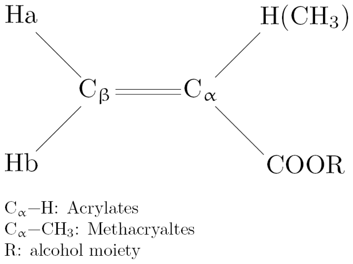
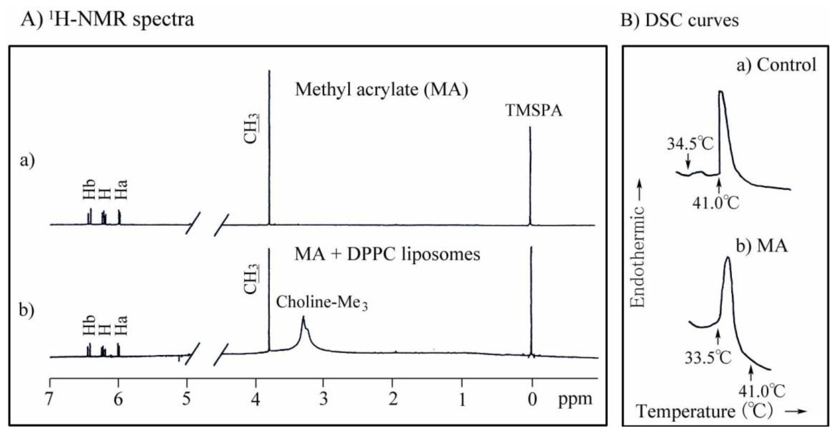
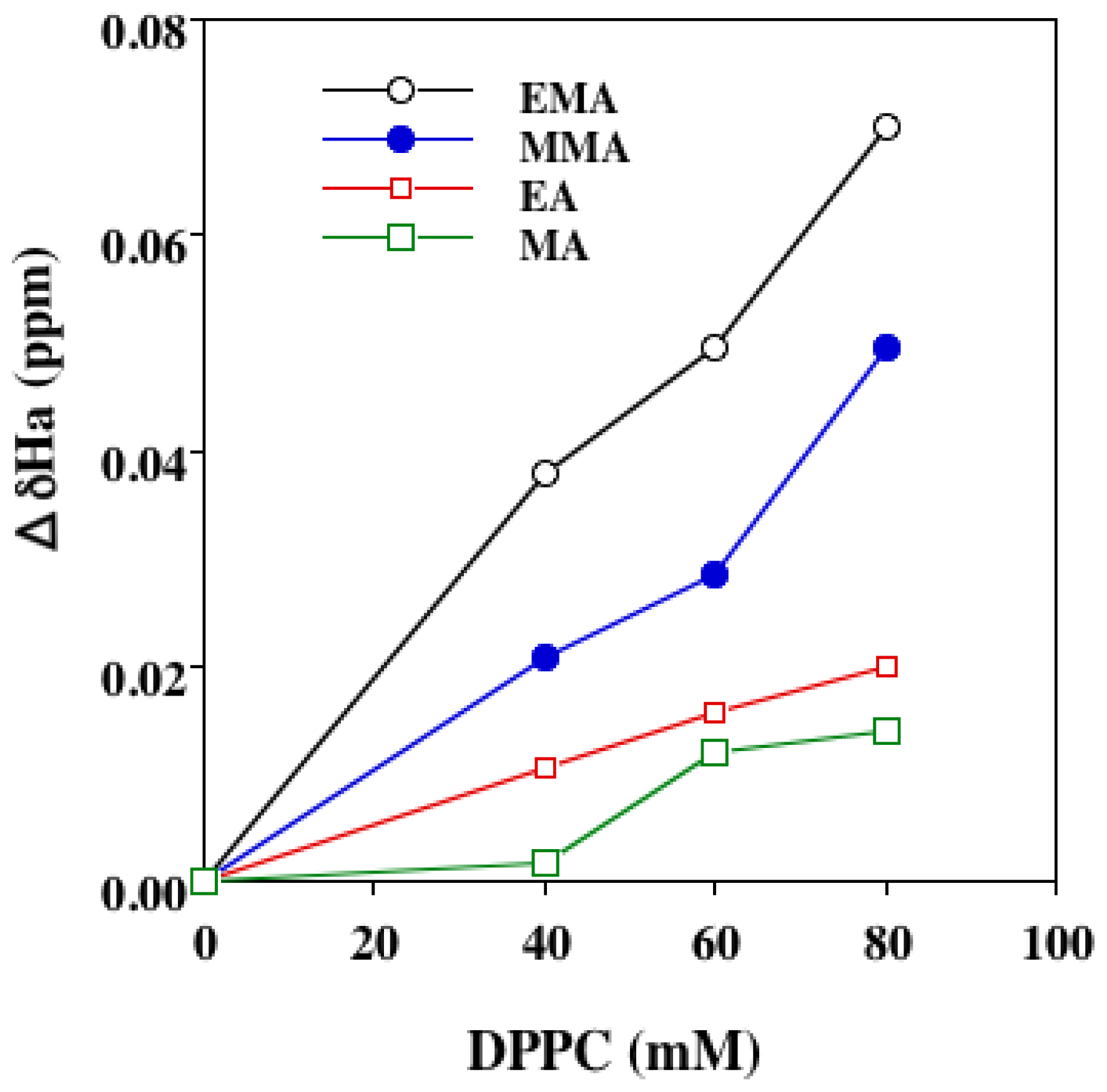
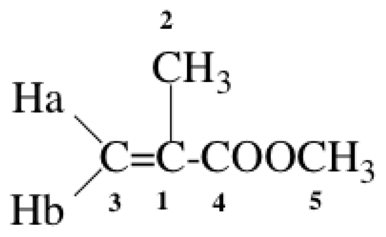
2.3.1. NMR Chemical Shifts of Ha</title>
<p>Liposomes have been employed in model systems to study the interaction of lipid-soluble drugs and monomers with biological membranes [<xref ref-type="bibr" rid="b17-ijms-13-00758">17</xref>,<xref ref-type="bibr" rid="b37-ijms-13-00758">37</xref>–<xref ref-type="bibr" rid="b40-ijms-13-00758">40</xref>]. Liposomes consist of lipid bilayers, and closely resemble the structure of biological membranes. Depending on their hydrophobicity, an exogenous hydrophobic compound will reside predominantly in liposomes and a hydrophilic one will be located in aqueous medium. NMR is one of the most powerful methods for studying not only the characterization of small unilamellar, large unilamellar and multilamellar liposomes [<xref ref-type="bibr" rid="b41-ijms-13-00758">41</xref>,<xref ref-type="bibr" rid="b42-ijms-13-00758">42</xref>] but also the interaction between (meth)acrylate monomers and liposomes as a model of cell membranes and transport phenomena across membranes [<xref ref-type="bibr" rid="b43-ijms-13-00758">43</xref>,<xref ref-type="bibr" rid="b44-ijms-13-00758">44</xref>]. We previously investigated the changes in <sup>1</sup>H and <sup>13</sup>C-NMR chemical shifts of methacrylates in DPPC liposomes. The chemical shift of Ha and β-carbon for methacrylates was shifted markedly to a higher field by their interaction with liposomes [<xref ref-type="bibr" rid="b38-ijms-13-00758">38</xref>–<xref ref-type="bibr" rid="b40-ijms-13-00758">40</xref>]. These findings may allow interpretation of the mechanism responsible for the hemolytic activity and cytotoxicity of monomers <italic>in vitro</italic> [<xref ref-type="bibr" rid="b39-ijms-13-00758">39</xref>,<xref ref-type="bibr" rid="b40-ijms-13-00758">40</xref>].</p>
<p>We investigated the interaction between unilamellar DPPC liposomes and MMA, EMA, EA or MA using <sup>1</sup>H-NMR spectroscopy. As an example, <sup>1</sup>H-NMR spectra of MA and DPPC liposome-bound MA are shown in <xref ref-type="fig" rid="f2-ijms-13-00758">Figure 2</xref>.</p>
<p>The difference in chemical shift (ΔδHa) between monomers and liposomal membrane-bound monomers was calculated. As a typical example, the shift for each proton of the MMA molecule at 25 and 50 °C is shown in <xref ref-type="table" rid="t6-ijms-13-00758">Table 6</xref>.</p>
<p>The <sup>1</sup>H-NMR-chemical shift (ppm) of MMA in D<sub>2</sub>O (free monomer) for Ha, Hb, 2H and 5H was 5.72, 6.13, 1.94 and 3.79, respectively. That for Ha, Hb, 2H and 5H derived from membrane-bound monomers was determined by varying the DPPC liposomal concentrations. The ΔδHa for each proton in the MMA molecule at a DPPC:MMA molar ratio of 10:1 was investigated, and this revealed that the ΔδHa for Ha at 25 °C was about 10 times greater than that for the corresponding protons in the MMA molecule, the value being even higher at 50 °C. Since the DPPC concentration exceeds that of MMA monomers, Langmuir’s adsorption isotherm probably holds well. It was clear that the Ha attached to the β-carbon of monomers was highly impregnated into liposomes in a DPPC concentration-dependent manner. The relationship between ΔδHa for the monomers EMA, MMA, EA and MA and increasing DPPC concentration is shown in <xref ref-type="fig" rid="f3-ijms-13-00758">Figure 3</xref>. The decline in the ΔδHa value was dependent on DPPC concentration in the order EMA &gt; MMA &gt; EA &gt; MA. The phase transition temperature (<italic>T</italic>) of DPPC liposomes was approximately 41 °C, and therefore the DPPC liposomes exist in a gel phase at 25 °C. We also examined the ΔδHa at 50 °C, at which DPPC liposomes exist in a liquid-crystalline phase. The ΔδHa at 50 °C declined in the order EMA &gt; MMA &gt; EA &gt; MA, which was identical to that at 25 °C (data not shown). We also examined the QSAR between the ΔδHa at 40 mM DPPC and log 1/H<sub>50</sub> for each monomer, and this revealed that the log 1/H<sub>50</sub> value for monomers was linearly correlated with their ΔδHa (QSAR 11). This suggested that the hemolytic activity of monomers may be related to their membrane permeation activity.</p></sec>
<sec>
<title>2.3.2. DSC Phase Transition Property</title>
<p>In earlier experiments, we examined changes in the phase transition properties of methacrylate-induced DPPC liposomes using DSC [<xref ref-type="bibr" rid="b38-ijms-13-00758">38</xref>–<xref ref-type="bibr" rid="b40-ijms-13-00758">40</xref>]. In this series, we investigated DSC changes in the phase transition temperature (<italic>T</italic>) (main transition peak), enthalpy (Δ<italic>H</italic>) and entropy (Δ<italic>S</italic>) of DPPC liposomes induced by (meth)acrylates. The results are shown in <xref ref-type="table" rid="t7-ijms-13-00758">Table 7</xref>. The Δ<italic>S</italic> value was calculated according to Δ<italic>S</italic> = Δ<italic>H</italic>/<italic>T</italic> (Equation 4) and that has been is also given in <xref ref-type="table" rid="t7-ijms-13-00758">Table 7</xref>. As an example of DSC scans, DSC curves of control and MA are also shown in <xref ref-type="fig" rid="f2-ijms-13-00758">Figure 2B</xref>. The <italic>T</italic>, Δ<italic>H</italic> and Δ<italic>S</italic> values for DPPC liposomes without any additives, as a control, were approximately 41.0 °C, 8.8 kcal/mol and 28.0 cal mol<sup>−1</sup> K<sup>−1</sup>, respectively. Control showed two peaks of pre-transition at 34.5 °C with a very small Δ<italic>H</italic> and main transition at 41.0 °C with a large Δ<italic>H</italic>, 8.8 kcal/mol. Acrylates, MA, EA and nBA, showed a shift of <italic>T</italic> to a much lower temperature range of 32–34 °C, whereas methacrylates, MMA and EMA showed a shift of <italic>T</italic> to a slightly lower temperature range of 39.5–40.5 °C. The decrease in Δ<italic>H</italic> for nBA and EMA was greater than that for MA, EA and MMA. Shifts of the <italic>T</italic> and Δ<italic>S</italic> values for acrylates were greater than those for methacrylates, possibly due to differences between the α-CH<sub>3</sub> and α-H substituents in the monomer molecule (<xref ref-type="fig" rid="f1-ijms-13-00758">Figure 1</xref>). The Δ<italic>S</italic> values for acrylates, MA and EA were greater than those for the corresponding methacrylates, MMA and EMA, possibly as a result of the effect of the steric factor of the α-substituent in the monomer molecule. The Δ<italic>H</italic> value for nBA was the lowest, probably in view of the hydrophobicity of its butyl substituent (<xref ref-type="table" rid="t2-ijms-13-00758">Table 2</xref>).</p>
<p>MA and EA, although showing great <italic>in vivo</italic> toxicity, possessed less hemolytic activity (<xref ref-type="table" rid="t1-ijms-13-00758">Table 1</xref>). Jain <italic>et al.</italic> [<xref ref-type="bibr" rid="b43-ijms-13-00758">43</xref>] previously reported that antihemolytic small molecules have the ability to expand the lipid bilayer of biomembranes and cause large changes in the phase transition properties of the DPPC bilayer. Marique-Moreno <italic>et al.</italic> [<xref ref-type="bibr" rid="b44-ijms-13-00758">44</xref>] investigated the effects of non-steroidal anti-inflammatory drug on human erythrocytes, and on liposomes as a cell membrane molecular model, and found that these drugs interacted strongly with dimyristoylphosphatidylcholine (DMPC) multilayers; DSC data also indicated a decrease in the melting (phase transition) temperature (<italic>T</italic>) of DMPC liposomes, which was attributed to destabilization of the gel phase. Taken together, it was concluded from these findings that the low hemolytic activity of MA, despite its high toxicity, may be attributable to its ability to expand the lipid bilayer of erythrocytes. The hemolytic activity of (meth)acrylates may be controlled by HF and hydrophobicity of the monomers, resulting from a good QSAR in the HF term and log <italic>P</italic>, whereas the <italic>in vivo</italic> toxicity of (meth)acrylates may be controlled by their π-electron density, σ-charge or <italic>η</italic> and <italic>χ</italic> reactivity principles, resulting from a good QSAR for these descriptors (<xref ref-type="table" rid="t4-ijms-13-00758">Table 4</xref>). <italic>In vivo</italic> toxicity may be controlled by the Michael-type reactivity of the monomers [<xref ref-type="bibr" rid="b11-ijms-13-00758">11</xref>]. As <italic>in vivo</italic> experiments are too complex to allow simple interpretation, liposome studies may help to clarify the mechanisms of <italic>in vitro</italic> and <italic>in vivo</italic> toxicity.</p></sec></sec></sec>
<sec>
<title>3. Experimental Section</title>
<sec>
<title>3.1. Chemicals</title>
<p>The following chemicals and reagents were obtained from the indicated sources. Methyl acrylate (MA), ethyl acrylate (EA), methyl methacrylate (MMA), ethyl methacrylate (EMA) and <italic>n</italic>-butyl acrylate (nBA)(Tokyo Chemical Industry Co., Ltd., Tokyo, Japan); <sc>l</sc>-α-dipalmitoylphosphatidylcholine (DPPC)(Sigma Chemical Co., USA); deuterium oxide, 3-(trimethylsilyl)propionic acid sodium salt (TMSPA)(Merck, Darmstadt, Germany).</p></sec>
<sec>
<title>3.2. NMR Spectra</title>
<p>The <sup>1</sup>H- and <sup>13</sup>C-NMR chemical shift data for various monomers in chloroform-<italic>d</italic> (CDCl<sub>3</sub>) were taken from the literature [<xref ref-type="bibr" rid="b18-ijms-13-00758">18</xref>,<xref ref-type="bibr" rid="b19-ijms-13-00758">19</xref>]. Briefly, the chemical shifts of the indicated monomers were measured in CDCl<sub>3</sub> at 35 °C at 125 and/or 500 MHz, respectively, using tetramethylsilane (TMS) as an internal standard.</p></sec>
<sec sec-type="methods">
<title>3.3. NMR Study</title>
<p>Preparation of liposomes: Briefly, the method of DPPC liposome preparation was as follows: DPPC was accurately weighed and dissolved in chloroform. The solution was evaporated to a dry thin film on the bottom of a round-bottom test tube and left under vacuum for 30 min. Deuterium oxide (D<sub>2</sub>O, Merck, Darmstadt, Germany) in 0.01 M phosphate buffer at pD 7.0 was then added, and sonication was performed under a nitrogen atmosphere for 10 min at 60 °C. After incubation for 20 min at room temperature, unilamelar DPPC liposomes were prepared by centrifugation at 20,000× g for 15 min. <sup>1</sup>H-NMR spectra were measured at 25 °C on a JEOL (Tokyo, Japan) JNM-GX 270 or ALPHA 500 instrument at a resolution of 0.01 ppm and 0.0013 ppm, respectively. An NMR sample tube with a coaxial capillary was used. The coaxial capillary with monomers was inserted into an NMR sample tube with the liposomes. Then, NMR spectra were measured at 25 °C and 50 °C, respectively. The external standard was TMSPA [<xref ref-type="bibr" rid="b17-ijms-13-00758">17</xref>,<xref ref-type="bibr" rid="b38-ijms-13-00758">38</xref>].</p></sec>
<sec sec-type="methods">
<title>3.4. DSC Study</title>
<p>An aliquot sample (20 μL) of DPPC and the indicated concentration of monomer in 0.01 M phosphate buffer solution at pH 7.0 was placed into a DSC specimen container. The specimen was allowed to equilibrate for 14 h at 5 °C, then the specimen was scanned in a sealed calorimetric container on a DSC-Rigaku calorimeter (Rigaku Denki Co. Ltd., Tokyo, Japan) at a heating rate of 5 °C min<sup>−1</sup> with a range setting of 0.5 mcal s<sup>−1</sup>. The instrument was calibrated with indium as a standard. The enthalpy (Δ<italic>H</italic>) was calculated from the area under the DSC curve [<xref ref-type="bibr" rid="b17-ijms-13-00758">17</xref>,<xref ref-type="bibr" rid="b38-ijms-13-00758">38</xref>,<xref ref-type="bibr" rid="b39-ijms-13-00758">39</xref>].</p></sec>
<sec>
<title>3.5. Hemolytic Activity</title>
<p>The concentration eliciting 50% (H<sub>50</sub>) hemolysis of rabbit erythrocytes for (meth)acrylates was taken from the literature [<xref ref-type="bibr" rid="b3-ijms-13-00758">3</xref>].</p></sec>
<sec>
<title>3.6. <italic>In Vivo</italic> Toxicity</title>
<p>LD<sub>50</sub> (50% lethal dose) data for intraperitoneal injection of mice with acrylate and methacrylate monomers were taken from the literature [<xref ref-type="bibr" rid="b3-ijms-13-00758">3</xref>]. Briefly, male albino ICR mice weighing 25 ± 5 g were used as the test animals, and the LD<sub>50</sub> dose for each compound was calculated in terms of 7-day mortality.</p></sec>
<sec>
<title>3.7. Theoretical Parameters</title>
<p>Parameters <italic>η</italic>, <italic>χ</italic> and <italic>ω</italic> were calculated using Equations 1–3, respectively (<xref ref-type="table" rid="t4-ijms-13-00758">Table 4</xref>). HOMO, LUMO, and heats of formation were taken from our reported studies [<xref ref-type="bibr" rid="b14-ijms-13-00758">14</xref>,<xref ref-type="bibr" rid="b15-ijms-13-00758">15</xref>]. Briefly, calculations of heats of formation were performed using the PM3/CONFLEX method. To obtain fine geometry details in the present study, initial geometry optimization was first performed using CONFLEX5 (Conflex, Tokyo, Japan). Thereafter, calculations using the PM3 method in the MOPAC 2000 program were carried out on a Tektronix CAChe workstation (Fujitsu Ltd., Japan).</p></sec></sec>
<sec sec-type="conclusions">
<title>4. Conclusions</title>
<p>QSARs between <italic>in vitro</italic> toxicity (1/H<sub>50</sub>) or <italic>in vivo</italic> toxicity (ipLD<sub>50</sub>) based on reported data and their NMR chemical shifts (δHa or δC<sub>β</sub>) or PM3-based theoretical parameters (HF, <italic>η</italic>, <italic>χ</italic>, <italic>ω</italic>) were investigated. There was a good linear QSAR between log 1/H<sub>50</sub> and log <italic>P</italic> or HF for (meth)acrylates. Also, a good QSAR between ipLD<sub>50</sub> and the δC<sub>β</sub>, <italic>η</italic>-, <italic>χ</italic>-, or <italic>ω</italic>-term for methacrylates was obtained, indicating that a common mechanism of action (Michael-type addition) of these monomers may be responsible for their <italic>in vivo</italic> toxicity. The interaction between DPPC liposomes and (meth)acrylates, investigated using NMR and DSC methods, indicated that the hemolytic activity of monomers may be controlled by their HF, which may be attributed to the monomer-induced phase transition properties of the erythrocyte lipid bilayer. NMR data may be an important tool for evaluating the biological activity of new vinyl monomers in medical and dental applications.</p></sec></body>
<back>
<ack>
<title>Acknowledgments</title>
<p>We thank M. Ishihara of Meikai University for semi-empirical calculations.</p></ack>
<ref-list>
<title>References</title>
<ref id="b1-ijms-13-00758"><label>1</label><citation citation-type="journal"><person-group person-group-type="author"><name><surname>Lawrence</surname><given-names>W.H.</given-names></name><name><surname>Bass</surname><given-names>G.E.</given-names></name><name><surname>Purcell</surname><given-names>W.P.</given-names></name><name><surname>Autian</surname><given-names>J.</given-names></name></person-group><article-title>Use of mathematical models in the study of structure-toxicity relationships of dental compounds: I. Esters of acrylic and methacrylic acids</article-title><source>J. Dent. Res</source><year>1972</year><volume>51</volume><fpage>526</fpage><lpage>535</lpage><pub-id pub-id-type="doi">10.1177/00220345720510024701</pub-id><pub-id pub-id-type="pmid">4501289</pub-id></citation></ref>
<ref id="b2-ijms-13-00758"><label>2</label><citation citation-type="journal"><person-group person-group-type="author"><name><surname>Tanii</surname><given-names>H.</given-names></name><name><surname>Hashimoto</surname><given-names>K.</given-names></name></person-group><article-title>Structure-toxicity relationship of acrylates and methacrylates</article-title><source>Toxicol. Lett</source><year>1982</year><volume>11</volume><fpage>125</fpage><lpage>129</lpage><pub-id pub-id-type="doi">10.1016/0378-4274(82)90116-3</pub-id><pub-id pub-id-type="pmid">7090003</pub-id></citation></ref>
<ref id="b3-ijms-13-00758"><label>3</label><citation citation-type="journal"><person-group person-group-type="author"><name><surname>Dillingham</surname><given-names>E.O.</given-names></name><name><surname>Lawrence</surname><given-names>W.H.</given-names></name><name><surname>Autian</surname><given-names>J.</given-names></name></person-group><article-title>Acrylate and methacrylate esters: Relationship of hemolytic activity and <italic>in vivo</italic> toxicity</article-title><source>J. Biomed. Mater. Res</source><year>1983</year><volume>17</volume><fpage>945</fpage><lpage>957</lpage><pub-id pub-id-type="doi">10.1002/jbm.820170606</pub-id><pub-id pub-id-type="pmid">6654932</pub-id></citation></ref>
<ref id="b4-ijms-13-00758"><label>4</label><citation citation-type="journal"><person-group person-group-type="author"><name><surname>Yoshii</surname><given-names>E.</given-names></name></person-group><article-title>Cytotoxic effects of acrylates and methacrylates: Relationships of monomer structures and cytotoxicity</article-title><source>J. Biomed. Mater. Res</source><year>1997</year><volume>37</volume><fpage>517</fpage><lpage>524</lpage><pub-id pub-id-type="doi">10.1002/(SICI)1097-4636(19971215)37:4&lt;517::AID-JBM10&gt;3.0.CO;2-5</pub-id><pub-id pub-id-type="pmid">9407300</pub-id></citation></ref>
<ref id="b5-ijms-13-00758"><label>5</label><citation citation-type="journal"><person-group person-group-type="author"><name><surname>Chan</surname><given-names>K.</given-names></name><name><surname>O’Brien</surname><given-names>P.J.</given-names></name></person-group><article-title>Structure-activity relationships for hepatocyte toxicity and electrophilic reactivity of α,β-unsaturated esters, acrylates and methacrylates</article-title><source>J. Appl. Toxicol</source><year>2008</year><volume>28</volume><fpage>1004</fpage><lpage>1015</lpage><pub-id pub-id-type="doi">10.1002/jat.1366</pub-id><pub-id pub-id-type="pmid">18615533</pub-id></citation></ref>
<ref id="b6-ijms-13-00758"><label>6</label><citation citation-type="journal"><article-title>Cosmetic Ingredient Review. Final report of the safety assessment of methacrylic acid</article-title><source>Int. J. Toxicol.</source><year>2005</year><volume>24</volume><fpage>33</fpage><lpage>51</lpage><pub-id pub-id-type="doi">10.1080/10915810500434191</pub-id><pub-id pub-id-type="pmid">16596768</pub-id></citation></ref>
<ref id="b7-ijms-13-00758"><label>7</label><citation citation-type="journal"><person-group person-group-type="author"><name><surname>Bakopoulou</surname><given-names>A.</given-names></name><name><surname>Papadopoulos</surname><given-names>T.</given-names></name><name><surname>Garefis</surname><given-names>P.</given-names></name></person-group><article-title>Molecular toxicology of substances related from resin-based dental restorative materials</article-title><source>Int. J. Mol. Sci</source><year>2009</year><volume>10</volume><fpage>3861</fpage><lpage>3899</lpage><pub-id pub-id-type="doi">10.3390/ijms10093861</pub-id><pub-id pub-id-type="pmid">19865523</pub-id></citation></ref>
<ref id="b8-ijms-13-00758"><label>8</label><citation citation-type="journal"><person-group person-group-type="author"><name><surname>Tsukeoka</surname><given-names>T.</given-names></name><name><surname>Suzuki</surname><given-names>M.</given-names></name><name><surname>Ohtsuki</surname><given-names>C.</given-names></name><name><surname>Sugino</surname><given-names>A.</given-names></name><name><surname>Tsuneizumi</surname><given-names>Y.</given-names></name><name><surname>Miyagi</surname><given-names>J.</given-names></name><name><surname>Kuramoto</surname><given-names>K.</given-names></name><name><surname>Moriya</surname><given-names>H.</given-names></name></person-group><article-title>Mechanical and histological evaluation of a PMMA-based bone cement modified with γ-methacryloxypropyltrimethoxysilane and calcium acetate</article-title><source>Biomaterials</source><year>2006</year><volume>27</volume><fpage>3897</fpage><lpage>3903</lpage><pub-id pub-id-type="doi">10.1016/j.biomaterials.2006.03.002</pub-id><pub-id pub-id-type="pmid">16563499</pub-id></citation></ref>
<ref id="b9-ijms-13-00758"><label>9</label><citation citation-type="journal"><person-group person-group-type="author"><name><surname>Hansch</surname><given-names>C.</given-names></name><name><surname>Fujita</surname><given-names>T.</given-names></name></person-group><article-title>p-σ-π Analysis. A method for the correlation of biological activity and chemical structure</article-title><source>J. Am. Chem. Soc</source><year>1964</year><volume>86</volume><fpage>1616</fpage><lpage>1626</lpage><pub-id pub-id-type="doi">10.1021/ja01062a035</pub-id></citation></ref>
<ref id="b10-ijms-13-00758"><label>10</label><citation citation-type="journal"><person-group person-group-type="author"><name><surname>Linhart</surname><given-names>I.</given-names></name><name><surname>Vosmanská</surname><given-names>M.</given-names></name><name><surname>Smejkal</surname><given-names>J.</given-names></name></person-group><article-title>Biotransformation of acrylates. Excretion of mercapturic acids and changes in urinary carboxylic acid profile in rat dosed with ethyl and 1-butyl acrylate</article-title><source>Xenobiotica</source><year>1994</year><volume>24</volume><fpage>1043</fpage><lpage>1052</lpage><pub-id pub-id-type="doi">10.3109/00498259409043301</pub-id><pub-id pub-id-type="pmid">7900410</pub-id></citation></ref>
<ref id="b11-ijms-13-00758"><label>11</label><citation citation-type="journal"><person-group person-group-type="author"><name><surname>Freidig</surname><given-names>A.P.</given-names></name><name><surname>Verhaar</surname><given-names>H.J.M.</given-names></name><name><surname>Hermens</surname><given-names>J.L.M.</given-names></name></person-group><article-title>Quantitative structure-property relationships for the chemical reactivity of acrylates and methacrylates</article-title><source>Environ. Toxicol. Chem</source><year>1999</year><volume>18</volume><fpage>1133</fpage><lpage>1139</lpage></citation></ref>
<ref id="b12-ijms-13-00758"><label>12</label><citation citation-type="journal"><person-group person-group-type="author"><name><surname>Cronin</surname><given-names>M.T.</given-names></name><name><surname>Walker</surname><given-names>J.D.</given-names></name><name><surname>Jaworska</surname><given-names>J.S.</given-names></name><name><surname>Comber</surname><given-names>M.H.</given-names></name><name><surname>Watts</surname><given-names>C.D.</given-names></name><name><surname>Worth</surname><given-names>A.P.</given-names></name></person-group><article-title>Use of QSARs in international decision-making frameworks to predict ecologic effects and environmental fate of chemical substances</article-title><source>Environ. Health Perspect</source><year>2003</year><volume>111</volume><fpage>1376</fpage><lpage>1390</lpage><pub-id pub-id-type="doi">10.1289/ehp.5759</pub-id><pub-id pub-id-type="pmid">12896861</pub-id></citation></ref>
<ref id="b13-ijms-13-00758"><label>13</label><citation citation-type="journal"><person-group person-group-type="author"><name><surname>Eroglu</surname><given-names>E.</given-names></name><name><surname>Palaz</surname><given-names>S.</given-names></name><name><surname>Oltulu</surname><given-names>O.</given-names></name><name><surname>Turkmen</surname><given-names>H.</given-names></name><name><surname>Ozaydın</surname><given-names>C.</given-names></name></person-group><article-title>Comparative QSTR study using semi-empirical and first principle methods based descriptors for acute toxicity of diverse organic compounds to the fathead minnow</article-title><source>Int. J. Mol. Sci</source><year>2007</year><volume>8</volume><fpage>1265</fpage><lpage>1283</lpage></citation></ref>
<ref id="b14-ijms-13-00758"><label>14</label><citation citation-type="journal"><person-group person-group-type="author"><name><surname>Ishihara</surname><given-names>M.</given-names></name><name><surname>Fujisawa</surname><given-names>S.</given-names></name></person-group><article-title>Quantum-chemical descriptors for estimating hemolytic activity of aliphatic and aromatic methacrylates</article-title><source>Chemosphere</source><year>2008</year><volume>70</volume><fpage>1898</fpage><lpage>1902</lpage><pub-id pub-id-type="doi">10.1016/j.chemosphere.2007.07.070</pub-id><pub-id pub-id-type="pmid">17850844</pub-id></citation></ref>
<ref id="b15-ijms-13-00758"><label>15</label><citation citation-type="journal"><person-group person-group-type="author"><name><surname>Ishihara</surname><given-names>M.</given-names></name><name><surname>Fujisawa</surname><given-names>S.</given-names></name></person-group><article-title>A structure-activity relationship study on the mechanisms of methacrylate-induced toxicity using NMR chemical shift of β-carbon, RP-HPLC log <italic>P</italic> and semiempirical molecular descriptor</article-title><source>Dent. Mater. J</source><year>2009</year><volume>28</volume><fpage>113</fpage><lpage>120</lpage><pub-id pub-id-type="doi">10.4012/dmj.28.113</pub-id><pub-id pub-id-type="pmid">19280976</pub-id></citation></ref>
<ref id="b16-ijms-13-00758"><label>16</label><citation citation-type="journal"><person-group person-group-type="author"><name><surname>Putz</surname><given-names>M.V.</given-names></name><name><surname>Ionaşcu</surname><given-names>C.</given-names></name><name><surname>Putz</surname><given-names>A.M.</given-names></name><name><surname>Ostafe</surname><given-names>V.</given-names></name></person-group><article-title>Alert-QSAR. Implications for electrophilic theory of chemical carcinogenesis</article-title><source>Int. J. Mol. Sci</source><year>2011</year><volume>12</volume><fpage>5098</fpage><lpage>5134</lpage><pub-id pub-id-type="doi">10.3390/ijms12085098</pub-id><pub-id pub-id-type="pmid">21954348</pub-id></citation></ref>
<ref id="b17-ijms-13-00758"><label>17</label><citation citation-type="journal"><person-group person-group-type="author"><name><surname>Fujisawa</surname><given-names>S.</given-names></name><name><surname>Kadoma</surname><given-names>Y.</given-names></name><name><surname>Ishihara</surname><given-names>M.</given-names></name><name><surname>Atsumi</surname><given-names>T.</given-names></name><name><surname>Yokoe</surname><given-names>I.</given-names></name></person-group><article-title>Dipalmitoylphosphatidylcholine (DPPC) and DPPC/cholesterol liposomes as predictors of the cytotoxicity of bis-GMA related compounds</article-title><source>J. Liposome Res</source><year>2004</year><volume>14</volume><fpage>39</fpage><lpage>49</lpage><pub-id pub-id-type="doi">10.1081/LPR-120039662</pub-id><pub-id pub-id-type="pmid">15461931</pub-id></citation></ref>
<ref id="b18-ijms-13-00758"><label>18</label><citation citation-type="journal"><person-group person-group-type="author"><name><surname>Hatada</surname><given-names>K.</given-names></name><name><surname>Kitayama</surname><given-names>T.</given-names></name><name><surname>Nishiura</surname><given-names>T.</given-names></name><name><surname>Shibuya</surname><given-names>W.</given-names></name></person-group><article-title>Relation between reactivities of vinyl monomers and their NMR spectra</article-title><source>Curr. Org. Chem</source><year>2002</year><volume>6</volume><fpage>121</fpage><lpage>153</lpage><pub-id pub-id-type="doi">10.2174/1385272023374454</pub-id></citation></ref>
<ref id="b19-ijms-13-00758"><label>19</label><citation citation-type="journal"><person-group person-group-type="author"><name><surname>Fujisawa</surname><given-names>S.</given-names></name><name><surname>Kadoma</surname><given-names>Y.</given-names></name></person-group><article-title>Prediction of the reduced glutathione (GSH) reactivity of dental methacrylate monomers using NMR spectra—Relationship between toxicity and GSH reactivity</article-title><source>Dent. Mater. J</source><year>2009</year><volume>28</volume><fpage>722</fpage><lpage>729</lpage><pub-id pub-id-type="doi">10.4012/dmj.28.722</pub-id><pub-id pub-id-type="pmid">20019424</pub-id></citation></ref>
<ref id="b20-ijms-13-00758"><label>20</label><citation citation-type="journal"><person-group person-group-type="author"><name><surname>McCarthy</surname><given-names>T.J.</given-names></name><name><surname>Hayes</surname><given-names>E.P.</given-names></name><name><surname>Schwartz</surname><given-names>C.S.</given-names></name><name><surname>Witz</surname><given-names>G.</given-names></name></person-group><article-title>The reactivity of selected acrylate esters toward glutathione and deoxyribonucleosides <italic>in vitro</italic>: Structure-activity relationships</article-title><source>Fundam. Appl. Toxicol</source><year>1994</year><volume>22</volume><fpage>543</fpage><lpage>548</lpage><pub-id pub-id-type="doi">10.1006/faat.1994.1061</pub-id><pub-id pub-id-type="pmid">8056201</pub-id></citation></ref>
<ref id="b21-ijms-13-00758"><label>21</label><citation citation-type="journal"><person-group person-group-type="author"><name><surname>Verhaar</surname><given-names>H.J.M.</given-names></name><name><surname>van Leeuwen</surname><given-names>C.J.</given-names></name><name><surname>Hermens</surname><given-names>J.L.M.</given-names></name></person-group><article-title>Classifying environmental pollutants. 1: Structure-activity relationships for prediction of aquatic toxicity</article-title><source>Chemosphere</source><year>1992</year><volume>25</volume><fpage>471</fpage><lpage>491</lpage><pub-id pub-id-type="doi">10.1016/0045-6535(92)90280-5</pub-id></citation></ref>
<ref id="b22-ijms-13-00758"><label>22</label><citation citation-type="journal"><person-group person-group-type="author"><name><surname>Russom</surname><given-names>C.L.</given-names></name><name><surname>Bradbury</surname><given-names>S.P.</given-names></name><name><surname>Broderius</surname><given-names>S.J.</given-names></name><name><surname>Hammermeister</surname><given-names>D.E.</given-names></name><name><surname>Drummond</surname><given-names>R.A.</given-names></name></person-group><article-title>Predicting modes of toxic action from chemical structure: Acute toxicity in the fathead minnow (<italic>Pimephales promelas</italic>)</article-title><source>Environ. Toxicol. Chem</source><year>1997</year><volume>16</volume><fpage>948</fpage><lpage>967</lpage></citation></ref>
<ref id="b23-ijms-13-00758"><label>23</label><citation citation-type="journal"><person-group person-group-type="author"><name><surname>Koleva</surname><given-names>Y.K.</given-names></name><name><surname>Madden</surname><given-names>J.C.</given-names></name><name><surname>Cronin</surname><given-names>M.T.</given-names></name></person-group><article-title>Formation of categories from structure-activity relationships to allow read-across for risk assessment: Toxicity of α,β-unsaturated carbonyl compounds</article-title><source>Chem. Res. Toxicol</source><year>2008</year><volume>21</volume><fpage>2300</fpage><lpage>2312</lpage><pub-id pub-id-type="doi">10.1021/tx8002438</pub-id><pub-id pub-id-type="pmid">19053326</pub-id></citation></ref>
<ref id="b24-ijms-13-00758"><label>24</label><citation citation-type="journal"><person-group person-group-type="author"><name><surname>del Re</surname><given-names>G.</given-names></name></person-group><article-title>A simple MO-LCAO method for the calculation of charge distributions in saturated organic molecules</article-title><source>J. Chem. Soc</source><year>1958</year><fpage>4031</fpage><lpage>4040</lpage><pub-id pub-id-type="doi">10.1039/JR9580004031</pub-id></citation></ref>
<ref id="b25-ijms-13-00758"><label>25</label><citation citation-type="journal"><person-group person-group-type="author"><name><surname>Talalay</surname><given-names>P.</given-names></name><name><surname>de Long</surname><given-names>M.J.</given-names></name><name><surname>Prochaska</surname><given-names>H.J.</given-names></name></person-group><article-title>Identification of a common chemical signal regulating the induction of enzymes that protect against chemical carcinogenesis</article-title><source>Proc. Natl. Acad. Sci. USA</source><year>1988</year><volume>85</volume><fpage>8261</fpage><lpage>8265</lpage><pub-id pub-id-type="doi">10.1073/pnas.85.21.8261</pub-id><pub-id pub-id-type="pmid">3141925</pub-id></citation></ref>
<ref id="b26-ijms-13-00758"><label>26</label><citation citation-type="web"><collab>The University of Oxford</collab><article-title>Department of Chemistry MSDS web resource</article-title><comment><ext-link xlink:href="http://msds.chem.ox.uk/AC/acrolein.html" ext-link-type="uri">http://msds.chem.ox.uk/AC/acrolein.html</ext-link></comment><access-date>accessed on 19 October 2011</access-date></citation></ref>
<ref id="b27-ijms-13-00758"><label>27</label><citation citation-type="web"><collab>The University of Oxford</collab><source>Department of Chemistry MSDS web resource</source><comment><ext-link xlink:href="http://msds.chem.ox.uk/AC/acrylonitrile.html" ext-link-type="uri">http://msds.chem.ox.uk/AC/acrylonitrile.html</ext-link></comment><access-date>accessed on 15 November 2011</access-date></citation></ref>
<ref id="b28-ijms-13-00758"><label>28</label><citation citation-type="web"><collab>The University of Oxford</collab><source>Department of Chemistry MSDS web resource</source><comment><ext-link xlink:href="http://msds.chem.ox.uk/AC/acrylamide.html" ext-link-type="uri">http://msds.chem.ox.uk/AC/acrylamide.html</ext-link></comment><access-date>accessed on 7 February 2005</access-date></citation></ref>
<ref id="b29-ijms-13-00758"><label>29</label><citation citation-type="journal"><person-group person-group-type="author"><name><surname>Dickinson</surname><given-names>D.A.</given-names></name><name><surname>Forman</surname><given-names>H.J.</given-names></name></person-group><article-title>Cellular glutathione and thiols metabolism</article-title><source>Biochem. Pharmacol</source><year>2002</year><volume>64</volume><fpage>1019</fpage><lpage>1026</lpage><pub-id pub-id-type="doi">10.1016/S0006-2952(02)01172-3</pub-id><pub-id pub-id-type="pmid">12213601</pub-id></citation></ref>
<ref id="b30-ijms-13-00758"><label>30</label><citation citation-type="journal"><person-group person-group-type="author"><name><surname>Geurtsen</surname><given-names>W.</given-names></name><name><surname>Leyhausen</surname><given-names>G.</given-names></name></person-group><article-title>Chemical-biological interactions of the resin monomer triethyleneglycol-dimethacrylate (TEGDMA)</article-title><source>J. Dent. Res</source><year>2001</year><volume>80</volume><fpage>2046</fpage><lpage>2050</lpage><pub-id pub-id-type="doi">10.1177/00220345010800120401</pub-id><pub-id pub-id-type="pmid">11808759</pub-id></citation></ref>
<ref id="b31-ijms-13-00758"><label>31</label><citation citation-type="journal"><person-group person-group-type="author"><name><surname>Schweikl</surname><given-names>H.</given-names></name><name><surname>Spagnuolo</surname><given-names>G.</given-names></name><name><surname>Schmalz</surname><given-names>G.</given-names></name></person-group><article-title>Genetic and cellular toxicology of dental resin monomers</article-title><source>J. Dent. Res</source><year>2006</year><volume>85</volume><fpage>870</fpage><lpage>877</lpage><pub-id pub-id-type="doi">10.1177/154405910608501001</pub-id><pub-id pub-id-type="pmid">16998124</pub-id></citation></ref>
<ref id="b32-ijms-13-00758"><label>32</label><citation citation-type="journal"><person-group person-group-type="author"><name><surname>Ishikawa</surname><given-names>T.</given-names></name><name><surname>Li</surname><given-names>Z.S.</given-names></name><name><surname>Lu</surname><given-names>Y.P.</given-names></name><name><surname>Rea</surname><given-names>P.A.</given-names></name></person-group><article-title>The GS-X pump in plant, yeast, and animal cells: Structure, function, and gene expression</article-title><source>Biosci. Rep</source><year>1997</year><volume>17</volume><fpage>189</fpage><lpage>207</lpage><pub-id pub-id-type="doi">10.1023/A:1027385513483</pub-id><pub-id pub-id-type="pmid">9217966</pub-id></citation></ref>
<ref id="b33-ijms-13-00758"><label>33</label><citation citation-type="journal"><person-group person-group-type="author"><name><surname>Tanel</surname><given-names>A.</given-names></name><name><surname>Averill-Bates</surname><given-names>D.A.</given-names></name></person-group><article-title>The aldehyde acrolein induces apoptosis via activation of the mitochondrial pathway</article-title><source>Biochim. Biophys. Acta</source><year>2005</year><volume>1743</volume><fpage>255</fpage><lpage>267</lpage><pub-id pub-id-type="doi">10.1016/j.bbamcr.2004.11.007</pub-id><pub-id pub-id-type="pmid">15843039</pub-id></citation></ref>
<ref id="b34-ijms-13-00758"><label>34</label><citation citation-type="journal"><person-group person-group-type="author"><name><surname>Zhang</surname><given-names>H.</given-names></name><name><surname>Kamendulis</surname><given-names>L.M.</given-names></name><name><surname>Jiang</surname><given-names>J.</given-names></name><name><surname>Xu</surname><given-names>Y.</given-names></name><name><surname>Klaunig</surname><given-names>J.E.</given-names></name></person-group><article-title>Acrylonitrile-induced morphological transformation in Syrian hamster embryo cells</article-title><source>Carcinogenesis</source><year>2000</year><volume>21</volume><fpage>727</fpage><lpage>733</lpage><pub-id pub-id-type="doi">10.1093/carcin/21.4.727</pub-id><pub-id pub-id-type="pmid">10753209</pub-id></citation></ref>
<ref id="b35-ijms-13-00758"><label>35</label><citation citation-type="journal"><person-group person-group-type="author"><name><surname>Zitting</surname><given-names>A.</given-names></name><name><surname>Heinonen</surname><given-names>T.</given-names></name></person-group><article-title>Decrease of reduced glutathione in isolated rat hepatocytes caused by acrolein, acrylonitrile, and the thermal degradation products of styrene copolymers</article-title><source>Toxicology</source><year>1980</year><volume>17</volume><fpage>333</fpage><lpage>341</lpage><pub-id pub-id-type="doi">10.1016/0300-483X(80)90014-1</pub-id><pub-id pub-id-type="pmid">7210018</pub-id></citation></ref>
<ref id="b36-ijms-13-00758"><label>36</label><citation citation-type="journal"><person-group person-group-type="author"><name><surname>Besaratinia</surname><given-names>A.</given-names></name><name><surname>Pfeifer</surname><given-names>G.P.</given-names></name></person-group><article-title>A review of mechanisms of acrylamide carcinogenicity</article-title><source>Carcinogenesis</source><year>2007</year><volume>28</volume><fpage>519</fpage><lpage>528</lpage><pub-id pub-id-type="pmid">17234719</pub-id></citation></ref>
<ref id="b37-ijms-13-00758"><label>37</label><citation citation-type="journal"><person-group person-group-type="author"><name><surname>Papahadjopoulos</surname><given-names>D.</given-names></name></person-group><article-title>Liposomes and their uses in biology and medicine</article-title><source>Ann. N. Y. Acad. Sci</source><year>1978</year><volume>308</volume><fpage>1</fpage><lpage>462</lpage><pub-id pub-id-type="doi">10.1111/j.1749-6632.1978.tb22009.x</pub-id><pub-id pub-id-type="pmid">29553</pub-id></citation></ref>
<ref id="b38-ijms-13-00758"><label>38</label><citation citation-type="journal"><person-group person-group-type="author"><name><surname>Fujisawa</surname><given-names>S.</given-names></name><name><surname>Kadoma</surname><given-names>Y.</given-names></name><name><surname>Masuhara</surname><given-names>E.</given-names></name></person-group><article-title>A calorimetric study of the interaction of synthetic phospholipid liposomes with vinyl monomers, acrylates and methacrylates</article-title><source>J. Biomed. Mater. Res</source><year>1984</year><volume>18</volume><fpage>1105</fpage><lpage>1114</lpage><pub-id pub-id-type="doi">10.1002/jbm.820180912</pub-id><pub-id pub-id-type="pmid">6544795</pub-id></citation></ref>
<ref id="b39-ijms-13-00758"><label>39</label><citation citation-type="journal"><person-group person-group-type="author"><name><surname>Fujisawa</surname><given-names>S.</given-names></name><name><surname>Atsumi</surname><given-names>T.</given-names></name><name><surname>Kadoma</surname><given-names>Y.</given-names></name></person-group><article-title>Cytotoxicity of methyl methacrylate (MMA) and related compounds and their interaction with dipalmitoylphosphatidylcholine (DPPC) liposomes as a model for biomembranes</article-title><source>Oral Dis</source><year>2000</year><volume>6</volume><fpage>215</fpage><lpage>221</lpage><pub-id pub-id-type="pmid">10918558</pub-id></citation></ref>
<ref id="b40-ijms-13-00758"><label>40</label><citation citation-type="journal"><person-group person-group-type="author"><name><surname>Fujisawa</surname><given-names>S.</given-names></name><name><surname>Atsumi</surname><given-names>T.</given-names></name><name><surname>Kadoma</surname><given-names>Y.</given-names></name></person-group><article-title>Cytotoxicity and phospholipid-liposome phase-transition properties of 2-hydroxyethyl methacrylate (HEMA)</article-title><source>Artif. Cells Blood Substit. Immobil. Biotechnol</source><year>2001</year><volume>29</volume><fpage>245</fpage><lpage>261</lpage><pub-id pub-id-type="doi">10.1081/BIO-100103048</pub-id><pub-id pub-id-type="pmid">11358040</pub-id></citation></ref>
<ref id="b41-ijms-13-00758"><label>41</label><citation citation-type="journal"><person-group person-group-type="author"><name><surname>Lasic</surname><given-names>D.D.</given-names></name></person-group><article-title>Magnetic resonance methods in the studies of liposomes</article-title><source>Bull. Magn. Reson</source><year>1991</year><volume>13</volume><fpage>3</fpage><lpage>13</lpage></citation></ref>
<ref id="b42-ijms-13-00758"><label>42</label><citation citation-type="journal"><person-group person-group-type="author"><name><surname>Cruciani</surname><given-names>O.</given-names></name><name><surname>Mannina</surname><given-names>L.</given-names></name><name><surname>Sobolev</surname><given-names>A.P.</given-names></name><name><surname>Cametti</surname><given-names>C.</given-names></name><name><surname>Segre</surname><given-names>A.</given-names></name></person-group><article-title>An improved NMR study of liposomes using 1-palmitoyl-2-oleoyl-<italic>sn</italic>-glycero-3-phospatidylcholine as model</article-title><source>Molecules</source><year>2006</year><volume>11</volume><fpage>334</fpage><lpage>344</lpage><pub-id pub-id-type="doi">10.3390/11050334</pub-id><pub-id pub-id-type="pmid">17962765</pub-id></citation></ref>
<ref id="b43-ijms-13-00758"><label>43</label><citation citation-type="journal"><person-group person-group-type="author"><name><surname>Jain</surname><given-names>M.K.</given-names></name><name><surname>Wu</surname><given-names>N.Y.</given-names></name><name><surname>Wray</surname><given-names>L.V.</given-names></name></person-group><article-title>Drug-induced phase change in bilayer as possible mode of action of membrane expanding drugs</article-title><source>Nature</source><year>1975</year><volume>255</volume><fpage>494</fpage><lpage>496</lpage><pub-id pub-id-type="doi">10.1038/255494a0</pub-id><pub-id pub-id-type="pmid">1138201</pub-id></citation></ref>
<ref id="b44-ijms-13-00758"><label>44</label><citation citation-type="journal"><person-group person-group-type="author"><name><surname>Marique-Moreno</surname><given-names>M.</given-names></name><name><surname>Suwalsky</surname><given-names>M.</given-names></name><name><surname>Villena</surname><given-names>F.</given-names></name><name><surname>Garidel</surname><given-names>P.</given-names></name></person-group><article-title>Effects of the nonsteroidal anti-inflammatory drug naproxen on human erythrocytes and on cell membrane molecular models</article-title><source>Biophys. Chem</source><year>2010</year><volume>147</volume><fpage>53</fpage><lpage>58</lpage><pub-id pub-id-type="doi">10.1016/j.bpc.2009.12.010</pub-id><pub-id pub-id-type="pmid">20083338</pub-id></citation></ref></ref-list>
<sec sec-type="display-objects">
<title>Figures and Tables</title>
<fig id="f1-ijms-13-00758" position="float">
<label>Figure 1</label>
<caption>
<p>The structure of acrylates and methacrylates.</p></caption>
<graphic xlink:href="ijms-13-00758f1.gif"/></fig>
<fig id="f2-ijms-13-00758" position="float">
<label>Figure 2</label>
<caption>
<p><sup>1</sup>H-NMR spectra (<bold>A</bold>) of: (a) MA and (b) DPPC liposome-bound MA (molar ratio: DPPC:MA = 10:1) in D<sub>2</sub>O (pD 7.0 phosphate buffer) at 25 °C, and DSC curves (<bold>B</bold>) of: (a) DPPC liposomes (control) and b) MA-treated DPPC liposomes (molar ratio: MA:DPPC = 1:1). The NMR chemical shift of Ha, Hb, H (α-C<underline>H</underline>) and CH<sub>3</sub> was derived from MA molecule. By contrast, that of choline-Me<sub>3</sub>, <italic>N</italic>-(CH<sub>3</sub>)<sub>3</sub> was derived from DPPC molecule [<xref ref-type="bibr" rid="b39-ijms-13-00758">39</xref>,<xref ref-type="bibr" rid="b40-ijms-13-00758">40</xref>].</p></caption>
<graphic xlink:href="ijms-13-00758f2.gif"/></fig>
<fig id="f3-ijms-13-00758" position="float">
<label>Figure 3</label>
<caption>
<p>The chemical shift difference (ΔδHa, ppm) between free monomers and DPPC liposome-bound monomers as a function of DPPC concentration. The concentration of each monomer was 4 mM. The δ (ppm) values for Ha were determined with external TMSPA at 25 °C using <sup>1</sup>H-NMR spectroscopy. The values represent the means of two or three separate experiments.</p></caption>
<graphic xlink:href="ijms-13-00758f3.gif"/></fig>
<table-wrap id="t1-ijms-13-00758" position="float">
<label>Table 1</label>
<caption>
<p>Hemolytic activity, <italic>in vivo</italic> toxicity and NMR chemical shifts for (meth)acrylates.</p></caption>
<table frame="hsides" rules="groups">
<thead>
<tr>
<th align="left" valign="middle">Compound</th>
<th align="center" valign="middle">log 1/H<sub>50</sub> (mole/L) <xref ref-type="table-fn" rid="tfn1-ijms-13-00758">a</xref></th>
<th align="center" valign="middle">7-days ipLD<sub>50</sub> (mole/10<sup>6</sup> g) <xref ref-type="table-fn" rid="tfn1-ijms-13-00758">a</xref></th>
<th align="center" valign="middle">δHa (ppm) <xref ref-type="table-fn" rid="tfn2-ijms-13-00758">b</xref></th>
<th align="center" valign="middle">δC<sub>β</sub> (ppm) <xref ref-type="table-fn" rid="tfn2-ijms-13-00758">b</xref></th></tr></thead>
<tbody>
<tr>
<td align="left" valign="top">Methyl acrylate (MA)</td>
<td align="center" valign="top">0.63</td>
<td align="right" valign="top">2.95</td>
<td align="left" valign="top">5.82</td>
<td align="center" valign="top">130.56</td></tr>
<tr>
<td align="left" valign="top">Ethyl acrylate (EA)</td>
<td align="center" valign="top">0.95</td>
<td align="right" valign="top">5.98</td>
<td align="left" valign="top">5.807</td>
<td align="center" valign="top">130.24</td></tr>
<tr>
<td align="left" valign="top"><italic>n</italic>-Propyl acrylate (nPA)</td>
<td align="center" valign="top">1.59</td>
<td align="right" valign="top">5.80</td>
<td align="left" valign="top">5.809</td>
<td align="center" valign="top">130.22</td></tr>
<tr>
<td align="left" valign="top"><italic>n</italic>-Butyl acrylate (nBA)</td>
<td align="center" valign="top">2.61</td>
<td align="right" valign="top">6.64</td>
<td align="left" valign="top">5.805</td>
<td align="center" valign="top">130.21</td></tr>
<tr>
<td align="left" valign="top">Isobutyl acrylate (IBA)</td>
<td align="center" valign="top">2.84</td>
<td align="right" valign="top">5.92</td>
<td align="left" valign="top">5.813</td>
<td align="center" valign="top">130.23</td></tr>
<tr>
<td align="left" valign="top">Methyl methacrylate (MMA)</td>
<td align="center" valign="top">1.05</td>
<td align="right" valign="top">10.88</td>
<td align="left" valign="top">5.555</td>
<td align="center" valign="top">125.23</td></tr>
<tr>
<td align="left" valign="top">Ethyl methacrylate (EMA)</td>
<td align="center" valign="top">1.44</td>
<td align="right" valign="top">7.89</td>
<td align="left" valign="top">5.541</td>
<td align="center" valign="top">124.97</td></tr>
<tr>
<td align="left" valign="top"><italic>n</italic>-Propyl methacrylate (nPMA)</td>
<td align="center" valign="top">2.17</td>
<td align="right" valign="top">11.63</td>
<td align="left" valign="top">5.54</td>
<td align="center" valign="top">124.95</td></tr>
<tr>
<td align="left" valign="top"><italic>n</italic>-Butyl methacrylate (nBMA)</td>
<td align="center" valign="top">3.42</td>
<td align="right" valign="top">10.47</td>
<td align="left" valign="top">5.532</td>
<td align="center" valign="top">124.70</td></tr></tbody></table>
<table-wrap-foot><fn id="tfn1-ijms-13-00758">
<label>a</label>
<p>Taken from Reference [<xref ref-type="bibr" rid="b3-ijms-13-00758">3</xref>]; isopropyl acrylate and methacrylates, and <italic>tert</italic>-butyl acrylate have been omitted because no known NMR data are available for them;</p></fn><fn id="tfn2-ijms-13-00758">
<label>b</label>
<p>Taken from Reference [<xref ref-type="bibr" rid="b18-ijms-13-00758">18</xref>].</p></fn></table-wrap-foot></table-wrap>
<table-wrap id="t2-ijms-13-00758" position="float">
<label>Table 2</label>
<caption>
<p>Physico-chemical parameters.</p></caption>
<table frame="hsides" rules="groups">
<thead>
<tr>
<th align="center" valign="middle">Com. <xref ref-type="table-fn" rid="tfn3-ijms-13-00758">a</xref></th>
<th align="center" valign="middle">Log <italic>P</italic> <xref ref-type="table-fn" rid="tfn4-ijms-13-00758">b</xref></th>
<th align="center" valign="middle">MR <xref ref-type="table-fn" rid="tfn4-ijms-13-00758">b</xref></th>
<th align="center" valign="middle">Vm (cm<sup>2</sup>/mole) <xref ref-type="table-fn" rid="tfn4-ijms-13-00758">b</xref></th>
<th align="center" valign="middle"><italic>Q</italic><sup>σ</sup> (C) <xref ref-type="table-fn" rid="tfn5-ijms-13-00758">c</xref></th>
<th align="center" valign="middle">Rate constant (<italic>k</italic><sub>app</sub>) (liter mol<sup>−1</sup>min<sup>−1</sup>) <xref ref-type="table-fn" rid="tfn6-ijms-13-00758">d</xref></th></tr></thead>
<tbody>
<tr>
<td align="center" valign="top">MA</td>
<td align="center" valign="top">0.625</td>
<td align="left" valign="top">21.85</td>
<td align="center" valign="top">49.02</td>
<td align="center" valign="top">0.1666</td>
<td align="center" valign="top">52.0</td></tr>
<tr>
<td align="center" valign="top">EA</td>
<td align="center" valign="top">1.165</td>
<td align="left" valign="top">26.03</td>
<td align="center" valign="top">59.25</td>
<td align="center" valign="top">0.1662</td>
<td align="center" valign="top">26.6</td></tr>
<tr>
<td align="center" valign="top">nPA</td>
<td align="center" valign="top">1.705</td>
<td align="left" valign="top">26.5</td>
<td align="center" valign="top">69.47</td>
<td align="center" valign="top">--</td>
<td align="center" valign="top">--</td></tr>
<tr>
<td align="center" valign="top">nBA</td>
<td align="center" valign="top">2.245</td>
<td align="left" valign="top">31.15</td>
<td align="center" valign="top">79.70</td>
<td align="center" valign="top">0.1662</td>
<td align="center" valign="top">38.7</td></tr>
<tr>
<td align="center" valign="top">IBA</td>
<td align="center" valign="top">2.245</td>
<td align="left" valign="top">31.15</td>
<td align="center" valign="top">79.70</td>
<td align="center" valign="top">0.1662</td>
<td align="center" valign="top">--</td></tr>
<tr>
<td align="center" valign="top">MMA</td>
<td align="center" valign="top">0.945</td>
<td align="left" valign="top">27.5</td>
<td align="center" valign="top">59.25</td>
<td align="center" valign="top">0.1638</td>
<td align="center" valign="top">0.325</td></tr>
<tr>
<td align="center" valign="top">EMA</td>
<td align="center" valign="top">1.485</td>
<td align="left" valign="top">31.68</td>
<td align="center" valign="top">69.48</td>
<td align="center" valign="top">0.1634</td>
<td align="center" valign="top">0.139</td></tr>
<tr>
<td align="center" valign="top">nPMA</td>
<td align="center" valign="top">2.025</td>
<td align="left" valign="top">32.15</td>
<td align="center" valign="top">79.70</td>
<td align="center" valign="top">--</td>
<td align="center" valign="top">--</td></tr>
<tr>
<td align="center" valign="top">nBMA</td>
<td align="center" valign="top">2.565</td>
<td align="left" valign="top">36.8</td>
<td align="center" valign="top">89.94</td>
<td align="center" valign="top">0.1634</td>
<td align="center" valign="top">No appreciable rate</td></tr></tbody></table>
<table-wrap-foot><fn id="tfn3-ijms-13-00758">
<label>a</label>
<p>For abbreviations see <xref ref-type="table" rid="t1-ijms-13-00758">Table 1</xref>;</p></fn><fn id="tfn4-ijms-13-00758">
<label>b</label>
<p>Taken from Reference [<xref ref-type="bibr" rid="b3-ijms-13-00758">3</xref>];</p></fn><fn id="tfn5-ijms-13-00758">
<label>c</label>
<p>Taken from Reference [<xref ref-type="bibr" rid="b1-ijms-13-00758">1</xref>];</p></fn><fn id="tfn6-ijms-13-00758">
<label>d</label>
<p>Taken from Reference [<xref ref-type="bibr" rid="b20-ijms-13-00758">20</xref>].</p></fn></table-wrap-foot></table-wrap>
<table-wrap id="t3-ijms-13-00758" position="float">
<label>Table 3</label>
<caption>
<p>Theoretical parameters.</p></caption>
<table frame="hsides" rules="groups">
<thead>
<tr>
<th align="left" valign="middle">Comp.</th>
<th align="center" valign="middle">Heat of formation (HF) kcal/mol</th>
<th align="center" valign="middle"><italic>E</italic><sub>HOMO</sub> eV</th>
<th align="center" valign="middle"><italic>η</italic> eV</th>
<th align="center" valign="middle"><italic>χ</italic> eV</th>
<th align="center" valign="middle"><italic>ω</italic> eV</th></tr></thead>
<tbody>
<tr>
<td align="left" valign="top">MA</td>
<td align="center" valign="top">−67.387</td>
<td align="center" valign="top">−11.066</td>
<td align="center" valign="top">5.492</td>
<td align="center" valign="top">5.574</td>
<td align="center" valign="top">2.829</td></tr>
<tr>
<td align="left" valign="top">EA</td>
<td align="center" valign="top">−72.173</td>
<td align="center" valign="top">−11.040</td>
<td align="center" valign="top">5.495</td>
<td align="center" valign="top">5.546</td>
<td align="center" valign="top">2.799</td></tr>
<tr>
<td align="left" valign="top">nPA</td>
<td align="center" valign="top">−77.404</td>
<td align="center" valign="top">−11.044</td>
<td align="center" valign="top">5.495</td>
<td align="center" valign="top">5.550</td>
<td align="center" valign="top">2.803</td></tr>
<tr>
<td align="left" valign="top">nBA</td>
<td align="center" valign="top">−82.791</td>
<td align="center" valign="top">−11.045</td>
<td align="center" valign="top">5.495</td>
<td align="center" valign="top">5.550</td>
<td align="center" valign="top">2.803</td></tr>
<tr>
<td align="left" valign="top">IBA</td>
<td align="center" valign="top">−82.435</td>
<td align="center" valign="top">−11.042</td>
<td align="center" valign="top">5.495</td>
<td align="center" valign="top">5.548</td>
<td align="center" valign="top">2.801</td></tr>
<tr>
<td align="left" valign="top">MMA</td>
<td align="center" valign="top">−74.768</td>
<td align="center" valign="top">−10.548</td>
<td align="center" valign="top">5.245</td>
<td align="center" valign="top">5.303</td>
<td align="center" valign="top">2.681</td></tr>
<tr>
<td align="left" valign="top">EMA</td>
<td align="center" valign="top">−79.542</td>
<td align="center" valign="top">−10.524</td>
<td align="center" valign="top">5.249</td>
<td align="center" valign="top">5.278</td>
<td align="center" valign="top">2.654</td></tr>
<tr>
<td align="left" valign="top">nPMA</td>
<td align="center" valign="top">−84.767</td>
<td align="center" valign="top">−10.529</td>
<td align="center" valign="top">5.248</td>
<td align="center" valign="top">5.281</td>
<td align="center" valign="top">2.657</td></tr>
<tr>
<td align="left" valign="top">nBMA</td>
<td align="center" valign="top">−90.156</td>
<td align="center" valign="top">−10.530</td>
<td align="center" valign="top">5.248</td>
<td align="center" valign="top">5.282</td>
<td align="center" valign="top">2.658</td></tr></tbody></table>
<table-wrap-foot><fn id="tfn7-ijms-13-00758">
<p>Values were taken from References [<xref ref-type="bibr" rid="b14-ijms-13-00758">14</xref>,<xref ref-type="bibr" rid="b15-ijms-13-00758">15</xref>].</p></fn></table-wrap-foot></table-wrap>
<table-wrap id="t4-ijms-13-00758" position="float">
<label>Table 4</label>
<caption>
<p>Quantitative structure-property relationship (QSPR) (<bold>A</bold>) and quantitative structure-activity relationship (QSAR) (<bold>B</bold>) for (meth)acrylates.</p></caption>
<table frame="hsides" rules="groups">
<tbody>
<tr>
<td colspan="2" align="left" valign="top">(<bold>A</bold>)</td></tr>
<tr>
<td colspan="2" align="left" valign="bottom">
<hr/></td></tr>
<tr>
<td align="left" valign="top">
<inline-formula>
<mml:math id="mm1">
<mml:semantics id="sm1">
<mml:mrow>
<mml:mtext>Chemical hardness</mml:mtext>
<mml:mo>:</mml:mo>
<mml:mi>η</mml:mi>
<mml:mo>=</mml:mo>
<mml:mo stretchy="false">(</mml:mo>
<mml:msub>
<mml:mrow>
<mml:mi>E</mml:mi></mml:mrow>
<mml:mrow>
<mml:mtext>LUMO</mml:mtext></mml:mrow></mml:msub>
<mml:mo>-</mml:mo>
<mml:msub>
<mml:mrow>
<mml:mi>E</mml:mi></mml:mrow>
<mml:mrow>
<mml:mtext>HOMO</mml:mtext></mml:mrow></mml:msub>
<mml:mo stretchy="false">)</mml:mo>
<mml:mo>/</mml:mo>
<mml:mn>2</mml:mn></mml:mrow></mml:semantics></mml:math></inline-formula></td>
<td align="center" valign="top">Equation (1)</td></tr>
<tr>
<td align="left" valign="top">
<inline-formula>
<mml:math id="mm2">
<mml:semantics id="sm2">
<mml:mrow>
<mml:mtext>Electronegativity</mml:mtext>
<mml:mo>:</mml:mo>
<mml:mi>χ</mml:mi>
<mml:mo>=</mml:mo>
<mml:mo>-</mml:mo>
<mml:mo stretchy="false">(</mml:mo>
<mml:msub>
<mml:mrow>
<mml:mi>E</mml:mi></mml:mrow>
<mml:mrow>
<mml:mtext>LUMO</mml:mtext></mml:mrow></mml:msub>
<mml:mo>+</mml:mo>
<mml:msub>
<mml:mrow>
<mml:mi>E</mml:mi></mml:mrow>
<mml:mrow>
<mml:mtext>HOMO</mml:mtext></mml:mrow></mml:msub>
<mml:mo stretchy="false">)</mml:mo>
<mml:mo>/</mml:mo>
<mml:mn>2</mml:mn></mml:mrow></mml:semantics></mml:math></inline-formula></td>
<td align="center" valign="top">Equation (2)</td></tr>
<tr>
<td align="left" valign="top">
<inline-formula>
<mml:math id="mm3">
<mml:semantics id="sm3">
<mml:mrow>
<mml:mtext>Electrophilicity</mml:mtext>
<mml:mo>:</mml:mo>
<mml:mi>ω</mml:mi>
<mml:mo>=</mml:mo>
<mml:msup>
<mml:mrow>
<mml:mi>χ</mml:mi></mml:mrow>
<mml:mn>2</mml:mn></mml:msup>
<mml:mo>/</mml:mo>
<mml:mn>2</mml:mn>
<mml:mi>η</mml:mi></mml:mrow></mml:semantics></mml:math></inline-formula></td>
<td align="center" valign="top">Equation (3)</td></tr>
<tr>
<td align="left" valign="top">
<inline-formula>
<mml:math id="mm4">
<mml:semantics id="sm4">
<mml:mrow>
<mml:mtext>Gibbs free energy</mml:mtext>
<mml:mo>:</mml:mo>
<mml:mi mathvariant="normal">Δ</mml:mi>
<mml:mi>G</mml:mi>
<mml:mo>=</mml:mo>
<mml:mi mathvariant="normal">Δ</mml:mi>
<mml:mi>H</mml:mi>
<mml:mo>-</mml:mo>
<mml:mi>T</mml:mi>
<mml:mi mathvariant="normal">Δ</mml:mi>
<mml:mi>S</mml:mi></mml:mrow></mml:semantics></mml:math></inline-formula></td>
<td align="center" valign="top">Equation (4)</td></tr>
<tr>
<td colspan="2" align="left" valign="top">For (meth)acrylates:</td></tr>
<tr>
<td align="left" valign="top"><italic>k</italic><sub>app</sub> = −941.33 (±9.43) + 7.53 (±1.64) δC<sub>β</sub> (<italic>n</italic> = 5, <italic>r</italic><sup>2</sup> = 0.88, <italic>p</italic> &lt; 0.05)</td>
<td align="center" valign="top">QSPR 1</td></tr>
<tr>
<td align="left" valign="top">For MA, EA, MMA and EMA at 40 mM DPPC:</td>
<td align="left" valign="top"/></tr>
<tr>
<td align="left" valign="top">ΔδHa = −0.320 (±0.012) − 0.005 (±0.001) HF (<italic>n</italic> = 4, <italic>r</italic><sup>2</sup> = 0.92, <italic>p</italic> &lt; 0.05)</td>
<td align="center" valign="top">QSPR 2</td></tr>
<tr>
<td colspan="2" align="left" valign="bottom">
<hr/></td></tr>
<tr>
<td colspan="2" align="left" valign="top">(<bold>B</bold>)</td></tr>
<tr>
<td colspan="2" align="left" valign="bottom">
<hr/></td></tr>
<tr>
<td colspan="2" align="left" valign="top">For (meth)acrylates:</td></tr>
<tr>
<td align="left" valign="top">Log 1/H<sub>50</sub> = −0.44 (±0.24) − 0.36 (±0.12) log <italic>P</italic> (<italic>n</italic> = 9, <italic>r</italic><sup>2</sup> = 0.95, <italic>p</italic> &lt; 0.001)</td>
<td align="center" valign="top">QSAR 1</td></tr>
<tr>
<td align="left" valign="top">Log 1/H<sub>50</sub> = −5.55 (±0.27) − 0.09 (±0.14) HF (<italic>n</italic> = 9, <italic>r</italic><sup>2</sup> = 0.86, <italic>p</italic> &lt; 0.001)</td>
<td align="center" valign="top">QSAR 2</td></tr>
<tr>
<td align="left" valign="top">ipLD<sub>50</sub> = 123.0 (±1.5) − 0.9 (±0.2) δC<sub>β</sub> (<italic>n</italic> = 9, <italic>r</italic><sup>2</sup> = 0.78, <italic>p</italic> &lt; 0.01)</td>
<td align="center" valign="top">QSAR 3</td></tr>
<tr>
<td align="left" valign="top">ipLD<sub>50</sub> = 109.0 (±1.5) − 17.8 (±3.6) δHa (<italic>n</italic> = 9, <italic>r</italic><sup>2</sup> = 0.78, <italic>p</italic> &lt; 0.01)</td>
<td align="center" valign="top">QSAR 4</td></tr>
<tr>
<td align="left" valign="top">ipLD<sub>50</sub> = 1.02 (±0.26) − 0.01 (±0.03) δC<sub>β</sub> + 1.40(±0.14) log <italic>P</italic> (<italic>n</italic> = 9, <italic>r</italic><sup>2</sup> = 0.94, <italic>p</italic> &lt; 0.001)</td>
<td align="center" valign="top">QSAR 5</td></tr>
<tr>
<td align="left" valign="top">ipLD<sub>50</sub> = −1.1 + 8.8 (±2.0) log <italic>P</italic> − 2.1 (±0.6) log <italic>P</italic><sup>2</sup> (<italic>n</italic> = 9, <italic>r</italic><sup>2</sup> = 0.78, <italic>p</italic> &lt; 0.01)</td>
<td align="center" valign="top">QSAR 6</td></tr>
<tr>
<td align="left" valign="top">ipLD<sub>50</sub> = 270.2 (±16) − 1592.8 (±429.9) <italic>Q</italic><sup>σ</sup>(C) (n = 7, <italic>r</italic><sup>2</sup> = 0.73, <italic>p</italic> &lt; 0.05)</td>
<td align="center" valign="top">QSAR 7</td></tr>
<tr>
<td align="left" valign="top">ipLD<sub>50</sub> = 111.2 (±1.5) − 19.3 (±4.2) <italic>η</italic> (<italic>n</italic> = 9, <italic>r</italic><sup>2</sup> = 0.75, <italic>p</italic> &lt; 0.01)</td>
<td align="center" valign="top">QSAR 8</td></tr>
<tr>
<td align="left" valign="top">ipLD<sub>50</sub> = 105.1 (±1.5) − 17.5 (±3.7) <italic>χ</italic> (<italic>n</italic> = 9, <italic>r</italic><sup>2</sup> = 0.75, <italic>p</italic> &lt; 0.01)</td>
<td align="center" valign="top">QSAR 9</td></tr>
<tr>
<td align="left" valign="top">ipLD50 = 98.5 (±1.4) − 33.1 (±6.6) <italic>ω</italic> (<italic>n</italic> = 9, <italic>r</italic><sup>2</sup> = 0.78, <italic>p</italic> &lt; 0.01)</td>
<td align="center" valign="top">QSAR 10</td></tr>
<tr>
<td colspan="2" align="left" valign="top">For MA, EA, MMA and EMA:</td></tr>
<tr>
<td align="left" valign="top">Log 1/H<sub>50</sub> = 0.57 (±0.13) + 117.55 (±27.48) ΔδHa (<italic>n</italic> = 4, <italic>r</italic><sup>2</sup> = 0.90, <italic>p</italic> &lt; 0.05)</td>
<td align="center" valign="top">QSAR 11</td></tr></tbody></table></table-wrap>
<table-wrap id="t5-ijms-13-00758" position="float">
<label>Table 5</label>
<caption>
<p>Concentration of double quinone reductase (QR) in Hepa 1clc7 cells, glutathione reactivity (<italic>k</italic><sub>app</sub>), <italic>in vivo</italic> oral or ipLD<sub>50</sub> (mouse) and NMR chemical shifts for reactive acrylates.</p></caption>
<table frame="hsides" rules="groups">
<thead>
<tr>
<th align="left" valign="middle" rowspan="3">Name</th>
<th align="left" valign="middle">Acrylate</th>
<th align="center" valign="middle">Concentration of QR</th>
<th align="center" valign="middle"><italic>k</italic><sub>app</sub></th>
<th align="center" valign="middle">Reported oral-LD<sub>50</sub>, (mg kg<sup>−1</sup>)</th>
<th align="center" valign="middle">NMR chemical shift <xref ref-type="table-fn" rid="tfn15-ijms-13-00758">h</xref></th></tr>
<tr>
<th colspan="5" align="left" valign="bottom">
<hr/></th></tr>
<tr>
<th align="left" valign="middle">Structure</th>
<th align="center" valign="middle">(mM) <xref ref-type="table-fn" rid="tfn8-ijms-13-00758">a</xref></th>
<th align="center" valign="middle">(M<sup>−1</sup>min<sup>−1</sup>) <xref ref-type="table-fn" rid="tfn9-ijms-13-00758">b</xref></th>
<th align="center" valign="middle">(ipLD<sub>50</sub>, mol kg<sup>−1</sup>) <xref ref-type="table-fn" rid="tfn10-ijms-13-00758">c</xref></th>
<th align="center" valign="middle">δHa(δC<sub>β</sub>), ppm</th></tr></thead>
<tbody>
<tr>
<td align="left" valign="top">MA</td>
<td align="left" valign="top">CH<sub>2</sub>=CHCOOCH<sub>3</sub></td>
<td align="center" valign="top">20</td>
<td align="center" valign="top">41.8</td>
<td align="center" valign="top">857 (5.5) <xref ref-type="table-fn" rid="tfn11-ijms-13-00758">d</xref></td>
<td align="center" valign="top">5.825(130.56)</td></tr>
<tr>
<td align="left" valign="top">MMA</td>
<td align="left" valign="top">CH<sub>2</sub>=C(CH<sub>3</sub>)COOCH<sub>3</sub></td>
<td align="center" valign="top">I</td>
<td align="center" valign="top">16.8</td>
<td align="center" valign="top">5,197 (10.3) <xref ref-type="table-fn" rid="tfn11-ijms-13-00758">d</xref></td>
<td align="center" valign="top">5.555(125.23)</td></tr>
<tr>
<td align="left" valign="top">Acrolein</td>
<td align="left" valign="top">CH<sub>2</sub>=CHCHO</td>
<td align="center" valign="top">130</td>
<td align="center" valign="top">94.6</td>
<td align="center" valign="top">40 (0.5) <xref ref-type="table-fn" rid="tfn12-ijms-13-00758">e</xref></td>
<td align="center" valign="top">6.495(137.57)</td></tr>
<tr>
<td align="left" valign="top">Acrylonitrile</td>
<td align="left" valign="top">CH<sub>2</sub>=CHC≡N</td>
<td align="center" valign="top">50</td>
<td align="center" valign="top">91.4</td>
<td align="center" valign="top">27 (0.8) <xref ref-type="table-fn" rid="tfn13-ijms-13-00758">f</xref></td>
<td align="center" valign="top">6.083(137.14)</td></tr>
<tr>
<td align="left" valign="top">Acrylamide</td>
<td align="left" valign="top">CH<sub>2</sub>=CHCONH<sub>2</sub></td>
<td align="center" valign="top">I</td>
<td align="center" valign="top">17.9</td>
<td align="center" valign="top">107 (8.4) <xref ref-type="table-fn" rid="tfn14-ijms-13-00758">g</xref></td>
<td align="center" valign="top">5.700(127.38)</td></tr></tbody></table>
<table-wrap-foot><fn id="tfn8-ijms-13-00758">
<label>a</label>
<p>Taken from Talalay <italic>et al</italic>. [<xref ref-type="bibr" rid="b25-ijms-13-00758">25</xref>]. I, inactive, &lt;20% increase in specific activity at 200 mM;</p></fn><fn id="tfn9-ijms-13-00758">
<label>b</label>
<p>Calculated using the QSPR 1;</p></fn><fn id="tfn10-ijms-13-00758">
<label>c</label>
<p>Calculated using the QSAR 3;</p></fn><fn id="tfn11-ijms-13-00758">
<label>d</label>
<p>Taken from Reference [<xref ref-type="bibr" rid="b2-ijms-13-00758">2</xref>];</p></fn><fn id="tfn12-ijms-13-00758">
<label>e</label>
<p>Taken from Reference [<xref ref-type="bibr" rid="b26-ijms-13-00758">26</xref>];</p></fn><fn id="tfn13-ijms-13-00758">
<label>f</label>
<p>Taken from Reference [<xref ref-type="bibr" rid="b27-ijms-13-00758">27</xref>];</p></fn><fn id="tfn14-ijms-13-00758">
<label>g</label>
<p>Taken from Reference [<xref ref-type="bibr" rid="b28-ijms-13-00758">28</xref>];</p></fn><fn id="tfn15-ijms-13-00758">
<label>h</label>
<p>Taken from Hatada <italic>et al.</italic> [<xref ref-type="bibr" rid="b18-ijms-13-00758">18</xref>].</p></fn></table-wrap-foot></table-wrap>
<table-wrap id="t6-ijms-13-00758" position="float">
<label>Table 6</label>
<caption>
<p>The chemical shift difference (ΔδHa, ppm) between free MMA and DPPC liposome-bound MMA at 25 and 50 °C.</p></caption>
<table frame="hsides" rules="groups">
<thead>
<tr>
<th align="center" valign="middle" rowspan="3">MMA, structure and numbering</th>
<th align="center" valign="middle" rowspan="3">H attached to the carbon</th>
<th colspan="2" align="center" valign="middle">ΔδHa, ppm</th></tr>
<tr>
<th colspan="2" align="left" valign="bottom">
<hr/></th></tr>
<tr>
<th align="center" valign="middle">25 °C</th>
<th align="center" valign="middle">50 °C</th></tr></thead>
<tbody>
<tr>
<td align="center" valign="middle" rowspan="4">
<graphic xlink:href="ijms-13-00758f4.gif"/></td>
<td align="center" valign="top">Ha</td>
<td align="left" valign="top">−0.01</td>
<td align="right" valign="top">−0.05</td></tr>
<tr>
<td align="center" valign="top">Hb</td>
<td align="left" valign="top">−0.005</td>
<td align="right" valign="top">−0.01</td></tr>
<tr>
<td align="center" valign="top">5H</td>
<td align="left" valign="top">−0.004</td>
<td align="right" valign="top">0.00</td></tr>
<tr>
<td align="center" valign="top">2H</td>
<td align="left" valign="top">−0.001</td>
<td align="right" valign="top">0.03</td></tr></tbody></table>
<table-wrap-foot><fn id="tfn16-ijms-13-00758">
<p>MMA, 4 mM; DPPC:MMA = 10:1 (molar ratio). The negative value for each proton in the MMA molecule exhibited a shift to a higher field, whereas the corresponding positive value exhibited a shift to a lower field.</p></fn></table-wrap-foot></table-wrap>
<table-wrap id="t7-ijms-13-00758" position="float">
<label>Table 7</label>
<caption>
<p>Changes in DSC phase transition properties of multilamellar DPPC liposomes induced by (meth)acrylates.</p></caption>
<table frame="hsides" rules="groups">
<thead>
<tr>
<th align="left" valign="middle">Compound</th>
<th align="center" valign="middle">Phase transition temperature (<italic>T</italic>) °C</th>
<th align="center" valign="middle">Enthalpy (Δ<italic>H</italic>) kcal/mol</th>
<th align="center" valign="middle">Entropy (Δ<italic>E</italic>) cal mo1<sup>−1</sup>K<sup>−1</sup></th></tr></thead>
<tbody>
<tr>
<td align="left" valign="top">Control</td>
<td align="center" valign="top">41.0</td>
<td align="center" valign="top">8.8</td>
<td align="center" valign="top">28.03</td></tr>
<tr>
<td align="left" valign="top">MA <xref ref-type="table-fn" rid="tfn18-ijms-13-00758">a</xref></td>
<td align="center" valign="top">33.5</td>
<td align="center" valign="top">7.9</td>
<td align="center" valign="top">25.77</td></tr>
<tr>
<td align="left" valign="top">MMA <xref ref-type="table-fn" rid="tfn18-ijms-13-00758">a</xref></td>
<td align="center" valign="top">39.5</td>
<td align="center" valign="top">6.7</td>
<td align="center" valign="top">21.44</td></tr>
<tr>
<td align="left" valign="top">Control</td>
<td align="center" valign="top">41.5</td>
<td align="center" valign="top">8.9</td>
<td align="center" valign="top">28.30</td></tr>
<tr>
<td align="left" valign="top">EA <xref ref-type="table-fn" rid="tfn19-ijms-13-00758">b</xref></td>
<td align="center" valign="top">32.5</td>
<td align="center" valign="top">7.7</td>
<td align="center" valign="top">25.20</td></tr>
<tr>
<td align="left" valign="top">nBA <xref ref-type="table-fn" rid="tfn19-ijms-13-00758">b</xref></td>
<td align="center" valign="top">31.5</td>
<td align="center" valign="top">4.0</td>
<td align="center" valign="top">13.13</td></tr>
<tr>
<td align="left" valign="top">EMA <xref ref-type="table-fn" rid="tfn19-ijms-13-00758">b</xref></td>
<td align="center" valign="top">40.5</td>
<td align="center" valign="top">5.7</td>
<td align="center" valign="top">18.18</td></tr></tbody></table>
<table-wrap-foot><fn id="tfn17-ijms-13-00758">
<p>Values are the means for two or three separate experiments. <italic>T</italic>: S.E. &lt; 0.01%; Δ<italic>H</italic>: S.E. &lt; 10%.; 75 mM DPPC.</p></fn><fn id="tfn18-ijms-13-00758">
<label>a</label>
<p>75 mM;</p></fn><fn id="tfn19-ijms-13-00758">
<label>b</label>
<p>25 mM.</p></fn></table-wrap-foot></table-wrap></sec></back></article>
