Article

Molecular Cloning and Functional Characterization of Tibetan Porcine STING

Key Laboratory for Animal Disease-Resistance Nutrition of Sichuan Province, Institute of Animal Nutrition, Sichuan Agricultural University, Yaan, Sichuan 625014, China
*
Author to whom correspondence should be addressed.
Int. J. Mol. Sci. 2012, 13(1), 506-515; https://doi.org/10.3390/ijms13010506
Submission received: 30 November 2011 / Revised: 22 December 2011 / Accepted: 22 December 2011 / Published: 4 January 2012
(This article belongs to the Special Issue Advances in Molecular Immunology)

Abstract

:
Tibetan pig is well known for its strong disease resistance. However, little is known about the molecular basis of its strong resistance to disease. Stimulator of interferon (IFN) genes (STING), also known as MPYS/MITA/ERIS/TMEM173, is an adaptor that functions downstream of RIG-I and MAVS and upstream of TBK1 and plays a critical role in type I IFN induction. Here we report the first cloning and characterization of STING gene from Tibetan pig. The entire open reading frame (ORF) of the Tibetan porcine STING is 1137 bp, with a higher degree of sequence similarity with Landrace pig (98%) and cattle (88%) than with chimpanzee (84%), human (83%) or mouse (77%). The predicted protein is composed of 378 amino acids and has 4 putative transmembrane domains. Real-time quantitative PCR analysis indicated that Tibetan pig STING mRNA was most abundant in the lung and heart. Overexpression of Tibetan porcine STING led to upregulation of IFN-β and IFN-stimulated gene 15 (ISG15) in porcine jejunal epithelial cell line IPEC-J2 cells. This is the first study investigating the biological role of STING in intestinal epithelial cells, which lays a foundation for the further study of STING in intestinal innate immunity.

1. Introduction

Innate immunity is the first line of host defense against invading pathogens. Innate immune responses are initiated by the host’s pattern recognition receptors (PRRs), which recognize conserved pathogen-associated molecular patterns (PAMPs) of microorganisms [13]. Upon recognition, PRRs trigger a series of signaling events leading to the expression of type I interferons (IFNs), IFN-stimulated genes (ISGs) and proinflammatory cytokines, which are all critical for the protection of a host suffering from microbial infection [4]. The host has developed at least two distinct mechanisms for the recognition of viral RNAs, which represents the first step of innate antiviral response [5]. One is mediated by Toll-like receptors (TLRs), such as TLR3, which recognizes viral double-stranded RNA (dsRNA) released by infected cells [6]. The other mechanism involves the RNA helicases RIG-I and MDA5, which function as intracellular viral RNA sensors [7,8].
Stimulator of IFN genes (STING), also known as TMEM173/MPYS/MITA/ERIS, has recently been shown to be an adaptor that functions downstream of RIG-I and MAVS and upstream of TBK1 [9,10]. Overexpression of STING activates interferon regulatory factor 3 (IRF3), leads to upregulation of type I IFNs as well as ISGs expression, and inhibits viral replication, whereas knockdown of endogenous STING has the opposite effects [1113]. These results demonstrate that STING is a key component in the virus-triggered IRF3 activation pathway and cellular antiviral response and plays a critical role in type I IFN induction.
Tibetan pig is a special Chinese indigenous pig breed, which is distributed in the Qinghai-Tibet Plateau at altitudes ranging from 2200 to 4400 m. It is well known for its remarkably strong disease resistance compared to other pig breeds. In this study, we cloned the Tibetan porcine STING cDNA and examined its tissue distribution. We also examined the effects of STING on IFN-β and ISG15 expressions at mRNA levels.

2. Results and Discussion

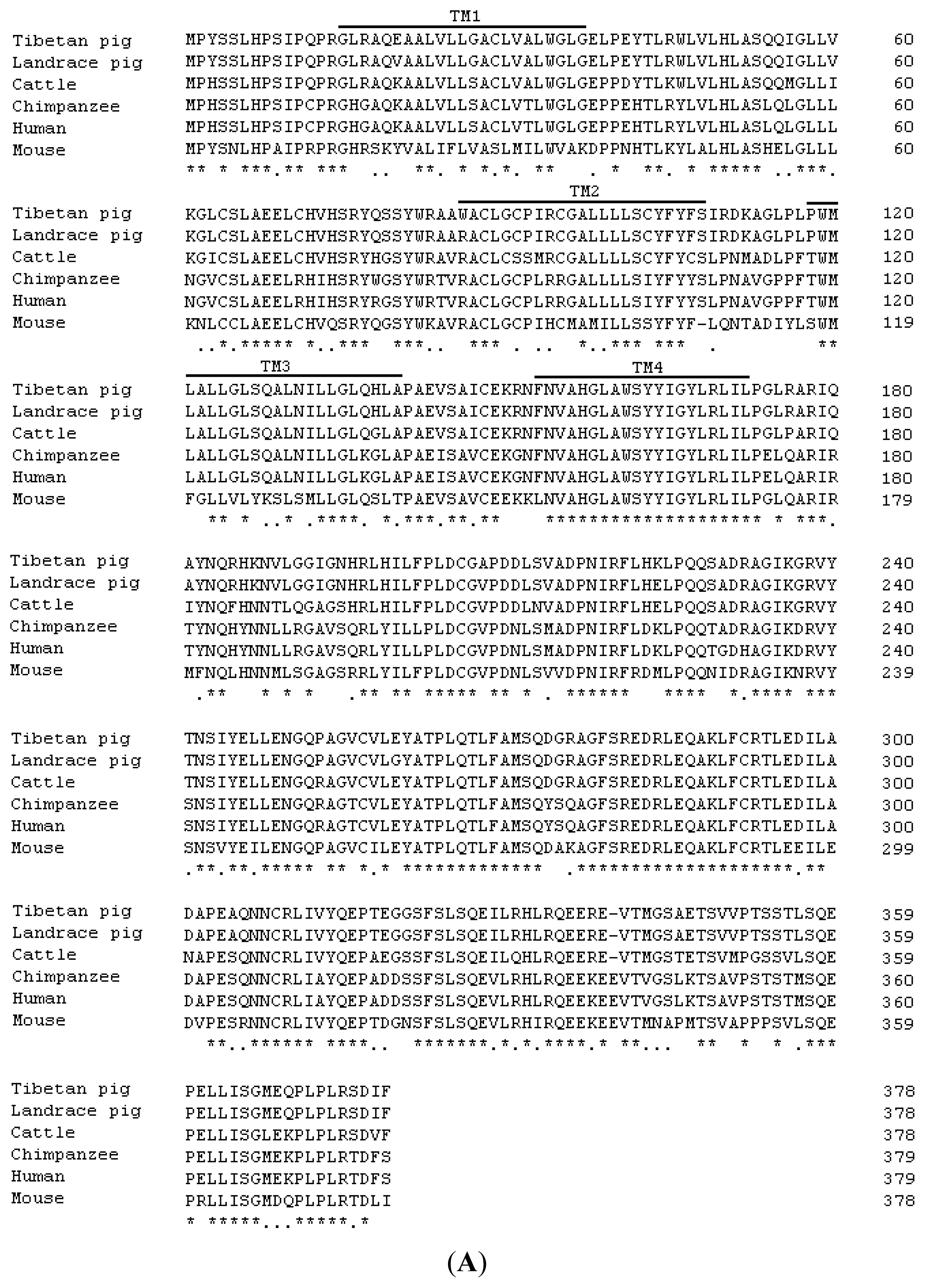
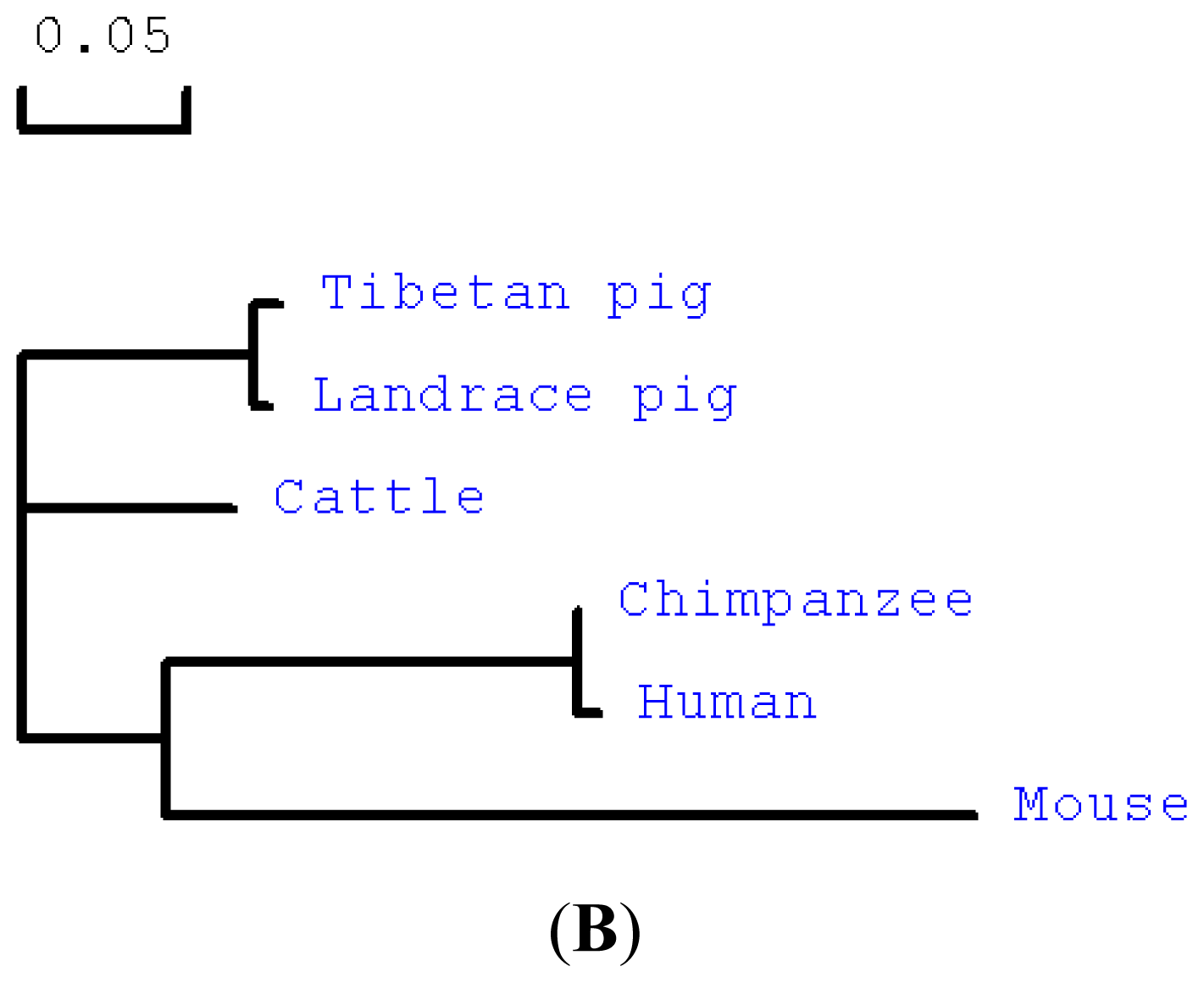
2.1. Cloning and Sequence Analysis of Tibetan Porcine STING

Based on the sequence of Landrace pig STING (GenBank ID: FJ455509), the primers pMD19-T-STING-F and pMD19-T-STING-R were designed and used to amplify the potential STING cDNA sequence from total RNA extracted from skeletal muscle of Tibetan pigs. The entire open reading frame (ORF) of Tibetan porcine STING contains 1137 bp (GenBank ID: JN226147) and encodes 378 amino acid residues (Figure S1). This nucleotide sequence shares 77%, 83%, 84%, and 88% homology with the known STING sequences of mouse (GenBank ID: NM_028261), human (GenBank ID: NM_198282), chimpanzee (GenBank ID: XM_001135484), and cattle (GenBank ID: NM_001046357), respectively. Not surprisingly, the nucleotide sequence of STING in the Tibetan pig is nearly identical to that of Landrace pig reported by Xie et al. [14], with 12 nucleotide differences. Whether some of these 12 differences may be due to single nucleotide polymorphisms need to be identified in future investigations.
The predicted amino acid sequence of the Tibetan porcine STING was compared with mammalian STING amino acid sequences available from GenBank (Figure 1A). There was 69%, 76%, 77% and 86% similarity to that of mouse (NP_082537), human (NP_938023), chimpanzee (XP_001135484) and cattle STING (NP_001039822), respectively. Not surprisingly, the Tibetan porcine STING protein sequence (AEL97644) had 98.68% homology with known Landrace pig sequence (ACJ70708), with 5 amino acid substitutions at residues 20 (Glu-Val), 86 (Trp-Arg), 208 (Ala-Val), 224 (Lys-Glu) and 260 (Glu-Gly). Structural analysis with the SMART program ( http://smart.embl-heidelberg.de/smart/set_mode.cgi?NORMAL=1) showed that Tibetan porcine STING contains four putative transmembrane (TM) domains (aa 15–37, aa 86–108, aa 118–140, and aa 153–172) at its N-terminus (Figure 1A). Similar transmembrane domains have also been reported in the STING sequences of human [13] and Landrace pig [14] and can be predicted in the mouse STING sequence, suggesting that transmembrane domains may be the common features of mammalian STING. In addition, the transmembrane domains of human STING have been demonstrated to be critical to retain this protein on the membrane of endoplasmic reticulum or mitochondria [12,13]. As mentioned above, there were 5 amino acid differences between Landrace and Tibetan porcine STINGs. Among them, an amino acid substitution at residues 20 (Glu-Val) was observed in its transmembrane domains. Whether this substitution may lead to the differences in disease resistance between Landrace and Tibetan pigs need further investigation. A phylogenetic analysis of the amino acid sequence was performed, and the resulting neighbor-joining tree showed that the Tibetan pig has a closer genetic relationship with Landrace pig and cattle than with chimpanzee, human and mouse (Figure 1B).

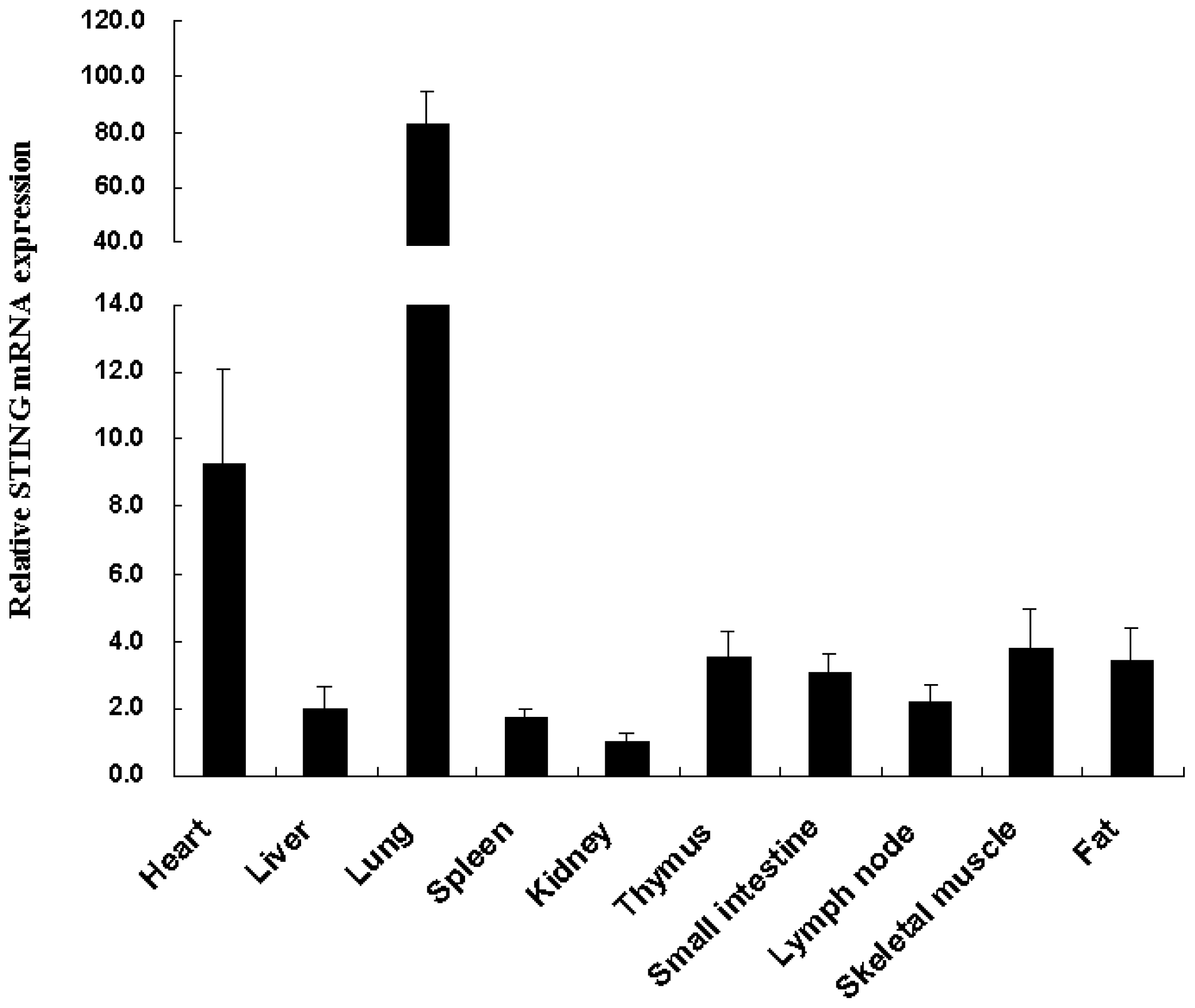
2.2. Tissue Distribution of the Tibetan Porcine STING mRNA

The mRNA expression profile of Tibetan porcine STING was determined by real-time quantitative PCR in various tissues. As shown in Figure 2, Tibetan porcine STING transcripts were most abundant in the lung and heart, followed by the skeletal muscle, thymus, fat, small intestine, lymph node and liver, and to a lesser extent in the spleen and kindey. The tissue distribution pattern of STING has been previously examined in several mammals, including human [11,13], mouse [12] and Landrace pig [14]. Landrace pig STING mRNA was mainly expressed in the spleen, lymph node and lung [14], while Tibetan pig STING mRNA was mainly detected in the lung and heart in the present study. The reason for this discrepancy may be due to the different age and health status of the animals. In addition to the lung and heart, Tibetan porcine STING was also detected in all other examined tissues, indicating that Tibetan porcine STING may be important in the innate immune system in various organs of the pigs.

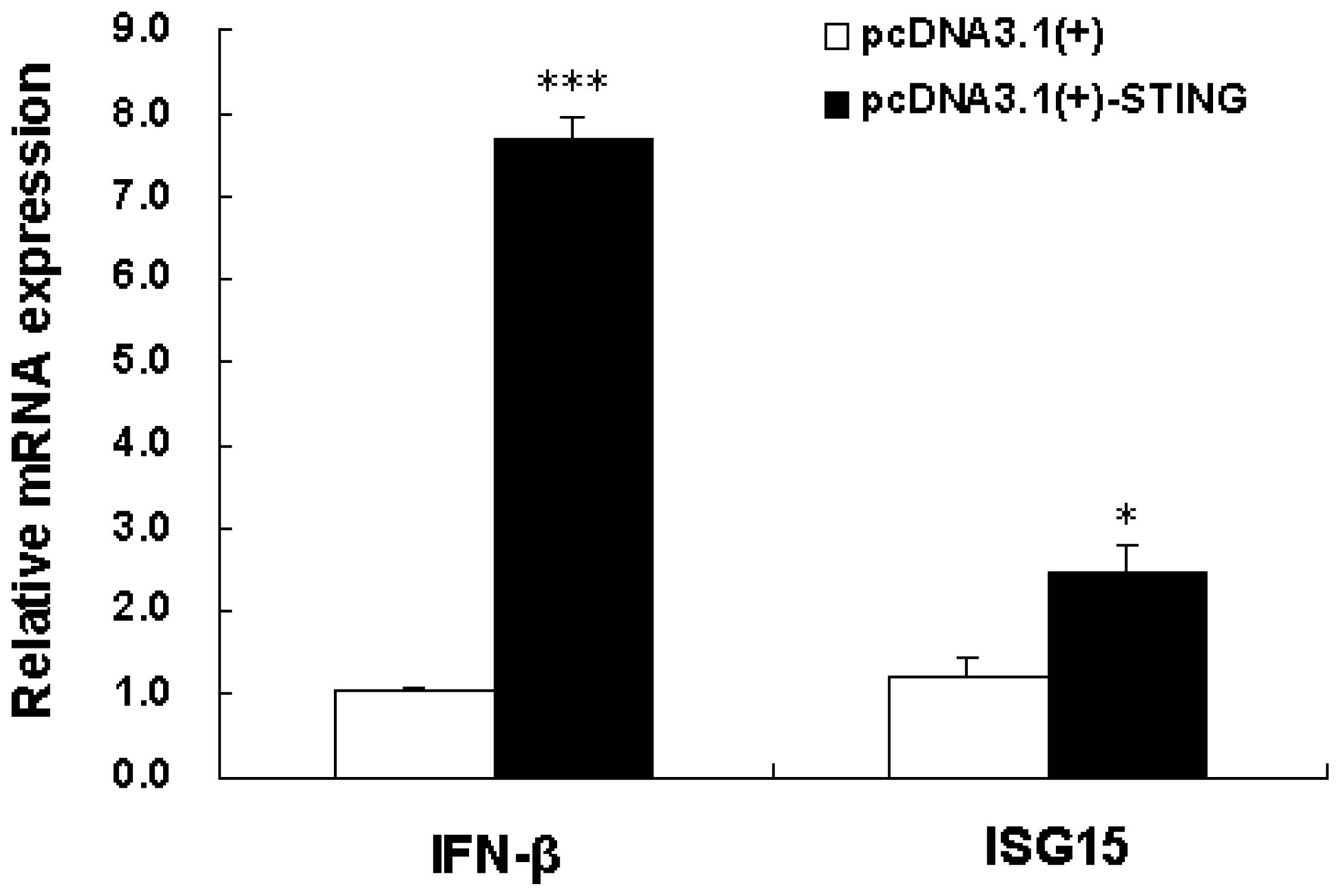
2.3. Overexpression of Tibetan Porcine STING Leads to Upregulation of IFN-β and ISG15 in IPEC-J2 Cells

An inconceivable number of microorganisms live in the intestinal tract of animals and humans [15]. Intestinal epithelial cells participate in the onset and regulation of the intestinal innate immune response to enteric virus infection [16]. During viral infection, IFN-α and IFN-β protect cells against viral invasion by interfering with viral replication. Although STING has been well-characterized as an IFN stimulator since it was cloned in 2008 [1114], there is no report of its function on intestinal epithelial cells. To examine whether Tibetan porcine STING is capable of stimulating expression of IFN-β in the porcine jejunal epithelial cell line IPEC-J2 cells, the Tibetan porcine STING expression plasmid was constructed and transfected into IPEC-J2 cells. Twenty-four hours after the transfection, the mRNA level of Tibetan porcine STING in IPEC-J2 cells transfected with the plasmid pcDNA3.1(+)-STING increased 443.56 ± 70.35 times (n = 3), compared with cells transfected with the empty vector pcDNA3.1(+) control (data not shown). Thus, the mRNA level of IFN-β was tested. As shown in Figure 3, IFN-β was significantly upregulated by Tibetan porcine STING overexpression. ISG15 (an IFN-stimulated gene) is strongly upregulated by type I IFN [17]. Here, we showed that ISG15 was also significantly up-regulated by overexpression of Tibetan porcine STING in IPEC-J2 cells (Figure 3). This is the first study investigating the biological role of STING in intestinal epithelial cells, which lays a foundation for the further study of STING in intestinal innate immunity.

3. Materials and Methods

3.1. Animals and Tissue Sample Collection

Three Tibetan female pigs (body weight of 15.47 ± 0.41 kg) were slaughtered by exsanguination according to protocols approved by the Animal Care Advisory Committee of Sichuan Agricultural University. The heart, liver, lung, spleen, kidney, thymus, small intestine (jejunum), lymph node, skeletal muscle and fat were removed and immediately snap frozen in liquid nitrogen before being stored at −80 °C for RNA isolation.

3.2. RNA Isolation and Reverse Transcription

Total RNA was extracted from collected tissue samples using RNAiso Plus reagent (TaKaRa, Dalian, China) according to the manufacturer’s instructions. The concentrations of total RNA were determined spectrophotometrically using a Beckman Coulter DU800 (Beckman Coulter, Fullerton, CA, USA). One microgram of total RNA from each sample was reverse transcribed in a final volume of 20 μL using a PrimeScript® RT reagent Kit with gDNA Eraser (TaKaRa) according to the manufacturer’s protocols. The first-strand cDNA was subsequently used as a template for PCR.

3.3. Cloning of STING cDNA

The full coding region of STING was obtained by PCR amplification from the total RNA extracted from skeletal muscle of Tibetan pigs. A pair of primers (pMD19-T-STING-F and pMD19-T-STING-R) were designed based on the sequence of Landrace pig STING (GenBank ID: FJ455509) and were as follows: 5′-ATGCCCTACTCCAGCCTGCATC-3′ (forward) and 5′-TCAGAAGATATCTGAGCGG AG-3′ (reverse). The PCR was performed in a 50 μL reaction volume containing 2 μL of the first-strand cDNA, 1 μL each of forward and reverse primers from 10 μM stocks, 21 μL DEPC-treated water, and 25 μL of 2 × Taq PCR MasterMix (Tiangen, Beijing, China). The thermal cycling conditions used were: 1 cycle of 94 °C for 3 min, then 35 cycles of 94 °C for 30 s, 63 °C for 30 s, and 72 °C for 1 min 30 s, and followed by 1 cycle of 72 °C for 7 min. The PCR product was purified, cloned into pMD19-T vector (TaKaRa) and sequenced, resulting in pMD19-T-STING.

3.4. Plasmid Construction

The DNA fragment containing the entire open reading frame (ORF) of Tibetan porcine STING was PCR amplified using the specific primers (pcDNA3.1(+)-STING-F: 5′-TACGAATTCATGCCCTACT CCAGC-3′ and pcDNA3.1(+)-STING-R: 5′-GCTCTAGATCAGAAGATATCTGAGCG-3′) and the plasmid pMD19-T-STING as a template. Primer pcDNA3.1(+)-STING-F introduced an EcoRI site, and primer pcDNA3.1(+)-STING-R contained an XbaI site (underlined). After digestion with EcoRI and XbaI, the PCR product was inserted into the vector pcDNA3.1(+) (Invitrogen, Carlsbad, CA, USA). Proper construction was confirmed by sequencing and was designated as pcDNA3.1(+)-STING.

3.5. Cell Culture

The IPEC-J2 cell line, originally derived from jejunal epithelia of a neonatal unsuckled piglet [18], was a kind gift from Dr. Junjun Wang (College of Animal Science and Technology, China Agricultural University, Beijing, China). IPEC-J2 cells were maintained in Dulbecco modified Eagle medium (DMEM)/Ham’s F-12 (1:1) medium (Invitrogen) supplemented with 10% fetal bovine serum (FBS) (Invitrogen), and antibiotics (100 U/mL penicillin and 100 μg/L streptomycin) (Invitrogen) at 37 °C, 5% CO2 in a humidified atmosphere. Medium was renewed every 2 days.

3.6. Transfection

IPEC-J2 cells were seeded in 6-well plates (Corning, NY, USA) at a density of 2.0 × 10⁵ cells/well 24 h prior to transfection. Two micrograms of the plasmid pcDNA3.1(+)-STING or an equal amount of the empty vector pcDNA3.1(+) were transfected using Lipofectamine 2000 (Invitrogen). At 24 h post-transfection, total RNA was extracted and reverse transcribed according to the above-mentioned method. All plasmids were prepared with Endo-free plasmid kit (Omega, Norcross, GA, USA) following the manufacture’s instruction.

3.7. Real-Time Quantitative PCR

Real-time quantitative PCR was performed in a CFX96 Real-Time PCR Detection System (Bio-Rad, Hercules, CA, USA). The gene-specific primers used are listed in Table 1. The PCR mixture consisted of 2 μL of the first-strand cDNA sample, 1 μL each of forward and reverse primers from 10 μM stocks, 6 μL DEPC-treated water, and 10 μL of 2 . SsoFast EvaGreen Supermix (Bio-Rad). The PCR cycling conditions used were: 45 cycles of 98 °C for 2 s and 60 °C for 5 s. Each primer pair used yielded a single peak in the melting curve and a single band with the expected size in agarose gel. Identities of the PCR products were confirmed by sequencing. Data were analyzed according to the efficiency-corrected comparative Ct method [19] and were normalized by β-actin expression in each sample.

3.8. Statistical Analysis

Data were expressed as mean ± SE. One-way ANOVA and Tukey’s tests (SPSS Inc., Chicago, IL, USA) were performed to assess the statistical significance between treatments. Statistical significance was set at P < 0.05.

4. Conclusions

Tibetan pig is well known for its strong disease resistance. However, little is known about the molecular basis of its strong resistance to disease. In this study, we first cloned Tibetan porcine STING, which encodes a 378-aa protein with 4 transmembrane domains. It is nearly identical to the Landrace pig STING sequence with 12 nucleotide and 5 amino acid differences. Overexpression of Tibetan porcine STING induced IFN-β expression. Future studies will be necessary to investigate whether the difference in STING sequence is related to the difference in disease resistance ability between Landrace and Tibetan pigs.

Supplementary Material

ijms-13-00506-s001.pdf

Acknowledgments

This work was supported by the Earmarked Fund for Modern Agro-industry Technology Research System of China (CARS-36) and the Scientific Research Fund of Sichuan Provincial Education Department (00924800). We thank Hong Hu, Hong Chen, Li Wang and Hui Diao for help in sample preparation.
  • Conflict of InterestThe authors declare no conflict of interest.

References

  1. Kawai, T.; Akira, S. The roles of TLRs, RLRs and NLRs in pathogen recognition. Int. Immunol 2009, 21, 317–337. [Google Scholar]
  2. Palm, N.W.; Medzhitov, R. Pattern recognition receptors and control of adaptive immunity. Immunol. Rev 2009, 227, 221–233. [Google Scholar]
  3. Takeuchi, O.; Akira, S. Innate immunity to virus infection. Immunol. Rev 2009, 227, 75–86. [Google Scholar]
  4. Akira, S.; Uematsu, S.; Takeuchi, O. Pathogen recognition and innate immunity. Cell 2006, 124, 783–801. [Google Scholar]
  5. Takeuchi, O.; Akira, S. Recognition of viruses by innate immunity. Immunol. Rev 2007, 220, 214–224. [Google Scholar]
  6. Alexopoulou, L.; Holt, A.C.; Medzhitov, R.; Flavell, R.A. Recognition of double-stranded RNA and activation of NF-κB by Toll-like receptor 3. Nature 2001, 413, 732–738. [Google Scholar]
  7. Yoneyama, M.; Kikuchi, M.; Natsukawa, T.; Shinobu, N.; Imaizumi, T.; Miyagishi, M.; Taira, K.; Akira, S.; Fujita, T. The RNA helicase RIG-I has an essential function in double-stranded RNA-induced innate antiviral responses. Nat. Immunol 2004, 5, 730–737. [Google Scholar]
  8. Yoneyama, M.; Kikuchi, M.; Matsumoto, K.; Imaizumi, T.; Miyagishi, M.; Taira, K.; Foy, E.; Loo, Y.M.; Gale, M., Jr; Akira, S.; Yonehara, S.; Kato, A.; Fujita, T. Shared and unique functions of the DExD/H-box helicases RIG-I, MDA5, and LGP2 in antiviral innate immunity. J. Immunol 2005, 175, 2851–2858. [Google Scholar]
  9. Nakhaei, P.; Hiscott, J.; Lin, R. STING-ing the antiviral pathway. J. Mol. Cell Biol 2010, 2, 110–112. [Google Scholar]
  10. Zeng, W.; Chen, Z.J. MITAgating viral infection. Immunity 2008, 29, 513–515. [Google Scholar]
  11. Ishikawa, H.; Barber, G.N. STING is an endoplasmic reticulum adaptor that facilitates innate immune signaling. Nature 2008, 455, 674–678. [Google Scholar]
  12. Sun, W.; Li, Y.; Chen, L.; Chen, H.; You, F.; Zhou, X.; Zhou, Y.; Zhai, Z.; Chen, D.; Jiang, Z. ERIS, an endoplasmic reticulum IFN stimulator, activates innate immune signaling through dimerization. Proc. Natl. Acad. Sci. USA 2009, 106, 8653–8658. [Google Scholar]
  13. Zhong, B.; Yang, Y.; Li, S.; Wang, Y.Y.; Li, Y.; Diao, F.; Lei, C.; He, X.; Zhang, L.; Tien, P.; Shu, H.B. The adaptor protein MITA links virus-sensing receptors to IRF3 transcription factor activation. Immunity 2008, 29, 538–550. [Google Scholar]
  14. Xie, L.; Liu, M.; Fang, L.; Su, X.; Cai, K.; Wang, D.; Chen, H.; Xiao, S. Molecular cloning and functional characterization of porcine stimulator of interferon genes (STING). Dev. Comp. Immunol 2010, 34, 847–854. [Google Scholar]
  15. Bäckhed, F.; Ley, R.E.; Sonnenburg, J.L.; Peterson, D.A.; Gordon, J.I. Host-bacterial mutualism in the human intestine. Science 2005, 307, 1915–1920. [Google Scholar]
  16. Shao, L.; Serrano, D.; Mayer, L. The role of epithelial cells in immune regulation in the gut. Semin. Immunol 2001, 13, 163–175. [Google Scholar]
  17. Martensen, P.M.; Justesen, J. Small ISGs coming forward. J. Interferon Cytokine Res 2004, 24, 1–19. [Google Scholar]
  18. Rhoads, J.M.; Chen, W.; Chu, P.; Berschneider, H.M.; Argenzio, R.A.; Paradiso, A.M. L-Glutamine and L-asparagine stimulate Na+-H+ exchange in porcine jejunal enterocytes. Am. J. Physiol 1994, 266, G828–G838. [Google Scholar]
  19. Pfaffl, M.W. A new mathematical model for relative quantification in real-time RT-PCR. Nucleic Acids Res 2001, 29. [Google Scholar] [CrossRef]
Figure 1. Alignment, phylogenetic tree, and architecture of the predicted amino acid sequence for Tibetan porcine STING. (A) Multiple comparison of amino acid sequences of Tibetan pig, Landrace pig, chimpanzee, cattle, human and mouse STING. Four putative transmembrane regions (TM) of Tibetan porcine STING are indicated by bold lines. The numbers on the right of every line refer to positions of the amino acid residues; (B) Phylogenetic analysis of Tibetan porcine STING. The neighbor-joining tree was constructed by DNAMAN. The sequences were derived from the predicted amino acid sequences of Tibetan porcine STING (AEL97644) and the GenBank entries with accession numbers ACJ70708 (Landrace pig), NP_001039822 (cattle), XP_001135484 (chimpanzee), NP_938023 (human), and NP_082537 (mouse). The scale bar is 0.05.
Ijms 13 00506f1aIjms 13 00506f1b
Figure 2. Relative mRNA expression of Tibetan porcine STING in different tissues. Total RNA from different tissues of 3 healthy Tibetan pigs was used to perform the real-time quantitative PCR. Samples were performed in duplicate. The amount of Tibetan porcine STING mRNA was normalized to the amount of β-actin mRNA. Data are presented as mean ± SE (n = 3), in arbitrary units.
Ijms 13 00506f2
Figure 3. Overexpression of Tibetan porcine STING leads to upregulation of IFN-β and ISG15. IPEC-J2 cells were transfected with 2 μg/well of the plasmid pcDNA3.1(+)-STING or the empty vector pcDNA3.1(+). IFN-β and ISG15 mRNA levels were determined using real-time quantitative PCR 24 h after transfection. The amount of IFN-β and ISG15 mRNA was normalized to the amount of β-actin mRNA. Results are the mean and standard errors from three independent experiments performed in duplicate. * P < 0.05 and *** P < 0.001 as compared with empty vector.
Ijms 13 00506f3
Table 1. Primers used in real-time quantitative PCR.
GenePrimerSequenceGenBank IDProduct size
IFN-βForward5′-AAATCGCTCTCCTGATGTGT-3′NM_00100392378 bp
Reverse5′-TGCTCCTTTGTTGGTATCG-3′
ISG15Forward5′-AGCAACGCCTATGAGGTC-3′EU584557101 bp
Reverse5′-AAAGTCAGCCAGAAATGGTC-3′
β-actinForward5′-CCACGAAACTACCTTCAACTCC-3′DQ845171132 bp
Reverse5′-GTGATCTCCTTCTGCATCCTGT-3′

