Communication

Spin Transition Sensors Based on β-Amino-Acid 1,2,4-Triazole Derivative

1
Institute of Condensed Matter and Nanosciences, Université Catholique de Louvain, Place L. Pasteur 1, 1348 Louvain-la-Neuve, Belgium
2
Department of Electrical Engineering and Computer Science, “Stefan cel Mare” University, University Street 13, Suceava 720229, Romania
*
Author to whom correspondence should be addressed.
Int. J. Mol. Sci. 2011, 12(8), 5339-5351; https://doi.org/10.3390/ijms12085339
Submission received: 11 July 2011 / Revised: 11 August 2011 / Accepted: 12 August 2011 / Published: 18 August 2011
(This article belongs to the Special Issue Recent Advances in Molecular Electronics)

Abstract

:
A β-aminoacid ester was successfully derivatized to yield to 4H-1,2-4-triazol-4-yl-propionate (βAlatrz) which served as a neutral bidentate ligand in the 1D coordination polymer [Fe(βAlatrz)3](CF3SO3)2·0.5H2O (1·0.5H2O). The temperature dependence of the high-spin molar fraction derived from 57Fe Mossbauer spectroscopy recorded on cooling below room temperature reveals an exceptionally abrupt single step transition between high-spin and low-spin states with a hysteresis loop of width 4 K (Tc = 232 K and Tc = 228 K) in agreement with magnetic susceptibility measurements. The material presents striking reversible thermochromism from white, at room temperature, to pink on quench cooling to liquid nitrogen, and acts as an alert towards temperature variations. The phase transition is of first order, as determined by differential scanning calorimetry, with transition temperatures matching the ones determined by SQUID and Mössbauer spectroscopy. The freshly prepared sample of 1·0.5H2O, dried in air, was subjected to annealing at 390 K, and the obtained white compound [Fe(βAlatrz)3](CF3SO3)2 (1) was found to exhibit a similar spin transition curve however much temperature was increased by (Tc = 252 K and Tc = 248 K). The removal of lattice water molecules from 1·0.5H2O is not accompanied by a change of the morphology and of the space group, and the chain character is preserved. However, an internal pressure effect stabilizing the low-spin state is evidenced.

Graphical Abstract

1. Introduction

Bistable molecular systems, particularly materials exhibiting captivating scenario of spin crossover (SCO) [1], are versatile switchable units in the thriving field of molecular electronics [2]. In a typical SCO material, the electron repositioning via singlet-quintet transitions is substantiated to be technologically significant [2]. Indeed, the reversible electron transfer from a diamagnetic low-spin (LS, 1A1g) state to a thermally populated paramagnetic high-spin (HS, 5T2g) state is recognized as an entropy driven process and could be addressed thermally, optically, electrically and under pressure/shock with highly profound spectroscopic, optical, magnetic, dielectric readout signal [3]. In the solid state, the presence of intra and intermolecular interactions acts as communication media between iron centers promoting cooperative first order spin transitions leading to a large memory domain [4] that can be suitable for potential applications [5]. Indeed, a SCO compound meeting display and data processing requirements would, in addition, have a good shelf life and an easily detectable optical response, and would ideally operate near room temperature [6,7].
Applications envisioned in these fields largely depend on molecular conformations precursors adopt during the coordination process which directs structure-properties relationships. In some cases, magnetic properties can even be modified by a structural perturbation as demonstrated on a 1D polyelectrolyte system [811]. Our interest in amino acid derivatization was fuelled by promising results shown by 1,2,4-triazole-carboxylate derivatives in synthetic chemistry [12], spin crossover area [13], nanoporous MOFs [14], biological interest in several metallo-proteases [15] and ‘soft’ sacrificial precursors to produce CdO with shape and phase selectivity [16]. As a continuation of our work on amino acid functionalization, [12] ethyl-4H-1,2,4-triazol-4-yl-acetate was used as a prospective precursor for magnetic sensors [13]. Here we introduce a new tailored 4-R-1,2,4-triazole from β-amino-acid, namely 4H-1,2,4-triazol-4-yl-propionate (βAlatrz) (Chart 1). The reason for introducing β-Alanine ethyl ester substituted on the 4 position of a 1,2,4-triazole core was to use an appropriate length of substituent which compromises the distance between 1D chains in complexes which can introduce supramolecular interactions with H-acceptors substituent. Thus, the spin transition (ST) properties of two one-dimensional FeII chains, [Fe(βAlatrz)3](CF3SO3)2·0.5H2O (1·0.5H2O) and its dehydrated form, [Fe(Alatrz)3](CF3SO3)2 (1) were studied.

2. Results and Discussion

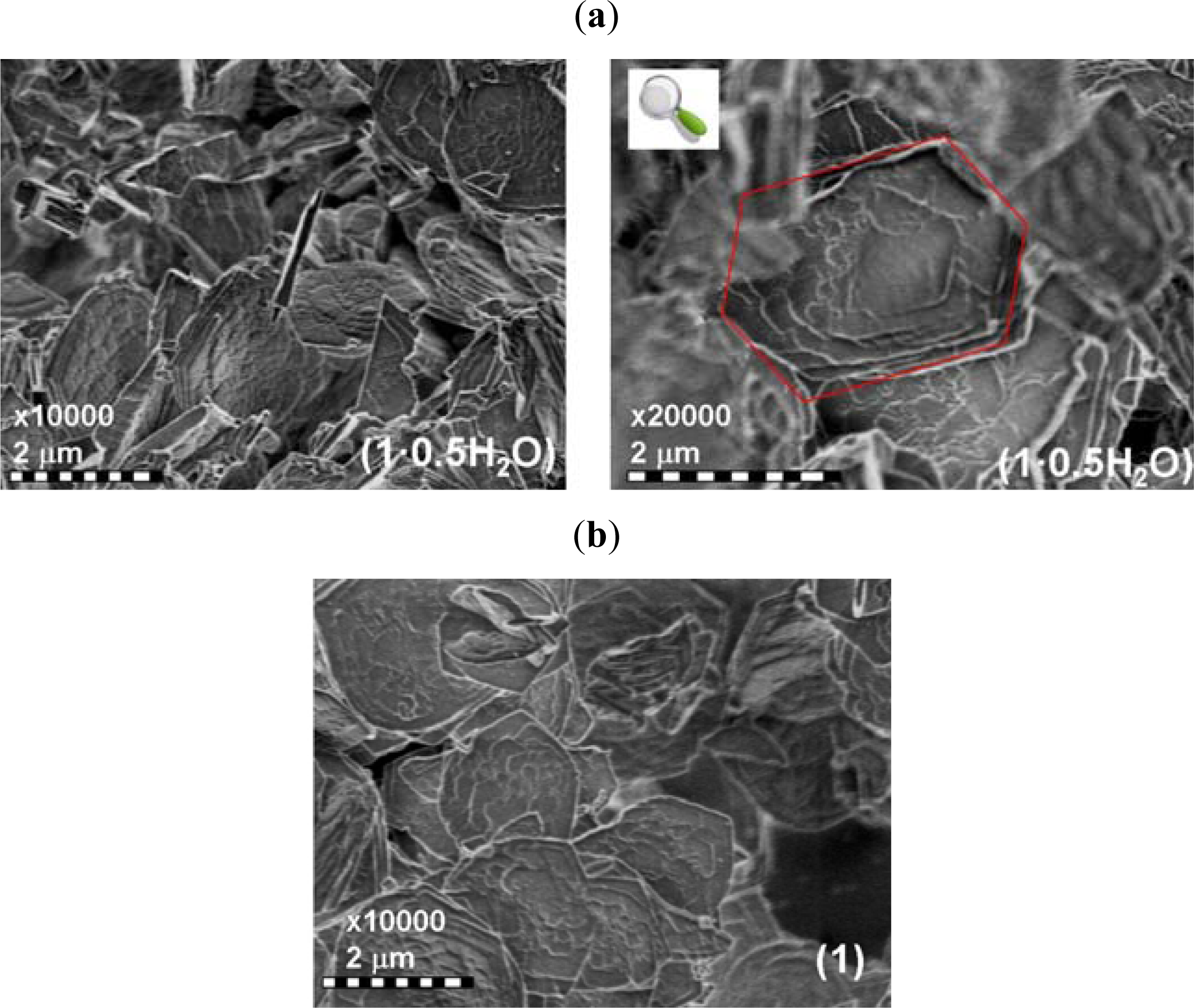
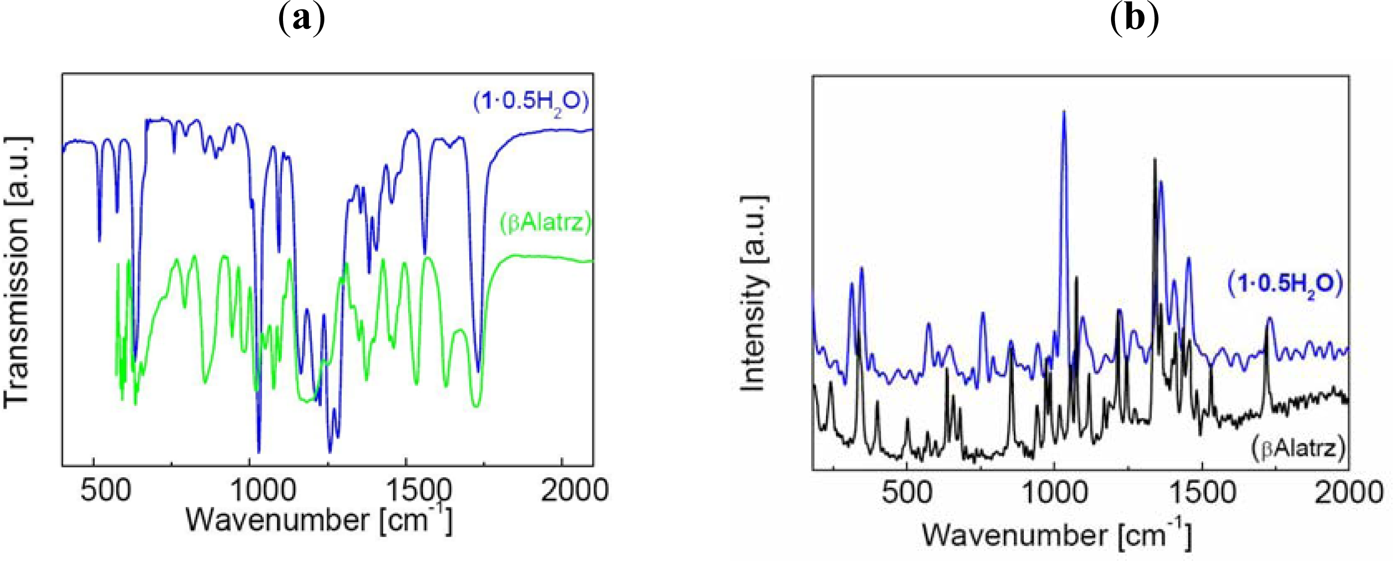
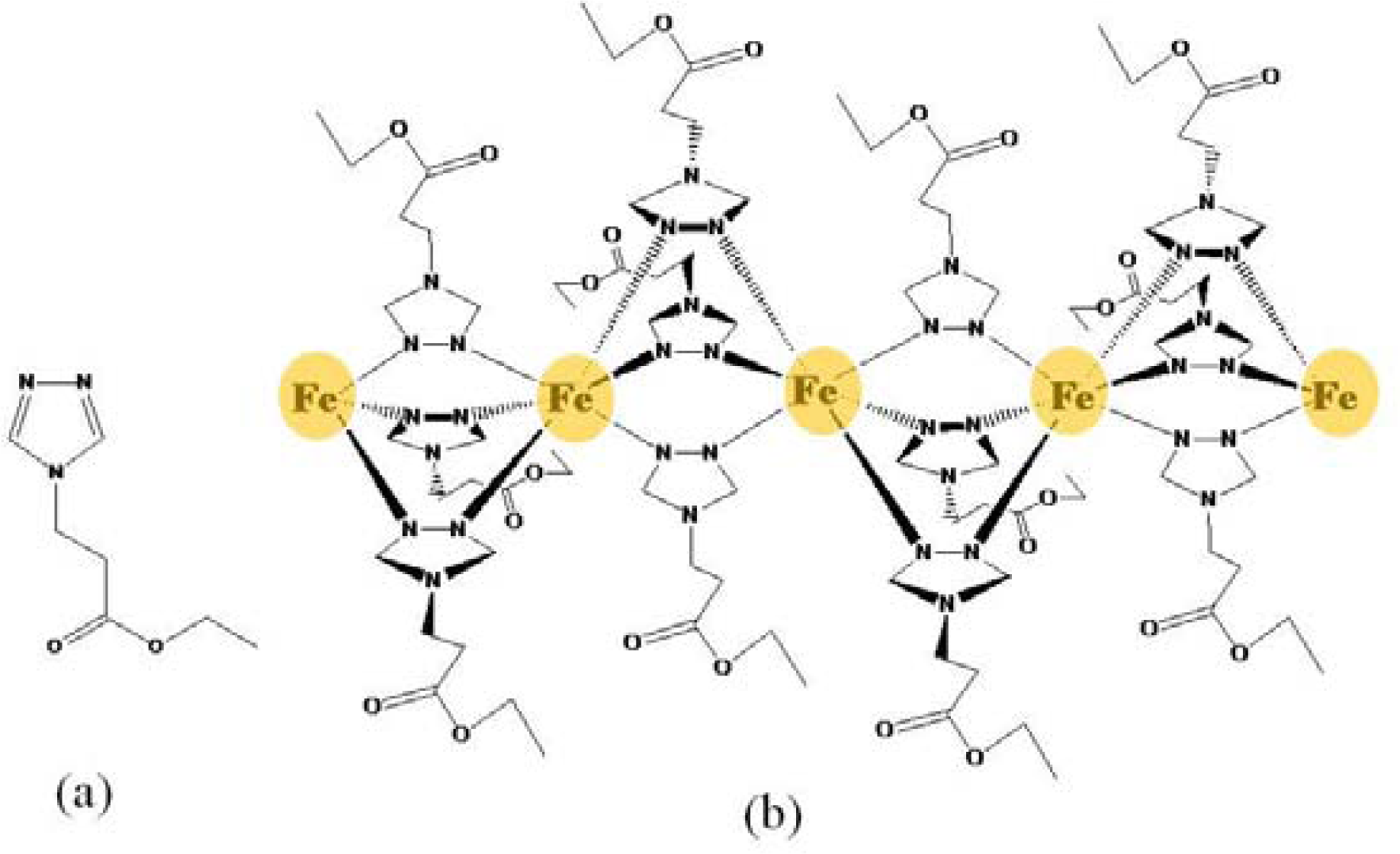
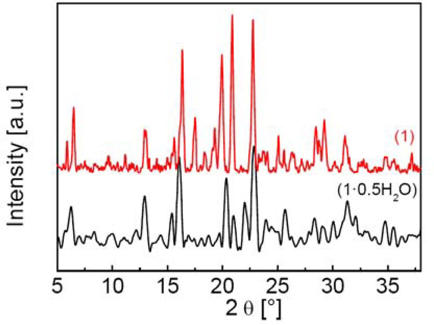
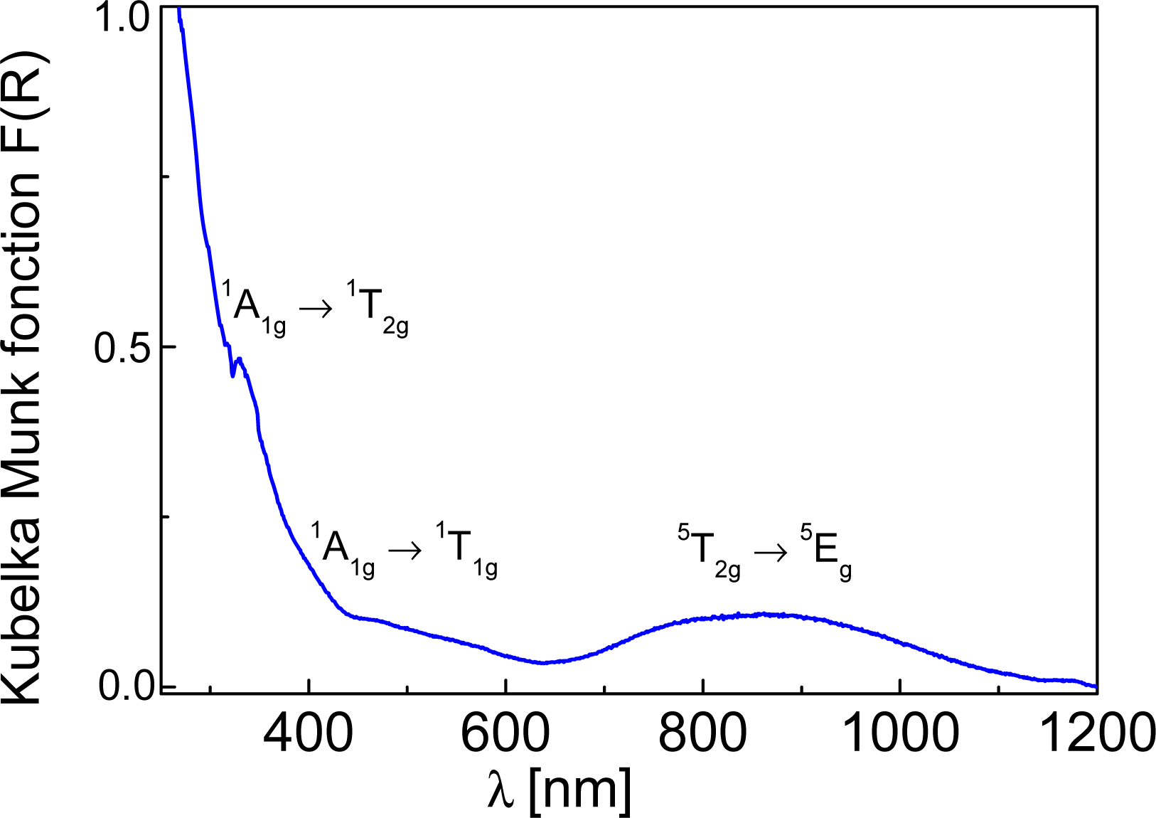
2.1. Preparation and Characterization of 1·0.5H2O and 1

The transamination reaction used in the synthesis of ethyl-4H-1,2,4-triazol-4-yl-acetate and 4H-1,2,4-triazol-4-yl acetic acid [12] proved to be a good synthetic strategy to build a β-amino-acid triazole. The synthetic route, starting with β-Alanine ethyl ester hydrochloride and N,N-Dimethylformamide-azine, helped us to obtain a new molecule, βAlatrz, with a good yield. The synthesis time is relatively longer than the synthesis of previous ligands and the final product had to be purified by “flash” column chromatography.
A 1D coordination polymer was obtained as a white powder by reaction of the corresponding FeII inorganic precursor, prepared in air, [Fe(H2O)6](CF3SO3)2 [17] with a methanolic solution of βAlatrz. This complex was successfully characterized by elemental analysis, TGA-DTA analyses, atomic absorption (AAS), X-Ray powder diffraction, IR, Raman, SEM, DSC, SQUID magnetometry and 57Fe Mössbauer spectroscopy. The thermogravimetric and elemental analyses reveal the presence of guest water molecules, affording the following general formula [Fe(βAlatrz)3](CF3SO3)2·0.5H2O (1·0.5H2O). Compound 1·0.5H2O was dehydrated by annealing, leading to a new compound, [Fe(βAlatrz)3](CF3SO3)2 (1), which was also characterized to reveal the influence of solvent molecules on the spin state. 1·0.5H2O presents a rather crystalline character as revealed from X-ray powder diffraction (XRPD) patterns (Figure 1).
This result is also illustrated by the SEM analysis with blocks of hexagonal shape of about 2 μm widths (Figure 2). FT-IR and Raman analyses were performed: (i) to confirm the identity of the ligand framework before and upon complexation as the ester functionality is susceptible to hydrolysis; (ii) to ascertain the coordination mode of iron to the ligand and the presence of the trifluoromethane sulfonate in the crystal lattice (Figure 3).
Two sharp IR bands at 3,118(s) and 2,983(m) cm−1 of βAlatrz, are assigned to the νCH2 modes [18]. The carboxylic group of ester shows two stretching vibrations corresponding to (C=O) and (C–O) at 1,728(s) and 1,205(s) cm−1, respectively, which are almost unchanged (1,732 and 1,212 cm−1) in 1·0.5H2O ruling out the possible ester hydrolysis and also confirming the non-involvement of carboxylic ester group in coordination. For βAlatrz, the band assigned to ring torsion of triazole at ν = 634(m) cm−1, the νC=N stretching vibration at 1,535(m) cm−1 and the N–N stretching band at ν = 1,022(s) cm−1, are all shifted upon complexation in 1·0.5H2O to 632 cm−1, νC=N = 1,560 cm−1, and νN-N = 1,028 cm−1, respectively.
These values confirm the coordination of the iron to the 1,2,4-triazole ring [19,20]. Indeed, the characteristic bands of the ligand are not only present in the spectra of the complexes but are also shifted towards larger wave numbers, from 3 to 15 cm−1, respectively. This energy increase is due to the deformation of the ligand upon coordination of the metal, the ligand molecule being indeed more constrained to perform vibration and twisting movements [19,21].
For a better understanding of the molecular structure of these ligands, Raman spectra were also collected (Figure 3). The band present at 1,728 cm−1 for βAlatrz is assigned to a C=O vibration. In complexes this band is retained and appears around 1,732 cm−1. The C–C stretching is also active in Raman as a medium band at 1,098 cm−1 for βAlatrz and appears in the same wavenumber in 1·0.5H2O. Presence of the monovalent counter-anion is also confirmed by IR and Raman: IR (cm−1) υ(S–O)∼1,283(s), (1·0.5H2O); Raman (cm−1) υ(S–O)∼1,034(s), 760(m) (1·0.5H2O) [22]. 57Fe Mössbauer spectroscopy confirm for 1·0.5H2O the presence of one FeIIN6 site, and exclude any oxidation product of iron. These spectroscopic data support a linear chain structure with FeII ions linked by triple N1,N2-1,2,4-triazole bridges [23,24].
1·0.5H2O was prepared as a white powders and presents a reversible thermochromism on cooling to pink (Figure 4). These colors depend on the spin state of the FeII centers, as the ST involves a change in the electronic configuration which modifies the absorption spectrum of the complex.
The white color of 1·0.5H2O, is due to the location of the spin-allowed lowest energy d-d transition, 5T2g5Eg, for the HS sites in the near infrared region (∼11,500 cm−1) [25,26]. Two supplementary bands, corresponding to the 1A1g1T1g and 1A1g1T2g d-d transitions of LS FeII sites are observed at ∼18,727 cm−1 and ∼30,300 cm−1, respectively suggesting that some proportion of FeII may be in the LS state at room temperature. The ligand field strengths for the HS and the LS state, given by equations 10DqHS = E(5E) − E(5T2) and 10DqLS = E(1T) − E(1A1) + (E(1T2) − E(1T1))/4, [25,26] allowed us to estimate 10DqHS and 10DqLS to be approximately 11,500 cm−1 and 26,302 cm−1, respectively. These values are characteristic for SCO complexes [25,26]. Presence of LS state at room temperature has been confirmed by 57Fe Mössbauer spectroscopy (vide infra).
The reversibility of the rehydration/dehydration process within a given framework structure with the change of crystallinity can play an important role on the SCO properties [27]. This process was investigated for 1·0.5H2O by thermogravimetric analysis, and the XRD powder pattern and SEM imaging recorded after annealing treatment. The sample was first deposited in a crucible and slowly heated at 1 K/min in air atmosphere (air flow 130 mL/min) to the temperature of complete dehydration (390 K) leading to 1. After a short standby period, the sample was cooled (1 K/min) slowly to reach room temperature revealing no rehydration. Thus the dehydration-rehydration process is irreversible and 1 is air stable which will ease any further physical studies on this compound. Comparison of the powder XRD patterns (Figure 1) and SEM images (Figure 2) of 1·0.5H2O and 1 allow to exclude any framework rupture. Indeed, these two compounds are isostructural, 1 showing higher peak intensities indicating a better crystalline character. No deterioration is observed by SEM.

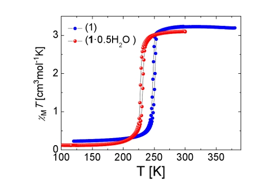
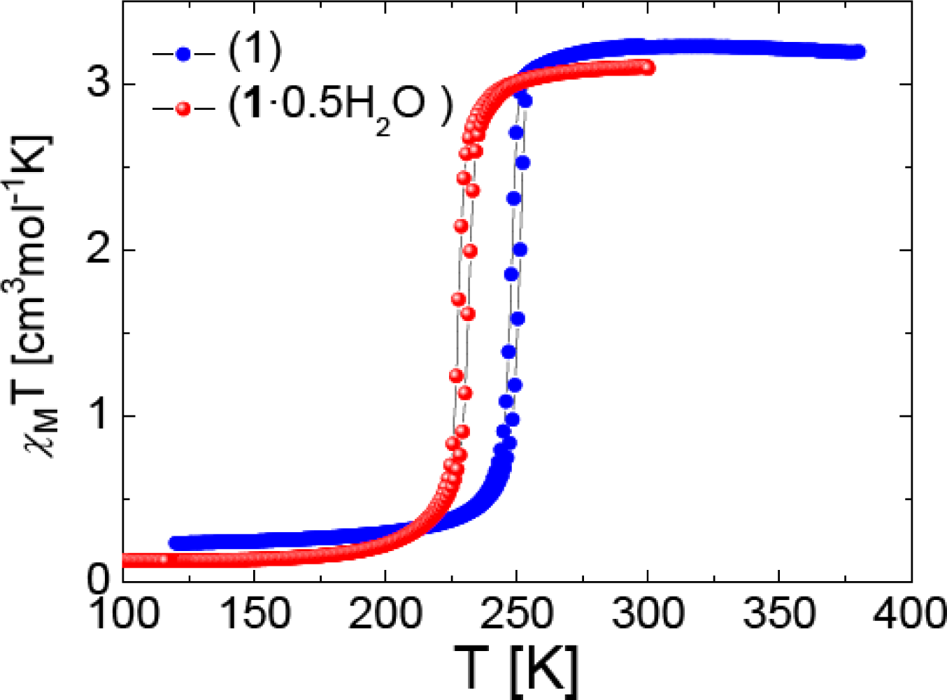
2.2. SQUID Magnetometry

The temperature dependent magnetic properties of 1·0.5H2O and 1 were determined by temperature-dependent susceptibility measurements using a SQUID magnetometer operating at 1000 Oe (Figure 5).
For complex 1·0.5H2O, χMT is 3.10 cm3 K mol−1 at 300 K, which is in the region expected for an FeII complex essentially in the HS state, with a small % of LS species. Upon cooling, χMT remains almost constant until 240 K where it drops sharply to Tc = 228 K down to ∼0.14 cm3 K mol−1 at 77 K, which is typical for a diamagnetic FeII complex with possibly a few HS ions.
Upon warming, the χMT curve differs slightly revealing a small hysteresis width (4 K) at Tc = 232 K for this very sharp spin transition. An hysteretic behavior of width 4 K was observed too for 1 with a ST shifted upwards (Tc = 248 K and Tc = 252 K). The same curve shape is observed too which confirms the preservation of the 1D chain character [27] and does not support the appearance of a structural phase transition following the annealing process. The stabilization of the LS state upon water release is uncommon for this family of complexes [28] and was only observed for the 1D chain compounds [Fe(4-(2′-hydroxyethyl)-1,2,4-triazole)3](anion)2·nH2O (anion = ClO4, n = 2, 0; anion = I, n = 1, 0) [29], [Fe(4-amino-1,2,4-triazole)3](anion)·nH2O (anion = TiF62−, n = 1, 0.5; anion = ZrF62−, n = 0.5, 0) [23] as well as for [Fe(ethyl-4H-1,2,4-triazol-4-yl-acetate)3](ClO4)2·nCH3OH (n = 1,0) but with methanol as solvent [13]. The effect of the release of non-coordinated solvent molecules on the spin state can be translated here as an internal positive pressure effect [5,23].

2.3. 57Fe Mössbauer Spectroscopy

An inspection of electronic and structural features for 1·0.5H2O and 1 using 57Fe Mössbauer spectroscopy was undertaken over the temperature range 77–300 K (Figure 6). At 77 K, the spectrum of 1·0.5H2O consists of two quadrupole doublets of different resonance area fractions. The major quadrupole doublet with isomer shift δLS = 0.51(2) mm/s and quadrupole splitting, ΔEQLS = 0.23(2) mm/s correspond to the LS state of FeII. The presence of an LS quadrupole splitting stems from a lattice contribution to the electric field gradient and therefore reveals a distorted character for the LS octahedron as expected within a chain, where constraints may not be negligible [30]. Another doublet, corresponding to HS FeII ions of weaker population (7%), with parameters (δHS = 1.14(1) mm/s and ΔEQHS = 3.15(2) mm/s) confirms the incomplete nature of the ST at 77 K. Upon warming to 300 K, the intensity of the HS doublet slowly increases to 8% at 225 K, after which it increases dramatically to 87.5% at 300 K, confirming an incomplete thermally induced LS → HS conversion for a single FeII site. The presence of LS ions at room temperature could be related to crystal defects or end of chains. The hysteresis effect is clearly evidenced at 225 K (Figure 6a).
At 80 K, the spectrum of 1 shows a single LS quadrupole doublet (δLS = 0.52(1) mm/s and ΔEQLS = 0.25(2) mm/s) indicating a complete spin transition. The compound remains mostly in the LS state on warming up to 250 K, after which a second quadrupole doublet attributed to HS FeII ions grows in intensity (e.g., at 297 K, δHS = 1.03(1) mm/s and ΔEQHS = 2.75(1) mm/s). The asymmetry of the lines observed in the HS state is attributed to a texture effect. Upon cooling to low temperature, the reverse situation is observed with a clear hysteresis effect at 250 K (Figure 6b).
The isomer shift (δLS∼0.51(1) mm/s at 77–80 K and ΔEQHS = 1.03(1) mm/s at 297–300 K) is not affected by the dehydration process which indicates that non coordinated water molecules are not H-bonded to the triazole ligand [31], but should be located at a remote position to the complex in the crystal lattice, either isolated or hydrogen bonded to the sulfonate group of the non coordinated anions. Their releases have no influence on the structural organization as demonstrated by the similarity in X-ray powder diffraction data. However, a clear influence on the transition temperatures, i.e., on the respective energy levels of the HS and LS states [25], has been detected.

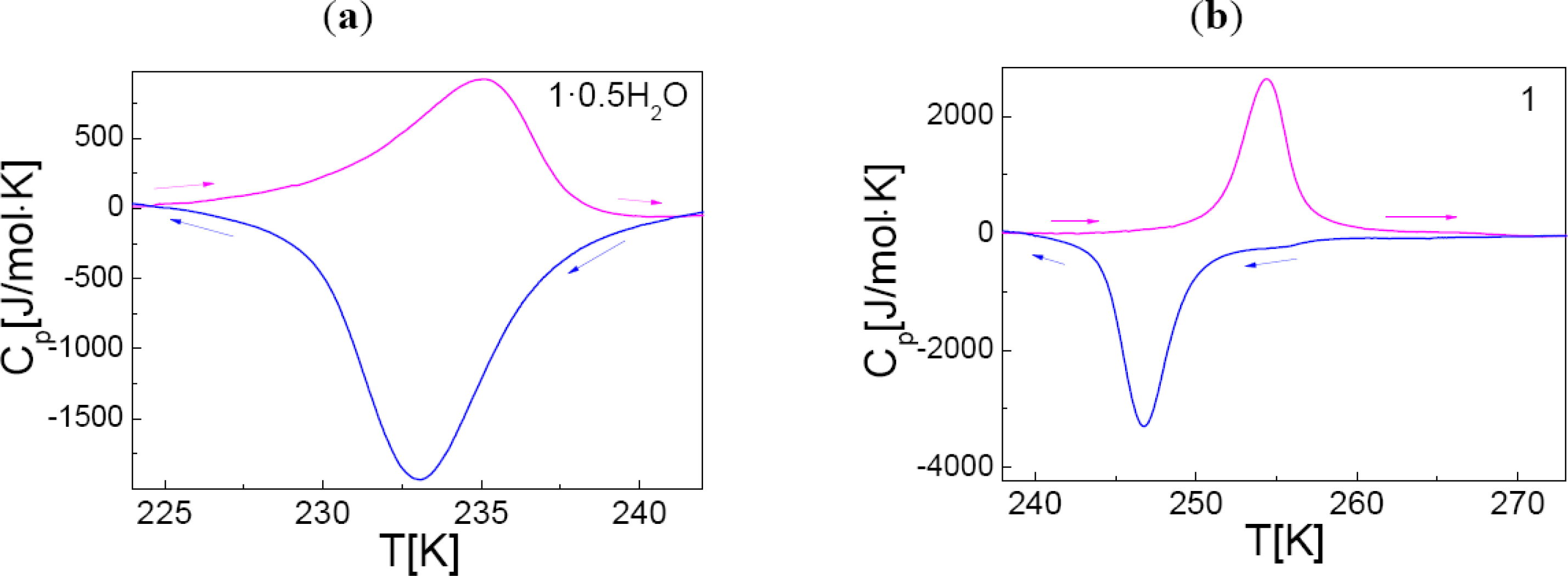
2.4. Differential Scanning Calorimetry

Compound 1·0.5H2O and 1 were investigated by differential scanning calorimetry over the temperature range 100–300 K, at 10 K/min for both cooling and heating modes (Figure 7).
An endothermic peak is observed on warming for 1·0.5H2O at Tmax = 235(1) K and an exothermic peak is recorded at Tmax = 233(1) K, on cooling. These peaks correspond to a first-order phase transition in agreement with the transition temperatures determined by both SQUID and Mössbauer spectroscopy. The enthalpy and entropy variations associated to the active SCO centers are ΔH = 12(2) kJ/mol and ΔS = 51(2) J/mol/K. A similar profile, although with more abrupt peaks, was detected for 1 with phase transitions shifted upwards to Tmax = 254(1) K and Tmax = 247(1) K. The peaks are separated by a narrow temperature domain, which is indicative of the presence of a hysteresis loop. The enthalpy and entropy associated to the ST, considering only the switching sites, were derived as follows: ΔH = 13(1) kJ/mol and ΔS = 59(2) J/mol/K.

3. Experimental Section

3.1. Chemicals

All reagents and solvents were used as received from commercial source: benzene (Fluka analytical), Methanol (VWR), SOCl2 (Sigma-Aldrich), glycine ethyl ester hydrochloride (ACROS), CF3SO3H (ACROS), Fe powder (Merck). Ethyl 4H-1,2,4-triazol-4-yl-propionate (βAlatrz) were prepared by a similar method described in reference [12].

3.2.1. Ethyl 4H-1,2,4-Triazol-4-yl-Propionate (βAlatrz)

N,N-Dimethylformamide azine dihydrochloride (I) and its free base (II) were obtained following the reported method [12]. The free base was recrystallized twice from benzene with charcoal and used in transamination reactions [12]. To a suspension of β-alanine ethyl ester hydrochloride (3 g, 19.53 mmol) in benzene (100 mL) at approx. 60 °C was added solid II (2.14 g, 15.04 mmol) with stirring, obtaining a transparent yellow solution. The mixture was refluxed then (at 130 °C) for 93 h with vigorous stirring. The reaction was monitored by NMR analysis at interval of time. Finally, the solvent was removed under vacuum and a chromatographic purification (SiO2, CH2Cl2 → 5% isopropanol in CH2Cl2) of the yellow oil gave pale yellow oil. Yield 2 g (60%). 1H NMR (300 MHz, CDCl3, 298 K): δ = 8.25 (s, 2H), 4.34 (t, 3H, J = 6.17 Hz), 4.14 (dd, 2H, J = 7.10 Hz & 7.15 Hz), 2.78 (t, 2H, J = 6.17 Hz), 1.23 (t, 3H, J = 7.16 Hz). 13C NMR (300 MHz, CDCl3 298 K): δ = 170.1, 143, 61.6, 40.6, 35.5, 14.1. MS: m/z = 170.03 (M + H+). FTIR (thin film from CH2Cl2 on ZnSe HATR through plate, cm−1): 3,118(s), 1,728(vs), 1,535(s), 1,236(s), 1,205(s), 1,186(s), 1,022(m), 637(s). Anal. Calcd. for C7H11N3O2 (169.18 g/mol): C, 49.70, H, 6.55, N, 24.84; Found C, 49.7, H, 6.81, N, 22.61.

3.2.2. [Fe(βAlatrz)3](CF3SO3)2·0.5H2O (1·0.5H2O)

[Fe(H2O)6](CF3SO3)2 was first synthesized as a very pale green powder using a described procedure [16], starting with an aqueous solution (1 mL) containing an iron powder in excess (2 g) carefully mixed to triflic acid (5 mL, 56.5 mmol). Yield: 9.8 g, 74%. [Fe(H2O)6](CF3SO3)2 (183.2 mg, 0.396 mmol) was dissolved in CH3OH (5 mL) with a pinch of ascorbic acid and added to the above solution of βAlatrz (205.7 mg, 1.216 mmol) dissolved in CH3OH (5 mL). The mixture was stirred for 15 min at room temperature, after which a white precipitate was obtained. It was filtered, washed with CH3OH (2 mL) and dried in air. Yield: 265.9 mg, 75.4%. Anal. for FeC23H34N9O12.5F6S2 (870.54 g/mol): calcd. C, 31.73; H, 3.94; N, 14.48; F, 13.09; S, 7.37; Fe, 6.42%. Found C, 31.64; H, 3.76; N, 14.41; F, 12.12; S, 6.37; Fe, 6.59%. IR (KBr, cm1): υ(C=O)∼1,732(vs), υ(C–O)∼1,211, 1,093(s), υ(C=N)∼1,560(m), υ(C–H out of plane)∼1,028(m), υ(C–H ring torsion)∼632(m), υ(S–O)∼1,282, 1,255 (vs, with shoulders).

3.3. Physical Measurements

Elemental analyses were performed at University College London (UK) and at S.C.A. CNRS Solaize (France). 1H and 13C NMR spectra were recorded at 300 MHz and 75 MHz, respectively, on a Bruker AC300 instrument. The residual solvent peak was used as internal reference. Mass spectral data were obtained on Thermo Finnigan LCQ Ion trap spectrometer (APCI mode). HRMS were carried out on a Micromass Q TOF 2 spectrometer in ESI mode, detecting positive mode. Raman spectra with 1,064 nm excitation were recorded between 2,300 to 400 cm−1 with a Bruker RFS 100/s FT-Raman spectrometer (I = 200 mW) at r.t using a diode-pumped, air-cooled Nd:YAG laser as the excitation source. IR spectra were collected on a Shimadzu FTIR-84005 spectrometer using KBr pellets. Thermogravimetric analyses (TGA) were performed in air (100 mL/min) at the heating rate of 1 °C/min from 293 K to 400 K using a Mettler Toledo TGA/SDTA 851e analyzer. Diffuse reflectance spectra on solids were recorded with a CARY 5E spectrophotometer using polytetrafluoroethylene as a reference. Powder X-ray diffraction patterns were recorded on a Siemens D5000 counter diffractometer working with Cu-Kα radiation and operating at room temperature. The samples were mounted on the support with silicon grease. 57Fe Mössbauer spectra were recorded in transmission geometry over the temperature range (78–300 K) with a conventional Mössbauer spectrometer equipped with a Cyclotron Ltd 57Co(Rh) radioactive source operating at room temperature. The samples were sealed in aluminum foil and mounted on an Oxford nitrogen bath cryostat. The spectra were fitted to the sum of Lorentzians by a least-squares refinement using Recoil 1.05 Mössbauer Analysis Software [32]. All isomer shifts refer to α-Fe at room temperature. Magnetic susceptibilities were measured in the temperature range 4–390 K using a MPMS-XL (7T) SQUID magnetometer. Data were corrected for magnetization of the sample holder and diamagnetic contributions, which were estimated from the Pascal constants. Differential scanning calorimetry measurements were carried out in a He(g) atmosphere using a Perkin-Elmer DSC Pyris 1 instrument equipped with a cryostat and operating down to 98 K. Aluminum capsules were loaded with 20–50 mg of sample and sealed. The heating and cooling rates were fixed at 10 K min−1. Temperatures and enthalpies were calibrated over the temperature range of interest (298–400 K) using the solid-liquid transitions of pure In (99.99%) [33], and the crystal-crystal transitions of pure cyclopentane (≥99%) [34], over the range 78–298 K. Scanning electron microscopy (SEM) was performed using a Gemini Digital Scanning Microscope 982 with 1 kV accelerating voltage with an aluminum sample holder.

4. Concluding Remarks

We have presented two novel FeII 1D ST chain compounds switching in the range 225–250 K. Release of non coordinated water molecules has a paramount effect on the LS state stabilization of 1. Its air stability and absence of solvent will ease further physical measurements, particularly using hydrostatic pressure [5] so as to shift its hysteresis loop towards the room temperature region.

Acknowledgments

We acknowledge financial support from IAP-VI (P6/17) INANOMAT, the Fonds National de la Recherche Scientifique (FNRS) (FRFC 2.4508.08, IISN 4.4507.10), a Concerted Research Action of the “Communauté Française de Belgique” allotted by the Académie Universitaire Louvain and from the European Social Fund through Sectorial Operational Program Human Resources: PRiDE (No POSDRU/89/1.5/S/57083. J. Marchand-Brynaert is a senior research associate of the F.R.S.-FNRS.

References

  1. Gütlich, P; Garcia, Y; Goodwin, HA. Spin crossover phenomena in Fe(II) complexes. Chem. Soc. Rev 2000, 29, 419–427. [Google Scholar]
  2. Bousseksou, A; Molnár, G; Salmon, L; Nicolazzi, W. Molecular spin crossover phenomenon: Recent achievements and prospects. Chem. Soc. Rev 2011, 40, 3313–3118. [Google Scholar]
  3. Gütlich, P; Garcia, Y; Spiering, H. Magnetism: From Molecules to Materials, 1st ed; Miller, JS, Drillon, M, Eds.; Wiley-VCH: Hoboken, NJ, USA, 2003; Volume IV. [Google Scholar]
  4. Weber, B; Bauer, W; Obel, J. An iron (II) spin-crossover complex with a 70 K wide thermal hysteresis loop. Angew. Chem. Int. Ed 2008, 47, 10098–10101. [Google Scholar]
  5. Garcia, Y; Ksenofontov, V; Gütlich, P. Spin transition molecular materials: New sensors. Hyperfine Interact 2002, 139–140, 543–551. [Google Scholar]
  6. Kahn, O; Krober, J; Jay, C. Spin Transition molecular materials for displays and data recording. Adv. Mater 1992, 4, 718–728. [Google Scholar]
  7. Kahn, O; Jay-Martinez, C. Spin-transition polymers: From molecular materials toward memory devices. Science 1998, 279, 44–48. [Google Scholar]
  8. Bodenthin, Y; Pietsch, U; Möhwald, H; Kurth, DG. Inducing spin crossover in Metallo-supramolecular polyelectrolytes through an amphiphilic phase transition. J. Am. Chem. Soc 2005, 127, 3110–3115. [Google Scholar]
  9. Bodenthin, Y; Pietsch, U; Grenzer, J; Geue, T; Möhwald, H; Kurth, DG. Structure and temperature behavior of metallo-supramolecular assemblies. J. Phys. Chem. B 2005, 109, 12795–12799. [Google Scholar]
  10. Bodenthin, Y; Schwarz, G; Tomkowicz, Z; Geue, T; Haase, W; Pietsch, U; Kurth, DG. Liquid crystalline phase transition induces spin crossover in a polyelectrolyte amphiphile complex. J. Am. Chem. Soc 2009, 131, 2934–2941. [Google Scholar]
  11. Schwarz, G; Bodenthin, Y; Tomkowicz, Z; Haase, W; Geue, T; Kohlbrecher, J; Pietsch, U; Kurth, DG. Tuning the structure and the magnetic properties of metallo-supramolecular polyelectrolyte-amphiphile complexes. J. Am. Chem. Soc 2011, 133, 547–558. [Google Scholar]
  12. Naik, AD; Marchand-Brynaert, J; Garcia, Y. A simplified approach to N- and N,N’-linked 1,2,4-triazoles by transamination. Synthesis 2008, 1, 149–154. [Google Scholar]
  13. Dîrtu, MM; Naik, AD; Marchand-Brynaert, J; Garcia, Y. Room temperature hysteretic spin transition in 1D iron(II) coordination polymers. J. Phys. Conf. Ser 2010, 217, 012085. [Google Scholar]
  14. Naik, AD; Dîrtu, MM; Léonard, A; Tinant, B; Marchand-Brynaert, J; Su, BL; Garcia, Y. Engineering three-dimensional chains of porous nanoballs from a 1,2,4-triazole-carboxylate supramolecular synthon. Cryst. Growth Des 2010, 10, 1798–1807. [Google Scholar]
  15. Naik, AD; Beck, J; Dîrtu, MM; Bebrone, C; Tinant, B; Robeyns, K; Marchand-Brynaert, J; Garcia, Y. Zinc complexes with 1,2,4-triazole functionalized amino acid derivatives: Synthesis, structure and beta-lactamase assay. Inorg. Chim. Acta 2011, 368, 21–28. [Google Scholar]
  16. Dîrtu, MM; Neuhausen, C; Naik, AD; Léonard, A; Robert, F; Marchand-Brynaert, J; Su, BL; Garcia, Y. Superlative scaffold of 1,2,4-Triazole derivative of glycine steering linear chain to a chiral helicate. Cryst. Growth Des 2011, 11, 1375–1384. [Google Scholar]
  17. Hagen, KS. Iron(II) Triflate salts as convenient substitutes for perchlorate salts: Crystal structures of [Fe(H2O)6](CF3SO3)2 and Fe(MeCN)4(CF3SO3)2. Inorg. Chem 2000, 39, 5867–5869. [Google Scholar]
  18. Chwaleba, D; Ilczyszyn, MM; Ilczyszyn, M; Ciunik, Z. Glycine-methanesulfonic acid (1:1) and glycine-p-toluenesulfonic acid (1:1) crystals: Comparison of structures, hydrogen bonds, and vibrations. J. Mol. Struct 2007, 831, 119–134. [Google Scholar]
  19. Haasnoot, JG; Vos, G; Groeneveld, WL. 1,2,4-Triazole complexes, III. Complexes of transition metal(II) nitrates and fluoroborates. Z. Naturforsch. B 1977, 32, 1421–1430. [Google Scholar]
  20. Adriaanse, JH; Askes, SHC; van Bree, Y; van Oudheusden, S; van den Bos, ED; Gunay, E; Mutikainen, I; Turpeinen, U; van Albada, GA; Haasnoot, JG; et al. Coordination chemistry of 5,6,7-trimethyl-[1,2,4]triazolo[1,5-a]pyrimidine with first-row transition-metal salts: Synthesis, spectroscopy and single-crystal structures, with counter-anion dependence of the structures. Polyhedron 2009, 28, 3143–3149. [Google Scholar]
  21. Sinditskii, VP; Fogelzang, AE; Dutov, MD; Sokol, VI; Serushkin, VV; Svetlov, BS; Poraikoshits, MA. Structure of complex-compounds of metal chlorides, sulfates, nitrates and perchlorates with carbohydrazine. Zh. Neorg. Khim 1987, 32, 1944–1949. [Google Scholar]
  22. Nakamoto, K. Infrared and Raman Spectra of Inorganic and Coordination Compounds, Part B, 5th ed; Wiley-Interscience: New York, NY, USA, 1997. [Google Scholar]
  23. Dîrtu, MM; Rotaru, A; Gillard, D; Linares, J; Codjovi, E; Tinant, B; Garcia, Y. Prediction of the spin transition temperature in FeII one-dimensional coordination polymers: An anion based database. Inorg. Chem 2009, 48, 7838–7852. [Google Scholar]
  24. Dîrtu, MM; Neuhausen, C; Naik, AD; Rotaru, A; Spinu, L; Garcia, Y. Insights into the origin of cooperative effects in the spin transition of [Fe(NH2trz)3](NO3)2: The role of supramolecular interactions evidenced in the crystal structure of [Cu(NH2trz)3](NO3)2·H2O. Inorg. Chem 2010, 49, 5727–5736. [Google Scholar]
  25. Gütlich, P; Hauser, A; Spiering, H. Thermal and optical switching of iron(II) complexes. Angew. Chem., Int. Ed 1994, 33, 2024–2054. [Google Scholar]
  26. Gütlich, P; Jung, J; Goodwin, HA. Molecular Magnetism: From Molecular Assemblies to the Devices; Coronado, E, Delhaès, P, Gatteschi, D, Miller, JS, Eds.; Springer: Berlin, Germany, 1996; p. 327. [Google Scholar]
  27. Garcia, Y; van Koningsbruggen, PJ; Lapouyade, R; Fournès, L; Rabardel, L; Kahn, O; Ksenofontov, V; Levchenko, G; Gütlich, P. Influences of temperature, pressure, and lattice solvents on the spin transition regime of the polymeric compound [Fe(hyetrz)3]A2·3H2O (hyetrz = 4-(2′-hydroxyethyl)-1,2,4-triazole and A = 3-nitrophenylsulfonate). Chem. Mater 1998, 10, 2426–2433. [Google Scholar]
  28. Garcia, Y; Niel, V; Muñoz, MC; Real, JA. Spin crossover in 1D, 2D and 3D polymeric Fe(II) networks; Spin crossover in transition metal compounds. Top. Curr. Chem 2004, 233, 229–257. [Google Scholar]
  29. Garcia, Y; van Koningsbruggen, PJ; Lapouyade, R; Rabardel, L; Kahn, O; Wieczorek, M; Bronisz, R; Ciunik, Z; Rudolf, MF. Synthesis and spin-crossover characteristics of polynuclear 4-(2′-hydroxyethyl)-1,2,4-triazole Fe(II) molecular materials. C R Acad Sci 1998, IIc, 523–532. [Google Scholar]
  30. Dîrtu, MM; Garcia, Y; Nica, M; Rotaru, A; Linares, J; Varret, F. Iron(II) spin transition 1,2,4-triazole chain compounds with novel inorganic fluorinated counteranions. Polyhedron 2007, 26, 2259–2263. [Google Scholar]
  31. Garcia, Y; Ksenofontov, V; Mentior, S; Dîrtu, MM; Gieck, C; Bhatthacharjee, A; Gütlich, P. Rapid cooling experiments and use of an anionic nuclear probe to sense the spin transition of the 1D coordination polymers [Fe(NH2trz)3]SnF6·nH2O (NH2trz = 4-amino-1,2,4-triazole). Chem. Eur. J 2008, 14, 3745–3758. [Google Scholar]
  32. Recoil, Mössbauer Spectral Analysis Software for Windows, Version 1.0; Department of Physics, University of Ottawa: Ottawa, ON, Canada, 1998.
  33. Breuer, KH; Eysel, W. The calorimetric calibration of differential scanning calorimetry cells. Thermochim. Acta 1982, 57, 317–329. [Google Scholar]
  34. Rotaru, A; Dîrtu, MM; Enachescu, C; Tanasa, R; Linares, J; Stancu, A; Garcia, Y. Calorimetric measurements of diluted spin crossover complexes [FexM1−x(btr)2(NCS)2]·H2O with MII = Zn and Ni. Polyhedron 2009, 28, 2531–2536. [Google Scholar]
Figure 1. Top: X-ray powder diffraction pattern of 1, (thermally treated compound in TGA); bottom: diffractogram of 1·0.5H2O (freshly prepared).
Ijms 12 05339f1
Figure 2. (a) SEM images of microcrystalline particles of 1·0.5H2O at 293 K; a selected crystal is highlighted in red; and (b) SEM analysis of 1 confirms the framework integrity preservation after thermal treatment.
Ijms 12 05339f2
Figure 3. (a) IR and (b) Raman spectra of the ligand (βAlatrz) and complex (1·0.5H2O), over the range 400–2,100 cm−1 and 180–2,000 cm−1, respectively. The IR and Raman spectra for 1·0.5H2O and 1 (not shown) are identical, except around 3,500 cm−1 with ν(O–H) of water molecules clearly identified for 1·0.5H2O.
Ijms 12 05339f3
Figure 4. UV-Vis diffuse reflectance spectrum of 1·0.5H2O showing d–d transitions at 293 K.
Ijms 12 05339f4
Figure 5. Thermal variation of χMT of 1·0.5H2O (depicted in red) and 1 (depicted in blue).
Ijms 12 05339f5
Figure 6. 57Fe Mössbauer spectra cycles for 1·0.5H2O (a) and 1 (b) at selected temperatures. Grey and dark grey correspond to the high-spin (HS) and low-spin (LS) doublets, respectively.
Ijms 12 05339f6
Figure 7. Heat capacity profiles for 1·0.5H2O and 1. The arrows indicate the cooling (←) and warming (→) modes, respectively.
Ijms 12 05339f7
Chart 1. Molecular structure of (a) ethyl 4H-1,2,4-triazol-4-yl-propionate (βAlatrz); and (b) complex [Fe(βAlatrz)3]2+.
Ijms 12 05339f8

