The Role of Bloom Index of Gelatin on the Interaction with Retinal Pigment Epithelial Cells

Abstract
:1. Introduction
2. Results and Discussion
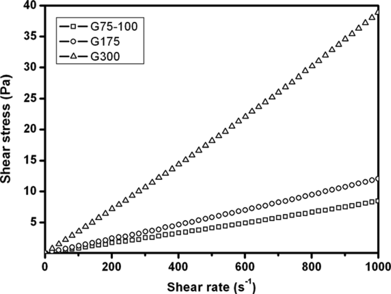
2.1. Rheological Measurements
2.2. Cell Proliferation Assays
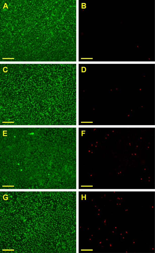
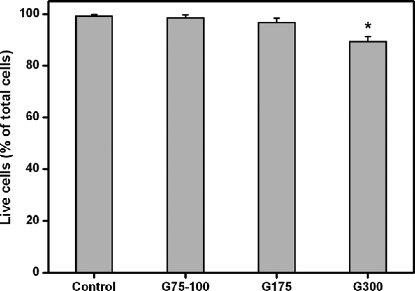
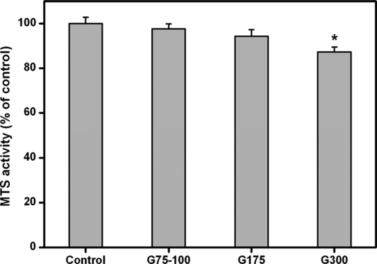
2.3. Cell Viability Assays
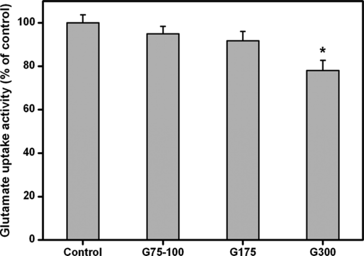
2.4. Glutamate Uptake Measurements
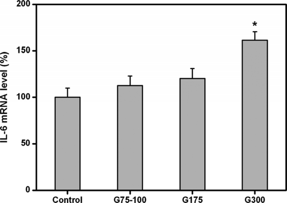
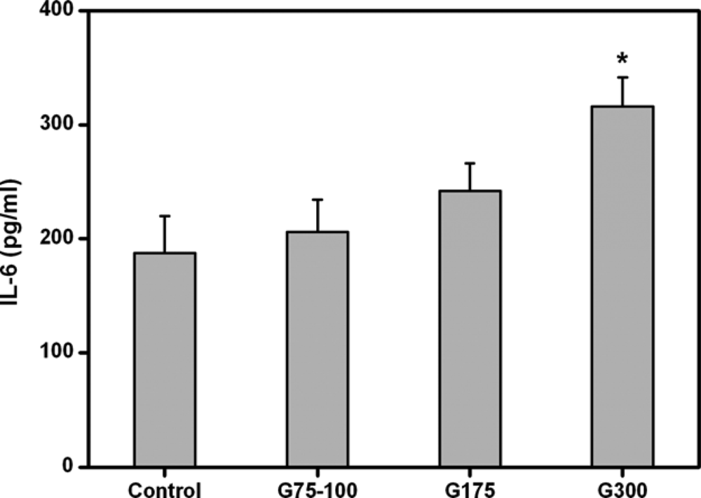
2.5. Cytokine Expression Analyses
3. Experimental Section
3.1. Materials
3.2. Preparation of Gelatin Membranes
3.3. Rheological Measurements
3.4. Human RPE Cell Line Cultures
3.5. In Vitro Biocompatibility Tests
3.5.1. Cell Proliferation Assays
3.5.2. Cell Viability Assays
3.5.3. Glutamate Uptake Measurements
3.5.4. Cytokine Expression Analyses
3.6. Statistics
4. Conclusions
Acknowledgments
References and Notes
- Djagny, KB; Wang, Z; Xu, S. Gelatin: A valuable protein for food and pharmaceutical industries: review. Crit. Rev. Food Sci. Nutr 2001, 41, 481–492. [Google Scholar]
- Bigi, A; Cojazzi, G; Panzavolta, S; Rubini, K; Roveri, N. Mechanical and thermal properties of gelatin films at different degrees of glutaraldehyde crosslinking. Biomaterials 2001, 22, 763–768. [Google Scholar]
- Tabata, Y; Ikada, Y. Protein release from gelatin matrices. Adv. Drug Deliv. Rev 1998, 31, 287–301. [Google Scholar]
- Bussemer, T; Dashevsky, A; Bodmeier, R. A pulsatile drug delivery system based on rupturable coated hard gelatin capsules. J. Control. Release 2003, 93, 331–339. [Google Scholar]
- Fan, H; Zhang, C; Li, J; Bi, L; Qin, L; Wu, H; Hu, Y. Gelatin microspheres containing TGF-β3 enhance the chondrogenesis of mesenchymal stem cells in modified pellet culture. Biomacromolecules 2008, 9, 927–934. [Google Scholar]
- Ethirajan, A; Schoeller, K; Musyanovych, A; Ziener, U; Landfester, K. Synthesis and optimization of gelatin nanoparticles using the miniemulsion process. Biomacromolecules 2008, 9, 2383–2389. [Google Scholar]
- Lai, JY; Chen, KH; Hsu, WM; Hsiue, GH; Lee, YH. Bioengineered human corneal endothelium for transplantation. Arch. Ophthalmol 2006, 124, 1441–1448. [Google Scholar]
- Lai, JY; Hsiue, GH. Functional biomedical polymers for corneal regenerative medicine. React. Funct. Polym 2007, 67, 1284–1291. [Google Scholar]
- Hsiue, GH; Lai, JY; Chen, KH; Hsu, WM. A novel strategy for corneal endothelial reconstruction with a bioengineered cell sheet. Transplantation 2006, 81, 473–476. [Google Scholar]
- Lai, JY; Lu, PL; Chen, KH; Tabata, Y; Hsiue, GH. Effect of charge and molecular weight on the functionality of gelatin carriers for corneal endothelial cell therapy. Biomacromolecules 2006, 7, 1836–1844. [Google Scholar]
- Lai, JY; Chen, KH; Hsiue, GH. Tissue-engineered human corneal endothelial cell sheet transplantation in a rabbit model using functional biomaterials. Transplantation 2007, 84, 1222–1232. [Google Scholar]
- Choi, YS; Hong, SR; Lee, YM; Song, KW; Park, MH; Nam, YS. Study on gelatin-containing artificial skin: I. Preparation and characteristics of novel gelatin-alginate sponge. Biomaterials 1999, 20, 409–417. [Google Scholar]
- Hsiue, GH; Lai, JY; Lin, PK. Absorbable sandwich-like membrane for retinal-sheet transplantation. J. Biomed. Mater. Res 2002, 61, 19–25. [Google Scholar]
- Lai, JY; Lin, PK; Hsiue, GH; Cheng, HY; Huang, SJ; Li, YT. Low Bloom strength gelatin as a carrier for potential use in retinal sheet encapsulation and transplantation. Biomacromolecules 2009, 10, 310–319. [Google Scholar]
- Guidoin, R; Marceau, D; Rao, TJ; King, M; Merhi, Y; Roy, PE; Martin, L; Duval, M. In vitro and in vivo characterization of an impervious polyester arterial prosthesis: The Gelseal Triaxial® graft. Biomaterials 1987, 8, 433–441. [Google Scholar]
- Lu, PL; Lai, JY; Tabata, Y; Hsiue, GH. A methodology based on the “anterior chamber of rabbit eyes” model for noninvasively determining the biocompatibility of biomaterials in an immune privileged site. J. Biomed. Mater. Res. A 2008, 86, 108–116. [Google Scholar]
- Usta, M; Piech, DL; MacCrone, RK; Hillig, WB. Behavior and properties of neat and filled gelatins. Biomaterials 2003, 24, 165–172. [Google Scholar]
- Bigi, A; Panzavolta, S; Rubini, K. Relationship between triple-helix content and mechanical properties of gelatin films. Biomaterials 2004, 25, 5675–5680. [Google Scholar]
- Saxena, A; Sachin, K; Bohidar, HB; Verma, AK. Effect of molecular weight heterogeneity on drug encapsulation efficiency of gelatin nano-particles. Colloid Surf. B-Biointerfaces 2005, 45, 42–48. [Google Scholar]
- Segtnan, VH; Isaksson, T. Temperature, sample and time dependent structural characteristics of gelatin gels studied by near infrared spectroscopy. Food Hydrocolloid 2004, 18, 1–11. [Google Scholar]
- Peng, HT; Martineau, L; Shek, PN. Hydrogel-elastomer composite biomaterials: 3. Effects of gelatin molecular weight and type on the preparation and physical properties of interpenetrating polymer networks. J. Mater. Sci.-Mater. Med 2008, 19, 997–1007. [Google Scholar]
- Lu, PL; Lai, JY; Ma, DHK; Hsiue, GH. Carbodiimide cross-linked hyaluronic acid hydrogels as cell sheet delivery vehicles: Characterization and interaction with corneal endothelial cells. J. Biomater. Sci.-Polym. Ed 2008, 19, 1–18. [Google Scholar]
- Wood, JPM; Chidlow, G; Graham, M; Osborne, NN. Energy substrate requirements of rat retinal pigmented epithelial cells in culture: Relative importance of glucose, amino acids, and monocarboxylates. Invest. Ophthalmol. Vis. Sci 2004, 45, 1272–1280. [Google Scholar]
- del Priore, LV; Tezel, TH; Kaplan, HJ. Survival of allogeneic porcine retinal pigment epithelial sheets after subretinal transplantation. Invest. Ophthalmol. Vis. Sci 2004, 45, 985–992. [Google Scholar]
- Ghosh, F; Juliusson, B; Arnér, K; Ehinger, B. Partial and full-thickness neuroretinal transplants. Exp. Eye Res 1999, 68, 67–74. [Google Scholar]
- Katz, EP; Li, ST. The intermolecular space of reconstituted collagen fibrils. J. Mol. Biol 1973, 73, 351–369. [Google Scholar]
- Sáenz, DA; Goldin, AP; Minces, L; Chianelli, M; Sarmiento, MIK; Rosenstein, RE. Effect of melatonin on the retinal glutamate/glutamine cycle in the golden hamster retina. Faseb. J 2004, 18, 1912–1913. [Google Scholar]
- Miyamoto, Y; del Monte, MA. Na+-Dependent glutamate transporter in human retinal pigment epithelial cells. Invest. Ophthalmol. Vis. Sci 1994, 35, 3589–3598. [Google Scholar]
- Brodbeck, WG; Voskerician, G; Ziats, NP; Nakayama, Y; Matsuda, T; Anderson, JM. In vivo leukocyte cytokine mRNA responses to biomaterials are dependent on surface chemistry. J. Biomed. Mater. Res. A 2003, 64, 320–329. [Google Scholar]
- Abe, T; Takeda, Y; Yamada, K; Akaishi, K; Tomita, H; Sato, M; Tamai, M. Cytokine gene expression after subretinal transplantation. Tohoku J. Exp. Med 1999, 189, 179–189. [Google Scholar]
- Kim, DH; Novak, MT; Wilkins, J; Kim, M; Sawyer, A; Reichert, WM. Response of monocytes exposed to phagocytosable particles and discs of comparable surface roughness. Biomaterials 2007, 28, 4231–4239. [Google Scholar]
- Dunn, KC; Aotaki-Keen, AE; Putkey, FR; Hjelmeland, LM. ARPE-19, a human retinal pigment epithelial cell line with differentiated properties. Exp. Eye Res 1996, 62, 155–169. [Google Scholar]


© 2009 by the authors; licensee Molecular Diversity Preservation International, Basel, Switzerland. This article is an open-access article distributed under the terms and conditions of the Creative Commons Attribution license (http://creativecommons.org/licenses/by/3.0/).
Share and Cite
Lai, J.Y. The Role of Bloom Index of Gelatin on the Interaction with Retinal Pigment Epithelial Cells. Int. J. Mol. Sci. 2009, 10, 3442-3456. https://doi.org/10.3390/ijms10083442
Lai JY. The Role of Bloom Index of Gelatin on the Interaction with Retinal Pigment Epithelial Cells. International Journal of Molecular Sciences. 2009; 10(8):3442-3456. https://doi.org/10.3390/ijms10083442
Chicago/Turabian StyleLai, Jui Yang. 2009. "The Role of Bloom Index of Gelatin on the Interaction with Retinal Pigment Epithelial Cells" International Journal of Molecular Sciences 10, no. 8: 3442-3456. https://doi.org/10.3390/ijms10083442