The Effect of Self-Assembling Peptide RADA16-I on the Growth of Human Leukemia Cells in Vitro and in Nude Mice

Abstract
:1. Introduction
2. Results and Discussion
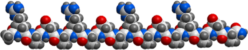
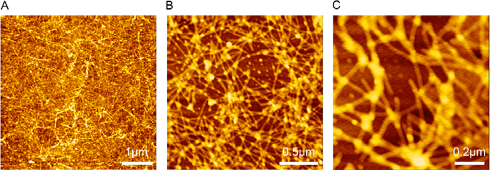
2.1. Self-assembly Assessment of RADA16-I
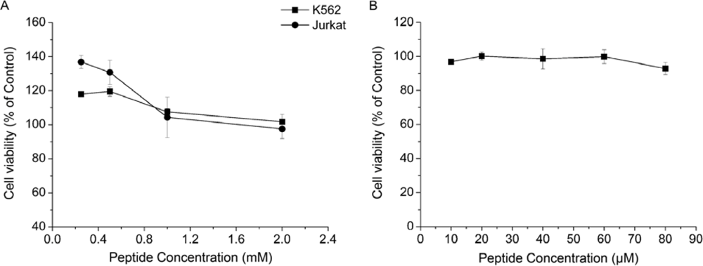
2.2. Cytotoxicity Assays in vitro
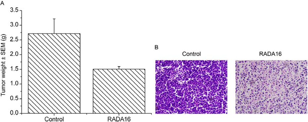
2.3. Inhibition of RADA16-I on K562 Tumor Growth in vivo
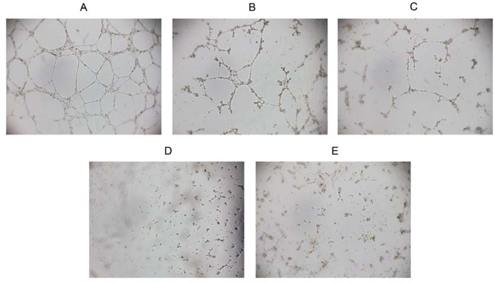
2.4. Inhibition of Vascular Tube-formation by RADA16-I
3. Experimental Section
3.1. Preparation of Peptide RADA16-I
3.2. Cell Cultures
3.3. Self-Assembly Assessment of RADA16-I with Atomic Force Microscopy
3.4. MTT Proliferation Assay
3.5. Inhibition of RADA16-I against K562 Tumor Growth in Mice
3.6. Vascular Tube Formation Assay in vitro
3.7. Statistical Analysis
4. Conclusions
Acknowledgments
References and Notes
- Chabner, BA; Roberts, TG, Jr. Timeline: Chemotherapy and the war on cancer. Nat. Rev. Cancer 2005, 5, 65–72. [Google Scholar]
- Smith, LL; Brown, K; Carthew, P; Lim, CK; Martin, EA; Styles, J; White, IN. Chemoprevention of breast cancer by tamoxifen: risks and opportunities. Crit. Rev. Toxicol 2000, 30, 571–594. [Google Scholar]
- Weaver, VM; Lelievre, S; Lakins, JN; Chrenek, MA; Jones, JC; Giancotti, F; Werb, Z; Bissell, MJ. Beta4 integrin-dependent formation of polarized three-dimensional architecture confers resistance to apoptosis in normal and malignant mammary epithelium. Cancer Cell 2002, 2, 205–216. [Google Scholar]
- Guo, YS; Jin, GF; Houston, CW; Thompson, JC; Townsend, CM, Jr. Insulin-like growth factor-I promotes multidrug resistance in MCLM colon cancer cells. J. Cell Physiol 1998, 175, 141–148. [Google Scholar]
- Sethi, T; Rintoul, RC; Moore, SM; MacKinnon, AC; Salter, D; Choo, C; Chilvers, ER; Dransfield, I; Donnelly, SC; Strieter, R; Haslett, C. Extracellular matrix proteins protect small cell lung cancer cells against apoptosis: a mechanism for small cell lung cancer growth and drug resistance in vivo. Nat. Med 1999, 5, 662–668. [Google Scholar]
- Zhang, S; Holmes, T; Lockshin, C; Rich, A. Spontaneous assembly of a self-complementary oligopeptide to form a stable macroscopic membrane. Proc. Natl. Acad. Sci. USA 1993, 90, 3334–3338. [Google Scholar]
- Zhang, S; Holmes, TC; DiPersio, CM; Hynes, RO; Su, X; Rich, A. Self-complementary oligopeptide matrices support mammalian cell attachment. Biomaterials 1995, 16, 1385–1393. [Google Scholar]
- Holmes, TC; de Lacalle, S; Su, X; Liu, G; Rich, A; Zhang, S. Extensive neurite outgrowth and active synapse formation on self-assembling peptide scaffolds. Proc. Natl. Acad. Sci. USA 2000, 97, 6728–6733. [Google Scholar]
- Kisiday, J; Jin, M; Kurz, B; Hung, H; Semino, C; Zhang, S; Grodzinsky, AJ. Self-assembling peptide hydrogel fosters chondrocyte extracellular matrix production and cell division: implications for cartilage tissue repair. Proc. Natl. Acad. Sci. USA 2002, 99, 9996–10001. [Google Scholar]
- Davis, ME; Motion, JP; Narmoneva, DA; Takahashi, T; Hakuno, D; Kamm, RD; Zhang, S; Lee, RT. Injectable self-assembling peptide nanofibers create intramyocardial microenvironments for endothelial cells. Circulation 2005, 111, 442–450. [Google Scholar]
- Zhang, S; Lockshin, C; Cook, R; Rich, A. Unusually stable beta-sheet formation in an ionic self-complementary oligopeptide. Biopolymers 1994, 34, 663–672. [Google Scholar]
- Caplan, MR; Moore, PN; Zhang, S; Kamm, RD; Lauffenburger, DA. Self-assembly of a beta-sheet protein governed by relief of electrostatic repulsion relative to van der Waals attraction. Biomacromolecules 2000, 1, 627–631. [Google Scholar]
- Caplan, MR; Schwartzfarb, EM; Zhang, S; Kamm, RD; Lauffenburger, DA. Control of self-assembling oligopeptide matrix formation through systematic variation of amino acid sequence. Biomaterials 2002, 23, 219–227. [Google Scholar]
- Schweigerer, L; Neufeld, G; Friedman, J; Abraham, JA; Fiddes, JC; Gospodarowicz, D. Capillary endothelial cells express basic fibroblast growth factor, a mitogen that promotes their own growth. Nature 1987, 325, 257–259. [Google Scholar]
- Malinda, KM; Sidhu, GS; Mani, H; Banaudha, K; Maheshwari, RK; Goldstein, AL; Kleinman, HK. Thymosin beta4 accelerates wound healing. J. Invest. Dermatol 1999, 113, 364–368. [Google Scholar]
- Zhang, S; Rich, A. Direct conversion of an oligopeptide from a beta-sheet to an alpha-helix: a model for amyloid formation. Proc. Natl. Acad. Sci. USA 1997, 94, 23–28. [Google Scholar]
- Yokoi, H; Kinoshita, T; Zhang, S. Dynamic reassembly of peptide RADA16 nanofiber scaffold. Proc. Natl. Acad. Sci. USA 2005, 102, 8414–8419. [Google Scholar]
- Ellis-Behnke, RG; Liang, YX; You, SW; Tay, DK; Zhang, S; So, KF; Schneider, GE. Nano neuro knitting: peptide nanofiber scaffold for brain repair and axon regeneration with functional return of vision. Proc. Natl. Acad. Sci. USA 2006, 103, 5054–5059. [Google Scholar]
- Jaffe, EA; Nachman, RL; Becker, CG; Minick, CR. Culture of human endothelial cells derived from umbilical veins. Identification by morphologic and immunologic criteria. J. Clin. Invest 1973, 52, 2745–2756. [Google Scholar]
- Tada, H; Shiho, O; Kuroshima, K; Koyama, M; Tsukamoto, K. An improved colorimetric assay for interleukin 2. J. Immunol. Methods 1986, 93, 157–165. [Google Scholar]


© 2009 by the authors; licensee Molecular Diversity Preservation International, Basel, Switzerland. This article is an open-access article distributed under the terms and conditions of the Creative Commons Attribution license (http://creativecommons.org/licenses/by/3.0/).
Share and Cite
Tang, C.; Shao, X.; Sun, B.; Huang, W.; Zhao, X. The Effect of Self-Assembling Peptide RADA16-I on the Growth of Human Leukemia Cells in Vitro and in Nude Mice. Int. J. Mol. Sci. 2009, 10, 2136-2145. https://doi.org/10.3390/ijms10052136
Tang C, Shao X, Sun B, Huang W, Zhao X. The Effect of Self-Assembling Peptide RADA16-I on the Growth of Human Leukemia Cells in Vitro and in Nude Mice. International Journal of Molecular Sciences. 2009; 10(5):2136-2145. https://doi.org/10.3390/ijms10052136
Chicago/Turabian StyleTang, Chengkang, Ximing Shao, Binbin Sun, Wenli Huang, and Xiaojun Zhao. 2009. "The Effect of Self-Assembling Peptide RADA16-I on the Growth of Human Leukemia Cells in Vitro and in Nude Mice" International Journal of Molecular Sciences 10, no. 5: 2136-2145. https://doi.org/10.3390/ijms10052136