Effect of pH on the Electronic Absorption Spectra of the Studied N-Heterocycles
This study is undertaken to gain some insight about the species that might be formed in solutions of different pHs, to explain the mechanism of ionization of such compounds and to evaluate their dissociation constants. The latter are readily obtained from the absorbance-pH curves by application of the half height [
22], modified limiting absorption [
23] and Colleter methods [
24].
The electronic absorption spectra of orotic acid gave two bands at 210 and 280 nm with a pKa of 9.3 and an isobestic point at 286 nm. Both bands are due to π-π* and n-π* electronic transitions, respectively.
Also, the UV spectrum of thiouracil gave two bands at 210 and 270 nm in the pH range 2-7 due to the π-π* transition, and a new band at 255 nm in the pH range 9-12. The position and intensity of this band is almost pH independent. The spectra gave a pKa value of 8.2 with the liberation of one proton due to the ionization of SH group.
Orotic acid has three potentially dissociable groups as indicated by conductometric titration [
25] with pK values as follows: pK
1~ 2.8, pK
2~ 9.45 and pK
3>13 [
26,
27]. The first ionization is due to the carboxylic group and the second is obtained by elimination of a second proton from N(1)-H or N(3)-H.
In the electronic absorption spectra of 1x10
-4M 5-carboxy-2-thiouracil (
1), in the pH range 2.03-11.03 three characteristic bands are observed at 210, 270 and 308 nm, with ε values that lie in the ranges 6900 – 18500, 9300 – 12700 and 12600 – 16500, respectively. The first band is probably due to a π-π* of the two sp
2 hybridized carbons of the exocyclic band in heterocyclic ring. The second band is assigned to the coupled π-π* of the two carbonyl and thiocarbonyl groups in the β-position [
28], and may be considered to be due to intramolecular hydrogen bond formation between N(3)-H with C=O and N(1)-H with C=S [
29]. The position of the first band is pH independent while its intensity increases up to pH 7.99 then decreases. The second band is more intense in acidic solutions and becomes less intense in neutral and basic solutions. The position of this band is independent of the pH over the range studied (2.03-7.01). In basic medium, this band is slightly shifted and decreased in intensity probably due to the formation of a protonated species overlapped with the π-π* electronic transition of uracil ring. However, the third band, due to presence of COOH group, is blue shifted in the pH range 2.03-7.01 but with the same intensity. This behavior suggests a tautomerism between the thione and thiol structures. When the pH is increased the band is strongly red shifted as a result of proton elimination. In addition to the former three bands, a new band appeared in the pH range 7.99-11.03 at 229 nm with ε values 6300-9800, representing the shift of the tautomeric equilibrium towards the enol form in basic solutions. The intensity of this band decreases as the pH increases. Three well-defined isobestic points appeared at 219, 306 and 324 nm. The first covers the pH values 3.13-4.99, 9.01, 10.02, the second covers the pH values 3.13-4.99, 7.01-10.02 and the third one is for the pH values 2.03-4.03, 7.99, 9.01. The isobestic points assign the presence of different absorbing species in equilibrium to each other [
30], i.e., the HN(1)C=S and HN(3)C=O groups are subjected to simultaneous ionization.
c) Colleter Method [24]:
This method is widely recognized for evaluation of the pK values of different classes of compounds. The calculation is applied on the basis that, for three values of absorbance at different H
+, the following equation is applied:
Where:
The absorbance values used in the calculations are selected from As-pH plots. The data are collected in
Table 1.
Table 1.
Dissociation Constants (pKa) of the Organic Compounds at λ = 335 nm.
Table 1.
Dissociation Constants (pKa) of the Organic Compounds at λ = 335 nm.
| Compound | Half Height | Modified Limiting | Colleter | n |
|---|
| 5-Carboxy-2-thiouracil (1) | 4.7
7.7 | 4.7
7.9 | 4.8
8.0 | 1.0
1.0 |
| 5,6-Diphenyl-3-hydroxy-1,2,4-triazine (2) | 4.3
7.6 | 4.4
7.5 | 4.1
7.5 | 0.5
1.0 |
| 1-Phenyl-3-methyl-5-pyrazolone (3) | 8.1 | 7.9 | 8.1 | 1.0 |
| 2-Mercapto-4,6-dimethylpyrimidine hydrochloride (4) | 3.6
8.1 | 3.6
8.3 | 3.5
8.3 | 1.0
1.0 |
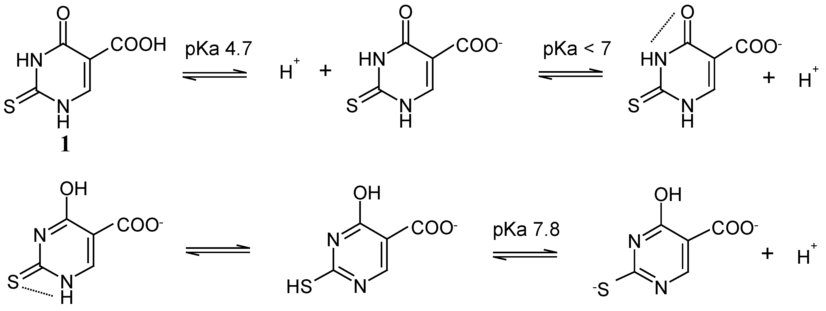
For 5-carboxy-2-thiouracil (
1) two pKa values were evaluated at 335 nm. The pKa
1 of 4.7 with the liberation of one proton through ionization is probably due to the removal of a proton from the –COOH group [
31]. The pKa
2 value of 7.8 with the liberation of a proton may be from the 2-SH group or the 4-OH group, where two modes of ionization could be represented:
The negative ferric chloride test for phenolic-OH and the pKa value lower than that reported for phenolic compounds [
32] lead us to argue that the mode of ionization (i) is more acceptable than (ii). Thus the ionization process is assumed to proceed as indicated in
Scheme 1.
Similarly, in the electronic absorption spectra of 1x10
-4M 5,6-diphenyl-3-hydroxy-1,2,4-triazine (
2) in solutions within the pH range 3.13 – 11.03, three electronic absorption bands appear at 216, 250 (both are intense) and 341 nm (broad) with ε values 6700–12600, 5300–12600 and 1600–4800, respectively. The first two bands are probably due to the π-π* transition while the third band may be due to intramolecular hydrogen bond between N (2)-H or N(4)-H with C=O [
29] .
When the pH increases in the 2.03–6.19 range, the intensity of the two bands at 216 and 250 nm decreases while the third one becomes more intense. However, the positions of these bands are pH independent. With a further increase of the pH to 11.02, the three bands become more intense with a slight red shift in the second and third bands.
Two isobestic points are seen at 288 and 318 nm. The first covers the pH values 4.03, 7.99–11.03 and the second one covers the pH values 7.99–11.03, indicating the presence of an equilibrium between different species in the basic media. Two pKa values were evaluated (4.3 and 7.6) at λ= 355 nm with the liberation of one proton in each step. The first one is due to the associated species H
2L
2 with n value of 0.5. The negative ferric chloride test reaction and the lower pKa value (7.6) than that of the reported for phenolic compounds [
32] assumed that the second proton is liberated from 4-NH or 2-NH groups and not from 3-OH group. The following mechanism of ionization is suggested:
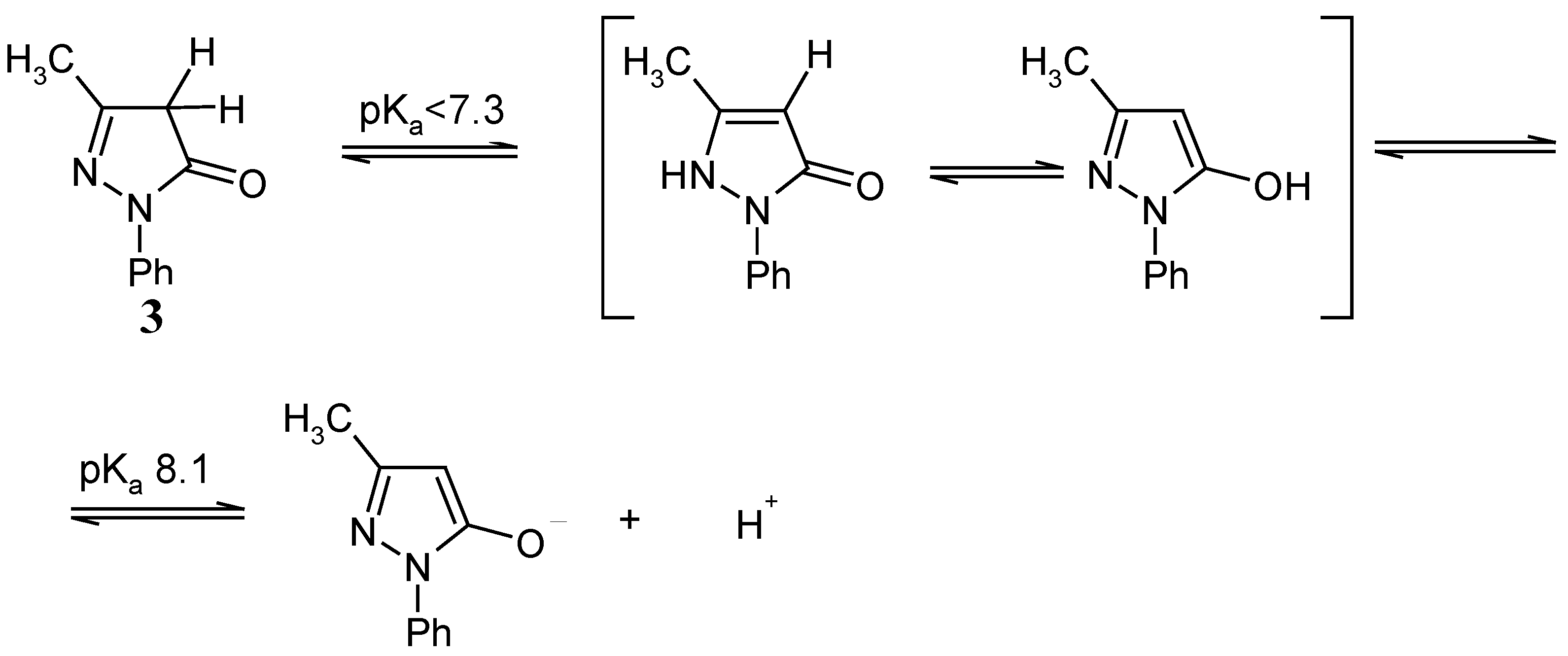
On the other hand, in the electronic absorption spectra of 1x10⁻⁴ M 1-phenyl-3-methyl-5-pyrazolone (3), in solutions within the pH range 2.01 – 7.37, one electronic absorption band appeared at 245 nm with ε values in the range 13900–16600, which is pH independent both in intensity and in position. On increasing the pH up to 9.12, two more bands appeared at 209 and 344 nm with ε values 1300–13900 and 1000–3400, respectively. The two bands at 209 and 245 nm become less intense on going from pH 8.13 to 9.12 while the 344 nm band becomes more intense. At the pHs 10.06 and 11.01, the first band disappeared, the second one becomes slightly more intense while the third band becomes less intense and broader. Accordingly, 1-phenyl-3-methyl-5-pyrazolone (3) exists in the keto form in acidic, neutral and strong basic solutions, however, it exists in the enol form only in solutions with 7.37 < pH <10.06.
Two well-defined isobestic points appeared at 242 nm (covering the pH’s 3.58, 9.12, 10.06) and 275 nm (covering the pH’s 3.58, 7.37, 8.13, 9.12) to indicate the presence of different absorbing species in equilibrium. One pKa value of 8.1 was evaluated at 335 nm in the basic medium (7.37 < pH < 10.06) with the liberation of one proton which may be (a) from the 2-NH group, or (b), the 5-OH group, where two modes of ionization could be presented (
Scheme 2).
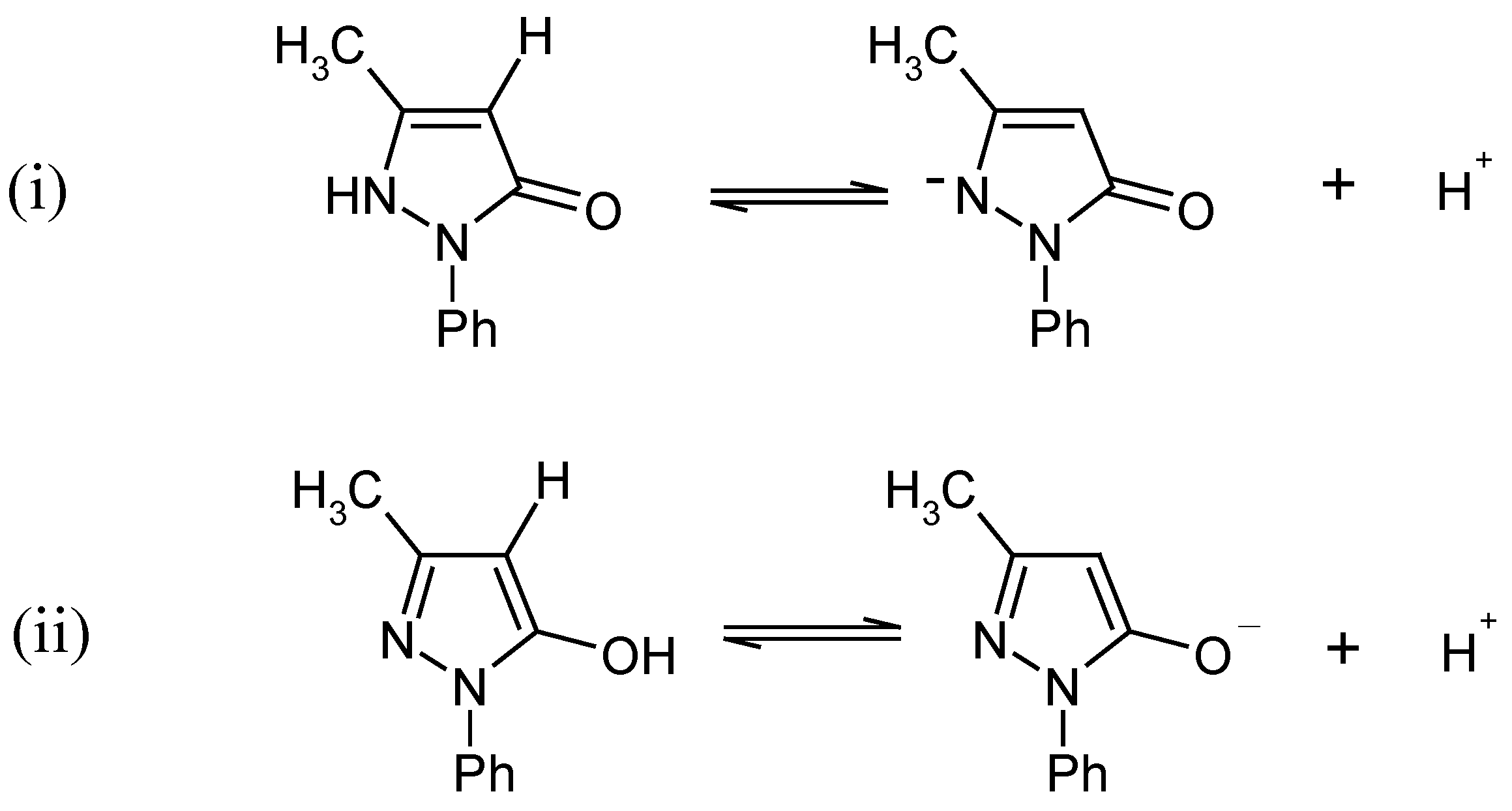
The positive ferric chloride test reaction for phenolic-OH and the closeness of the pKa value to that reported for phenolic compounds [
32] lead us to propose that the mode of ionization (ii) is more acceptable than (i). Therefore the ionization process is assumed to proceed as indicated in
Scheme 3:
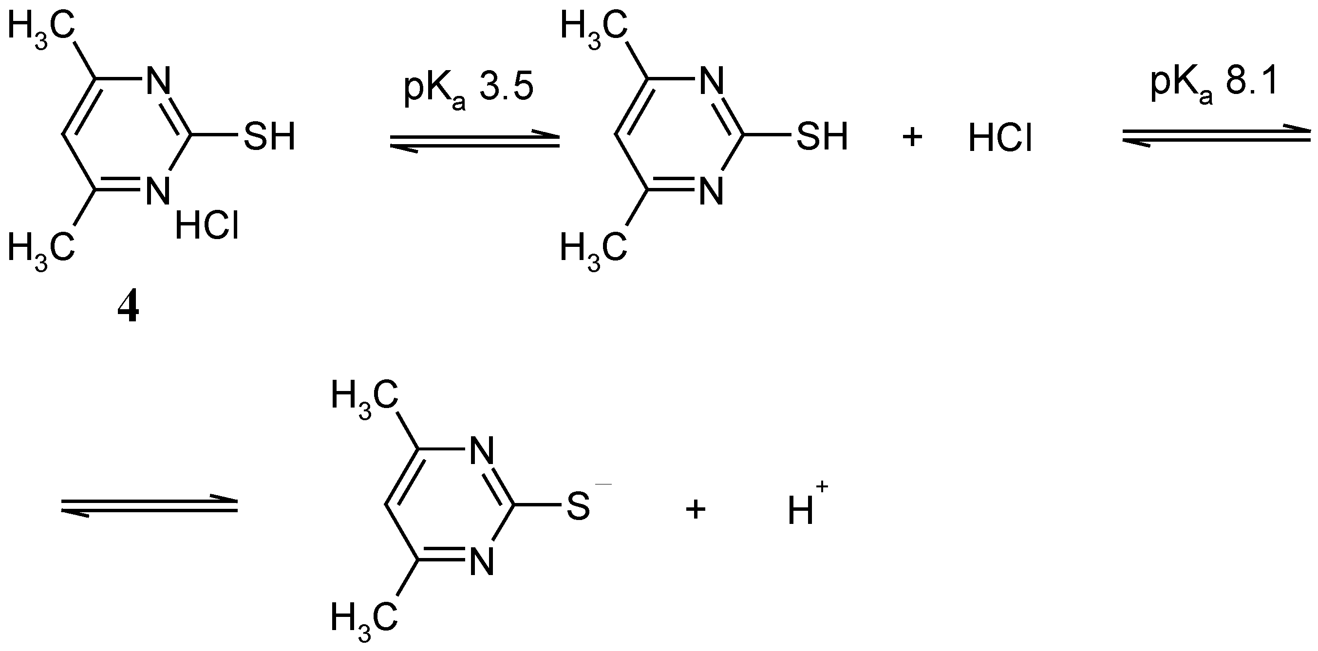
The electronic absorption spectra of 1x10
-4M 2-mercapto-4,6-dimethylpyrimidine hydrochloride (
4), in the 2.00–11.03 pH range displayed three electronic absorption bands at 216, 274 and 331 nm with ε values amount 3200–10400, 16700–20800 and 1600– 5100, respectively. In the pH 2.00– 4.99 range, the first band becomes more intense and as the pH increases, it becomes broad and less intense. However, in the pH 2.00–7.37 range, the second one is pH independent, both in intensity or in position, while it becomes less intense with a slight blue shift in the pH 8.13–11.01 range. Meanwhile, the intensity of the third band increases in the pH 2.00–4.99 range, then decreases as the pH increases stepwise, until it completely vanishes in strong basic media. Two isobestic points appear at 268 and 272 nm. The first covered all the pH range, while the second one covers the pH values 2.00–7.37, 11.01. However, two pKa values were evaluated (3.5 and 8.1), with the liberation of one proton in each step. The ionization mechanism seen in
Scheme 4 is proposed:


