Article

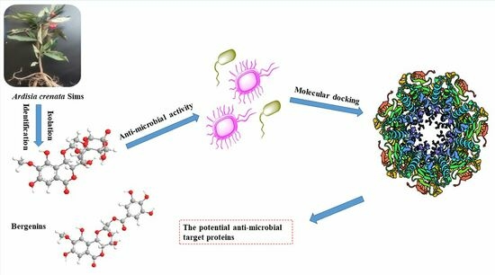
Discovery of Potential Anti-Microbial Molecules and Spectrum Correlation Effect of Ardisia crenata Sims via High-Performance Liquid Chromatography Fingerprints and Molecular Docking

1
College of Pharmacy, Guizhou University of Traditional Chinese Medicine, Guiyang 550025, China
2
Kunming Institute of Botany, Chinese Academy of Sciences, Kunming 650201, China
*
Author to whom correspondence should be addressed.
These authors contributed equally to this work.
Molecules 2024, 29(5), 1178; https://doi.org/10.3390/molecules29051178
Submission received: 14 January 2024 / Revised: 22 February 2024 / Accepted: 1 March 2024 / Published: 6 March 2024
(This article belongs to the Special Issue Antimicrobial Activity of Natural Extracts)

Abstract

:
Ardisia crenata Sims, an important ethnic medicine, is recorded in the Chinese Pharmacopoeia for treating laryngeal diseases and upper respiratory tract infections. This study aimed to evaluate the antimicrobial effect of extracts and potential antimicrobial compounds of A. crenata Sims. It was found that the roots of A. crenata Sims have a potential inhibitory effect on Candida albicans and Aspergillus flavus, with MICs of 1.56 mg/mL and 0.39 mg/mL, and the leaves of A. crenata Sims have a potential inhibitory effect on Pseudomonas aeruginosa and Staphylococcus aureus, with MICs of 3.12 mg/mL and 6.77 mg/mL, respectively. Meanwhile, five compounds including one catechin and four bergenins were obtained from roots. These components were identified on the fingerprint spectrum, representing chromatographic peaks 16, 21, 22, 23, and 25, respectively. Among these, 11-β-d-glucopyranosyl-bergenin and (−)-gallocatechin showed potential inhibition for Staphylococcus aureus and Pseudomonas aeruginosa with MIC of 0.26 and 0.33 mg/mL, respectively. The roots, stems, and leaves of A. crenata Sims are very similar in chemical composition, with large differences in content. Principal component analysis (PCA) and Hierarchical cluster analysis (HCA) showed that 16 batches of A. crenata Sims could be divided into four main production areas: Guizhou, Jiangsu, Guangxi, and Jiangxi. Furthermore, molecular docking results showed that 11-β-d-glucopyranosyl-bergenin had a better affinity for Casein lytic proteinase P (ClpP), and (−)-gallocatechin possessed a strong affinity for LasA hydrolysis protease and LasB elastase. These findings suggest catechin and bergenins from A. crenata Sims can be used as antimicrobial activity molecules.

Graphical Abstract

1. Introduction

As the most common human bacterial pathogen, Staphylococcus aureus often asymptotically colonizes the nasal mucosa of humans, causing superficial infections of the skin and mucosa, and even life-threatening systemic infections [1]. People with damaged skin and mucosal barrier or impaired immune systems are particularly vulnerable to S. aureus infection, which can cause a variety of diseases, including pneumonia, sepsis, serious skin infection, and respiratory system infection [2,3]. Among the various virulence factors of S. aureus, Casein Hydrolase (ClpP) is the key virulence factor that determines the pathogenicity of S. aureus and plays a crucial role in the pathogenicity of bacteria. Therefore, ClpP has been identified as a new candidate antibacterial target for screening and discovering inhibitors of important virulence factors of S. aureus [4,5]. Pseudomonas aeruginosa can colonize various surfaces and tissues with strong adaptability, invasiveness, and pathogenicity and cause various acute and chronic infections such as burn wounds, urinary tract infections (UTI), and lung infections [6]. This can be attributed not only to its highly endogenous nature and acquired resistance but also to various virulence factors [7]. P. aeruginosa can secrete different kinds of extracellular proteases, such as LasA protease, LasB elastase, alkaline protease, and protease IV. Among them, AprA and LasB can alter the mucosal cilia clearance rate, degrade lung tissue, and disrupt the host immune system, thereby strongly promoting lung diseases [8]. P. aeruginosa is also the main cause of chronic lung infection in patients with cystic fibrosis. The emergence and spread of widely resistant or multidrug-resistant P. aeruginosa isolates pose significant risks to human health [9].
Antibiotics are one of the greatest inventions of the 20th century and are widely used in the treatment of infectious diseases. However, approximately 50% of antibiotics are abused and misused globally every year, leading to strong antibiotic resistance in bacteria [10,11]. Traditional Chinese medicine (TCM) has been used clinically for thousands of years, with characteristics such as less toxicity, fewer side effects, and multiple targets of action. It has played a crucial role in human efforts to overcome major epidemics. The effective active ingredients of Chinese herbal medicine mainly contain polysaccharides, essential oils, and phenolic compounds [12,13,14]. These components often have certain antibacterial effects and are not prone to developing compound resistance, and they even reverse the compound resistance of bacteria. They have received increasing attention in clinical and scientific research [15]. Ardisia crenata Sims, a plant of the genus Ardisia, also called zhu sha gen is mainly distributed in Guizhou, China [16]. It is also used as an important Miao medicine called ba zhua jin long with antimicrobial, anti-viral, anti-inflammatory, and anti-tumor effects [17,18]. In addition, A. crenata Sims has a significant antibacterial effect on type A, type B hemolytic streptococcus, and Staphylococcus aureus [19]. Clinically, as the main drug of Kaihoujian spray (child type), it is mainly used to treat respiratory tract infections, tonsillitis, rheumatic bone pain, and other diseases without general toxicity or adverse effects [20,21]. At present, studies have found that A. crenata Sims mainly contains coumarins, triterpenoid saponins, flavonoids, and other chemical components [22,23]. Especially, bergenin as one kind of main coumarin, has inhibitory effects on the growth of microbes [24].
Modern analytical techniques have played an important role in the quality identification of TCM, including the detection of hydrazine in real water and soil samples from the growing areas of TCM [25]. In particular, integrated metagenomics and meta-transcriptomics sequencing can also be used to examine the abundance of microbial consortiums and their metabolites [26,27,28]. Macromolecular phase separation was also used to deliver bioactive compounds [29]. The chromatography-mass spectrometry technology can detect the active components of TCM and food, as well as analyze and confirm the structure of unknown active substances [30]. In addition, research has shown that the anti-mold secondary amine bond of soy protein can effectively and environmentally improve the anti-mold properties of its adhesive [31].
In this paper, we established the fingerprints of A. crenata Sims roots, stems, and leaves, and assigned their common peaks and characteristic peaks. Meanwhile, five compounds were isolated and purified from A. crenata Sims. The contents of these compounds were also determined to illustrate the differences in different medicinal parts of A. crenata Sims. Furthermore, we also compare the antimicrobial activity of different batches of A. crenata Sims. Moreover, these compounds were evaluated by molecular docking analysis, obtaining good antimicrobial activity to elucidate the possible mechanism.

2. Results and Discussion

2.1. Anti-Microbial Activity Evaluation of A. crenata Sims

The different parts of A. crenata Sims were used to evaluate antimicrobial activities against two kinds of fungi and six kinds of bacteria. As shown in Table 1, the extracts of root, stem, and leaf exhibited inhibitory activity on these test strains with an inhibition zone diameter (IZD) of 6.08~20.84 mm. The results showed that the roots of A. crenata Sims had good activity against Candida albicans and Aspergillus flavus. Leaves had good antibacterial activity against Pseudomonas aeruginosa and Staphylococcus aureus. In addition, the results indicated that the Minimum inhibitory concentration (MIC) values of root to Candida albicans and Aspergillus flavus were 1.56 mg/mL and 0.39 mg/mL, respectively. The MIC values of leaves against Pseudomonas aeruginosa and Staphylococcus aureus were 3.12 mg/mL and 6.77 mg/mL, respectively (Table 2).

2.2. Structural Identification of Known Compounds from A. crenata Sims

The known compounds (15) (Table 3) were identified on the basis of detailed spectroscopic interpretation (Figure S6–S8) and comparison with the previously reported data on (−)-gallocatechin (1) [32], 11-α-d-galactopyranoside bergenin (2) [33], 11-β-d-glucopyranosyl bergenin (3) [34], bergenin (4) [35] and 11-O-galloybergenin (5) [36]. Compounds (2), (3), and (5) were found in A. crenata Sims for the first time.

2.3. Analysis of HPLC Fingerprint

The HPLC fingerprint of 16 batches and the reference fingerprint from A. crenata Sims are presented in Figure 1 and Figure 2. Ten common peaks are shown as peaks 1, 4, 5, 6, 16, 19, 21, 22, 23, and 25 in Figure 1a–c. Among these, the identified compounds belonged to peaks 16, 21, 22, 23, and 25, respectively.
The similarity analysis was conducted with A. crenata Sims in the S1 production area as a reference (Table 4). As a result, the root similarity was more than 0.97 in different producing areas, while, many differences in the fingerprints of the leaves and stems of A. crenata Sims from different regions. The aboveground part of A. crenata Sims was different due to being affected by the environment.

2.4. PCA and HCA Analysis of 16 Batches of A. crenata Sims

As shown in Figure 3, HCA analysis found that the roots of A. crenata Sims from 16 production areas have high similarity, and the differences in chemical composition of stems and leaves due to different climatic environments in different production areas. The results of PCA analysis showed that the roots, stems, and leaves of A. crenata Sims in the four production areas could be divided into four categories, representing the major production areas, including Guizhou, Jiangsu, Guangxi, and Jiangxi (Figure 4).

2.5. Effect of Compounds 15 on Anti-Microbial Activities

The isolated and identified compounds were used to evaluate the antimicrobial activities against the tested strains. As shown in Table 5, compounds 15 exhibited inhibitory activity on these microorganisms with inhibition zone diameters (IZD) of 6.11~9.28 mm. The results showed that 11-β-d-glucopyranosyl-bergenin, 11-α-d-galactopyrnside-bergenin, and 11-O-galloybergenin had good activity against Staphylococcus aureus. (−)-gallocatechin and bergenin had good antibacterial activity against Pseudomonas aeruginosa. In addition, the results indicated that the MIC values of these compounds against Staphylococcus aureus and Pseudomonas aeruginosa ranged from 0.26 to 0.39 mg/mL, respectively.

2.6. Spectrum–Effect Relationship

The spectrum–effect relationship between chromatographic peaks and anti-microbial activity was established by GRA and PLSR models. As a result, GRA analysis showed that the correlation data of all the common peaks were greater than 0.7. This indicated that the antimicrobial activities of A. crenata Sims were caused by the compounds represented by all these peaks (Table 6).
VIP value > 1 was used as the standard to screen the key components of the anti-microbial effect of roots from A. crenata Sims. As a result, PLSR analysis showed that peaks of 19, 21, 26, 27, and 29 were the main components of A. crenata Sims against Candida albicans (Figure 5a and Figure 6a). Peaks of 19, 25, 26, 27, and 29 were the main components of roots from A. crenata Sims against Aspergillus flavus (Figure 5b and Figure 6b). Peaks 16 and 22 were the main components of leaves from A. crenata Sims against Pseudomonas aeruginosa (Figure 5c and Figure 6c). Peaks 19, 23, and 8 were the main components of leaves from A. crenata Sims against Staphylococcus aureus (Figure 5d and Figure 6d). These results indicated that the antimicrobial effect of A. crenata Sims was jointly influenced by multiple components.

2.7. Quantitative Analysis of Anti-Microbial Ingredients in A. crenata Sims

As shown in Table 7, the HPLC analysis showed that the contents of bergenin, and (−)-gallocatechin were much higher than 5.5 mg/g in the roots. Moreover, these two components in roots were much higher quantities than in the stems and leaves. In addition, 3.84 mg/g of 11-O-galloybergenin was found in the leaves. Meanwhile, 0.38 mg/g of 11-β-d-glucopyranosyl-bergenin was found in the stems, which was much higher than the content in the roots and leaves. The chromatograms of standards solution of these compounds were showed in Figures S1–S5 (Supplementary Materials).

2.8. Molecular Docking of Active Compounds on Key Target Proteins of Bacteria

The ClpP protease in S. aureus is responsible for stress tolerance and participates in virulence regulation [37]. It plays an important role in maintaining homeostasis and pathogenicity in bacteria. Therefore, inhibition of this protease can reduce the overall virulence level of pathogenic bacteria and kill them [38]. The interaction between the active compounds and target proteins, the ClpP PRs (PDB ID: 3V5e), LasA PRs (PDB ID: 3IT7), LasB PRs (PDB ID: 3DBK), DNA ligase (2XCQ), DNA gyrase (3JSN), and MurF ligase (4CVL), with the receptors and active compounds as ligands, was explored using Schrödinger Suite. As shown in Table 8 and Figure 7, 11-β-d-glucopyranosyl-bergenin (A), 11-α-d-galactopyranoside-bergenin (B), and 11-O-galloybergenin (C) provided a strong binding affinity of −9.84~−8.34 to ClpP protease. Among them, eight hydrogen bonds were formed between 11-β-d-glucopyranosyl-bergenin and ClpP PR (Figure 7A), including between 8-hydroxyl and residue MET-31; between 4-hydroxyl and residue ASN-39; between 2′-hydroxyl, 3′-hydroxyl group of glucose, 3-hydroxyl and residue ILE-4; and between 6′-hydroxyl group of glucose and residue ILE-4, ASP-19. For 11-α-d-galactopyranoside-bergenin, six hydrogen bonds were formed between hydroxyl groups and residues ILE-4, ASP-37, ASN-39, ASN-42, and MET-31 (Figure 7B). For 11-O-galloybergenin, five hydrogen bonds were formed between hydroxyl groups and residues ILE-4, ASP-19, THR-6, and MET-31 (Figure 7C).
As shown in Table 8 and Figure 8, bergenin (A), (−)-gallocatechin (B), and ceftazidime (C) provided a good binding affinity of −4.17–−5.63 to DNA gyrase. Among them, four hydrogen bonds were formed between bergenin and DNA gyrase (Figure 8A), including between 3-hydroxyl, 4-hydroxyl, and residue GLU264; between 6-carbonyl and residue GLU261; and between 10-hydroxyl and residue GLN-269. For (−)-gallocatechin, three hydrogen bonds were formed between 3-hydroxyl and residues GLN-269 and LYS-265; and between 3′-hydroxyl and residues GLU264 (Figure 8B). For the positive drug ceftazidime, five hydrogen bonds were formed between hydroxyl groups and residues GLN-269, LYS-265, GLU-261, and GLU-264 (Figure 8C).
As shown in Table 8 and Figure 9, bergenin (A), 11-O-galloybergenin (B), and ceftazidime (C) provided a good binding affinity of −4.22–−6.72 to DNA ligase. Among them, five hydrogen bonds were formed between bergenin and DNA ligase (Figure 9A), including between 3-hydroxyl and residue GLU-264; between 12-methoxy and residue GLN-269; between 10-hydroxyl and residue LYS-265; and between 11-hydroxyl and residue LYS-265, SER-268. For 11-O-galloybergenin, three hydrogen bonds were formed between 11-ester and residues SER-268; between 6-carbonyl and residues GLU264; and between 10-hydroxyl and residue THR-267 (Figure 9B). For the positive drug ceftazidime, five hydrogen bonds were formed between hydroxyl groups and residues THR-306, PHE-86, and GLU-88 (Figure 9C).
The protease of P. aeruginosa is mainly regulated by the Las pathway of Quorum sensing (QS). Among them, the LasA hydrolytic protease induces respiratory infections by assisting in the colonization and digestion of host tissues [39]. For LasA PRs, as shown in Table 8 and Figure 10, (−)-gallocatechin, bergenin, and 11-O-galloybergenin exhibited good binding activity to LasA PRs with a docking score of more than −5.0. Among them, eight hydrogen bonds are formed between (−)-gallocatechin and LasA PRs (Figure 10A), including between 5-hydroxyl and residue VAL-87; between 7-hydroxyl and residue TYR-30; between 4′-hydroxyl and residue ILE-85, ASP-83, and GLN-84; between 3′-hydroxyl and residue GLN-84 and ASP-83; and between 5′-hydroxyl and residue ARG-64. For bergenin, four hydrogen bonds were formed between 3-hydroxyl and residue TYR-49; between 6-carbonyl and residue SER-16; between 8-hydroxyl and residue SER-50; and between 12-methoxy and residue ARG-12 (Figure 10B). For 11-O-galloybergenin, seven hydrogen bonds were formed between 3-hydroxyl and residue ILE-85; between 12-methoxy and residue ASP-83, GLN-84; and between 8-hydroxyl and residue ASP-83, GLN-84, and ARG-64 (Figure 10C).
The QS system of P. aeruginosa regulates the production of virulence factor LasB elastase. Additionally, it can damage and decompose human cellular tissues [8]. For LasB PRs, as shown in Table 8 and Figure 11, (−)-gallocatechin, bergenin, and 11-O-galloybergenin showed good binding affinity to LasB PRs with a docking score of more than −5.5. Among them, six hydrogen bonds are formed between (−)-gallocatechin and LasB PRs (Figure 11A), including between 7-hydroxyl and residue GLY-157; between 5-hydroxyl and residue TYR-216, GLY-29; between 3′, 4′-hydroxyls and residue GLY-219; and between 5′-hydroxyl and residue ASP-221. For bergenin, five hydrogen bonds were formed between 10-hydroxyl and residue GLU-164; between 4-hydroxyl and residue TRP-115; and between 3-hydroxyl and residue HIS-144, GLU-141 (Figure 11B). For 11-O-galloybergenin, eight hydrogen bonds were formed between 3′, 4′-hydroxyls and residue ASN-61; between 4′-hydroxyls and residue THR-62; between 6-carbonyl and residue ASP-41; and between 12-methoxy, 10-hydroxyl, and residue ARG-55 (Figure 11C).
As shown in Table 8 and Figure 12, 11-α-d-galactopyranoside-bergenin (A), 11-O-galloybergenin (B), and (−)-gallocatechin (C) provided a good binding affinity of −4.19~−5.03 to MurF ligase. Among them, three hydrogen bonds were formed between 11-α-d-galactopyranoside-bergenin and MurF ligase, including between 4-hydroxyl and residue ALA-217; between 2′-hydroxyl of galactose and residue ARG-240; and between 3′-hydroxyl of galactose and residue SER-238 (Figure 12A). For 11-O-galloybergenin, five hydrogen bonds were formed between 8-hydroxyl and residue PHE-100; between 4-hydroxyl and residue ARG-181; between 5′-hydroxyl and residue ARG-98; and between 6-carbonyl and residue ARG-98, ARG-181 (Figure 12B). For (−)-gallocatechin, three hydrogen bonds were formed between 1-oxygen and residues ARG-181; between 7-hydroxyl and residues ALA-174; and between 3′-hydroxyl and residue GLY-214 (Figure 12C).

2.9. Molecular Docking of Active Compounds on Key Target Proteins of Fungi

The mechanisms of antifungal drugs mainly include inhibiting fungal cell wall synthesis and affecting cell membrane function. 1,3-β-glucan synthase and chitin synthase are the key fungal target proteins involved in the cell wall and squalene synthase (SQS) in the cell membrane. For SQS PRs, as shown in Table 9 and Figure 13, 11-O-galloybergenin (A), and bergenin (B) provided a good binding activity to SQS PRs with docking scores of 4.23 and 4.61, respectively. Among them, six hydrogen bonds were formed between 11-O-galloybergenin and SQS PRs, including between 3′, 4′-hydroxyls and residue LYS-498; between 2′-hydroxyl and residue LYS-73; between 10-hydroxyl and residue ASP-494; between 8-hydroxyl and residue LEU-493; and between 1-oxygen and residue GLN-317 (Figure 13A). For bergenin, four hydrogen bonds were formed between 3, 4-hydroxyls and residue ASN-76; and between 6-carbonyl and residue ARG-102 (Figure 13B).
As shown in Table 9 and Figure 14, bergenin (A), (−)-gallocatechin (B), and Nystatin (C) provided a good binding affinity of −4.62–−6.38 to 1,3-β-glucan synthase. Among them, three hydrogen bonds were formed between bergenin and 1, 3-β-glucan synthase, including between 11-hydroxyl and residue MET-1266, GLY-1267; and between 6-carbonyl and residue THR-444 (Figure 14A). For (−)-gallocatechin, three hydrogen bonds were formed between 5-hydroxyl and residue LYS-384, ASN-385; and between 3′-hydroxyl and residue ARG-520 (Figure 14B). For the positive drug Nystatin, four hydrogen bonds were formed between hydroxyl groups and residues ARG-443 and SER-516 (Figure 14C).
As shown in Table 9 and Figure 15, 11-α-d-galactopyranoside-bergenin (A) and 11-β-d-glucopyranosyl-bergenin (B) showed high-level binding affinities of −4.13 and −4.51 for chitin synthase, respectively. Among them, two hydrogen bonds were formed between 11-α-d-galactopyranoside-bergenin and chitin synthase, including between 10-hydroxyl and residue LYS-889; and between 4′-hydroxyl of galactose and residue ILE-887 (Figure 15A). For 11-β-d-glucopyranosyl-bergenin, three hydrogen bonds were formed between 10-hydroxyl, 9-methoxy, and residue ARG-785 (Figure 15B).

3. Materials and Methods

3.1. Instruments and Chemicals

In this study, we used the following instruments: Agilent-1260 High-Performance Liquid Chromatographer (Agilent, Santa Clara, CA, USA); DRX-500 AVANCE III-600MHz superconducting nuclear magnetic resonance imager (Bruker, Bremen, Germany); RID-20A differential refractive detector (Shimadzu, Kyoto, Japan); SB-600DTY ultrasonic Multi-frequency cleaning machine (Ningbo, China); Hve-50 autoclave; HCB-1300V medical ultra clean table (Haier, Qingdao, China). We used the following chemicals and materials: 11-α-d-galactopyranoside-bergenin, bergenin, (−)-gallocatechin, 11-O-galloybergenin, and 11-β-d-glucopyranosyl-bergenin (standard laboratory- and self-made products); UN1648 Acetonitrile (GR, Thermo Scientific, Waltham, MA, USA); Methanol (GR, BCR, USA); Phosphoric acid (GR, Tianjin Kemel, Tianjin, China); C-18 reversed-phase column packing ODS-A-HG (YMC, Kyoto, Japan); Sephadex LH-20 (Beijing Solarbio, Beijing, China); Semi-preparative column (250 mm × 10 mm, 5 μm, Shimadzu, Kyoto, Japan): Blank drug-sensitive paper (Jining Best Micro, Jining, China); Ceftazidime (Hangzhou Microbial Reagent, Hangzhou, China); Nystatin (Hefei BASF, Hefei, China).

3.2. Plant Materials and Test Strains

Sixteen batches of samples from different regions of China were identified as Ardisia crenata Sims by Professor Shenghua Wei of Guizhou University of Traditional Chinese Medicine, as shown in Table S1. Candida albicans (BNCC 186382), Aspergillus flavus (A1142B), Staphylococcus aureus (ATCC 6538P), Bacillus subtilis (ATCC 6633), Enterococcus faecalis (ATCC 19433), Escherichia coli (CICC 10389), Pseudomonas aeruginosa (ATCC 9027), and Proteus vulgaris (ACCC 11002) were provided by the laboratory of the College of Life Sciences, Guizhou University (Guiyang, Guizhou).

3.3. Extraction and Isolation of Roots from A. crenata Sims

The dried roots of A. crenata Sims were crushed to obtain 10 kg powder, and extracted with a 70% (v/v) ethanol/water mixture at reflux three times for 2 h each. The extraction solution was extracted with petroleum ether, ethyl acetate, and n-butanol successively, and dried to obtain the petroleum ether layer (30.2 g), ethyl acetate layer (102.42 g), and n-butanol layer (879.6 g) extracts. The soluble fraction of the n-butanol (312.01 g) was eluted by dichloromethane/methanol (10:1–0:1) via silica gel column chromatography to obtain seven fractions (Fr.1~Fr.7). Compound 4 (5.6 g) was precipitated colorless crystals from Fr.1. to Fr.3 (8.27 g) was subjected to ODS column chromatography with MeOH-H2O (2:8 to 1:0) to obtain sub-fractions A1–A11. A2 was purified by semi-preparative HPLC (MeOH-H2O, 20:80; flow rate: 3 mL/min) to obtain compound 2 (19 mg, tR = 8 min) and compound 3 (17 mg, tR = 11 min). The soluble fraction of the ethyl acetate (87.01 g) was eluted by dichloromethane-methanol (100:1–0:1) via silica gel column chromatography to obtain 11 fractions (Fr.1~Fr.11). Fr.5 (7.24 g) was chromatographed by ODS column with MeOH-H2O (2:8 to 1:0) to obtain sub-fractions E1–E11. E2 was purified by semi-preparative HPLC (MeOH-H2O, 40:60; flow rate: 3 mL/min) to obtain compound 5 (30 mg, tR = 15 min). E1 was purified by semi-preparative HPLC (MeOH-H2O, 20:80; flow rate: 3 mL/min) to obtain compound 1 (19 mg, tR = 13 min).

3.4. Sample Preparation

The roots, stems, and leaves of A. crenata Sims were dried and crushed into powder. Sample powders (2.0 g) were accurately weighed and extracted with 50 mL methanol in a stoppered Erlenmeyer flask with an ultrasonic multi-frequency cleaning machine (frequency 40 kHz) for 40 min. The obtained extracts were filtered, concentrated under reduced pressure and vacuum dried. Then, the extracts were mixed with 10% DMSO to configure with a concentration of 100 mg/mL as the test solution for the anti-bacterial experiment. The 11-α-d-galactopyranoside-bergenin, 11-β-d-glucopyranosyl-bergenin, bergenin, 11-O-galloybergenin, and (−)-gallocatechin were dissolved in 30% MeOH-H2O to for content determination.

3.5. Chromatographic Conditions

Chromatography was performed on an Agilent-1260 HPLC (Agilent, USA) equipment with a diode array detector (DAD) and a YMC-Pack ODS-A (250 mm × 4. 6 mm, 5 μm) at 25 °C. The mobile phase consisted of acetonitrile (A) and 0.1% phosphoric acid (B) with a gradient elution mode as follows: 0–15 min, 5%–10% A; 15–35 min, 10%–10% A; 35–65 min, 10%–24% A; 65–80 min, 24%–40% A; 80–85 min, 40%–50% A; 85–90 min, 50%–5% A; 90–95 min, 5%–5% A. The flow rate was 0.5 mL/min with an injection volume of 5 μL and the DAD detection wavelength was set at 214 nm.

3.6. Validation of Methodology

The precision test was evaluated by six consecutive injections of the same sample (S3) solution, and the repeatability was evaluated by repeating six times with samples (S3) from the same place of origin. The stability tests were analyzed within 0, 2, 4, 6, 8, 10, 12, and 24 h, respectively.

3.7. Analysis of HPLC Fingerprint

The similarity of the Chinese medicine chromatographic fingerprints was analyzed and evaluated using the 2012A version system under the optimized HPLC conditions. Hierarchical cluster analysis (HCA) and multivariate principal component analysis (PCA) were used to divide the samples into different groups based on the similarity of their measured properties [40].

3.8. Anti-Bacterial Activity Evaluation

The antimicrobial doses were set according to the pharmacological dosage of A. crenata Sims in Kaihoujian spray (child type) and the antimicrobial concentration gradient selected in the pre-experiment. The blank drug-sensitive paper (6 mm × 1 mm) was soaked in the compounds solution (0.4 mg/mL) and extracts solution of roots, stems, and leaves (100 mg/mL) of A. crenata Sims and the sterile 10% DMSO solution was used as the blank control. The ceftazidime and nystatin were set as positive drugs with a concentration of 1.0 mg/mL. Candida albicans and Aspergillus flavus were placed in an incubator at 28 °C while the other six bacteria were cultured in an incubator at 37 °C for 24 h to observe the growth of the bacteria and fungi. The diameter of the inhibition zone was measured three times, and the data were recorded.
The minimum inhibitory concentration (MIC) of extracts and compounds from A. crenata Sims was performed according to the two-fold serial dilution method. The dilution concentrations of roots, stems and leaves extracts for S. aureus were 55.50 to 0.22, 66.90 to 0.26, and 54.20 to 0.21 mg/mL, respectively. The dilution concentrations of roots, stems and leaves extracts for B. subtilis were 55.00 to 0.21, 66.30 to 0.26, and 60.20 to 0.24 mg/mL, respectively. The dilution concentrations of root, stem, and leaf extracts for E. faecalis were 66.60 to 0.26, 71.40 to 0.28, and 70.60 to 0.27 mg/mL, respectively. The dilution concentrations of root, stem, and leaf extracts for E. coli were 66.60 to 0.26, 71.40 to 0.28, and 70.60 to 0.27 mg/mL, respectively. The dilution concentrations of root, stem, and leaf extracts for P. aeruginosa were 54.70 to 0.21, 60.40 to 0.24, and 100.00 to 0.19 mg/mL, respectively. The dilution concentrations of root, stem, and leaf extracts for P. vulgaris were 67.80 to 0.26, 67.50 to 0.26, and 55.50 to 0.22 mg/mL, respectively. The dilution concentrations of root, stem, and leaf extracts for C. albicans were 100 to 0.19, 98 to 0.19, and 100 to 0.19 mg/mL, respectively. The dilution concentrations of root, stem, and leaf extracts for A. flavus were 100 to 0.19, 88.6 to 0.34, and 98.8 to 0.38 mg/mL, respectively. The dilution concentration of 11-β-d-glucopyranosyl-bergenin for S. aureus was 1.04 to 0.03 mg/mL and (−)-gallocatechin for P. aeruginosa was 1.32 to 0.04 mg/mL. The dilution concentration of ceftazidime for bacteria was 0.64 to 0.02 mg/mL and nystatin for fungi was 0.84 to 0.025 mg/mL. Bacteria were cultured at 37 °C for 24 h and fungi were kept at 28 °C for 48 h, respectively. Then, 10 μL of 2,3,5-triphenyl tetrazolium chloride (TTC) was added to the plates and incubated. The MIC was determined as the highest dilution of extracts and compounds exhibiting no growth visibility of bacteria and fungi. All the tests were performed in replicates three times.

3.9. Spectrum-Effect Relationship

3.9.1. Gray Relational Analysis (GRA)

GRA analysis could be used to determine the contribution of fingerprint-shared peaks to anti-microbial activity. Sixteen batches of A. crenata Sims were used as reference sequences to determine the inhibition zone diameter of the four sensitive strains. The common peak area data in the HPLC fingerprint of the corresponding batches of A. crenata Sims were taken as the comparison sequence. The gray correlation analysis method was used to establish a spectral efficacy correlation mathematical statistical model and the correlation degree of each common peak to the efficacy indicators was calculated, and the resolution coefficient was ξ = 0.5 [41].

3.9.2. Partial Least Squares Regression (PLSR)

The peak area of each common peak in the fingerprint of A. crenata Sims was set as the independent variable (X) and the antibacterial activity of A. crenata Sims against strains as the dependent variable (Y), using these, the regression models were built sequentially. Then SIMCA-P 14 was used for PLSR analysis and the regression coefficient of X to Y and the variable importance projection (VIP) value were calculated [42].

3.10. Molecular Docking Analysis

Molecular docking is currently one of the important means for studying the interaction between small molecules and proteins in traditional Chinese medicine. It can be used to identify targets with a high affinity for speculating the mechanism of traditional Chinese medicine in treating diseases. In this paper, the key target proteins related to antimicrobial activity were used for docking with ingredients of A. crenata Sims. The molecular docking analyses of anti-microbial compounds to target proteins were conducted according to the Ligand docking module of Schrödinger Suite 2021-1 (Schrödinger, LLC, New York, NY, USA). The Crystal structures of ClpP PR (PDB ID: 3V5e), LasA PR (PDB ID: 3IT7), LasB PR (PDB ID: 3DBK), DNA ligase (2XCQ), DNA gyrase (3JSN) and MurF ligase (4CVL) were chosen for the docking analysis [43].

4. Conclusions

In summary, we innovatively established fingerprints, for the first time, of the roots, stems, and leaves of A. crenata Sims from different origins, and screened the main regions as sources of medicinal materials. In addition, we also speculated on the antimicrobial active ingredients in the chemical composition of A. crenata Sims via combining antimicrobial experiments with fingerprint analysis. Furthermore, we isolated and identified five phenolic compounds and quantified them from A. crenata Sims. Among these, 11-β-d-glucopyranosyl-bergenin and (−)-gallocatechin showed better inhibition for Staphylococcus aureus and Pseudomonas aeruginosa, respectively. Moreover, 11-β-d-glucopyranosyl-bergenin had a much better affinity to ClpP PR and (−)-gallocatechin showed the best affinity to LasA PR and LasB PR. These results confirmed that unvalidated molecular docking analysis may suggest a possible mechanism of antimicrobial activity. And the phenolic components could be used as anti-microbial activity molecules for developing new anti-microbial agents. In our next work, we will conduct molecular biology experiments to verify the antimicrobial target proteins screened by molecular docking.

Supplementary Materials

The following are available online at https://www.mdpi.com/article/10.3390/molecules29051178/s1, Figure S1: The chromatogram of standards solution of bergenin; Figure S2: The chromatogram of standards solution of (−)-gallocatechin; Figure S3: The chromatogram of standards solution of 11-α-d-galactopyranoside-bergenin; Figure S4: The chromatogram of standards solution of 11-β-d-glucopyranosyl-bergenin; Figure S5: The chromatogram of standards solution of 11-O-galloybergenin; Figure S6: The HR-ESI-MS spectrum of 11-α-d-galactopyranosidebergenin; Figure S7: The HR-ESI-MS spectrum of 11-β-d-glucopyranosyl-bergenin; Figure S8: The HR-ESI-MS spectrum of 11-O-galloybergenin; Table S1: Origin of Ardisia crenata Sims.

Author Contributions

Y.Z. (Ying Zhou) and Y.Z. (Yongqiang Zhou) designed the experiments. C.Z. and J.L. determined the anti-microbial activities and collected a sample of A. crenata Sims. C.W. isolated, identified the compounds, and analyzed spectrum–effect relationship. X.L., Y.Z. (Yan Zhang) and T.H. performed molecular docking. C.Z., C.W. and Y.Z. (Yongqiang Zhou) wrote the article, while critical revision of the publication was performed by all authors. All authors have read and agreed to the published version of the manuscript.

Funding

The project was supported by the Science and Technology Program of Guizhou Province [QianKe He Foundation ZK [2023] General 419], [QianKe He Foundation ZK [2022] General 468], and [QianKe He Foundation ZK [2021] General 539], Subtopics of National Key R&D Program of China (No. 2018YFC170810301); Doctor Start-up Fund of Guizhou Universty of Tradtional Chinse Medicine ([2019] NO.70).

Institutional Review Board Statement

Not applicable.

Informed Consent Statement

Not applicable.

Data Availability Statement

Data are contained within the article and Supplementary Materials.

Conflicts of Interest

The authors declare no conflicts of interest.

References

  1. Schmidt, T.; Kock, M.M.; Ehlers, M.M. Molecular characterization of Staphylococcus aureus isolated from bovine mastitis and close human contacts in South African dairy herds: Genetic diversity and inter-species host transmission. Front. Microbiol. 2017, 8, 511. [Google Scholar] [CrossRef] [PubMed]
  2. Tong, S.Y.; Davis, J.S.; Eichenberger, E.; Holland, T.L.; Fowler, V.G., Jr. Staphylococcus aureus infections: Epidemiology, pathophysiology, clinical manifestations, and management. Clin. Microbiol. Rev. 2015, 28, 603–661. [Google Scholar] [CrossRef] [PubMed]
  3. Luo, W.; Tian, L.; Tan, B.; Shen, Z.H.; Xiao, M.W.; Wu, S.; Meng, X.R.; Wu, X.; Wang, X.Y. Update: Innate Lymphoid Cells in Inflammatory Bowel Disease. Dig. Dis. Sci. 2022, 67, 56–66. [Google Scholar] [CrossRef] [PubMed]
  4. Jing, S.S.; Kong, X.R.; Wang, L.; Wang, H.M.; Feng, J.X.; Wei, L.; Meng, Y.; Liu, C.; Chang, X.R.; Qu, Y.S.; et al. Quercetin reduces the virulence of S. aureus by targeting ClpP to protect mice from MRSA-induced lethal pneumonia. Microbiol. Spectr. 2022, 10, e02340-21. [Google Scholar] [CrossRef] [PubMed]
  5. Wei, B.Y.; Zhang, T.; Wang, P.Y.; Pan, Y.H.; Li, J.H.; Chen, W.Z.; Zhang, M.; Ji, Q.J.; Wu, W.J.; Lan, L.F.; et al. Anti-infective therapy using species-specific activators of Staphylococcus aureus ClpP. Nat. Commun. 2022, 13, 6909. [Google Scholar] [CrossRef] [PubMed]
  6. Driscoll, J.A.; Brody, S.L.; Kollef, M.H. The epidemiology, pathogenesis and treatment of Pseudomonas aeruginosa infections. Drugs 2007, 67, 351–368. [Google Scholar] [CrossRef] [PubMed]
  7. Qin, S.G.; Xiao, W.; Zhou, C.M.; Pu, Q.Q.; Deng, X.; Lan, L.F.; Liang, H.H.; Song, X.R.; Wu, M. Pseudomonas aeruginosa: Pathogenesis, virulence factors, antibiotic resistance, interaction with host, technology advances and emerging therapeutics. Signal Transduct. Target. Ther. 2022, 7, 199. [Google Scholar] [CrossRef]
  8. Casilag, F.; Lorenz, A.; Krueger, J.; Klawonn, F.; Weiss, S.; Häussler, S. The LasB elastase of Pseudomonas aeruginosa acts in concert with alkaline protease AprA to prevent flagellin-mediated immune recognition. Infect. Immun. 2016, 84, 162–171. [Google Scholar] [CrossRef]
  9. Horcajada, J.P.; Montero, M.; Oliver, A.; Sorlí, L.; Luque, S.; Gómez-Zorrilla, S.; Benito, N.; Grau, S. Epidemiology and treatment of multidrug-resistant and extensively drug-resistant Pseudomonas aeruginosa infections. Clin. Microbiol. Rev. 2019, 32, e00031-19. [Google Scholar] [CrossRef]
  10. Uddin, T.M.; Chakraborty, A.J.; Khusro, A.; Zidan, B.R.M.; Mitra, S.; Emran, T.B.; Koirala, N. Antibiotic resistance in microbes: History, mechanisms, therapeutic strategies and future prospects. J. Infect. Public Health 2021, 14, 1750–1766. [Google Scholar] [CrossRef]
  11. Durand, G.A.; Raoult, D.; Dubourg, G. Antibiotic discovery: History, methods and perspectives. Int. J. Antimicrob. Agents 2019, 53, 371–382. [Google Scholar] [CrossRef]
  12. Sun, S.J.; Deng, P.; Peng, C.E.; Ji, H.Y.; Mao, L.F.; Peng, L.Z. Extraction, structure and immunoregulatory activity of low molecular weight polysaccharide from Dendrobium officinale. Polymers 2022, 14, 2899. [Google Scholar] [CrossRef] [PubMed]
  13. Wang, K.; Ma, J.; Li, Y.; Han, Q.; Yin, Z.; Zhou, M.; Luo, M.Y.; Chen, J.Y.; Xia, S.T. Effects of essential oil extracted from Artemisia argyi leaf on lipid metabolism and gut microbiota in high-fat diet-fed mice. Front. Nutr. 2022, 9, 1024722. [Google Scholar] [CrossRef]
  14. Ibrahim, M.; Riaz, M.; Ali, A.; Shaheen, M.; ur Rahman, S.; Aziz, R.; Alamri, A.S.; Alhomrani, M.; Dablool, A.S.; Alghamdi, S.; et al. Evaluating the total phenolic, protein contents, antioxidant and pharmacological effects of Cynodon dactylon extracts against Escherichia coli and Staphylococcus aureus. Pol. J. Chem. Technol. 2023, 25, 110–119. [Google Scholar] [CrossRef]
  15. Jiang, Q.J.; Yang, F.; Yang, A.D.; Wu, Y.; Lu, W.T.; Lei, S.Y.; Wu, X.L. Research progress of bacteriostatic mechanism of traditional Chinese medicine based on chemical components. Chin. J. Antibiot. 2023, 43, 855–861. [Google Scholar]
  16. Liu, F.; Peng, J.; Feng, Y.; Ma, Y.; Ren, Y.; Sun, P.; Zhao, Y.X.; Liu, S.; Wu, F.M.; Xie, J. An ethnobotanical study on the medicinal herb practices of the gelao ethnic minority in North Guizhou, China: An exploration of traditional knowledge. Front. Pharmacol. 2023, 14, 1217599. [Google Scholar] [CrossRef]
  17. Tao, H.; Zhou, Y.; Yin, X.; Wei, X.; Zhou, Y. Two New Phenolic Glycosides with Lactone Structural Units from Leaves of Ardisia crenata Sims with Antibacterial and Anti-Inflammatory Activities. Molecules 2022, 27, 4903. [Google Scholar] [CrossRef]
  18. Podolak, I.; Mynarski, A.; Wróbel, D.; Grabowska, K.; Galanty, A. Bioactive benzoquinones content variability in red-berry and white-berry varieties of Ardisia crenata Sims and assessment of cytotoxic activity. Nat. Prod. Res. 2021, 35, 157–161. [Google Scholar] [CrossRef]
  19. Tian, Z.H.; He, Y.; Luo, H.M.; Huang, Y.Q. Antibacterial and anti-inflammatory effects of Ardisia crenata Sims. Northwest Pharm. J. 1998, 13, 109–110. [Google Scholar]
  20. Ye, Q.; Chen, J.P.; Ling, Y.; Liu, Y.M.; Liu, Y.; Gai, X.H.; Tian, C.W.; Cheng, C.Q. Research progress on chemical constituents and pharmacological effects of Ardisiae Crenatae Radi. Chin. Tradit. Herb. Drugs 2022, 53, 2851–2860. [Google Scholar]
  21. Ding, S.; Tang, C.P.; Wu, C.Q.; Dong, Y.S.; Dong, X.; Wu, C.B.; Gu, C.B.; Fan, X.B.; Wang, Q.J.; Jiang, T. Toxicity study of thirteen⁃week repeated dose of Kaihoujian spray (child type) in juvenile rats. Mil. Med. 2021, 45, 516–525. [Google Scholar]
  22. Song, N.N.; Yang, L.M.; Zhang, M.J.; An, R.F.; Liu, W.; Huang, X.F. Triterpenoid saponins and phenylpropanoid glycoside from the roots of Ardisia crenata and their cytotoxic activities. Chin. J. Nat. Med. 2021, 19, 63–69. [Google Scholar] [CrossRef] [PubMed]
  23. Li, X.; Shi, H.; Ding, J.X.; Feng, T.T.; Liu, X.W.; Liu, C.; Zhou, Y. Analysis of chemical constituents asflavonoids and coumarins in Radix Ardisiae from different sources. China Pharm. 2021, 32, 443–452. [Google Scholar]
  24. Salimo, Z.M.; Yakubu, M.N.; da Silva, E.L.; de Almeida, A.C.G.; Chaves, Y.O.; Costa, E.V.; da Silva, F.M.A.; Tavares, J.F.; Monteiro, W.M.; de Melo, G.C.; et al. Chemistry and pharmacology of bergenin or its derivatives: Apromising molecule. Biomolecules 2023, 13, 403. [Google Scholar] [CrossRef] [PubMed]
  25. Lu, G.; Yu, S.; Duan, L.; Meng, S.; Ding, S.; Dong, T. New 1, 8-naphthalimide-based colorimetric fluorescent probe for specific detection of hydrazine and its multi-functional applications. Spectrochim. Acta Part A Mol. Biomol. Spectrosc. 2024, 305, 123450. [Google Scholar] [CrossRef] [PubMed]
  26. Ahmad, H.A.; Ahmad, S.; Gao, L.; Ismail, S.; Wang, Z.; El-Baz, A.; Ni, S.Q. Multi-omics analysis revealed the selective enrichment of partial denitrifying bacteria for the stable coupling of partial-denitrification and anammox process under the influence of low strength magnetic field. Water Res. 2023, 245, 120619. [Google Scholar] [CrossRef] [PubMed]
  27. Zhao, Y.; Dong, Y.; Chen, X.; Wang, Z.; Cui, Z.; Ni, S.Q. Using sulfide as nitrite oxidizing bacteria inhibitor for the successful coupling of partial nitrification-anammox and sulfur autotrophic denitrification in one reactor. Chem. Eng. J. 2023, 475, 146286. [Google Scholar] [CrossRef]
  28. Serrano, L.; Lacerda, J.W.; Moura, M.S.; Ali, A.; Vasconcelos, L.G.D.; Sousa Junior, P.T.; Bellete, B.S.; Soares, M.A.; Vieira, L.C.C.; Sampaio, O.M. Metabolomics Analysis of Combretum lanceolatum Roots in the Presence of Its Endophytic Fungi. J. Braz. Chem. Soc. 2023, 34, 234–241. [Google Scholar]
  29. Wang, Q.; Guo, Q.; Niu, W.; Wu, L.; Gong, W.; Yan, S.; Nishinari, K.; Zhao, M. The pH-responsive phase separation of type-A gelatin and dextran characterized with static multiple light scattering (S-MLS). Food Hydrocoll. 2022, 127, 107503. [Google Scholar] [CrossRef]
  30. Tang, Y.; Yang, G.; Liu, X.; Qin, L.; Zhai, W.; Fodjo, E.K.; Shen, X.S.; Wang, Y.; Lou, X.Y.; Kong, C. Rapid sample enrichment, novel derivatization, and high sensitivity for determination of 3-chloropropane-1, 2-diol in soy sauce via high-performance liquid chromatography-tandem mass spectrometry. J. Agric. Food Chem. 2023, 71, 15388–15397. [Google Scholar] [CrossRef]
  31. Kan, Y.; Kan, H.; Bai, Y.; Zhang, S.; Gao, Z. Effective and environmentally safe self-antimildew strategy to simultaneously improve the mildew and water resistances of soybean flour-based adhesives. J. Clean. Prod. 2023, 392, 136319. [Google Scholar] [CrossRef]
  32. Cui, Y.J.; Liu, P.; Chen, R.Y. Study on the effective constituents of Caulis Spatholobi. China J. Chin. Mater. Med. 2005, 02, 42–44. [Google Scholar]
  33. Li, Y.J.; Xu, C.T.; Lin, D.D.; Qin, J.K.; Ye, G.J.; Deng, Q.H. Anti-inflammatory polyphenol constituents derived from Cissus pteroclada Hayata. Bioorg. Med. Chem. Lett. 2016, 26, 3425–3428. [Google Scholar] [CrossRef] [PubMed]
  34. Zhang, H.; Su, Y.; Wu, Z.; Gao, X. Bergenin glycosides from Rodgersia aesculifolia. Phytochem. Lett. 2015, 13, 114–118. [Google Scholar] [CrossRef]
  35. Kopanski, L.; Schnelle, G. Isolation of bergenin from barks of Syzygium cumini. Planta Med. 1988, 54, 572. [Google Scholar] [CrossRef]
  36. Ye, H.Y.; Chen, C.X.; Hao, X.J. The chemical constituents from Ostryopsis nobilis. Acta Bot. Yunnanica 1996, 18, 473–475. [Google Scholar]
  37. Frees, D.; Andersen, J.H.; Hemmingsen, L.; Koskenniemi, K.; Bæk, K.T.; Muhammed, M.K.; Gudeta, D.D.; Nyman, T.A.; Sukura, A.; Varmanen, P.; et al. New insights into Staphylococcus aureus stress tolerance and virulence regulation from an analysis of the role of the ClpP protease in the strains Newman, COL, and SA564. J. Proteome Res. 2012, 11, 95–108. [Google Scholar] [CrossRef]
  38. Gao, P.; Ho, P.L.; Yan, B.; Sze, K.H.; Davies, J.; Kao, R.Y.T. Suppression of Staphylococcus aureus virulence by a small-molecule compound. Proc. Natl. Acad. Sci. USA 2018, 115, 8003–8008. [Google Scholar] [CrossRef]
  39. Luo, J.; Dong, B.; Wang, K.; Cai, S.; Liu, T.; Cheng, X.; Lei, D.; Chen, Y.; Li, Y.; Kong, J.; et al. Baicalin inhibits biofilm formation, attenuates the quorum sensing-controlled virulence and enhances Pseudomonas aeruginosa clearance in a mouse peritoneal implant infection model. PLoS ONE 2017, 12, e0176883. [Google Scholar] [CrossRef]
  40. Cui, L.L.; Zhang, Y.Y.; Shao, W.; Gao, D.M. Analysis of the HPLC fingerprint and QAMS from Pyrrosia species. Ind. Crop. Prod. 2016, 85, 29–37. [Google Scholar] [CrossRef]
  41. Liu, W.; Liu, Y.; Liu, J.B.; Guo, H.J.; Zheng, L.Z.; Zhou, L.Y.; Zhong, Y.N.; Qin, J.P. Study on the spectrum effect relationship of anti-inflammatory effect of different polar parts of Ampelopsis grossedentata in mice based on grey correlation analysis. Chin. Pharm. 2020, 31, 2382–2386. [Google Scholar]
  42. Zhang, X.F.; Chen, J.; Yang, J.L.; Shi, Y.P. UPLC-MS/MS analysis for antioxidant components of Lycii Fructus based on spectrum-effect relationship. Talanta 2020, 180, 389–395. [Google Scholar] [CrossRef]
  43. Zhang, H.; He, X.M.; Wang, X.T.; Yu, B.; Zhao, S.Q.; Jiao, P.L.; Jin, H.W.; Liu, Z.M.; Wang, K.W.; Zhang, L.R.; et al. Design, synthesis and biological activities of piperidinespirooxadiazole derivatives as α7 nicotinic receptor antagonists. Eur. J. Med. Chem. 2020, 207, 112774–112790. [Google Scholar] [CrossRef]
Figure 1. The HPLC reference fingerprint of the roots (a), stems (b), and leaves (c) from A. crenata Sims.
Molecules 29 01178 g001
Figure 2. The HPLC fingerprint of the roots (a), stems (b), and leaves (c) from A. crenata Sims.
Molecules 29 01178 g002
Figure 3. HCA analysis of roots (a), stems (b), and leaves (c) from A. crenata Sims.
Molecules 29 01178 g003
Figure 4. PCA analysis of roots (a), stems (b), and leaves (c) from A. crenata Sims.
Molecules 29 01178 g004
Figure 5. The common peaks of A. crenata Sims and the VIP values. (a) Candida albicans, (b) Aspergillus flavus, (c) Pseudomonas aeruginosa, and (d) Staphylococcus aureus.
Molecules 29 01178 g005
Figure 6. The partial expression coefficient of the common peaks. (a) Candida albicans, (b) Aspergillus flavus, (c) Pseudomonas aeruginosa, and (d) Staphylococcus aureus.
Molecules 29 01178 g006
Figure 7. The molecular docking of 11-β-d-glucopyranosyl-bergenin (A), 11-α-d-galactopyranoside-bergenin (B), and 11-O-galloybergenin (C) on ClpP.
Molecules 29 01178 g007
Figure 8. The molecular docking of bergenin (A), (−)-gallocatechin (B), and ceftazidime (C) on DNA gyrase.
Molecules 29 01178 g008
Figure 9. The molecular docking of bergenin (A), 11-O-galloybergenin (B), and ceftazidime (C) on DNA ligase.
Molecules 29 01178 g009
Figure 10. The molecular docking results of (−)-gallocatechin (A), bergenin (B), and 11-O-galloybergenin (C) on LasA.
Molecules 29 01178 g010
Figure 11. The molecular docking results of (−)-gallocatechin (A), bergenin (B), and 11-O-galloybergenin (C) on LasB.
Molecules 29 01178 g011
Figure 12. The molecular docking results of 11-α-d-galactopyranoside-bergenin (A), 11-O-galloybergenin (B), and (−)-gallocatechin (C) on MurF ligase.
Molecules 29 01178 g012
Figure 13. The molecular docking results of 11-O-galloybergenin (A) and bergenin (B) on SQS PRs.
Molecules 29 01178 g013
Figure 14. The molecular docking results of bergenin (A), (−)-gallocatechin (B) and Nystatin (C) on 1, 3-β-glucan synthase.
Molecules 29 01178 g014
Figure 15. The molecular docking results of 11-α-d-galactopyranoside-bergenin (A) and 11-β-d-glucopyranosyl-bergenin (B) on chitin synthase.
Molecules 29 01178 g015
Table 1. Anti-microbial activities of different parts from A. crenata Sims (x ± s, n = 3, d, mm).
No.StrainsRoot
(100 mg/mL)
Stem
(100 mg/mL)
Leaf
(100 mg/mL)
Ceftazidime a
(1.0 mg/mL)
1Staphylococcus aureus (ATCC 6538P)8.08 ± 0.276.50 ± 0.338.98 ± 1.5622.89 ± 2.78
2Bacillus subtilis (ATCC 6633)8.60 ± 1.196.54 ± 0.237.13 ± 0.7230.07 ± 3.11
3Enterococcus faecalis (ATCC 19433)6.15 ± 0.086.04 ± 0.026.10 ± 0.0726.42 ± 2.71
4Escherichia coli (CICC 10389)6.10 ± 0.066.41 ± 0.126.89 ± 0.6829.57 ± 2.63
5Pseudomonas aeruginosa (ATCC 9027)8.83 ± 0.117.13 ± 0.8910.48 ± 1.9728.00 ± 2.74
6Proteus vulgaris (ACCC 11002)6.42 ± 0.276.46 ± 0.328.71 ± 1.3523.85 ± 2.89
Nystatin b
(1.0 mg/mL)
7Candida albicans (BNCC 186382)10.89 ± 1.586.11 ± 0.036.08 ± 0.5425.75 ± 2.67
8Aspergillus flavus (A1142B)20.84 ± 1.767.25 ± 0.626.15 ± 0.0812.27 ± 1.32
a,b Positive control.
Table 2. MIC of different parts from A. crenata Sims on different strains (x ± s, n = 3, mg/mL).
No.StrainsRoot StemLeafCeftazidime a
1Staphylococcus aureus (ATCC 6538P)6.94 ± 0.418.36 ± 0.296.77 ± 0.370.16 ± 0.01
2Bacillus subtilis (ATCC 6633)6.87 ± 0.128.29 ± 0.247.53 ± 0.330.04 ± 0.003
3Enterococcus faecalis (ATCC 19433)8.33 ± 0.318.93 ± 0.168.82 ± 0.180.08 ± 0.005
4Escherichia coli (CICC 10389)8.85 ± 0.148.41 ± 0.238.11 ± 0.120.04 ± 0.003
5Pseudomonas aeruginosa (ATCC 9027)6.84 ± 0.157.55 ± 0.343.12 ± 0.110.08 ± 0.006
6Proteus vulgaris (ACCC 11002)8.47 ± 0.248.44 ± 0.326.94 ± 0.240.16 ± 0.02
Nystatin b
7Candida albicans (BNCC 186382)1.56 ± 0.153.06 ± 0.243.13 ± 0.120.05 ± 0.004
8Aspergillus flavus (A1142B)0.39 ± 0.0232.77 ± 0.0153.09 ± 0.110.21 ± 0.02
8Aspergillus flavus (A1142B)20.84 ± 1.767.25 ± 0.626.15 ± 0.0812.27 ± 1.32
a,b Positive control.
Table 3. Structures of compounds 15 from the roots of A. crenata Sims.
No.Rt [min]NameMolecular WeightStructuresFormula
119.25(−)-gallocatechin306.27Molecules 29 01178 i001C15H14O7
223.8411-α-d-galactopyranoside bergenin490.41Molecules 29 01178 i002C20H26O14
327.1311-β-d-glucopyranosyl bergenin490.41Molecules 29 01178 i003C20H26O14
430.57bergenin328.27Molecules 29 01178 i004C14H16O9
562.7211-O-galloybergenin480.38Molecules 29 01178 i005C21H20O13
Table 4. The fingerprint similarities of root, stem, and leaf from A. crenata Sims.
BatchRootStemLeaf
S11.0001.0001.000
S20.9980.9970.997
S30.9990.9940.580
S40.9980.6200.674
S50.9880.9970.975
S60.9790.9960.986
S70.9830.9960.979
S80.9990.9950.920
S90.9980.9960.995
S100.9980.5430.959
S110.9980.5430.959
S120.9980.6940.751
S130.9990.5280.959
S140.9870.8900.700
S150.9970.9040.600
S160.9920.6430.775
Reference fingerprint0.9980.9720.980
Table 5. Anti-microbial activities of compounds from A. crenata Sims (x ± s, n = 3, d, mm).
Microorganisms11-β-d-Glucopyranosyl-bergenin11-α-d-Galacto pyrnside-bergenin11-O-GalloybergeninBergenin(−)-GallocatechinCeftazidime a
Staphylococcus aureus (ATCC 6538P)9.28 ± 0.938.13 ± 0.887.91 ± 0.916.83 ± 0.746.54 ± 0.6622.73 ± 2.66
Bacillus subtilis (ATCC 6633)----6.28 ± 0.6830.02 ± 3.32
Enterococcus faecalis (ATCC 19433)---6.22 ± 0.75-26.31 ± 2.88
Escherichia coli (CICC 10389)6.23 ± 0.726.08 ± 0.61---29.42 ± 2.77
Pseudomonas aeruginosa (ATCC 9027)6.19 ± 0.746.41 ± 0.787.18 ± 0.876.11 ± 0.788.23 ± 0.8928.13 ± 2.81
Proteus vulgaris (ACCC 11002)--6.11 ± 0.85--23.52 ± 2.68
Nystatin b
Candida albicans (BNCC 186382)--6.34 ± 0.766.74 ± 0.92-25.61 ± 2.46
Aspergillus flavus (A1142B)--6.17 ± 0.756.21 ± 0.83-12.39 ± 1.24
a,b Positive control. “” No activity.
Table 6. Grey correlation analysis between the fingerprint of A. crenata Sims and anti-microbial activity.
Peak Number RootPeak NumberLeaf
Candida
albicans
Aspergillus
flavus
Pseudomonas aeruginosaStaphylococcus
aureus
10.770.7510.830.82
40.790.7830.840.83
50.820.8340.820.81
60.880.8850.830.85
90.820.8260.840.87
110.820.8270.720.76
120.850.8480.770.75
150.830.83100.730.74
160.780.79130.880.92
190.830.84160.860.85
210.810.79190.800.73
220.840.84210.730.78
230.870.90220.860.84
250.730.71230.720.71
260.890.86240.810.79
270.890.88260.750.73
280.680.70
290.870.84
Table 7. The contents of active ingredients in different parts (mg/g) of A. crenata Sims.
No.Rt (min)NameRootStemLeafRegression Equation
119.25(−)-gallocatechin5.553.362.82Y = 13,065x − 294.98
R2 = 0.9995
223.8411-α-d-galactopyranoside-bergenin0.01--Y = 8866x + 92.139
R2 = 0.9994
327.1311-β-d-glucopyranosyl-bergenin0.210.380.16Y = 13,851x + 105.93
R2 = 0.9993
430.57bergenin19.1117.808.10Y = 42,625x + 310.13
R2 = 0.9991
562.7211-O-galloybergenin2.011.483.84Y = 28,407x − 1084.7
R2 = 0.9990
-: not detected under the current conditions.
Table 8. Molecular docking score of the active compounds on key target proteins of bacteria.
CompoundsClpP PRs
(3V5e)
LasA PRs
(3IT7)
LasB PRs
(3DBK)
DNA Gyrase
(2XCQ)
DNA Ligase
(3JSN)
MurF Ligase
(4CVL)
11-β-d-glucopyranosyl-bergenin−9.84−1.87−3.58−1.18−1.82−1.53
11-α-d-galactopyranoside-bergenin−8.58−2.93−3.90−1.56−2.28−4.19
11-O-galloybergenin−8.34−5.08−5.66−3.41−4.91−4.73
bergenin−7.79−5.49−5.59−4.17−4.22−1.21
(−)-gallocatechin−7.65−6.61−6.81−4.55−1.14−5.03
Ceftazidime a−3.98−3.17−3.86−5.63−6.72−3.19
a Positive control.
Table 9. Molecular docking score of the active compounds on key target proteins of fungi.
CompoundsSQS PRs (7WG1)1,3-β-Glucan
Synthase (8JZN)
Chitin Synthase
(7STL)
11-β-d-glucopyranosyl-bergenin−1.39−2.23−4.51
11-α-d-galactopyranoside-bergenin−1.52−2.42−4.13
11-O-galloybergenin−4.23−1.18−2.92
bergenin−4.61−4.93−2.56
(−)-gallocatechin−3.02−4.62−1.33
Nystatin a−3.19−6.38−3.11
a Positive control.
Disclaimer/Publisher’s Note: The statements, opinions and data contained in all publications are solely those of the individual author(s) and contributor(s) and not of MDPI and/or the editor(s). MDPI and/or the editor(s) disclaim responsibility for any injury to people or property resulting from any ideas, methods, instructions or products referred to in the content.

