
New Anti-Glycative Lignans from the Defatted Seeds of Sesamum indicum

Department of Food Science and Biotechnology, Daegu University, Gyeongsan 38453, Republic of Korea
*
Author to whom correspondence should be addressed.
†
Current address: Research Division for Biotechnology, Advanced Radiation Technology Institute (ARTI), Korea Atomic Energy Research Institute (KAERI), Jeongeup 56212, Republic of Korea.
Molecules 2023, 28(5), 2255; https://doi.org/10.3390/molecules28052255
Submission received: 26 January 2023
/
Revised: 22 February 2023
/
Accepted: 24 February 2023
/
Published: 28 February 2023
(This article belongs to the Section Natural Products Chemistry)
Abstract
:Seven known analogs, along with two previously undescribed lignan derivatives sesamlignans A (1) and B (2), were isolated from a water-soluble extract of the defatted sesame seeds (Sesamum indicum L.) by applying the chromatographic separation method. Structures of compounds 1 and 2 were elucidated based on extensive interpretation of 1D, 2D NMR, and HRFABMS spectroscopic data. The absolute configurations were established by analyzing the optical rotation and circular dichroism (CD) spectrum. Inhibitory effects against the formation of advanced glycation end products (AGEs) and peroxynitrite (ONOO−) scavenging assays were performed to evaluate the anti-glycation effects of all isolated compounds. Among the isolated compounds, (1) and (2) showed potent inhibition towards AGEs formation, with IC50 values of 7.5 ± 0.3 and 9.8 ± 0.5 μM, respectively. Furthermore, the new aryltetralin-type lignan 1 exhibited the most potent activity when tested in the in vitro ONOO− scavenging assay.

1. Introduction
Among naturally occurring bioactive polyphenolics, lignan is a class of diphenolic secondary metabolites that are distributed in the plant kingdom and biosynthesized by oxidative dimerization of two phenylpropanoid units [1]. This class of natural polyphenols possesses a broad range of structural diversity and biological potencies, including anti-tumor, antioxidant, anti-inflammatory, anti-neurodegenerative, and antiviral activities [2]. Recent interest in novel lignans originated from natural foodstuffs has continuing increase due to their biological benefits correlated with disease prevention and health promotion. Sesame (Sesamum indicum L.), one of the most important oilseed crops, is a rich source of lignans such as sesamol, sesamin, sesamolin, and sesaminol [3]. Sesame seeds produce a highly stable oil and provide several food nutritional benefits [4]. Studies have reported good antioxidant activities [5] as well as numerous valuable biological effects [6]. Sesame cake obtained from oil extraction is mainly used as a feed ingredient for domestic animals or is discarded. However, defatted sesame seeds are reported to exert various biological properties, including antioxidant [7], anti-diabetic [8,9], and inhibition of brain edema formation [10], and were found to be rich in lignan and polyphenolic constituents [11,12]. Especially, the major lignan glucoside (sesaminol triglucoside) isolated from defatted sesame cake exhibits potent biological activities such as radical scavenging and cytochrome P450 enzyme inhibition [13,14,15]. Current research interests in the biologically active lignans of S. indicum continue because of their inherent ability to prevent diseases and improve health [16,17].
Persistent hyperglycemia is a critical cause associated with the pathogenesis of diabetic complications [18], which can arise from protein kinase C (PKC) isoform activation, increased aldose reductase (AR)-related polyol pathway flux, increased hexosamine pathway flux, and the formation of advanced glycation end products (AGEs) [19]. The formation and accumulation of AGEs are closely implicated in diabetic complication-associated diseases such as arteriosclerosis, kidney disease, neuropathy, osteoporosis, and Alzheimer’s disease [20]. The AGE accumulation reaction is further accelerated with the increased formation of reactive oxygen species (ROS) and free radicals [21]. Thus, suppressing the formation of AGEs and oxidative stress is recognized as an effective therapeutic strategy for diabetic complications in humans.
As part of our continuing search to discover naturally occurring anti-glycation compounds from various natural sources, the ethyl acetate-soluble extract of defatted sesame seeds was found to inhibit activities against the in vitro formation of AGEs and radical scavenging assays. The subsequent bioactivity-guided isolation of this extract led to the isolation of a new aryltertralin-type (1) and a tertrahydrofuran-type lignan (2) along with seven known compounds (3−9), using successive column chromatography over Toyopearl HW-40 gel and ODS AQ gel together with semipreparative HPLC. The new structure elucidation, inhibition of AGEs formation, and ONOO− scavenging activity of compounds 1 and 2 were conducted and are presented in this study (Figure 1).
2. Results and Discussion
2.1. Structure Elucidation of New Compounds and Characterization of Known Compounds
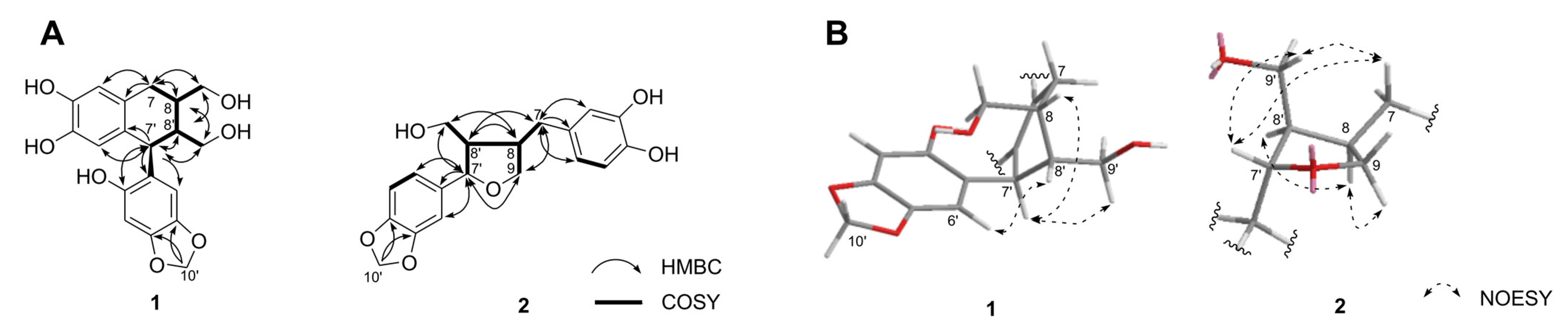
The new structure compound 1 was isolated as a yellow amorphous powder, [α]20D +21.6° (c 0.1, MeOH). A pseudomolecular ion peak at m/z 360.1206 [M]+, observed in the HRFABMS spectrum of 1 in conjunction with 13C NMR spectroscopic data, corresponded to the molecular formula C19H20O7. The 1H NMR spectrum of 1 displayed four aromatic proton signals at δH 6.52 (1H, s, H-2), 6.40 (1H, s, H-3′), 6.29 (1H, s, H-6′), and 6.18 (1H, s, H-5), revealing the presence of two 1,3,4,6-tetrasubstituted aromatic rings. Signals for a methylenedioxy group at δH 5.78 (2H, s, H-10′), two oxygenated methylene proton signals at δH 3.69 (1H, m, H-9′), 3.64−3.62 (2H, m, H-9), and 3.42 (1H, dd, J = 11.4, 4.8 Hz, H-9′), one methylene at δH 2.68 (2H, d, J = 7.8 Hz, H-7), and three methines at δH 4.17 (1H, d, J = 10.2 Hz, H-7′), 1.94 (1H, m, H-8), and 1.73 (1H, m, H-8′) were also obtained on the 1H NMR spectrum (Table 1). The 13C NMR and HSQC spectra of 1 showed 19 carbon signals: 12 aromatic carbon signals at δC 151.2 (C-2′), 147.5 (C-5′), 144.3 (C-4), 144.2 (C-3), 142.4 (C-4′), 132.1 (C-1), 129.4 (C-6), 125.3 (C-1′), 117.2 (C-5), 115.7 (C-2), 109.9 (C-3′), and 98.4 (C-6′), one methylenedioxy group at δC 102.0 (C-10′), two oxygenated methylenes at δC 66.2 (C-9) and 63.3 (C-9′), one methylene at δC 33.4 (C-7), and three methine carbons at δC 47.8 (C-8′), 40.9 (C-7′), and 40.8 (C-8) (Table 1). Analogous resonances consistent with the presence of these functionalities were displayed in the 13C NMR data of 1 (Table 1) [22,23]. The 1H-1H COSY correlations of H-7/H-8/H-9, H-7′/H-8′/H-9′, and H-8/H-8′, and the HMBC correlations of H-8 to C-1,-7,-7′ and H-7′ to C-1′,-2,-2′,-8,-8′,-9′, indicate that 1 is an aryltetralin-type lignan [24]. The HMBC correlations from the methylenedioxy moiety (H-10′) indicate that this is located at C-4′ (δC 142.4) and -5′ (δC 147.5) (Figure 2A) [24].
The relative configuration of 1, established on the basis of analyzing the NOESY correlations observed of H-6′/H-8′, H-7′/H-8, and H-7′/H-9′ along with the large-coupling constants (J7′, 8′ = 10.2 Hz), confirmed the trans-trans arrangement (Figure 2B) [25,26]. Compared to previous reports, the circular dichroism (CD) spectrum of 1 showed positive Cotton effects at 238 (Δε + 4.7) and 282 (Δε + 2.4) nm and a negative Cotton effect at 301 (Δε −8.7) nm (Figure S9) [27]. Consequently, the absolute configuration of 1 was determined to be a 7′S, 8R, 8′R-configuration. Compound 1 was identified as a new naturally occurring arytetralin-type lignan, and this compound was assigned the trivial name sesamlignan A (Figure S30).
Compound 2 was isolated as a brown amorphous optically active powder ([α]20D +58.9°) and showed a pseudomolecular ion at m/z 344.1262 [M]+, corresponding to the molecular formula C19H20O6 in the HRFABMS. The 1H NMR data of 2 exhibited resonances for two sets of ABX-type aromatic rings at δH 6.82 (1H, d, J = 2.4 Hz, H-2′), 6.77 (1H, dd, J = 7.8, 2.4 Hz, H-6′), 6.74 (1H, d, J = 7.8 Hz, H-6′), 6.67 (1H, d, J = 8.4 Hz, H-5), 6.62 (1H, d, J = 2.4 Hz, H-2), and 6.51 (1H, dd, J = 8.4, 2.4 Hz, H-6), and one methylenedioxy group at δH 5.90 (2H, s, H-10′). The spectrum also included signals attributable to three methine protons at δH 4.76 (1H, d, J = 6.0 Hz, H-7′), 2.68 (1H, m, H-8), and 2.29 (1H, m, H-8′), one methylene proton at δH 2.81 (1H, dd, J = 13.8 Hz, 5.4, H-7) and 2.42 (1H, dd, J = 13.8, 10.2 Hz, H-7), one oxygenated methylene protons at 3.97 (1H, dd, J = 8.4, 6.6 Hz, H-9), 3.69 (1H, dd, J = 8.4, 6.6 Hz, H-9), and one hydroxymethyl group at δH 3.81 (1H, dd, J = 10.8, 7.2 Hz, H-9′) and 3.61 (1H, dd, J = 10.8, 7.2 Hz, H-9′) (Table 1). These NMR data observations indicate that 2 contains a three-ring system: two aromatic rings and one tetrahydrofuran moiety. The above data, together with the observation of 13C NMR and HSQC spectra, implied that 2 is a tetrahydrofuran-type lignan [28]. The connective positions of each methylenedioxyphenyl and a dihydroxyphenyl group by the HMBC technique, which demonstrated a three-bond correlation between the oxygenated methine proton (H-7′) to C-1′ (δC 138.6), -2′ (δC 107.2), and -6′ (δC 121.0), and methylene proton signals (H-7) to C-1 (δC 133.5), -2 (δC 108.6), -6 (δC 120.3), -8 (δC 43.7), and -9 (δC 73.7) positions, respectively (Figure 2A). The linkage point of hydroxymethyl residue (H-9′) on the furan moiety (C-7′, -8, -8′) in 2 was determined unambiguously from key HMBC correlations in Figure 2A. These spectroscopic features are comparable to those reported for acuminatin [29], previously isolated from the aerial parts of Helichrysum acuminatum, except for the signals of a methoxyl group at C-3 present in 2.
Moreover, the proposed relative stereochemistry of 2 in the tetrahydrofuran moiety was confirmed by the spatial correlations between H-7′, H-7, and H-9′, H-8 and H-8′, and H-8 and H-9 as observed in the NOESY spectrum (Figure 2B) [30,31]. The absolute configuration of the tetrahydrofuran moiety in 2 was determined as a 7′S, 8R, 8′R-configuration based on the positive specific optical rotation value {[α]20D +58.9° (c 0.1, MeOH)} as well as negative Cotton effects at 229 (Δε −0.3) and 288 (Δε −0.7) nm in the CD spectral comparison using authentic analogs (Figure S17) [32]. Although the planar structure of 2 has previously been reported as an intermediate of sesamin metabolites identified in rat urine by GC-MS data [33], this is the first report of isolation and determination of the absolute structure using spectroscopic interpretation (Figure S30).
Based on the spectroscopic analysis and comparison of the data with literature values, the previously reported compounds 3−9 were identified as (+)-sesaminol 2′-O-glucopyranosyl(1→2)-O-[glucopyranosyl(1→6)]-O-glucopyranoside (3) [11,34], (+)-sesaminol 2′-O-glucopyranosyl(1→2)-O-glucopyranoside (4) [13,35], (+)-sesaminol (5) [36], (+)-epipinoresinol 4′-O-glucopyranoside (6) [37], (+)-epipinoresinol (7) [38], apocynin (8) [39], and vanillic acid (9) [10] (Figures S18–S29). The methyl-substituted vanillic acid, apocynin (8), was first isolated from Sesamum indicum (Figure 1).
2.2. Inhibition of Formation of AGEs and ONOO− Scavenging Effects
All pure isolated compounds 1−9 were evaluated for their capacity to inhibit the formation of AGEs using aminoguanidine as the positive control (Table 2). Compared to the positive control aminoguanidine (IC50: 995.3 ± 3.6 μM), the most potent inhibitory effects against AGEs formation were exhibited by the new lignans sesamelignans A (1) and B (2) with IC50 values of 7.5 ± 0.3 and 9.8 ± 0.5 μM, respectively. The IC50 values of the furfuran-type lignans 3−7 were obtained in the range 17.8 to 65.8 μM for AGEs formation ability. The simple phenolic compounds 8 and 9 were considerably less effective compared to other lignan derivatives. In addition, we further evaluated the anti-oxidant effects of the isolated compounds using the previously reported ONOO− scavenging assay [40]. The novel aryltetralin lignan 1 showed maximum scavenging activity against the ONOO− scavenging assay (IC50: 8.1 ± 0.5 μM) compared to the positive control L-penicillamine (IC50: 15.0 ± 1.0 μM). Vanillic acid (9) has previously been described as a powerful ONOO− scavenging substance isolated from Panax ginseng, and our results are in agreement with those findings [41]. Although various lignan analogs from natural products have been reported as anti-glycation inhibitors [42], the current study is the first to validate the new aryltetralin-type lignan 1 with potent inhibitory effects of AGEs formation and ONOO− scavenging activity. Taken together, our results indicate the potential to develop sesamelignan A (1) as a therapeutic for diabetic complications and related diseases.
3. Materials and Methods
3.1. General Experimental Procedures
The ultraviolet (UV) spectrum was measured on a T-60 spectrophotometer (PG Instrument, Leicestershire, UK), and the circular dichroism (CD) spectrum was run on a JASCO J-1500 spectrometer (JASCO, Tokyo, Japan). 1H-, 13C-NMR, 1H-1H COSY, HSQC, HMBC, and NOESY spectra were measured on a Varian VNS-600 MHz spectrometer (Varian, Palo Alto, CA, USA) equipment using CD3OD (δH 3.35, δC 49.0) as the solvent and tetramethylsilane (TMS) as the internal standard. Fast atom bombardment mass spectrometer (FABMS) was recorded on a JMS-700 GC-HRMS spectrometer (JEOL, Tokyo, Japan), and optical rotation was obtained using a JASCO P-2000 polarimeter. Toyopearl HW-40C gel (Tosho Co. Tokyo, Japan) and ODS gel (ODS AQ 120-50S, YMC Co., Kyoto, Japan) were used for column chromatography.
3.2. Plant Material and Preparation
Sesame seeds (Sesamum indicum L.) were collected in June 2017 from Yecheon-gun, Republic of Korea, and identified by Prof. Tae Hoon Kim. A voucher specimen was deposited at the Natural Products Chemistry Laboratory of Daegu University. The dried sesame seeds (20 kg) were roasted in an electric frying pan (D-1692, Dongkwang oil machine Co., Seoul, Korea) at 300 °C for 12 min. Oil was extracted from the roasted sesame seeds using an electric oil squeezer (D-1880, Dongkwang oil machine Co., Seoul, Korea), and the remaining sesame byproducts were used in the experiment.
3.3. Extraction and Isolation
Defatted sesame seeds (8.0 kg) were powdered and extracted with distilled water (40 L) at 70 °C for 3 h, after which the extract solution was concentrated in vacuo to yield the solid extract (726.0 g). The dried extract (720.0 g) was suspended in 10% MeOH in H2O (1 L) and partitioned sequentially using organic solvents to yield n-hexane—(2.3 g), EtOAc—(32.2 g), n-BuOH—(66.9 g), and H2O—(425.1 g) soluble fractions. The EtOAc−soluble fraction was found to be active in the AGEs formation inhibition assay, with an IC50 value of 154.8 ± 2.4 μg/mL (Table S1). One portion of the EtOAc−fraction (23.5 g) was chromatographed over a Toyopearl HW-40 column (4 cm i.d. × 40 cm, coarse grade) eluted with gradient systems of H2O-MeOH increasing polarity (0% to 100%, followed by 70% acetone) to yield eleven sub-fractions (SE01-SE11). Fraction SE03 (480.1 mg) was subjected to ODS gel column chromatography (1 cm i.d. × 40 cm, particle size 50 μm) with a MeOH/H2O system, resulting in the isolation of compounds 3 (55.3 mg, tR 20.9 min), 4 (54.1 mg, tR 23.2 min), and 6 (37.4 mg, tR 19.0 min). Similar fractionation of SE04 (306.6 mg) on ODS gel chromatography (1 cm i.d. × 42 cm) yielded the pure compounds 8 (13.8 mg, tR 16.4 min) and 9 (15.5 mg, tR 10.9 min). Finally, the sub-fraction SE08 (155.9 mg) was subjected to ODS gel column chromatography (1 cm i.d. × 42 cm) with aqueous MeOH to give the pure compounds 1 (4.7 mg, tR 16.8 min), 2 (2.6 mg, tR 22.6 min), 5 (3.4 mg, tR 31.0 min), and 7 (3.9 mg, tR 25.0 min). HPLC (Shimadzu, Tokyo, Japan) analysis was performed using the YMC-Pack ODS A-302 column (4.6 mm i.d. × 150 mm, particle size 5 μm; YMC Co., Kyoto, Japan) and mobile phase comprising 0.1% HCOOH in H2O (Solvent A) and MeCN (Solvent B). A gradient system was performed with a linear gradient of 5% to 100% solvent B for 35 min with the flow rate set at 1.0 mL/min.
Sesamlignan A (1): Yellow amorphous powder. [α]20D +21.6° (c 0.1, MeOH). UV λmax MeOH (log ε): 205 (3.54), 235 (sh), 295 (1.19) nm. CD (MeOH) Δε (nm): 211 (+21.3), 238 (+4.7), 282 (+2.4), 301 (−8.7) nm. 1H- and 13C-NMR: see Table 1. FABMS m/z 360 [M]+. HRFABMS m/z 360.1203 [M]+ (calc. for C19H20O7, 360.1209) (Figures S1–S9).
Sesamlignan B (2): Brown amorphous powder. [α]20D +58.9° (c 0.1, MeOH). UV λmax MeOH (log ε): 204 (3.75), 234 (sh), 285 (1.10) nm. CD (MeOH) Δε (nm): 210 (−4.3), 229 (−0.3), 288 (−0.7) nm. 1H- and 13C-NMR: see Table 1. FABMS m/z 344 [M]+. HRFABMS m/z 344.1262 [M]+ (calc. for C19H20O6, 344.1260) (Figures S10–S17).
(+)-Sesaminol 2′-O-glucopyranosyl(1→2)-O-[glucopyranosyl(1→6)]-O-glucopyrano side (3): Yellow amorphous powder. [α]20D −47.0° (c 0.1, MeOH). 1H-NMR (CD3OD, 600 MHz): δ 6.91 (1H, s, H-3′), 6.85 (1H, d, J = 1.2 Hz, H-2″), 6.83 (1H, s, H-6′), 6.81 (1H, dd, J = 7.8, 1.2 Hz, H-6″), 6.76 (1H, d, J = 7.8 Hz, H-5″), 5.91 (2H, s, H-7″), 5.89 (2H, s, H-7′), 5.20 (1H, d, J = 4.8 Hz, H-2), 5.02 (1H, d, J = 7.2 Hz, H-1″′), 4.87 (1H, d, J = 7.2 Hz, H-1″″), 4.69 (1H, d, J = 4.8 Hz, H-6), 4.34 (1H, d, J = 7.2 Hz, H-1″″′), 4.24 (1H, dd, J = 9.0, 6.6 Hz, H-4eq), 4.21 (1H, d, J = 10.8 Hz, H-8ax), 4.19 (1H, d, J = 10.8 Hz, H-8eq), 4.13 (1H, dd, J = 10.8, 1.2 Hz, H-6″′), 3.81 (1H, overlap, H-4ax), 3.86–3.19 (17H, overlap, glucose), 3.00 (1H, m, H-5), 2.90 (1H, m, H-1); 13C-NMR (CD3OD, 150 MHz): δ 149.9 (C-2′), 149.3 (C-5′), 148.5 (C-4″), 148.4 (C-3″), 144.0 (C-4′), 136.5 (C-1″), 125.5 (C-1′), 120.7 (C-6″), 109.0 (C-5″), 107.6 (C-2″), 105.9 (C-6′), 104.9 (C-1″″′), 104.5 (C-1″″), 102.6 (C-7′), 102.4 (C-7″), 101.2 (C-1″′), 99.3 (C-3′), 86.5 (C-6), 82.8 (C-2), 81.8 (C-2″′), 78.4 (C-3″′), 78.1 (C-4″″), 78.0 (C-3″′), 77.9 (C-3″″′), 77.8 (C-5″″), 76.9 (C-5″′), 76.0 (C-2″″′), 75.1 (C-2″″), 73.8 (C-8), 72.8 (C-4), 71.6 (C-4″″′), 71.2 (C-5″″′), 71.1 (C-4″′), 70.3 (C-6″′), 62.8 (C-6″″′), 62.2 (C-6″″), 55.8 (C-1), 55.6 (C-5). FABMS m/z 856 [M]+ (Figures S18 and S19).
(+)-Sesaminol 2′-O-glucopyranosyl(1→2)-O-glucopyranoside (4): Yellow amorphous powder. [α]20D −26.7° (c 0.1, MeOH). 1H-NMR (CD3OD, 600 MHz): δ 6.91 (1H, s, H-3′), 6.85 (1H, d, J = 1.8 Hz, H-2″), 6.81 (1H, s, H-6′), 6.80 (1H, dd, J = 7.8, 1.8 Hz, H-6″), 6.75 (1H, d, J = 7.8 Hz, H-5″), 5.90 (2H, s, H-7″), 5.89 (2H, s, H-7′), 5.18 (1H, d, J = 4.8 Hz, H-2), 4.84 (1H, d, J = 7.8 Hz, H-1″′), 4.62 (1H, d, J = 4.8 Hz, H-6), 4.34 (1H, d, J = 7.8 Hz, H-1″″), 4.27 (1H, dd, J = 9.6, 7.8 Hz, H-8eq), 4.17 (1H, dd, J = 9.6, 6.0 Hz, H-4eq), 4.13 (1H, m, H-6″′), 4.06 (1H, dd, J = 9.6, 4.8 Hz, H-8ax), 3.84 (1H, dd, J = 9.6, 4.8 Hz, H-4ax), 3.86–3.21 (11H, overlap, glucose), 3.00 (1H, m, H-5), 2.95 (1H, m, H-1); 13C-NMR (CD3OD, 150 MHz): δ 150.4 (C-2′), 149.3 (C-5′), 148.6 (C-4″), 148.5 (C-3″), 144.1 (C-4′), 136.4 (C-1″), 125.5 (C-1′), 120.7 (C-6″), 109.0 (C-5″), 107.6 (C-2″), 106.0 (C-6′), 104.8 (C-1″″), 103.2 (C-7′), 102.6 (C-7″), 102.4 (C-1″′), 99.9 (C-3′), 86.9 (C-6), 82.9 (C-2), 78.1 (C-5″′), 78.0 (C-5″″), 77.1 (C-3″′), 75.0 (C-4″′), 74.9 (C-2″″), 74.2 (C-2″′), 72.4 (C-3″″), 71.6 (C-4″″), 71.3 (C-4), 71.2 (C-8), 70.8 (C-6″′), 62.8 (C-6″″), 55.6 (C-1), 55.2 (C-5). FABMS m/z 694 [M]+ (Figures S20 and S21).
(+)-Sesaminol (5): White amorphous powder. [α]20D +73.6° (c 0.1, MeOH). 1H-NMR (CD3OD, 600 MHz): δ 6.86 (1H, d, J = 1.2 Hz, H-2″), 6.82 (1H, dd, J = 7.2, 1.2 Hz, H-6″), 6.76 (1H, d, J = 7.2 Hz, H-5″), 5.91 (2H, s, H-7″), 6.75 (1H, s, H-3′), 6.35 (1H, s, H-6′), 5.81 (2H, s, H-7′), 4.98 (1H, d, J = 4.2 Hz, H-2), 4.67 (1H, d, J = 4.2 Hz, H-6), 4.21 (1H, dd, J = 9.6, 7.8 Hz, H-8eq), 4.24 (1H, dd, J = 9.6, 6.0 Hz, H-4eq), 4.01 (1H, dd, J = 9.6, 4.2 Hz, H-8ax), 3.85 (1H, dd, J = 9.6, 4.2 Hz, H-4ax), 3.00 (1H, m, H-5), 2.95 (1H, m, H-1); 13C-NMR (CD3OD, 150 MHz): δ 151.2 (C-2′), 148.2 (C-5′), 147.7 (C-4″), 146.4 (C-3″), 145.0 (C-4′), 135.4 (C-1″), 126.1 (C-1′), 122.1 (C-6″), 109.2 (C-5″), 107.8 (C-2″), 106.5 (C-6′), 102.9 (C-7′), 101.4 (C-7″), 99.8 (C-3′), 85.8 (C-6), 81.0 (C-2), 71.6 (C-4), 70.5 (C-8), 53.2 (C-1), 51.0 (C-5). FABMS m/z 370 [M]+ (Figure S22).
(+)-Epipinoresinol 4′-O-glucopyranoside (6): Brown amorphous powder. [α]20D +37.7° (c 0.1, MeOH). 1H-NMR (CD3OD, 600 MHz): δ 7.13 (1H, d, J = 8.4 Hz, H-5″), 7.02 (1H, d, J = 1.8 Hz, H-2″), 6.63 (1H, d, J = 1.8 Hz, H-2′), 6.91 (1H, dd, J = 8.4, 1.8 Hz, H-6″), 6.80 (1H, dd, J = 7.8, 1.8 Hz, H-6′), 6.75 (1H, d, J = 7.8 Hz, H-5′), 4.82 (1H, d, J = 7.8 Hz, H-1″′), 4.74 (1H, d, J = 4.8 Hz, H-2), 4.69 (1H, d, J = 7.2 Hz, H-6), 4.23 (1H, overlap, H-8eq), 4.22 (1H, overlap, H-4eq), 3.86 (3H, s, OCH3-3″), 3.84 (3H, s, OCH3-3′), 3.82 (1H, m, H-8ax), 3.80 (1H, m, H-4ax), 3.69 (1H, dd, J = 11.4, 4.2 Hz, H-6″′), 3.68 (1H, m, H-6″′), 3.46 (1H, m, H-2″′), 3.45 (1H, m, H-4″′), 3.30 (1H, m, H-3″′), 3.29 (1H, m, H-5″′), 3.12 (1H, m, H-5), 3.10 (1H, m, H-1); 13C-NMR (CD3OD, 150 MHz): δ 151.1 (C-3″), 149.1 (C-3′), 147.5 (C-4″), 147.3 (C-4′), 137.5 (C-1″), 133.8 (C-1′), 120.1 (C-6′), 119.8 (C-6″), 118.1 (C-5″), 116.1 (C-5′), 111.7 (C-2′), 111.0 (C-2″), 102.9 (C-1″′), 87.5 (C-6), 87.1 (C-2), 78.2 (C-5″′), 77.9 (C-3″′), 74.9 (C-2″′), 73.1 (C-4), 72.7 (C-8), 71.3 (C-4″′), 62.5 (C-6″′), 56.8 (OCH3-3′), 56.4 (OCH3-3″), 55.5 (C-1), 55.4 (C-5). FABMS m/z 520 [M]+ (Figures S23 and S24).
(+)-Epipinoresinol (7): Brown amorphous powder. [α]20D +84.5° (c 0.1, MeOH). 1H-NMR (CD3OD, 600 MHz): δ 6.94 (1H, d, J = 1.8 Hz, H-2″), 6.96 (1H, d, J = 1.8 Hz, H-2′), 6.81 (1H, dd, J = 8.4, 1.8 Hz, H-6″), 6.80 (1H, d, J = 8.4 Hz, H-5′), 6.77 (1H, d, J = 8.4 Hz, H-5″), 6.76 (1H, dd, J = 8.4, 1.8 Hz, H-6′), 4.85 (1H, d, J = 4.8 Hz, H-2), 4.41 (1H, d, J = 7.2 Hz, H-6), 4.09 (1H, d, J = 9.6 Hz, H-8eq), 3.83 (1H, d, J = 9.6 Hz, H-8ax), 3.86 (3H, s, OCH3-3″), 3.85 (3H, s, OCH3-3′), 3.78 (1H, t, J = 9.0 Hz, H-4eq), 3.38 (1H, m, H-4ax), 3.28 (1H, m, H-5), 2.93 (1H, m, H-1); 13C-NMR (CD3OD, 150 MHz): δ 149.1 (C-3″), 148.8 (C-3′), 147.4 (C-4″), 146.6 (C-4′), 133.9 (C-1″), 131.3 (C-1′), 120.1 (C-6′), 119.4 (C-6″), 116.1 (C-5″), 116.0 (C-5′), 110.9 (C-2′), 111.6 (C-2″), 89.5 (C-6), 83.5 (C-2), 71.9 (C-4), 70.6 (C-8), 56.4 (OCH3-3′), 55.6 (OCH3-3″), 51.2 (C-1), 59.8 (C-5). FABMS m/z 358 [M]+ (Figures S25 and S26).
Apocynin (8): White amorphous powder. 1H-NMR (CD3OD, 600 MHz): δ 7.56 (1H, dd, J = 8.4, 1.8 Hz, H-6), 7.55 (1H, d, J = 1.8 Hz, H-2), 6.85 (1H, d, J = 8.4 Hz, H-5), 3.86 (3H, s, OCH3-3), 2.53 (3H, s, CH3-8); 13C-NMR (CD3OD, 150 MHz): δ 199.4 (C-7), 153.4 (C-4), 149.0 (C-3), 130.6 (C-1), 125.2 (C-6), 115.8 (C-5), 112.0 (C-2), 56.3 (OCH3-3), 26.2 (CH3-8). FABMS m/z 166 [M]+ (Figures S27 and S28).
Vanillic acid (9): White amorphous powder. 1H-NMR (CD3OD, 600 MHz): δ 7.55 (1H, d, J = 1.8 Hz, H-2), 7.55 (1H, dd, J = 8.4, 1.8 Hz, H-6), 6.83 (1H, d, J = 8.4 Hz, H-5), 3.87 (3H, s, OCH3-3). FABMS m/z 168 [M]+ (Figure S29).
3.4. Evaluation of AGEs Formation Inhibitory Effects
Using a previously reported method [43] with minor modification, we evaluated the AGEs formation inhibitory potential of the isolated compounds. Briefly, the reaction mixture was prepared by adding 10 mg/mL BSA in 50 mM phosphate buffer (pH 7.4) containing 0.02% sodium azide to a sugar solution (200 mM D-fructose and 200 mM D-glucose). The reaction mixture (800 μL) was then combined with various concentrations of either the test compounds (200 μL) or the positive control (aminoguanidine) dissolved in 5% DMSO. After incubation at 37 °C for 7 days, the fluorescent reaction products were determined using an ELISA reader (Infinite F200; Tecan Austria GmBH, Grodig, Austria), with excitation and emission maxima at 350 and 450 nm, respectively. The concentration required for 50% inhibition (IC50 value) of the fluorescence intensity was determined by linear regression analysis. All measurements were obtained in triplicate.
3.5. Evaluation of ONOO− Scavenging Activities
The ONOO− scavenging ability was evaluated by observing the extremely fluorescent dihydrorhodamine 123 (DHR 123) that is rapidly generated from non-fluorescent DHR 123 in the presence of ONOO− [40]. The dihydrorhodamine buffer (pH 7.4) comprises 50 mM sodium phosphate monobasic, 50 mM sodium phosphate dibasic, 90 mM sodium chloride, 5 mM potassium chloride, and 100 μM DTPA, and the final DHR 123 concentration used was 5.0 μM. The test sample was dissolved in 5% DMSO. The final fluorescent intensities of the treated samples were observed 5 min after treatment with and without the addition of authentic ONOO− (10 μM) dissolved in 0.3 N NaOH. The fluorescence intensity of the oxidized DHR 123 was estimated with a fluorescence ELISA reader at emission and excitation wavelengths of 530 and 480 nm, respectively. Results of the ONOO− scavenging effect were evaluated as the final fluorescence intensity minus the background fluorescence, determined by the detection of DHR 123 oxidation. The 50% inhibition (IC50 value) was measured by linear regression analysis of the scavenging activity under the above assay conditions. L-Penicillamine was used as a positive control. All measurements were obtained in triplicate.
3.6. Statistical Analysis
Data for the in vitro analyses of AGEs formation and ONOO− scavenging activity were analyzed using the Proc GLM procedure of SAS software (version 9.3, SAS Institute Inc., Cary, NC, USA). The results are reported as the least square mean values and standard deviation. Statistical significance was considered at p < 0.05.
4. Conclusions
This paper reported two previously undescribed lignans (1 and 2) along with seven known compounds (3−9) isolated from the defatted sesame cake. The new chemical structures of 1 and 2 were characterized by detailed NMR, MS, and CD spectra data analysis. All compounds were evaluated for their inhibitory potential against AGEs formation and ONOO− scavenging properties. The unusual aryltetralin-type (1) and tertrahydrofuran-type lignan (2) showed the most potent inhibitory effects of AGEs formation compared to the positive control. In addition, the newly discovered sesamlignan A (1) exhibited a maximum potency for ONOO− scavenging capacity. Thus, we propose that sesamlignans A and B have the potential to be developed as therapeutic agents for treating diabetic complications and related diseases.
Supplementary Materials
The following are available online at https://www.mdpi.com/article/10.3390/molecules28052255/s1, Figure S1: 1H NMR spectrum of compound 1 in CD3OD. Figure S2: 13C NMR spectrum of compound 1 in CD3OD: Figure S3. 1H-1H COSY spectrum of compound 1 in CD3OD. Figure S4: HSQC spectrum of compound 1 in CD3OD. Figure S5: HMBC spectrum of compound 1 in CD3OD. Figure S6: Expanded key HMBC correlation of compound 1 (H-7′ to C-5). Figure S7: NOESY spectrum of compound 1 in CD3OD. Figure S8: HRFABMS spectrum of compound 1. Figure S9: CD spectrum of compound 1. Figure S10: 1H NMR spectrum of compound 2 in CD3OD. Figure S11: 13C NMR spectrum of compound 2 in CD3OD. Figure S12: 1H-1H COSY spectrum of compound 2 in CD3OD. Figure S13: HSQC spectrum of compound 2 in CD3OD. Figure S14: HMBC spectrum of compound 2 in CD3OD. Figure S15: NOESY spectrum of compound 2 in CD3OD. Figure S16: HRFABMS spectrum of compound 2. Figure S17: CD spectrum of compound 2. Figure S18: 1H NMR spectrum of compound 3 in CD3OD. Figure S19: 13C NMR spectrum of compound 3 in CD3OD. Figure S20: 1H NMR spectrum of compound 4 in CD3OD. Figure S21: 13C NMR spectrum of compound 4 in CD3OD. Figure S22: 1H NMR spectrum of compound 5 in CD3OD. Figure S23: 1H NMR spectrum of compound 6 in CD3OD: Figure S24: 13C NMR spectrum of compound 6 in CD3OD. Figure S25: 1H NMR spectrum of compound 7 in CD3OD. Figure S26: 13C NMR spectrum of compound 7 in CD3OD. Figure S27: 1H NMR spectrum of compound 8 in CD3OD. Figure S28: 13C NMR spectrum of compound 8 in CD3OD. Figure S29: 1H NMR spectrum of compound 9 in CD3OD. Figure S30: Chemical structures of the new compounds 1 and 2 isolated from defatted Sesame cake. Table S1: AGEs formation inhibitory effects on the fraction from defatted Sesame cake.
Author Contributions
G.H.J. and T.H.K. performed the experiments and data and wrote the article. T.H.K. was the project leader who conceived and designed the experiments. All authors have read and agreed to the published version of the manuscript.
Funding
This research was supported by Daegu University Research Grant, 2022.
Institutional Review Board Statement
Not applicable.
Informed Consent Statement
Not applicable.
Data Availability Statement
The data presented in this study are available on request from the corresponding author.
Conflicts of Interest
The authors declare no conflict of interest.
Sample Availability
Not applicable.
References
- Saleem, M.; Kim, H.J.; Ali, M.S.; Lee, Y.S. An update on bioactive plant lignans. Nat. Prod. Rep. 2005, 22, 696–716. [Google Scholar] [CrossRef] [PubMed]
- Zálešák, F.; Bon, D.J.Y.D.; Pospíšil, J. Lignans and Neolignans: Plant secondary metabolites as a reservoir of biologically active substances. Pharmacol. Rev. 2019, 146, 104284. [Google Scholar] [CrossRef] [PubMed]
- Grougnet, R.; Magiatis, P.; Laborie, H.; Lazarou, D.; Papadopoulos, A.; Skaltsounis, A.L. Sesamolinol glucoside, disaminyl ether, and other lignans from sesame seeds. J. Agric. Food Chem. 2012, 60, 108–111. [Google Scholar] [CrossRef] [PubMed]
- Namiki, M. The chemistry and physiological functions of sesame. Food Rev. Int. 1995, 11, 281–329. [Google Scholar] [CrossRef]
- Suja, K.P.; Jayalekshmy, A.; Arumughan, C. Free radical scavenging behavior of antioxidant compounds of sesame (Sesamum indicum L.) in DPPH• system. J. Agric. Food Chem. 2004, 52, 912–915. [Google Scholar] [CrossRef]
- Dar, A.A.; Arumugam, N. Lignans of sesame: Purification methods, biological activities and biosynthesis—A review. Bioorg. Chem. 2013, 50, 1–10. [Google Scholar] [CrossRef]
- Jeong, S.M.; Kim, S.Y.; Kim, D.R.; Nam, K.C.; Ahn, D.U.; Lee, S.C. Effect of seed roasting conditions on the antioxidant activity of defatted sesame meal extracts. J. Food Sci. 2004, 69, C377–C381. [Google Scholar] [CrossRef]
- Takeuchi, H.; Mooi, L.Y.; Inagaki, Y.; He, P. Hypoglycemic effect of a hot-water extract from defatted sesame (Sesamum indicum L.) seed on the blood glucose level in genetically diabetic KK-Ay mice. Biosci. Biotehcnol. Biochem. 2001, 65, 2318–2321. [Google Scholar] [CrossRef] [Green Version]
- Wikul, A.; Damsud, T.; Kataoka, K.; Phuwapraisirisan, P. (+)-Pinoresinol is a putative hypoglycemic agent in defatted sesame (Sesamum indicum) seeds though inhibiting α-glucosidase. Bioorg. Med. Chem. Lett. 2012, 22, 5215–5217. [Google Scholar] [CrossRef]
- Lee, K.; Jo, I.Y.; Park, S.H.; Kim, K.S.; Bae, J.; Park, J.W.; Bu, Y. Defatted sesame seed extract reduces brain oedema by regulating aquaporin 4 expression in acute phase of transient focal cerebral ischaemia in rat. Phytother. Res. 2012, 26, 1521–1527. [Google Scholar] [CrossRef]
- Jeon, J.S.; Park, C.L.; Syed, A.S.; Kim, Y.M.; Cho, I.J.; Kim, C.Y. Preparative separation of sesamin and sesamolin from defatted sesame meal via centrifugal partition chromatography with consecutive sample injection. J. Chromatogr. B 2016, 1011, 108–113. [Google Scholar] [CrossRef] [PubMed]
- Lee, S.C.; Jeong, S.M.; Kim, S.Y.; Nam, K.C.; Ahn, D.U. Effect of far-infrared irradiation on the antioxidant activity of defatted sesame meal extracts. J. Agric. Food Chem. 2005, 53, 1495–1498. [Google Scholar] [CrossRef]
- Moazzami, A.A.; Andersson, R.E.; Kamal-Eldin, A. HPLC analysis of sesaminol glucosides in sesame seeds. J. Agric. Food Chem. 2006, 54, 633–638. [Google Scholar] [CrossRef] [PubMed]
- Jan, K.C.; Ho, C.T. Inhibitory activity of sesaminol and sesaminol triglycoside on cytochrome P450 enzymes and their pharmacokinetics in rats. J. Funct. Foods 2014, 7, 142–149. [Google Scholar] [CrossRef]
- Shyu, Y.S.; Hwang, L.S. Antioxidative activity of the crude extract of lignan glycosides from unroasted Burma black sesame meal. Food Res. Int. 2002, 35, 357–365. [Google Scholar] [CrossRef]
- Majdalawieh, A.F.; Massri, M.; Nasrallah, G.K. A comprehensive review on the anti-cancer properties and mechanisms of action of sesamin, a lignan in sesame seeds (Sesamum indicum). Eur. J. Pharmacol. 2017, 815, 512–521. [Google Scholar] [CrossRef] [PubMed]
- Park, S.H.; Ryu, S.N.; Bu, Y.; Kim, H.; Simon, J.E.; Kim, K.S. Antioxidant components as potential neuroprotective agents in sesame (Sesamum indicum L.). Food Rev. Int. 2010, 26, 103–121. [Google Scholar] [CrossRef]
- Reddy, M.A.; Zhang, E.; Natarajan, R. Epigenetic mechanisms in diabetic complications and metabolic memory. Diabetologia 2015, 58, 443–455. [Google Scholar] [CrossRef] [Green Version]
- Forbes, J.M.; Cooper, M.E. Mechanisms of diabetic complications. Physiol. Rev. 2013, 93, 137–188. [Google Scholar] [CrossRef]
- Negre-Salvayre, A.; Salvayre, R.; Augé, N.; Pamplona, R.; Portero-Otin, M. Hyperglycemia and glycation in diabetic complications. Antioxid. Redox Sign. 2009, 11, 3071–3109. [Google Scholar] [CrossRef]
- Giacco, F.; Brownlee, M. Oxidative stress and diabetic complications. Circ. Res. 2010, 107, 1058–1070. [Google Scholar] [CrossRef] [PubMed] [Green Version]
- Kiem, P.V.; Cuong, L.C.V.; Tai, B.H.; Nhiem, N.X.; Anh, H.L.T.; Quang, T.H.; Ngan, N.T.T.; Oh, H.; Kim, Y.C. New lignans from Antidesma hainanensis inhibit NO production in BV2 microglial cells. Chem. Pharm. Bull. 2016, 64, 1707–1712. [Google Scholar] [CrossRef] [PubMed] [Green Version]
- Zang, Y.N.; Liu, Z.Z.; Feng, Z.M.; Jiang, J.S.; Zhang, P.C. Lignans from the root of Rhodiola crenulata. J. Agric. Food. Chem. 2012, 60, 964–972. [Google Scholar]
- Yoshikawa, K.; Tani, S.; Baba, C.; Hashimoto, T. Phenylpropanoid, sapnol A, lignan and neolignan sophorosides, saposides A and B, isolated from Canadian sugar maple sap. Molecules 2013, 18, 9641–9649. [Google Scholar] [CrossRef] [PubMed] [Green Version]
- Latté, K.P.; Kaloga, M.; Schäfer, A.; Kolodziej, H. An ellagitannin, n-butyl gallate, two aryltetralin lignans, and an unprecedented diterpene ester from Pelargonium reniforme. Phytochemistry 2008, 69, 820–826. [Google Scholar] [CrossRef]
- Suh, W.S.; Kim, K.H.; Kim, H.K.; Choi, S.U.; Lee, K.R. Three new lignan derivatives from Lindera glauca (Siebold et Zucc.) Blume. Helv. Chim. Acta 2015, 98, 1087–1094. [Google Scholar] [CrossRef]
- Zhong, X.N.; Ide, T.; Otsuka, H.; Hirata, E.; Takeda, Y. (+)-Isolarisiresinol 3a-O-sulphate from leaves of Myrsine seguinii. Phytochemistry 1998, 49, 1777–1778. [Google Scholar] [CrossRef]
- Li, N.; Wu, J.L.; Hasegawa, T.; Sakai, J.I.; Bai, L.M.; Wang, L.Y.; Tomida, A. Bioactive lignans from Peperomia duclouxii. J. Nat. Prod. 2007, 70, 544–548. [Google Scholar] [CrossRef]
- Jakupovic, J.; Pathak, V.P.; Bohlmann, F.; King, R.M.; Robinson, H. Obliquin derivatives and other constituents from Australian Helichrysum species. Phytochemistry 1987, 26, 803–807. [Google Scholar] [CrossRef]
- Nhiem, N.X.; Lee, H.Y.; Kim, N.Y.; Park, S.J.; Kim, E.S.; Han, J.E.; Kim, S.H. Stereochemical assignment of five new lignan glycosides from Viscum album by NMR study combined with CD spectroscopy. Magn. Reson. Chem. 2012, 50, 772–777. [Google Scholar] [CrossRef]
- Ullah, N.; Ahmad, S.; Anis, E.; Mohammad, P.; Rabnawaz, H.; Malik, A. A lignan from Daphne oleoides. Phytochemistry 1999, 50, 147–149. [Google Scholar] [CrossRef]
- Xie, L.H.; Akao, T.; Hamasaki, K.; Deyama, T.; Hattori, M. Biotransformation of pinoresinol diglucoside to mammalian lignans by human intestinal microflora, and isolation of Enterococcus faecalis strain PDG-1 responsible for the transformation of (+)-pinoresinol to (+)-lariciresinol. Chem. Pharm. Bull. 2003, 51, 508–515. [Google Scholar] [CrossRef] [PubMed] [Green Version]
- Liu, Z.; Saarinen, N.M.; Thompson, L.U. Sesamin is one of the major precursors of mammalian lignans in sesame seed (Sesamum indicum) as observed in vitro and in rats. J. Nutr. 2006, 136, 906–912. [Google Scholar] [CrossRef] [PubMed] [Green Version]
- Katsuzaki, H.; Kawakishi, S.; Osawa, T. Sesaminol glucosides in sesame seeds. Phytochemistry 1994, 35, 773–776. [Google Scholar] [CrossRef] [PubMed]
- Noguchi, A.; Fukui, Y.; Iuchi-Okada, A.; Kakutani, S.; Satake, H.; Iwashita, T.; Nakao, M.; Umezawa, T.; Ono, E. Sequential glucosylation of a furofuran lignan,(+)-sesaminol, by Sesamum indicum UGT71A9 and UGT94D1 glucosyltransferases. Plant J. 2008, 54, 415–427. [Google Scholar] [CrossRef]
- Nagata, M.; Osawa, T.; Namiki, M.; Fukuda, Y.; Ozaki, T. Stereochemical structures of antioxidative bisepoxylignans, sesaminol and its isomers, transformed from sesamolin. Agric. Biol. Chem. 1987, 51, 1285–1289. [Google Scholar]
- Rahman, M.M.; Dewick, P.M.; Jackson, D.E.; Lucas, J.A. Lignans of Forsythia intermedia. Phytochemistry 1990, 29, 1971–1980. [Google Scholar] [CrossRef]
- Nishibe, S.; Tsukamoto, H.; Hisada, S. Effects of O-methylation and O-glucosylation on carbon-13 nuclear magnetic resonance chemical shifts of matairesinol,(+)-pinoresinol and (+)-epipinoresinol. Chem. Pharm. Bull. 1984, 32, 4653–4657. [Google Scholar] [CrossRef]
- Finnemore, H. CXLVI—The constituents of Canadian hemp. part I. apocynin. J. Chem. Soc. Trans. 1908, 93, 1513–1519. [Google Scholar] [CrossRef] [Green Version]
- Kooy, N.W.; Royall, J.A.; Ischiropoulos, H.; Beckman, J.S. Peroxynitrite-mediated oxidation of dihydrorhodamine 123. Free Radic. Biol. Med. 1994, 6, 149–156. [Google Scholar] [CrossRef]
- Kang, K.S.; Tanaka, T.; Cho, E.J.; Yokozawa, T. Evaluation of the peroxynitrite scavenging activity of heat-processed ginseng. J. Med. Food 2009, 12, 124–130. [Google Scholar] [CrossRef] [PubMed]
- Yeh, W.J.; Hsia, S.M.; Lee, W.H.; Wu, C.H. Polyphenols with antiglycation activity and mechanisms of action: A review of recent findings. J. Food Drug. Anal. 2017, 25, 84–92. [Google Scholar] [CrossRef] [PubMed] [Green Version]
- Vinson, J.A.; Howard, T.B., III. Inhibition of protein glycation and advanced glycation end products by ascorbic acid and other vitamins and nutrients. J. Nutr. Biochem. 1996, 7, 659–663. [Google Scholar] [CrossRef]
Figure 1.
Chemical structures of compounds 1–9 isolated from defatted sesame seeds.
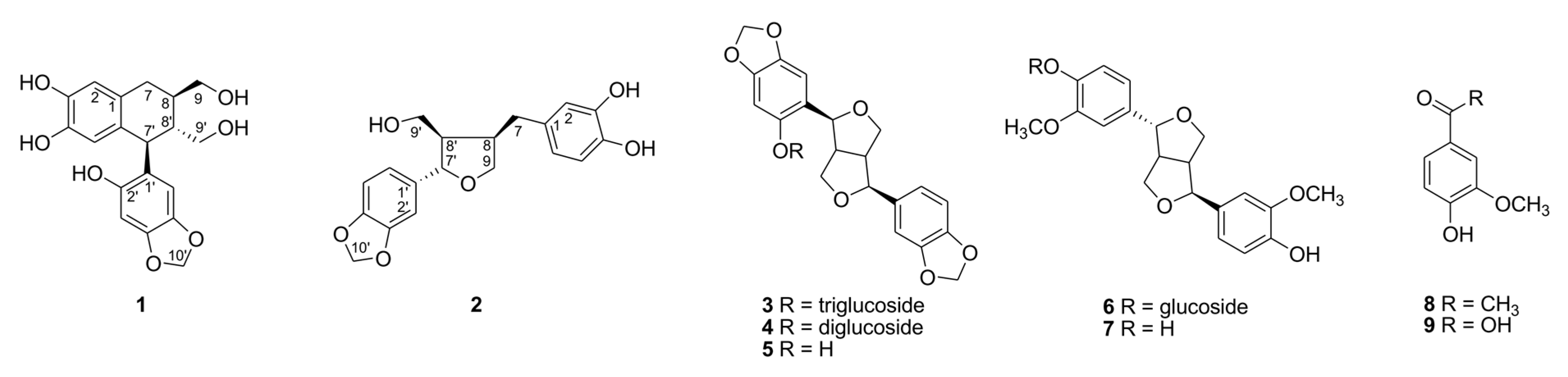
Figure 2.
1H-1H COSY (A), key HMBC (A), and NOESY (B) correlations of 1 and 2.

Table 1.
1H and 13C NMR data of compounds 1 and 2 in CD3OD 1.
| 1 | 2 | |||
|---|---|---|---|---|
| Positions | δH (J in Hz) 2 | δC, Type | δH (J in Hz) | δC, Type |
| 1 | ― | 132.1, C | ― | 133.5, C |
| 2 | 6.52 (s) | 115.7, CH | 6.62 (d, 2.4) | 108.8, CH |
| 3 | ― | 144.2, C | ― | 144.6, C |
| 4 | ― | 144.3, C | ― | 146.3, C |
| 5 | 6.18 (s) | 117.2, CH | 6.67 (d, 8.4) | 116.4, CH |
| 6 | ― | 129.4, C | 6.51 (dd, 8.4, 2.4) | 120.3, CH |
| 7 | 2.69 (d, 7.8) | 33.4, CH2 | 2.81 (dd, 13.8, 5.4) 2.42 (dd, 13.8, 10.2) | 33.3, CH2 |
| 8 | 1.94 (m) | 40.8, CH | 2.68 (m) | 43.7, CH |
| 9 | 3.64 (m) 3.62 (m) | 66.2, CH2 | 3.97 (dd, 8.4, 6.6) 3.69 (dd, 8.4, 6.6) | 73.7, CH2 |
| 1′ | ― | 125.3, C | ― | 138.6, C |
| 2′ | ― | 151.2, C | 6.82 (d, 2.4) | 107.2, CH |
| 3′ | 6.40 (s) | 98.4, CH | ― | 148.4, C |
| 4′ | ― | 142.4, C | ― | 149.3, C |
| 5′ | ― | 147.5, C | 6.74 (d, 7.8) | 116.7, CH |
| 6′ | 6.29 (s) | 109.9, CH | 6.77 (dd, 7.8, 2.4) | 121.0, CH |
| 7′ | 4.17 (d, 10.2) | 40.9, CH | 4.76 (d, 6.6) | 84.0, CH |
| 8′ | 1.73 (m) | 47.8, CH | 2.29 (m) | 54.1, CH |
| 9′ | 3.69 (m) 3.42 (dd, 11.4, 4.8) | 63.3, CH2 | 3.81 (dd, 10.8, 7.2) 3.61 (dd, 10.8, 7.2) | 60.4, CH2 |
| 10′ | 5.78 (s) | 102.0, CH2 | 5.90 (s) | 102.3, CH2 |
1 Assignments of chemical shifts are based on the analysis of HSQC and HMBC spectra. 2 J values (Hz) are given in parentheses.
Table 2.
Effects on the inhibition of AGEs formation and ONOO− scavenging activities of the isolated compounds 1–9.
Table 2.
Effects on the inhibition of AGEs formation and ONOO− scavenging activities of the isolated compounds 1–9.
| Compounds | IC50 Value (μM) 1 | |
|---|---|---|
| Inhibition of AGEs Formation 2 | ONOO− Scavenging Activity 2 | |
| 1 | 7.5 ± 0.3 e | 8.1 ± 0.5 e |
| 2 | 9.8 ± 0.5 d | 20.8 ± 0.6 c |
| 3 | 17.8 ± 0.9 c | 35.1 ± 0.8 c |
| 4 | 41.7 ± 1.3 b | 52.2 ± 1.5 b |
| 5 | 65.8 ± 2.9 b | 82.1 ± 2.2 a |
| 6 | 29.0 ± 1.7 c | 51.9 ± 1.7 b |
| 7 | 52.4 ± 2.7 b | 75.9 ± 2.0 a |
| 8 | >300 a | 59.2 ± 1.2 b |
| 9 | >300 a | 15.5 ± 0.5 d |
| Aminoguanidine 3 | 995.3 ± 3.6 a | ― |
| L-Penicillamine 3 | ― | 15.0 ± 1.0 d |
1 All compounds were examined in triplicate experiments. 2 Different letters (a–e) within the same column indicate significant differences (p < 0.05). 3 Aminoguanidine and L-penicillamine were used as positive controls.
Disclaimer/Publisher’s Note: The statements, opinions and data contained in all publications are solely those of the individual author(s) and contributor(s) and not of MDPI and/or the editor(s). MDPI and/or the editor(s) disclaim responsibility for any injury to people or property resulting from any ideas, methods, instructions or products referred to in the content. |
© 2023 by the authors. Licensee MDPI, Basel, Switzerland. This article is an open access article distributed under the terms and conditions of the Creative Commons Attribution (CC BY) license (https://creativecommons.org/licenses/by/4.0/).
Share and Cite
MDPI and ACS Style
Jeong, G.H.; Kim, T.H. New Anti-Glycative Lignans from the Defatted Seeds of Sesamum indicum. Molecules 2023, 28, 2255. https://doi.org/10.3390/molecules28052255
AMA Style
Jeong GH, Kim TH. New Anti-Glycative Lignans from the Defatted Seeds of Sesamum indicum. Molecules. 2023; 28(5):2255. https://doi.org/10.3390/molecules28052255
Chicago/Turabian StyleJeong, Gyeong Han, and Tae Hoon Kim. 2023. "New Anti-Glycative Lignans from the Defatted Seeds of Sesamum indicum" Molecules 28, no. 5: 2255. https://doi.org/10.3390/molecules28052255