
Photodynamic Therapy: From the Basics to the Current Progress of N-Heterocyclic-Bearing Dyes as Effective Photosensitizers


1
CQ-VR—Chemistry Centre of Vila Real, University of Trás-os-Montes and Alto Douro, Quinta de Prados, 5001-801 Vila Real, Portugal
2
CICS-UBI—Health Sciences Research Centre, University of Beira Interior, Av. Infante D. Henrique, 6201-506 Covilhã, Portugal
*
Author to whom correspondence should be addressed.
Molecules 2023, 28(13), 5092; https://doi.org/10.3390/molecules28135092
Submission received: 1 June 2023
/
Revised: 16 June 2023
/
Accepted: 27 June 2023
/
Published: 29 June 2023
(This article belongs to the Special Issue Recent Developments in the Synthesis and Functionalization of Nitrogen Heterocycles)
Abstract
:Photodynamic therapy, an alternative that has gained weight and popularity compared to current conventional therapies in the treatment of cancer, is a minimally invasive therapeutic strategy that generally results from the simultaneous action of three factors: a molecule with high sensitivity to light, the photosensitizer, molecular oxygen in the triplet state, and light energy. There is much to be said about each of these three elements; however, the efficacy of the photosensitizer is the most determining factor for the success of this therapeutic modality. Porphyrins, chlorins, phthalocyanines, boron-dipyrromethenes, and cyanines are some of the N-heterocycle-bearing dyes’ classes with high biological promise. In this review, a concise approach is taken to these and other families of potential photosensitizers and the molecular modifications that have recently appeared in the literature within the scope of their photodynamic application, as well as how these compounds and their formulations may eventually overcome the deficiencies of the molecules currently clinically used and revolutionize the therapies to eradicate or delay the growth of tumor cells.
1. Introduction
Photodynamic therapy (PDT) is a minimally invasive therapeutic strategy that intends to be applied to the most diverse problems of our society’s daily lives: as an anticancer therapy [1,2,3], as a way of combating multi-resistant bacteria [4] or certain fungi [5] that may compromise human health, and, more recently, as a way of eradicating certain viral strains [6,7]. PDT already plays a role in treating several non-oncological and oncological diseases, mainly of dermatological origin [8]. Its slow clinical progression in cancer treatment is primarily due to the lack of approved photosensitizers (PSs). Nevertheless, researchers have tried to find new attractive molecules and their formulations that perform better than those currently used.
Focusing on cancer, this term refers to a wide range of diseases that trigger abnormal growth and division due to cell proliferation and regulation mechanism modifications. Given their uncontrolled behavior, they become highly invasive and can even reach the bloodstream and migrate to places where extravasation allows the formation of metastases [9]. In addition to environmental and hereditary factors, lifestyle, and exposure to carcinogens, aging is one of the factors that most contribute to the onset of this disease [10,11]. Although the scientific, pharmaceutical, and medical communities have made efforts to improve the available treatments, the long-term response of patients treated with radiotherapy and chemotherapy is still insufficient due to the severe side effects [12,13]. These gaps highlight the need to explore emerging therapies such as PDT. Clinical studies have shown that this therapeutic modality can be curative, especially in less advanced tumors [14,15]. In addition, it can prolong survival in cases of inoperable tumors and improve patients’ quality of life [3,15].
Featuring heterocyclic units accompanied by nitrogen atoms in their molecular structure as well as conjugated double bonds that allow the design of chromophores, N-heterocyclic-bearing dyes constitute several families of compounds that, thanks to their relevant “photoproperties”, have been addressed concerning their potential application as PDT photosensitizing agents. Hundreds of new synthetic or hemisynthetic origin molecules have been reported in recent years to find the compound that combines desirable characteristics for its phototherapeutic application and better understand how and why certain structural modifications result in such diverse photobiological effects.
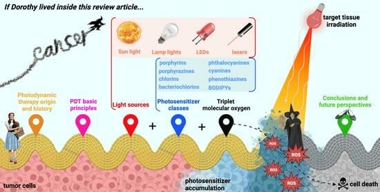
This review aims to present the reader with a concise approach to what PDT is and its fundamental principles, its importance as a therapeutic approach to complement or even replace conventional therapies in the treatment of diseases of oncological origin, several parameters to take into account to guarantee better efficiency and efficacy, as well as some of the families of N-heterocyclic-bearing dyes and the structural modifications or their formulation strategies that have been recently carried out to create new light-absorbing potential drugs with photodynamic interest (Figure 1).
2. Photodynamic Therapy
2.1. A Piece of Photodynamic Therapy’s Origin and History
Dating back to 1550 B.C., the Ebers papyrus is the oldest medical document that reports the use of sunlight as a treatment, using powder from various plants such as bishop’s flower (Ammi majus), wild parsnip (Pastinaca sativa), parsley (Petroselinum crispum), and St. John’s wort (Hypericum perforatum) for the application and treatment of depigmented skin lesions [16]. Also in Egyptian culture, pharaohs called for the construction of roofless temples dedicated to the Aton god of the sun for body light exposure and to benefit from its healing powers, and the use of Ammi majus plant extracts was common in the treatment of diseases of a dermatological nature [17]. Indian medical literature dated 1400 BC, the Atharva Veda, reports treating vitiliginous skin with babchi (Psoralea corylifolia) extracts, a plant from which it is now known that 8-methoxypsoralen is the furan-containing coumarin-based molecule that induced the photodynamic effect [16,18]. Even today, this core of photosensitizing molecules is widely studied for its potential phototherapeutic use [19,20]. Also, in Greek, Chinese, and Roman civilizations, there is evidence of the use of energy from the sun as a remedy for the treatment of various diseases.
Arnold Rikli, a Swiss healer and physician, reintroduced the use of the powers of sunlight during the late 1800s and early 1900s. A believer that nature was the basis for treating many of the complications related to human health (mentioning his famous phrase, “Water is good; air is better and light is the best of all”), Rikli trusted in the power of light in therapy, introducing “sunbathing” as a therapy for chronic diseases and functional disorders in an attempt to apply the knowledge that had been known for hundreds of years but had been forgotten [16,21].
Until then, the effectiveness of using sunlight to treat various diseases had been proven, namely for vitiligo, lupus, acne, pulmonary tuberculosis, and rickets, using (or not) extracts and powders from plant species administered topically or orally. However, it was only in 1900 that the concept of a “photosensitizing molecule” emerged, with the potential use of dyes in this therapy having been recognized: Oscar Raab, a medical student, reported that the light factor was lethal for species of Paramecium caudatum in the presence of the acridine dye and that these two elements, individually, did not cause any consequences to this unicellular organism [22]. Taking advantage of these discoveries, von Tappeiner continued Raab’s research with the dermatologist Jesionek, who published works detailing clinical trials using dyes such as eosin, fluorescein, and sodium dichloroanthracene disulfonate in the treatment of topical disorders, including skin cancer [23,24]. At the same time, they also reported data on the possible use of eosin in facial basal cell carcinoma after sun exposure or using light from arc lamps. Von Tappeiner was the first to speculate about the need for triplet molecular oxygen, introducing the term “photodynamic therapy” [16].
Despite the significant findings of Raab and von Tappeiner, Niels Finsen, a Danish physician, is considered by many to be the pioneer of modern clinical PDT. Finsen owned a medical institute in Copenhagen, to which he attached a “sun garden” to allow his patients to benefit from the healing powers of sunlight. Later, he abandoned the sunlight to start using artificial lights and noticed that exposure to red or ultraviolet light could have therapeutic effects since red light prevented the formation of pustules in patients with smallpox and that ultraviolet light allowed the treatment of cutaneous tuberculosis [16,23,25]. Due to the impact of his discoveries, Finsen was awarded the Medicine Nobel Prize in 1903.
A noteworthy milestone demonstrating the acceptance of PDT as a therapeutic modality was the approval of Photofrin in 1993 for treating bladder tumors in Canada and later in the United States, Japan, and some European countries [26,27]. Today, the Photofrin oligomer is used worldwide in treating oesophageal, endobronchial, and lung cancer, in addition to being used in Japan for gastric cancer. Since its discovery, new molecules have been clinically approved, namely 5-aminolevulinic acid and methyl 5-aminolevulinate (Levulan and Metvix, respectively), precursors of porphyrins, in the treatment of basal cell carcinoma and squamous cell carcinoma; in Japan, Talaporfin chlorin (also known as Laserphyrin) in the treatment of glioblastoma and lung cancer; in Russia, the chlorin mixture Radachlorin in basal cell carcinoma and the sulfonated phthalocyanine Photosens for lung, liver, breast, skin and gastrointestinal cancer; and in the European Union, Foscan chlorin and Redaporfin bacteriochlorin have gained a role in the therapy of head and neck cancer, and Tookad bacteriochlorin in prostate cancer [28].
However, from then on, there was a growing need to synthesize new molecules with greater efficacy and fewer side effects, giving rise to new “second- and third-generation PSs” [29,30].
These and other milestones in the history and progress of PDT to date are highlighted in the timeline in Figure 2.
2.2. Principles of Photodynamic Therapy
Clinically, the PS is administered through the most convenient route, depending on its pharmacokinetic and pharmacodynamic properties and the location of the target tissue. When administered intravenously, the photosensitizing agent is distributed throughout the body, ideally directing and accumulating selectively in the tissue to be treated. At the time of administration, the patient must be deprived of any light source to avoid premature activation. Retained by the target tissue and before being eliminated from the body, the same tissue is irradiated using a suitable light source, preferably emitting where radiation permeates tissues more efficiently, at wavelengths coincident with the molecule absorption, and with sufficient energy for its activation. Activation of the compound culminates in cell death and consequent ablation of the target tissue [3,31,32].
According to how the photosensitizing molecule acts, in general, it can produce reactive oxygen species (ROS) [33] that, despite being essential for the normal functioning of the cell [34,35], in high concentrations, become cytotoxic [34]. Thus, this therapeutic approach results from combining three individually innocuous components that are lethal when combined: the PS, light, and triplet state oxygen molecules.
The Perrin-Jablonski energy diagram explains photophysically and photochemically the activity of PS compounds (Figure 3). This diagram elucidates that these molecules, in their ground state, are in the lowest possible energy configuration with opposite spins. When appropriate radiation is applied, an electron is transferred to a higher energy state, transiting the PS to the excited singlet state, a highly unstable energy configuration that easily returns to its fundamental state, emitting a photon in the form of fluorescence. The ground state PS does not result in therapeutic activity; however, the ability of dyes to fluoresce can be harnessed to create diagnostic or even theranostic techniques. Only if the PS transitions from the excited singlet state to the excited triplet state by intersystem crossing (ISC), that is, the inversion of the excited electron spin, is the drug capable of carrying out photodynamic reactions (of type I or type II). The loss of energy resulting from the transition of the PS from the triplet state to the ground state occurs through phosphorescence emission [1,31].
Type I reactions are characterized by the interaction of the triplet excited state PS with cellular substrates and the consequent formation of free radicals through the transfer of electrons or protons to form anion or cation substrates, respectively [36,37]. An electron transfer from these radical substrates to molecular oxygen in the triplet state results in superoxide anions, which can produce hydrogen peroxide when reacting with water. If the production of hydrogen peroxide and superoxide anions is exacerbated, highly cytotoxic hydroxyl radicals may be formed through the Haber–Weiss reaction. Hydroxyl radicals can also be produced in the presence of, for example, iron or copper metal ions by the well-known Fenton reaction. The production of these radicals is desirable for photodynamic activity due to their distinct reactivity and ability to oxidize any molecule of biological interest [38,39].
Type II reactions are responsible for forming singlet oxygen through direct energy transfer between the triplet state PS and the triplet state molecular oxygen. Despite its high cytotoxicity, singlet oxygen has a minimal lifetime, so its action is mainly limited to the place of its generation [40]. Type I and type II reactions can coexist, and the prevalence of one type over the other is mainly related to the intrinsic properties of the PS [41]. Other conditioning factors are how the PS is distributed in the cellular environment, its proximity to biological substrates, and the triplet molecular oxygen concentration around it.
Photosensitizing molecules capable of tissue destruction through mechanisms independent of the presence of molecular oxygen in the triplet state (type III reactions) have also been reported. These type III PSs have the intrinsic property of specific targeting for biomolecules, such as proteins and nucleic acids, among other biomacromolecules. As a result, these target biological elements are efficiently destroyed when irradiated, compromising normal functioning and survival [42]. Recently, new type III N-heterocyclic dyes called “NBEX” were reported, which, after self-assembling and forming NBEX nanoparticles, bind specifically to ribonucleic acid (RNA) molecules, destroying them after irradiation and inhibiting the normal synthesis of proteins essential for vital cellular processes [43] (Figure 4).
2.3. Photodynamic Therapy Clinical Applications
The clinical application of PDT began in dermatology. In this medical specialty, its purpose is much easier since drugs and their formulations can be administered topically, and access to the lesion is facilitated [44]. In addition, given the restricted penetration of light into biological tissues and the difficulty in delivering the energy required for PS activation to deeper tissues [45], the cutaneous surface is an organ of excellence concerning phototherapeutics. Photodynamic treatments in dermatology have shown promising results, with aesthetic improvements and similar therapeutic outcomes to surgery or radiotherapy in treating actinic keratosis, Bowen’s disease, and basal cell carcinoma [46]. This reinforces the need to expand PDT into other areas of medicine.
As such, strategies were gradually created for treating diseases of various medical natures, such as in the field of ophthalmology, in the fight against age-related macular degeneration, pachychoroid disease, and oncological disorders such as choroidal hemangioma, and retinal hemangioblastoma [47,48]; urology, in the treatment of bladder, prostate, urethra, ureter, penis and kidney malignancies [49,50]; otorhinolaryngology, in the control of tumors in the head and neck [51,52], including carcinomas of the mouth, pharynx, larynx and ear; gynecology, in the treatment of carcinoma of the cervix, vulva and vagina [53,54,55]; and being applied to tissues of the gastrointestinal tract, such as in the therapy of high-grade Barrett’s esophagus and esophageal cancer [56,57,58].
Despite being recognized worldwide for their comparative promise with conventional strategies, all potential drugs, before being clinically applied, must undergo clinical trials that prove their safety, effectiveness, and possible side effects in the short and long term. For PDT, clinical approval is difficult, as it is necessary to consider not only the therapeutic properties of the PS but also its combination with an energy radiation application system and its light dosimetry.
2.4. Photodynamic Therapy Advantages and Disadvantages Compared to Other Strategies
Chemotherapy, a therapeutic strategy based on the systemic administration of a cytotoxic product, despite allowing it to reach tumor cells in different parts of the body and efficiently eradicate them, has the most significant disadvantage of causing damage to healthy cells [59]. Furthermore, numerous side effects have been observed depending on the chemotherapeutic agent used, such as nephrotoxicity, hepatotoxicity, ototoxicity, neurotoxicity, cytopenia (leukopenia, neutropenia, anemia, and thrombocytopenia), nausea, vomiting, and alopecia [60]. Similarly, radiotherapy, in addition to the numerous adverse effects it causes in patients (fatigue, hair loss, damage to the epidermal layer, swelling, loss of memory and tenderness, fertility issues, and cardiovascular complications) [61], is unable to induce death in tumor cells that cannot be seen in imaging exams [62], is ineffective in the treatment of tumors with little blood supply [63], and needs to be administered for a continuous period of 1 to 2 months for best results [64].
To improve these negative features, research has been guided toward searching for more viable, safer, and more successful alternatives for treating cancer. Photodynamic therapy has precisely this objective, offering advantages such as:
- Double selectivity: It is spatially selective, as the light beam is focused on the precise location of the target tissue, and, as the PS preferentially accumulates and retains at the site to be treated, systemic toxicity is minimized [66]. Furthermore, as the half-life of the ROS produced, such as singlet oxygen and hydroxyl radicals, is less than a microsecond, reactions of an oxidative nature hardly occur in adjacent healthy tissues;
- Combinable with other therapies: If PDT does not produce the desired effect, it can be used in combination with other therapeutic modalities, such as chemotherapy, radiotherapy, surgery, and immunotherapy, among other more invasive means, since drug interactions do not occur as therapeutic targets are different from those of PDT, and irradiation treatment does not interfere with the activity of other antineoplastics [67,68]. Therapeutic targets differ, making their combination non-competitive and capable of producing improved effects compared to single treatments;
- Reduced recovery time [72];
- Repeatable in case of tumor recurrence: Photodynamic treatment can be repeated in the event of the appearance of a new primary tumor in a previously treated area without risk of damage to surrounding normal tissues or development of resistance to therapy, mechanisms recurrently observed and reported in the literature regarding conventional medicines, which limit their effectiveness [66,73,74];
- Lead to better aesthetic results: Especially in cases of skin cancer, good aesthetic results are observed compared to surgical methods [75]. Photodynamic treatment should not cause an increase in the temperature of the tissues, causing no destruction of the connective tissue and allowing the anatomical and functional integrity of the tissues to be maintained [76].
On the other hand, and like any therapeutic approach, PDT has several limitations that condition it clinically:
- Few commercially available PSs: Not many PS molecules are approved for clinical use. Furthermore, most PS currently applied are derived from porphyrins, a family of molecules whose molar absorptivity at higher wavelengths, in the regions of the electromagnetic spectrum where tissues are more transparent to light, is relatively low [77];
- Dependence on the presence of triplet molecular oxygen: PSs whose mechanisms of action are type I and type II reactions depend on triplet molecular oxygen since this is necessary for forming reactive species that induce cell death [80]. Developing new type III PSs could circumvent this limitation [43];
- Absence of contact with light sources for significant periods: Depending on the pharmacokinetic and pharmacodynamic properties of the PS, the time it takes to reach and accumulate preferentially in the target tissue is variable. However, during this period, designated by many as the “drug-light interval”, the patient cannot be exposed to any light, as this would activate the compound prematurely [26,81];
- Persistent cutaneous photosensitivity: Even after photodynamic treatment, the patient should not be exposed directly to light sources since the PS takes some time to be eliminated from the body. From light incidence until the patient can be exposed to radiation, it depends mainly on the photosensitizing molecule. Early exposure results in skin photosensitivity, an effect that may last over time [82];
- Dosimetry is difficult to prescribe because several parameters must be optimized before applying therapy. The PS dose to be administered, the “drug-light interval”, as well as the light source to be used, the irradiation area, and the dose of light to be applied must be determined considering factors such as the size and location of the tumor and the level of oxygenation of the tissues to be eliminated [83].
3. The “Three Elements” of Photodynamic Therapy
3.1. Light Source
Illumination is a critical element of PDT, as its clinical effectiveness depends on the accuracy of light delivery at an appropriate dose [84]. Thanks to technological advances, delivering light accurately and in adequate amounts to a large part of the human body is possible. Therefore, this therapeutic approach can be applied to a wide range of diseases [78]. The choice of radiation source (Table 1) should depend on the location of the tumor and its depth. However, its dosimetry is still difficult to estimate, including the time of radiation exposure, the fluence of light, and the mode of light delivery (whether continuous or fractionated). Nevertheless, dosimetry is crucial for therapeutic success, knowing that delivering a high dose of light in a short period of time can lead to a rapid depletion of triplet molecular oxygen, compromising the destruction of the target tissue [31,85]. There is evidence that the most viable methodology is to carry out prolonged photodynamic treatments using lower irradiation energies [86,87].
More recently, alternatives to conventional light sources have been studied to create a therapeutic strategy independent of external light that overcomes the fact that light cannot permeate tissues as efficiently as desired [94]. An example of these alternatives is chemiluminescence-mediated PDT (CL-PDT), based on the joint use of the PS and a chemiluminescent molecule [95] (Figure 5). These chemiluminescent molecules can emit energy in the form of radiation directly or indirectly: directly when the chemiluminescent molecule is activated by oxidation, forming a high-energy intermediate that dissipates energy by emitting photons until it returns to the ground state, and indirectly when these intermediates release energy that is subsequently absorbed by nearby fluorophores [96]. Luminol and its derivatives are direct chemiluminescent compounds that emit energy in the presence of oxidizing agents [97]. They were already studied for potential application in PDT [98]. To ensure that luminol irradiates the photosensitizing molecule, unique molecules resulting from the conjugation by covalent bonds of the chemiluminescent agent and PS were recently reported [99].
3.2. Triplet Molecular Oxygen
Triplet molecular oxygen plays a significant role in the PDT process, and its availability in the target tissue critically affects the treatment outcome when using PSs whose mechanism of action concerns mainly type I or type II reactions.
Since triplet molecular oxygen is essential for therapeutic success, hypoxia conditions can considerably jeopardize the effectiveness of the treatment [102]. One of the consequences inherent to PDT is tumor hypoxia itself since this strategy is based on the consumption of oxygen present in the tissues for its destruction and, in several cases, the vasculature that irrigates it [103]. Furthermore, tissue molecular oxygen concentration can vary significantly between tumors and regions of the same tumor, depending on the density of the vasculature that irrigates it and the rates of diffusion and consumption of oxygen [104]. Thus, irradiation with high-power light sources can be disadvantageous as it leads to total oxygen consumption [86,105].
Monitoring the concentration of oxygen in the tissues is a remarkable tool that allows the adjustment of the power of the incident light so that, ideally, the amount of oxygen consumed is equivalent to that diffused to the tumor tissue to guarantee the continued production of ROS during the period of irradiation without depletion of the component from which they are produced [106,107]. In addition, in tumors of a hypoxic nature, it is predictable that there will be evidence of resistance to therapy when type I or type II PSs are administered [108,109], so the use of PSs that produce type III reactions is desirable in this circumstance [43]. Alternatively, PDT experiments carried out with patients in hyperbaric chambers significantly improves the efficiency of the treatment of tissue ablation [110,111].
3.3. Photosensitizer
Thousands of compounds, dyes, and pigments with photosensitizing capacity have already been identified, either of natural [112], synthetic [113], or hemi-synthetic origin [114]. Porphyrins are the most widely studied of the various classes of PSs reported in the literature. However, due to several limitations that these dyes have, such as the low absorption in the red and NIR regions and higher absorption in blue and green, where tissues are less transparent to light, new alternatives have been sought to improve them [115].
Proof that efficient photoreactive molecules are present in nature is that insects that fed on the green fungus Cortinarius austrovenetus, after exposure to solar radiation, ended up dying, revealing the presence of compounds with photocytotoxic activity. It was discovered that this is a fungus-specific mechanism: damaged tissues of the green mushroom become violet due to the oxidation of the pigment austrovenetin in hypericin [116,117], one of the most studied PSs today [118]. Nowadays, it is known that, in addition to Cortinarius austrovenetus, a greater diversity of plants, such as Matricaria chamomilla, Spinacia oleracea, and Aloe vera, have similar defense strategies [116]. With advances in analytical technology, photosensitizing substances are becoming more easily discovered in natural extracts. As the identification of these “biophotoactive” molecules continues to increase, it is expected that many more will be found in the coming years [119,120]. Therefore, using PSs derived from plants can be considered an eco-friendly method of PDT [120].
Because families of natural compounds exhibit desirable properties for their phototherapeutic application, structural modifications have been carried out to enhance them, make them more biologically active, or give them more attractive photophysicochemical properties [121,122]. In addition to the porphyrins themselves, widely found in nature in a wide variety of organisms, synthetic analogs of pyranoanthocyanines, dyes that form during the maturation of red wine, have also been explored for their photobiological effects with promising results [122].
Regardless of their natural or synthetic nature, the photosensitizing candidate must exhibit specific properties for their phototherapeutic applicability. Table 2 presents the characteristics of an ideal PS for PDT, on which research has focused when looking for new candidates to be applied in this therapeutic strategy. However, it is crucial to remember that finding a PS with all these qualities is difficult. Furthermore, depending on the application, the most efficient PSs will likely compromise these properties.
4. N-Heterocyclic-Bearing Dyes as Photodynamic Therapy Photosensitizers
In an endless search for molecule structures that combine the most significant number of properties inherent to those of an ideal PS, several dyes have been described in the literature, namely N-heterocyclic-bearing dyes (Figure 6). These dyes have the characteristic of having at least one heterocyclic unit in their molecular structure, that is, at least one cyclic structure with at least two different elements, in this case, carbon and nitrogen. Thus, this nucleus of dyes comprises a wide variety of families of compounds of porphyrin- and non-porphyrin nature. Several structural modifications and formulations, including their encapsulation in nanoparticles, have been studied to understand and make known to the scientific community how advantageous they are for their potential phototherapeutic applicability and to create diagnostic or therapeutic approaches.
4.1. Most Recent Studies Regarding Photosensitizing Candidates’ Discovery
4.1.1. Porphyrin-Based Photosensitizers
Porphyrins
Porphyrins are a class of organic compounds whose chemical structure is composed of a ring formed by four N-heterocyclic pyrrole-type units connected by four methine bridges [130]. Its presence in nature and its role in various biological processes are well documented [131,132]. Generally, these dyes family can efficiently absorb visible light, preferentially in the blue and green regions, and produce singlet oxygen and free radicals proficiently [113,133]. However, due to their low solubility in water, short circulation time, and lack of specificity for tumors, traditional porphyrins have limited effectiveness as PDT PSs and diagnostic tools [134,135,136]. To address these shortcomings, scientists have developed various methods to improve the efficiency of porphyrins through chemical modification and nanofunctionalization techniques [137].
Concerning structural modifications carried out in porphyrins, the research focus has been directed to the functionalization of methine protons in the meso position by groups of diverse natures and the coordination of free base protons by metallic ions. Additionally, since it is the oldest known class of PSs and given that structure-activity effects are already relatively well known, the preparation of nanoparticles that incorporate these compounds and allow improving specific gaps in these candidates for PSs has been highly reported in recent literature.
The preparation of metalloporphyrins can significantly improve the phototherapeutic effects of analogous free-base porphyrins. For example, very recently, in the study by Hou et al. [138], the activity of water-soluble porphyrins coordinated with zinc and copper was compared with that of uncoordinated porphyrins, and it was verified that the one bearing zinc exhibited increased singlet oxygen and peroxide formation. Contrary to zinc, the copper coordinate did not reveal any therapeutic effect.
In addition, several investigators have studied biological effect variations for porphyrins coordinated with other metals such as palladium, manganese, and nickel [139,140,141,142,143]. According to these studies, coordination with palladium was not as advantageous as with platinum [139], and for porphyrin dyes complexed with copper, nickel, and zinc ions, the copper derivative showed the worst photodynamic effects [140]. In contrast to most research involving metalloporphyrins, Frant et al. [142] conclude in their work that manganese-metalized porphyrin is, by itself, cytotoxic, while its free-base analog induces effects only when irradiated on colorectal cancer cells. N-heterocyclic carbene, gold, meso-functionalized zinc, and palladium coordinated porphyrins prepared by Scoditti et al. [143] showed no cumulative impact on the ISC rate, revealing that the introduction of metals at different porphyrin positions does not induce a synergistic effect.
Regarding the modifications made to the meso porphyrin position, several functional groups have been reported, depending mainly on the therapeutic or functional aim:
- Inferring the functioning of specific cell receptors, Manathanath et al. [144] synthesized a series of tetrahydroxiphenyl-derived porphyrins (THPP) appended with the 4,6-diamino-1,3,5-triazine group. This moiety was introduced since triazines are known for their attractive bioactivity due to their kinase receptor inhibitory nature, particularly their ability to inhibit the epidermal growth factor receptor-tyrosine kinase, overexpressed in tumor cells, which is involved in tumor proliferation, metastasis, and angiogenesis processes [145,146];
- Responsiveness to elements overexpressed in the tumor environment: Huang et al. [147] designed a “dual response” porphyrin-based PS capable of responding to the typical increase in glutathione (GSH) and hydrogen sulfide concentration in tumor cells. For this purpose, they prepared a reversible derivative of the already known THPP esterified with 2,4-dinitrobenzosulfonyl chloride, which presented low to moderate toxicity and zero formation of singlet oxygen. Furthermore, the investigators testified that only in the presence of GSH and hydrogen sulfide occurs the photosensitizing agent activation by the quenching group departure, stating that it has a high clinical potential in reducing the effect of cutaneous photosensitivity;
- Improvement of functional properties of this family of PSs: Certain functional groups can significantly contribute to refining physiological and chemical stability and biocompatibility [148]. The introduction of polyethylene glycol (PEG) moieties into porphyrin cores, for example, has been reported several times in the literature [149,150,151]. Lazewski et al. [151] demonstrated that, irrespective of the coordinated metal, short-PEGylated porphyrins reveal reduced dark cytotoxicity, increased ability to produce singlet oxygen, and that the structural location where the polymer is introduced can result in the variation of the biological effects;
- As an interface between single-PSs and nano-PSs, Li et al. [152] demonstrate that the chain conjugation of PEG-bearing porphyrins with perylene diimide units resulted, by self-assembly, in a kind of nanoparticle with attractive properties: (i) high biocompatibility; (ii) intense absorption in the visible and NIR regions; (iii) therapeutic efficiency in vitro and in vivo with reduced side effects; (iv) potential use as a theranostic agent by obtaining second near-infrared (NIR-II) fluorescence images.
The incorporation of porphyrins in nanostructures is also recurrently reported in the literature to improve these first- and second-generation PSs, which, due to their several shortcomings that make their therapeutic efficacy unfeasible in their single form, when encapsulated, improve many properties related to their phototherapeutic effects.
As an example of a nanosystem that incorporates porphyrins, Shang et al. [153] reported a new theranostic nanomaterial targeting mitochondria called “PTPF-MitP”, by combining three functional groups: a lipocationic selective peptide as a mitochondrial targeting unit (MitP), meso-tetra(4-carboxyphenyl)porphyrin (TCPP) as a NIR fluorescent signaling unit, and platinum nanoparticles functionalized with polydopamine and TCPP as photothermal and PDT agents, respectively. In A549 human lung adenocarcinoma cells, the nanomaterial was in the mitochondria, proving the usefulness of the MitP unit in its targeting. In addition, the authors showed that this targeting was essential for the best therapeutic activity since nanomaterials not functionalized with MitP saw their photocytotoxicity reduced by half under 650 nm laser irradiation. Thus, its improved cytotoxicity, the fact that the death pathway adopted after photodynamic treatment is apoptotic given the release of cytochrome C, the increase in temperature observed after irradiation, and its visualization in real-time by imaging encourage the use of this “nanomissile” as a theranostic agent for lung adenocarcinoma.
The conception of porphyrin-containing nanoparticles may also contribute, for example, to better cell permeation and increased efficiency in a tumor environment poor in triplet molecular oxygen. This is proof of the study by Jiao et al. [154], in which they couple porphyrin metallacages capable of concentrating triplet molecular oxygen to proteolytic enzymes to break down hyaluronic acid (HA) moieties in an amphiphilic polymeric nanostructure 1,2-distearoyl-sn-glycero-3-phosphoethanolamine-N-(methoxy-polyethylene glycol)-2000 (DSPE-PEG2000; Figure 7). In this way, the incorporation of proteases allowed better permeation of the drug in cancer tissues since HA is overexpressed in tumor cells and is one of the main components of the extracellular matrix [155]; after irradiation at 660 nm, porphyrin was capable of producing singlet oxygen more proficiently than porphyrin in the free state, and the assembly of amphiphilic molecules made it possible to overcome the hydrophobicity of the PS candidate.
Dozens of other highly innovative strategies have been recently reported within the scope of porphyrin encapsulation, such as the design of triple action nanostructures (PDT, chemotherapy, and immunotherapy) [156], the combination of graphene quantum dots given their recognized photostability, biocompatibility, and minimal toxicity [157], as well as a vast array of techniques that make it possible to complement the therapeutic branch with prevention and diagnosis in the design of new and increasingly attractive porphyrin-based theranostic methods [158,159,160].
Porphyrazines
Porphyrazines are synthetic derivatives of the porphyrin class, which, instead of the typical methine carbons, have nitrogen atoms in their structural places. Although less known than porphyrins in their phototherapeutic application, these derivatives already have well-defined synthesis pathways and a wide range of structures associated with this family of dyes [161,162]. Like porphyrins, porphyrazines also have the ability to absorb light at specific wavelengths and generate ROS after light activation, which can lead to the destruction of cancer cells [163].
The cellular location of new photosensitizing molecules is usually one of the focuses of investigation since they should accumulate in organelles of high significance for cellular integrity and maintenance, guaranteeing the occurrence of cell death after light activation. The most desired target is usually the mitochondria, since this is crucial for the cell’s energy metabolism, and variations in the permeability of its membrane can trigger the release of cytochrome C and the consequent occurrence of the apoptotic death mechanism [164,165]. Despite this, studies prove that other organelles, including the Golgi apparatus, lysosomes, peroxisomes, and the cytosol, can be sites where photosensitization successfully occurs [166,167]. Additionally, demonstrating that mitochondrial uptake is not necessary for photodynamic success, in 2021, Tasso et al. [168] synthesized a series of five magnesium porphyrazine complexes in which photobleaching inside cellular plasmatic membranes occurs through an electron abstraction of the lipid double bond, resulting in irreversible damage to the membrane. Interestingly, they observed that the higher the PS photobleaching rate, the faster the membrane leakage was induced. This research also challenges the commonly held belief that photostability is a crucial requirement when developing new PSs and suggests that a defined photobleaching rate can be beneficial for developing dosimetry in hypoxic target regions and drug elimination.
A particularly studied subclass of porphyrazines is the cyanoarylporphyrazines, whose researchers have been introducing different aryl groups and complexing them with various metals, as is the example of the study by Yuzhakova et al. [169]. They prepared two cyanoporphyrazines complexed with gadolinium, differing in the side chains: one functionalized with fluorobenzene and the other with (benzyloxy)benzene. In vivo, both dyes showed intense fluorescence in tumor regions compared to healthy tissues, indicating selective accumulation. The authors also showed that these porphyrazines have promise as contrast agents and can be used in magnetic resonance imaging. Despite being interesting from a diagnostic point of view, only the (benzyloxy)benzene derivative caused moderate tumor growth inhibition. Complexation with iron of other analogs of tetracyanoporphyrazines [170], this time functionalized with naphthyl groups, despite seeing their cellular uptake reduced, proves to have distinct photodynamic interest since much higher photodynamic indices were observed for the metal-complexed dye. In addition to their interest as potential PSs, the authors show that they may serve as possible fluorescent probes for determining the viscosity of solutions [171]. Other recent studies report other aryl groups that potentiate this subclass of porphyrazines [166,172].
The encapsulation of porphyrazines has also been the subject of study [173,174], one of the most attractive studies recently reported being that of Mlynarczyk et al. [175]. These authors report the synthesis of a seco-porphyrazine, a derivative possessing one of the pyrrole units opened by oxidative processes. These derivatives are little studied due to their complex synthesis and isolation, as well as their high chemical instability. However, once obtained with high purity, it was studied for its potential in PDT in CAL27 and HSC-3 oral squamous cell carcinoma cells and HeLa cervical epithelial adenocarcinoma cells. With distinct phototherapeutic activity in its free form, it was encapsulated in different liposomes made up of four types of lipids: phosphatidylcholine (POPC), dioleoylphosphatidylethanolamine (DOPE), phosphatidylglycerol (PG), and 1,2-dioleoyl-3-trimethylammonium-propane chloride (DOTAP). Mixtures of DOTAP:POPC or PG:DOPE lipids showed more attractive effects than PG:POPC or DOTAP:DOPE, with researchers demonstrating that the lipid constitution of these nanostructures can condition the photoactivity of the compounds internalized in them.
Assigning a particular favorable characteristic to the phototherapeutic application can be challenging, generally requiring a large set of molecules so that it can be confidently stated that the introduction of a specific functional group confers a particular property. Yagodin et al. [176], in their recent study and based on all their knowledge about porphyrin-based molecules, have prepared a tetraquinoxalineporphyrazine that has attractive photodynamic properties: maximum absorption at wavelengths around 760–770 nm, improved stability compared to other derivatives, and a high relative singlet oxygen quantum yield. The authors explain that the bathochromic shift is justified by the π-extension of the quinoxaline group directly linked to the porphyrazinic ring, that light stability is conferred particularly by pyrazine, and that the eight anionic carboxylate substituents explain its increased water solubility. Based on the well-known Warburg effect, which describes the efficient uptake of glucose by tumor tissues to produce energy via glycolysis, several authors, including Klein and Ziegler [163], prepared candidates for glycoconjugate PSs to increase the uptake of these molecules by the tumor cells, “deceiving” them. Despite exhibiting appealing properties, their biological activity has not been evaluated.
Chlorins
Chlorins, also known as dihydroporphyrins, are porphyrinic dyes with attractive photophysical characteristics, namely improved absorption in the red region compared to traditional porphyrins [177]. The preparation of these molecules is usually initiated by derivatizing porphyrins; however, their purification is hampered by the fact that they are critically unstable, either because of the ease of retro-oxidation in porphyrins or because they are not very stable to light, as well as because, generally, multi-step synthesis is necessary, making their preparation even more challenging [178].
Clinically, chlorins already play a role, some of the most important being Temoporfin, Verteporfin, and Radachlorin. These commercial molecules are still a source of inspiration for creating new chlorins with improved properties and developing formulations that can improve them therapeutically or promote their theranostic application. Chlorophyll itself, a photosynthetic pigment present in the chloroplasts of plants and some algae, is used as a starting point for the synthesis of various chlorins [179,180], namely the well-known and recognized chlorin-e6 [181].
An example of this inspiration is, for example, the study by Qin et al. [182], where the researchers present a derivative of chlorin-e6, a PS whose hydrophobic nature and reduced activity in hypoxic environments strongly compromise its therapeutic action. Thus, based on these two shortcomings, Qin had two challenges that resulted in the preparation of a nanoplatform of undoubted significance: functionalizing structural chlorin with amphiphilic D-α-tocopherol polyethylene glycol 1000 succinate (TPGS) to modulate its hydrophobicity and, additionally, to induce the production of ROS by the interaction of TPGS with mitochondrial complex II, as well as its assembly into α-cyano-4-hydroxycinnamate units for the formulation of nanostructures capable of intervening in the regulation of lactate metabolism and therefore saving intracellular molecular oxygen (Figure 8). Another example that shows the relevance of working on already known PSs is the study by Kawasaki et al. [183], in which, through chlorin-e6 self-assembly in polymeric polysaccharides from maltotriose units, they prepared a PS system 780 times more potent than Photofrin. However, a disadvantage of chlorins, and most classes of PSs, is their poor targeting. As such, in a study of extreme significance, Yang et al. [184] developed chlorin-e6-loaded silica nanoparticles and, to specifically target gastric cancer cells, covered them with a cell membrane derived from cells of the same disease, obtaining excellent results both in vitro and in vivo. Other strategies have been adopted, such as integrating chlorins in maltotriose-functionalized nanoparticles [185] and HA-based carbon nanotubes [186].
The conjugation of dyes with molecules used as chemotherapeutic agents in a fusion approach of PDT with chemotherapy is a therapeutic strategy called chemophotodynamic therapy (chemoPDT). As an example, one of the most recent studies addressing this subject is that of Yang et al. [187], in which chondroitin sulfate-based nanoparticles co-charged with chlorin-e6 and paclitaxel are prepared. These nanosystems showed efficient photodynamic and chemotherapeutic synergy in vivo, releasing paclitaxel after the destruction of the nanostructure by laser irradiation for chlorin activation.
Structurally, several adaptations in the core of chlorins have been carried out, from the preparation of new pyrazolopyridine-bearing diphenyl chlorins designed from molecules with very relevant theranostic properties [178,188], their functionalization with fatty acids such as myristic acid to increase their cellular permeability [189], the introduction of groups with sulfur atoms such as thiophenes in order to improve their ability to form singlet oxygen [190], to their structural conjugation with sugar molecules and other compounds of biological interest [191,192].
Bacteriochlorins
Plant photosynthesis mainly involves chlorophylls, which have been extensively studied due to their spectroscopic and photophysical properties [193,194,195]. However, bacteriochlorophylls, natural bacteriochlorins found in some photosynthetic bacteria species, are also relevant in understanding fundamental processes and potential applications [196,197]. Structurally, bacteriochlorins differ from chlorins as they present two reduced pyrroles on opposite sides of the porphyrin-derived macrocycle (Figure 6).
Bacterichlorins can absorb electronic radiation in the visible to NIR region of 700 to 900 nm, penetrating deep into tissues [198]. While naturally occurring metal complexes derived from bacteriochlorophylls are unstable and have limited use, there are now synthetic compounds recently described that belong to the bacteriochlorin family and are highly photostable [199,200]. Therefore, two significant issues with bacteriochlorophylls must be addressed for their phototherapeutic application: firstly, they are prone to instability, which can lead to unintended dehydrogenation and subsequently the formation of a chlorin; and secondly, the rigidity of their macrocycle makes it challenging to perform synthetic transformations [198,201,202].
In the recent literature, one of the most reported strategies is the functionalization of bacteriochlorins with naphthalimides since these last molecules have an excellent ability to fluoresce, are endowed with good light stability, and have high Stokes shifts. Morozova et al. [203] showed that this conjugation resulted in the selective accumulation of malignant cells from murine tumor cells of S37 sarcoma, that excitation at the maximum wavelengths of the bacteriochlorin unit resulted in activity as high as that of individual bacterio-chlorin, and that the additional excitation of the naphthalamide unit results in increased therapeutic efficiency due to the transfer of energy from the fluorophore to the PS. In addition, the fluorescence properties of the fluorophore unit enhance the use of these molecules for theranostics. Other similar studies based on naphtalimide-bearing bacteriochlorins are also reported in recent original articles [202,204,205].
Current research is focused on developing new therapeutic strategies using Redaporfin and understanding its mechanism of action. Studies by Lobo et al. have shown that low doses of light-activated PS can destroy the primary tumor and reduce the formation of metastases while also triggering antitumor immune responses in mice with CT26 tumors [206]. In addition, Karwicka et al. found that depriving the tumor of blood supply using this PS molecule led to a highly effective antineoplastic response in vivo, with tumors not reappearing in 67% of mice after more than a year [207]. Lastly, Mendes and Arnaut explored the potential of combining Redaporfin with glycolysis inhibitors 3-bromopyruvate and 2-deoxyglucose [208] (Figure 9). The authors explain that glucose transporters serve cells to absorb glucose and 2-deoxyglucose, which is analogous to glucose but cannot be metabolized. 2-Deoxyglucose acts as an inhibitor of hexokinase and phosphoglucose isomerase. Meanwhile, 3-bromopiruvato is taken up by cells through monocarboxylate transporters, which are overexpressed in tumor cells, inhibiting hexokinase and glyceraldehyde-3-phosphate dehydrogenase, crucial enzymes in the glycolytic pathway. The combined action, particularly of 3-bromopyruvate, with the excellent photobiological properties of the PS, studied in mice with large subcutaneous CT26 tumors, showed cure rates that were 22% higher than using Redaporfin alone and 33% higher than using only the glycolysis inhibitor.
Conjugation with chemotherapeutics such as erlotinib [209], the introduction of groups that allow improvements in their biocompatibility through hydrophilicity enhancement (e.g., Trizma and spermine groups [210,211]), and the functionalization of bacteriochlorins of natural origin or the design of new bioinspired bacteriochlorins [199,212], are other approaches described in recent works that deserve the scientific community’s attention.
In the field of oncology, and among the previously mentioned works, the study by Cheruku et al. is one of the most attractive. The authors prepared a series of chlorophyll-a and bacteriochlorophyll-a derivatives structurally conjugated with the erlotinib chemotherapeutic agent in different positions of the macrocycles through linking groups of different nature, having concluded that these two latter factors interestingly conditioned in a significant way their in vivo tumor selectivity and therapeutic effects.
Phthalocyanines
Phthalocyanines (Pcs) are porphyrin-based dyes composed of four isoindole nuclei joined by four nitrogen atoms. Among the porphyrinic PSs, Pcs may have been most recently studied due to their superior photochemical and photophysical properties [213]. Compared to conventional porphyrins such as Photofrin and Foscan, for which treatments are typically associated with skin photosensitivity, the fact that Pcs exhibit strong and sharp absorption wavelengths in the range of 650 to 800 nm causes this to be minimized, allowing for better light penetration into tissues and consequently the treatment of deeper pathologies [214]. Highlighting this class as potential PSs, several Pcs and their formulations are in clinical trials, namely the aluminum-complexed Photosens [215] and the liposome-loaded ZnPc “CGP55847” formulation [216].
Photosens sees an improvement compared to most Pcs in its biocompatibility. Structurally, this property is explained by the sulfonic groups, which indicated neurotoxicity in in vivo studies for tetrasubstituted dyes. Interestingly, the sulfonic groups’ number reduction contributed to filling this gap, with those disubstituted with sulfonic groups in opposite positions showing greater effectiveness. Phthalocyanines with sulfonyl groups are still studied nowadays [217].
Summarizing several examples of the structural changes made over the last few years, Table 3 gives a general overview of their influence on the therapeutic potential of Pcs.
The mediation of the therapeutic activity of PSs by other molecules of biological interest is also reported in the literature as an approach that significantly increases their cytotoxicity. Two of the most recent examples in the literature involve biomolecules, such as catechins [229] and cannabidiol [230], and chemotherapeutic agents, for example, doxorubicin [231] and dacarbazine [232].
Within the scope of the use of biomolecules, Nkune et al. [230] report the synthesis of a tetra 2-mercaptoacetate, ZnPc, that accumulates into the lysosome and mitochondria of A375 human metastatic melanoma cells, which, combined with cannabidiol, proved to be lethal for the cells under investigation, reducing cell viability by about 20%. This value becomes interesting since the photosensitizing agent, by itself, when irradiated, like cannabidiol, decreases only 50% of cell viability under the same experimental conditions.
Concerning chemoPDT, Doustvandi et al. [231] demonstrated that by combining doxorubicin and ZnPc and applying low doses of radiation, synergistic effects occur: death by autophagy and apoptosis, cell cycle arrest in G2/M, and a significant reduction in cell migration capacity were some of the phenomena observed with a higher incidence compared to treatments with a PS or chemotherapeutic agent separately.
Researchers have also focused on the encapsulation of Pcs in drug delivery systems, such as micelles [233], liposomes [234], nanocapsules [235], nanoemulsions [236], and, interestingly, in “nanodiamonds” [237,238].
These so-called “nanodiamonds”, as they contain several functional groups on the surface, such as amine-, amide-, hydroxyl-, carbonyl-, and carboxyl-groups, some of which are not present in ordinary graphene quantum dots, facilitate the binding of potential PSs. In 2019, Matshitse et al. [237] published a study involving nanodiamonds covalently bonded to boron-dipyrromethenes (BODIPYs) by amide bonds and stacked on a ZnPc derivative. This nanodiamond structure saw a very significant increase in its singlet oxygen quantum yield compared to individual molecules. Evaluated against MCF-7 cells, the nanodiamond bearing both PSs showed harmlessness in the dark and high photocytotoxicity. However, modest differences from nanodiamonds functionalized exclusively with BODIPYs or ZnPc were witnessed. Three years later, the same investigation group presented a new study evaluating benzothiazole-substituted ZnPcs with zero, three, or four positive charges linked or not covalently to the nanodiamonds [238]. The asymmetry and cationic character resulted in superior PDT activity.
Finally, another strategy of sublime significance that has been used to improve targeting is the loading of nanostructures with specific biomolecules, such as lipid carriers [239] and titanium dioxide nanoparticles [240], with folic acid, explained by the difference in expression of its receptors between normal cells and tumor cells, in which they are overexpressed, namely in prostate, breast, and ovarian tumors.
4.1.2. Cyanine-Based Photosensitizers
Cyanines are a class of photosensitizing dyes consisting of two heterocyclic rings containing carbon atoms and at least one nitrogen atom, which are connected by a polymethine chain comprising an odd number of carbon atoms necessary for the resonance of the lone pair of electrons between the two nitrogen atoms of heterocyclic rings [241,242].
Although they are structurally simpler than molecules derived from porphyrin-based dyes, a wide range of molecules can be built from their skeleton. In their design, there are several constraints to consider for them to perform a potential phototherapeutic activity, namely:
- Polymethine chain length: The number of carbons present in the polymethine chain is the property that defines which class cyanines belong to. Cyanine-based compounds with a single carbon in the methine chain are called monomethine cyanines (Cy1), with three trimethines (Cy3), five pentamethines (Cy5), and seven heptamethine cyanines (Cy7). Cy5 and Cy7 are the most noted in the literature in the study of their photobiology properties since they efficiently absorb at higher wavelengths: while Cy1 absorbs at around 400 nm, Cy7 sees its maximum absorption peaks around 800 nm [243];
- Heterocycle units: For PDT purposes, the preparation of cyanines bearing heterocycles derived from indolenine, benzoindole, benzothiazole, benzoselenazole, benzoxazole, and quinolines is the most common. Among those mentioned above, indolenine and benz[e]indole derivatives are the most highlighted in the recent literature. Furthermore, anthracene units serving as heterocyclic units have been reported [244,245]. The nature of the heterocycles may condition the biological activity of cyanines, varying, for example, the stability of dyes to light [246];
- Central ring: The introduction of cyclic structures in the center of cyanines is a strategy that allows for improving the rigidity and chemical stability of this class of molecules. It is especially common in the case of Cy5, where there are rings with four or five members (squaraine and croconaine dyes, respectively [247,248]) and, in the case of Cy7, cyclohexane rings or their derivatives (for example, a boron fluoride complex within the core structure [128]). The functionalization of Cy7’s cyclohexane ring with halogenated atoms such as bromine and chloride is common [249], which serve as excellent leaving groups for functionalization with other groups of interest [250,251,252];
- N-alkyl chains: Since cyanines are intrinsically lipophilic compounds, this characteristic can be modulated by increasing or decreasing the length of N-aliphatic chains. This modulation of biocompatibility is also carried out, for example, by introducing sulfonic groups [249,252], carboxylic acids [253,254], pyridines [255], or even more attractively, by functionalizing them with PEG chains [253] or HA units [256]. The conjugation of cyanines with porphyrin-based dyes, adding them via N-alkyl chains, is another recently reported strategy to formulate new promising PSs [257]. A further attractive approach is the dimerization of these dyes, building molecules with two covalently linked cyanine nuclei, these chains serving as bridges between units [258]. The targeting of these compounds can also be enhanced, for example, by introducing specific antibodies to these chains [259];
- Halogenation and the so-called “heavy atom effect”: The introduction of halogens and other heavy atoms, such as selenium [260,261], contributes above all to more efficient anticancer activity and, by increasing the lifetime of these dyes in the triplet state, to the improvement of their singlet oxygen production. The presence of iodine atoms is highlighted as the form of halogenation that most actively increases therapeutic activity [249,254,262]. Interestingly, Semenova et al. provide evidence that, effectively, there is no linearity between the therapeutic effects and the number of iodine atoms introduced, reporting that the “ideal” number for the Cy7 they prepared is two iodine atoms [263]. Despite this, for indolenine-based Cy5, bromination proved to be more advantageous [264]. However, for benz[e]indole-based brominated Cy5, the same research group shows that there is no advantage in these halogenated ones compared to non-halogenated ones, indicating that the heterocycle may play a role [265]. Intending to improve its bioavailability, Shi et al. introduce the trifluoromethyl group into the Cy7 N-alkyl chains, stating that this structural modification can improve the cellular uptake of the dye [266].
Concerning cyanines, two very recent works should be highlighted: the first in which the authors take advantage of the fact that the prepared Cy3 forms J-aggregates in a strategy that serves to enhance therapeutic efficacy [267], and the second in which analogous iodinated and non-iodinated cyanines with an antibody covalently joined to them through an amide linker are synthesized to develop a methodology that also allows simultaneous treatment and monitoring of tumors (Figure 10) [259].
In the first work, Li et al. [267] prepared an iodo-indolenine- and quinoline-based Cy3, which, in its free form, has maximum absorption at 630 nm. However, as soon as it is administered intravenously, the dye aggregates under the control of anions with maximum absorption at 700 nm and is directed towards the tumor via the enhanced permeability and retention (EPR) effect, a mechanism in which macromolecular compounds can accumulate progressively in the vascularized cancer area. In an intracellular environment, RNA molecules constituted the negatively charged microenvironment for reconstructing these aggregates, accumulating preferentially and gradually in the nucleoli of tumor cells. The authors predict that this “smart J-aggregation” can be used in future studies since it integrates the ability of tumor targeting and improves the therapeutic efficacy of the potential PS molecule.
In the second study, Kobzev et al. [259] combine PDT with immunotherapy through the design of new photoimmunotherapeutic agents resulting from the conjugation of Cy7s, iodinated and non-iodinated, with the antibody trastuzumab (Figure 10). The iodinated conjugate demonstrates a greater capacity to produce singlet oxygen, as expected according to the effect of the heavy atom. The non-iodinated conjugate did not show significant phototoxicity under 730 nm light-emitting diode (LED) irradiation, acting only as an immunotherapeutic agent. In contrast, the iodinated one, in addition to exhibiting intense fluorescence signals that allow monitoring of the conjugate’s distribution in the body, suppresses tumor growth about five times. The immunotherapeutic effect is demonstrated when comparing tumor suppression with the dye without antibody conjugation, which only suppresses about 1.4 times. With such impressive results, the researchers believe that PS-antibody conjugation could be a tool further explored in other cyanine-based PSs.
Incorporating central rings in Cy5 is also common in preparing squaraine dyes, with a four-membered central ring derived from squaric acid, and croconaine dyes, with a five-membered ring derivative from croconic acid. Between these two sub-classes, squaraines are the most discussed in the recent literature, showing interesting photoproperties. Furthermore, compared to Cy5 with conventional methine chains, these structures, as they contain carbon rings, are known to be more chemically stable.
Regarding the photodynamic activity of squaraines, our last review focuses on the effect of the various structural modifications carried out on them on their biological activity [247]. Nonetheless, it should be noted that the main structural changes carried out more recently in these molecules are mostly the replacement of one of the oxygen atoms of the four-membered central ring by amines (for example, ethanolamines, picolylamines, and sulfonamides) [268,269,270] and the functionalization of that same ring with barbituric acid derivatives [271].
Since their debut in 1970, croconaine dyes have been mentioned in the scientific and patent literature [248]. However, the level of research conducted on croconaine dyes has not matched that of squaraines. Croconaines can absorb in the NIR region at higher wavelengths than squaraines and exhibit enhanced fluorescence properties, making them outstanding for biomedical applications [272]. As such, they are described in the recent literature as extraordinary molecules with potential use in diagnostic techniques (for example, brain tumors [273]), as well as in PDT approaches and photothermal [274,275] and, consequently, as theranostic agents [272].
An example of work that validates the theranostic potential of croconaines by addressing their photodynamic activity is that of Sun et al. [276] (Figure 11). Because there are few amphiphilic PSs with absorption capacity in the NIR range and pH-responsive NIR-II fluorescence, the researchers combined all these properties in a croconaine dye. The dye was coordinated with ferric ions, resulting in its absorption loss, fluorescence ability, and photocytotoxicity. It was subsequently encapsulated with a pH-responsive polymer connected to the glypican-3 (GPC-3) receptor-specific peptide GP2633. By EPR, croconaine-containing nanoparticles were routed to the target tissue and endocytosed by GPC-3 receptors. Given the acidic pH characteristic of the tumor microenvironment, the polymer was degraded and the coordination bonds were broken, causing its NIR-II fluorescence activation. After 808 nm laser irradiation, the croconaine showed singlet oxygen generation ability. The released ferric cations induce ferroptosis as, in addition to being used for nuclear magnetic resonance imaging, they are reduced to ferrous by the consumption of GSH, inhibiting tumor growth, and through Fenton’s reaction, the production of cytotoxic hydroxyl radicals is promoted.
4.1.3. Phenothiazine-Based Photosensitizers
Phenothiazines are dyes with a central thiazine core and two benzene rings condensed to the thiazine ring. Given their biological, photochemical, and photophysical qualities [277,278], these dyes have recently been studied as PSs, more focused on the inactivation of bacterial, viral, and parasite strains [279,280,281,282]. Two of the best-known phenothiazines are methylene blue and toluidine blue O, with methylene blue recurrently reported in clinical cases in medical approaches to the treatment of bacterial infections, including for dental diseases, proving to be effective, safe, and with interesting aesthetic results [280,283]. In addition, derivatives of methylene blue, that is, 3,7-disubstituted dyes of the phenothiazine nucleus, have been synthesized, functionalizing these positions with several amines to be studied for photobiological and optoelectronic applications (for example, as sensitizers of organic solar cells and for the construction of dye-based lasers) [278]. Inspired by these molecules’ core, sonodynamic therapy agents have been investigated [284].
Although less common than its antibacterial activity, some molecules derived from phenothiazines are still created within the scope of their application in cancer PDT. One of the most attractive examples is that of Yang et al. [285], where they conjugate methylene blue with camptothecin through an activatable linker that contains a disulfide bond susceptible to breakage by GSH. Administered in vivo, the authors did not observe physiological toxicity, suggesting safety and biocompatibility, making them believe that they created a molecule capable of overcoming the limitations of methylene blue and camptothecin in their individuality.
4.1.4. Boron-Dipyrromethene-Based Photosensitizers
In recent years, research has significantly increased on the 4,4-difluoro-4-bora-3a,4a-diaza-s-indacene dyes, boron-dipyrromethenes, or BODIPYs. This has resulted in a notable rise in publications related to this chromophore core, showing its high versatility concerning its technological [286,287] and biomedical applications [99,288,289,290]. Within the scope of its phototherapeutic application, it is important to emphasize that BODIPYs offer a viable alternative to porphyrin-based compounds, owing to their flexibility in synthesis and photochemical properties [291], and consequently exhibit unique properties when modified and functionalized (Table 4).
One of the most attractive and vaguely reported approaches is the creation of structures made up of BODIPY PS units designed by self-assembly directed by the coordination of metallic ions, such as platinum and ruthenium, called supramolecular coordination complexes. These complexes can exhibit various shapes, including prisms, rectangles, hexagons, and triangles, among others, with the geometry they adopt highly influencing their photodynamic success [319,320]. Furthermore, the fact that they are coordinated prevents their aggregation and, consequently, their more efficient generation of ROS, and given the metal “heavy atom effect”, singlet oxygen production is also augmented [321]. Additionally, coordinating metals can also serve as chemotherapeutic agents, this being a chemoPDT modality [320].
There is nothing more thoughtful than taking the chemical and biological differences between tumor and healthy cells and, from these, building systems that allow their accumulation and activation only at the target site. Although not exclusive to BODIPYs, many “on/off” strategies, such as the one previously detailed on croconaines (Figure 11), have been extensively reported for this class of dyes [322,323]: the development of polymers, nanoparticles, or pH-responsive PSs that are only activated in acidic media characteristic of the tumor microenvironment [324,325]; the creation of GSH-cleavable quencher moieties containing PSs since the concentration of GSH in tumors is exacerbated higher than in normal cells [326,327]; and the design of systems that undergo activation by enzymatic degradation [328,329].
On the last topic, and by way of example, Juang et al. [329] took advantage of the fact that the level of tyrosinase, the enzyme that regulates melanin production through melanogenesis, is closely associated with melanoma severity by functionalizing a BODIPY dye with its inhibitor phenylthiourea to improve therapeutic efficacy against a tyrosinase-positive melanoma cell line. The authors verified an increase in its cellular uptake compared to a phenylthiourea-free compound, as well as the ability to produce ROS and significant effects on cell viability when irradiated. These results denote that this is a successful method and could be used to create even more efficient PSs for treating melanoma in the future.
5. Conclusions and Future Perspectives
Photodynamic therapy is a therapeutic strategy that, in recent years, has been growing exponentially in the most diverse areas that concern it to create new approaches that allow solving problems in today’s society, such as diseases of oncological origin. In this review, the authors intended to show how multidisciplinary PDT is, depending on knowledge about several scientific subjects, and whose combination is essential to its development and progression. Thus, based on this knowledge, we present the PDT basic principles from a clinical and photophysical-chemical point of view, showing how, theoretically, PS molecules act on the target tissue and what mechanisms they can adopt to produce cytotoxicity, the role of the three essential elements to improve phototherapeutic results, as well as the progression and alternatives that have been explored in recent years for the conception of improved therapeutic modalities to overcome the PDT weaknesses.
Regarding PSs, we emphasize that the slow progression of PDT is mainly due to the lack of clinically approved molecules, so the need to design new compounds that potentially exhibit properties inherent to those of an ideal PS is highly pertinent. As such, the concerns of current research in the preparation of new PSs are related to photophysical properties such as singlet oxygen production and absorption capacity at red and NIR wavelengths, modulation of lipophilicity inherent to most of the nuclei of these classes of compounds, as well as their direction to the precise location to be treated. To this end, approaches of high scientific interest have been created, with desirable results and elucidating how prominent the progress in oncology and medicine can be, especially when chemistry and nanotechnology subjects are merged.
Thus, given the colossal range of results that reinforce the proficiency of this therapeutic approach, there is a need to make “leads” the most relevant and best-founded based on encouraging results to be applied clinically soon. Furthermore, the development of new PS classes as well as the analysis of the therapeutic effects of the hitherto less explored ones, the creation of new delivery systems, and new targeting methods are equally desired since a more extensive and comprehensive spectrum of treatment methodologies will allow for a more successful fight against this very heterogeneous disease.
Author Contributions
Writing—original draft preparation, E.L.; writing—review and editing, L.V.R. All authors have read and agreed to the published version of the manuscript.
Funding
The authors are thankful to the Portuguese Foundation for Science and Technology and the European Regional Development Fund for financial support to the research centres the Chemistry Centre of Vila Real (UID/QUI/UI0616/2019) and the Health Sciences Research Centre (UIDB/00709/2020 and Project POCI-01-0145-FEDER-007491). E.L. is grateful to Portuguese Foundation for Science and Technology for his Ph.D. grant SFRH/BD/147645/2019. Figure 1, Figure 2, Figure 3, Figure 4, Figure 5, Figure 7, Figure 8, Figure 9, Figure 10 and Figure 11 were created with Biorender.
Conflicts of Interest
The authors declare no conflict of interest.
References
- Correia, J.H.; Rodrigues, J.A.; Pimenta, S.; Dong, T.; Yang, Z. Photodynamic Therapy Review: Principles, Photosensitizers, Applications, and Future Directions. Pharmaceutics 2021, 13, 1332. [Google Scholar] [CrossRef] [PubMed]
- Allison, R.R. Photodynamic therapy: Oncologic horizons. Futur. Oncol. 2014, 10, 123–124. [Google Scholar] [CrossRef] [PubMed]
- Agostinis, P.; Berg, K.; Cengel, K.A.; Foster, T.H.; Girotti, A.W.; Gollnick, S.O.; Hahn, S.M.; Hamblin, M.R.; Juzeniene, A.; Kessel, D.; et al. Photodynamic therapy of cancer: An update. CA Cancer J. Clin. 2011, 61, 250–281. [Google Scholar] [CrossRef] [PubMed]
- Jia, Q.; Song, Q.; Li, P.; Huang, W. Rejuvenated Photodynamic Therapy for Bacterial Infections. Adv. Healthc. Mater. 2019, 8, e1900608. [Google Scholar] [CrossRef]
- Wu, X.; Hu, Y. Photodynamic Therapy for the Treatment of Fungal Infections. Infect. Drug Resist. 2022, 15, 3251–3266. [Google Scholar] [CrossRef]
- Mfouo-Tynga, I.S.; Mouinga-Ondeme, A.G. Photodynamic Therapy: A Prospective Therapeutic Approach for Viral Infections and Induced Neoplasia. Pharmaceuticals 2022, 15, 1273. [Google Scholar] [CrossRef]
- Conrado, P.C.; Sakita, K.M.; Arita, G.S.; Galinari, C.B.; Gonçalves, R.S.; Lopes, L.D.; Lonardoni, M.V.; Teixeira, J.J.; Bonfim-Mendonça, P.S.; Kioshima, E.S. A systematic review of photodynamic therapy as an antiviral treatment: Potential guidance for dealing with SARS-CoV-2. Photodiagnosis Photodyn. Ther. 2021, 34, 102221. [Google Scholar] [CrossRef]
- Wan, M.T.; Lin, J. Current evidence and applications of photodynamic therapy in dermatology. Clin. Cosmet. Investig. Dermatol. 2014, 7, 145–163. [Google Scholar] [CrossRef] [Green Version]
- Gensbittel, V.; Kräter, M.; Harlepp, S.; Busnelli, I.; Guck, J.; Goetz, J.G. Mechanical Adaptability of Tumor Cells in Metastasis. Dev. Cell 2021, 56, 164–179. [Google Scholar] [CrossRef]
- Laconi, E.; Marongiu, F.; DeGregori, J. Cancer as a disease of old age: Changing mutational and microenvironmental landscapes. Br. J. Cancer 2020, 122, 943–952. [Google Scholar] [CrossRef]
- Anisimov, V.N. The relationship between aging and carcinogenesis: A critical appraisal. Crit. Rev. Oncol. 2003, 45, 277–304. [Google Scholar] [CrossRef] [PubMed] [Green Version]
- Gebauer, J.; Higham, C.; Langer, T.; Denzer, C.; Brabant, G. Long-Term Endocrine and Metabolic Consequences of Cancer Treatment: A Systematic Review. Endocr. Rev. 2019, 40, 711–767. [Google Scholar] [CrossRef] [Green Version]
- Hodgson, D.C. Long-term toxicity of chemotherapy and radiotherapy in lymphoma survivors: Optimizing treatment for individual patients. Clin. Adv. Hematol. Oncol. 2015, 13, 103–112. [Google Scholar] [PubMed]
- Gu, B.; Wang, B.; Li, X.; Feng, Z.; Ma, C.; Gao, L.; Yu, Y.; Zhang, J.; Zheng, P.; Wang, Y.; et al. Photodynamic therapy improves the clinical efficacy of advanced colorectal cancer and recruits immune cells into the tumor immune microenvironment. Front. Immunol. 2022, 13, 1050421. [Google Scholar] [CrossRef] [PubMed]
- Alsaab, H.O.; Alghamdi, M.S.; Alotaibi, A.S.; Alzhrani, R.; Alwuthaynani, F.; Althobaiti, Y.S.; Almalki, A.H.; Sau, S.; Iyer, A.K. Progress in Clinical Trials of Photodynamic Therapy for Solid Tumors and the Role of Nanomedicine. Cancers 2020, 12, 2793. [Google Scholar] [CrossRef]
- Abdel-Kader, M.H. (Ed.) History of Photodynamic Therapy. In Photodynamic Therapy; Springer: Berlin/Heidelberg, Germany, 2014; pp. 3–22. ISBN 978-3-642-39628-1. [Google Scholar]
- Abdel-kader, M.H. CHAPTER 1. The Journey of PDT Throughout History: PDT from Pharos to Present. In Comprehensive Series in Photochemical & Photobiological Sciences; Kostron, H., Hasan, T., Eds.; Royal Society of Chemistry: Cambridge, UK, 2016; pp. 1–21. ISBN 978-1-78262-451-6. [Google Scholar]
- Daniell, M.D.; Hill, J.S. A History of Photodynamic Therapy. ANZ J. Surg. 1991, 61, 340–348. [Google Scholar] [CrossRef]
- Hashimoto, A.; Takamura-Enya, T.; Oda, Y. Synthesis and In Vitro Biological Evaluation of Psoralen-Linked Fullerenes. Photochem. Photobiol. 2019, 95, 1403–1411. [Google Scholar] [CrossRef]
- Hübinger, L.; Runge, R.; Rosenberg, T.; Freudenberg, R.; Kotzerke, J.; Brogsitter, C. Psoralen as a Photosensitizers for Photodynamic Therapy by Means of In Vitro Cherenkov Light. Int. J. Mol. Sci. 2022, 23, 15233. [Google Scholar] [CrossRef]
- Grzybowski, A.; Sak, J.; Pawlikowski, J. A brief report on the history of phototherapy. Clin. Dermatol. 2016, 34, 532–537. [Google Scholar] [CrossRef]
- Spikes, J.D. The origin and meaning of the term “photodynamic” (as used in “photodynamic therapy”, for example). J. Photochem. Photobiol. B Biol. 1991, 9, 369–371. [Google Scholar] [CrossRef]
- Moan, J.; Peng, Q. An Outline of the History of PDT. In Photodynamic Therapy; Patrice, T., Ed.; The Royal Society of Chemistry: London, UK, 2003; pp. 1–18. ISBN 978-0-85404-306-4. [Google Scholar]
- Szeimies, R.-M.; Dräger, J.; Abels, C.; Landthaler, M. Chapter 1 History of Photodynamic Therapy in Dermatology. In Comprehensive Series in Photosciences; Elsevier: Amsterdam, The Netherlands, 2001; Volume 2, pp. 3–15. ISBN 978-0-444-50828-7. [Google Scholar]
- Finsen, N.R. REMARKS on the RED-LIGHT TREATMENT of SMALL-POX: Is the Treatment of Small-pox Patients in Broad Daylight Warrantable? BMJ 1903, 1, 1297–1298. [Google Scholar] [CrossRef] [PubMed] [Green Version]
- Hamblin, M.R. Photodynamic Therapy for Cancer: What’s Past is Prologue. Photochem. Photobiol. 2020, 96, 506–516. [Google Scholar] [CrossRef] [PubMed] [Green Version]
- Dougherty, T.J.; Gomer, C.J.; Henderson, B.W.; Jori, G.; Kessel, D.; Korbelik, M.; Moan, J.; Peng, Q. Photodynamic Therapy. Gynecol. Oncol. 1998, 90, 889–905. [Google Scholar] [CrossRef] [PubMed] [Green Version]
- Karges, J. Clinical Development of Metal Complexes as Photosensitizers for Photodynamic Therapy of Cancer. Angew. Chem. Int. Ed. 2022, 61, e202112236. [Google Scholar] [CrossRef] [PubMed]
- Wöhrle, D.; Hirth, A.; Bogdahn-Rai, T.; Schnurpfeil, G.; Shopova, M. Photodynamic therapy of cancer: Second and third generations of photosensitizers. Russ. Chem. Bull. 1998, 47, 807–816. [Google Scholar] [CrossRef]
- Mfouo-Tynga, I.S.; Dias, L.D.; Inada, N.M.; Kurachi, C. Features of third generation photosensitizers used in anticancer photodynamic therapy: Review. Photodiagnosis Photodyn. Ther. 2021, 34, 102091. [Google Scholar] [CrossRef]
- Celli, J.P.; Spring, B.Q.; Rizvi, I.; Evans, C.L.; Samkoe, K.S.; Verma, S.; Pogue, B.W.; Hasan, T. Imaging and Photodynamic Therapy: Mechanisms, Monitoring, and Optimization. Chem. Rev. 2010, 110, 2795–2838. [Google Scholar] [CrossRef] [Green Version]
- Li, X.; Lee, S.; Yoon, J. Supramolecular photosensitizers rejuvenate photodynamic therapy. Chem. Soc. Rev. 2018, 47, 1174–1188. [Google Scholar] [CrossRef]
- Ming, L.; Cheng, K.; Chen, Y.; Yang, R.; Chen, D. Enhancement of tumor lethality of ROS in photodynamic therapy. Cancer Med. 2021, 10, 257–268. [Google Scholar] [CrossRef]
- Patel, R.; Rinker, L.; Peng, J.; Chilian, W.M. Reactive Oxygen Species: The Good and the Bad. In Reactive Oxygen Species (ROS) in Living Cells; Filip, C., Albu, E., Eds.; InTech: London, UK, 2018; ISBN 978-1-78923-134-2. [Google Scholar]
- Milkovic, L.; Cipak Gasparovic, A.; Cindric, M.; Mouthuy, P.-A.; Zarkovic, N. Short Overview of ROS as Cell Function Regulators and Their Implications in Therapy Concepts. Cells 2019, 8, 793. [Google Scholar] [CrossRef] [Green Version]
- Juarranz, Á.; Jaén, P.; Sanz-Rodríguez, F.; Cuevas, J.; González, S. Photodynamic therapy of cancer. Basic principles and applications. Clin. Transl. Oncol. 2008, 10, 148–154. [Google Scholar] [CrossRef] [PubMed]
- Baptista, M.S.; Cadet, J.; Greer, A.; Thomas, A.H. Practical Aspects in the Study of Biological Photosensitization Including Reaction Mechanisms and Product Analyses: A Do’s and Don’ts Guide. Photochem. Photobiol. 2023, 99, 313–334. [Google Scholar] [CrossRef] [PubMed]
- Plaetzer, K.; Krammer, B.; Berlanda, J.; Berr, F.; Kiesslich, T. Photophysics and photochemistry of photodynamic therapy: Fundamental aspects. Lasers Med. Sci. 2009, 24, 259–268. [Google Scholar] [CrossRef] [PubMed]
- Yoo, J.O.; Ha, K.S. New insights into the mechanisms for photodynamic therapy-induced cancer cell death. Int. Rev. Cell. Mol. Biol. 2012, 295, 139–174. [Google Scholar]
- Maharjan, P.S.; Bhattarai, H.K. Singlet Oxygen, Photodynamic Therapy, and Mechanisms of Cancer Cell Death. J. Oncol. 2022, 2022, 7211485. [Google Scholar] [CrossRef] [PubMed]
- Huang, L.; Xuan, Y.; Koide, Y.; Zhiyentayev, T.; Tanaka, M.; Hamblin, M.R. Type I and Type II mechanisms of antimicrobial photodynamic therapy: An in vitro study on gram-negative and gram-positive bacteria. Lasers Surg. Med. 2012, 44, 490–499. [Google Scholar] [CrossRef] [Green Version]
- Dong, C.; Yi, Q.; Fang, W.; Zhang, J. A mini review of nanomaterials on photodynamic therapy. J. Photochem. Photobiol. C Photochem. Rev. 2023, 54, 100568. [Google Scholar] [CrossRef]
- Yao, Q.; Fan, J.; Long, S.; Zhao, X.; Li, H.; Du, J.; Shao, K.; Peng, X. The concept and examples of type-III photosensitizers for cancer photodynamic therapy. Chem 2022, 8, 197–209. [Google Scholar] [CrossRef]
- Nguyen, K.; Khachemoune, A. An update on topical photodynamic therapy for clinical dermatologists. J. Dermatol. Treat. 2019, 30, 732–744. [Google Scholar] [CrossRef]
- Mallidi, S.; Anbil, S.; Bulin, A.-L.; Obaid, G.; Ichikawa, M.; Hasan, T. Beyond the Barriers of Light Penetration: Strategies, Perspectives and Possibilities for Photodynamic Therapy. Theranostics 2016, 6, 2458–2487. [Google Scholar] [CrossRef] [Green Version]
- Lucena, S.R.; Salazar, N.; Gracia-Cazaña, T.; Zamarrón, A.; González, S.; Juarranz, Á.; Gilaberte, Y. Combined Treatments with Photodynamic Therapy for Non-Melanoma Skin Cancer. Int. J. Mol. Sci. 2015, 16, 25912–25933. [Google Scholar] [CrossRef] [PubMed] [Green Version]
- Lin, Z.; Zhou, L.; Huang, C.; Li, Z.; Lu, T.; Cong, Q.; Liang, J.; Zhong, X.; Lu, L.; Jin, C. Twenty-year outcome in neovascular age-related macular degeneration treated with photodynamic therapy and intravitreal bevacizumab/ranibizumab injections: A case report. Photodiagnosis Photodyn. Ther. 2023, 42, 103349. [Google Scholar] [CrossRef] [PubMed]
- Di Nicola, M.; Williams, B.K. Current Indications for Photodynamic Therapy in Retina and Ocular Oncology. Curr. Ophthalmol. Rep. 2021, 9, 107–116. [Google Scholar] [CrossRef]
- Kubrak, T.; Karakuła, M.; Czop, M.; Kawczyk-Krupka, A.; Aebisher, D. Advances in Management of Bladder Cancer—The Role of Photodynamic Therapy. Molecules 2022, 27, 731. [Google Scholar] [CrossRef] [PubMed]
- Bozzini, G.; Colin, P.; Betrouni, N.; Nevoux, P.; Ouzzane, A.; Puech, P.; Villers, A.; Mordon, S. Photodynamic therapy in urology: What can we do now and where are we heading? Photodiagnosis Photodyn. Ther. 2012, 9, 261–273. [Google Scholar] [CrossRef]
- Ibarra, A.M.C.; Cecatto, R.B.; Motta, L.J.; Franco, A.L.D.S.; Silva, D.D.F.T.D.; Nunes, F.D.; Hamblin, M.R.; Rodrigues, M.F.S.D. Photodynamic therapy for squamous cell carcinoma of the head and neck: Narrative review focusing on photosensitizers. Lasers Med. Sci. 2021, 37, 1441–1470. [Google Scholar] [CrossRef]
- Zhan, Q.; Wu, C.; Ding, H.; Huang, Y.; Jiang, Z.; Liao, N.; Wang, K.; Li, Y. Emerging trends in photodynamic therapy for head and neck cancer: A 10-year bibliometric analysis based on CiteSpace. Photodiagnosis Photodyn. Ther. 2022, 38, 102860. [Google Scholar] [CrossRef]
- Maździarz, A. Successful pregnancy and delivery following selective use of photodynamic therapy in treatment of cervix and vulvar diseases. Photodiagnosis Photodyn. Ther. 2019, 28, 65–68. [Google Scholar] [CrossRef]
- Matoba, Y.; Banno, K.; Kisu, I.; Aoki, D. Clinical application of photodynamic diagnosis and photodynamic therapy for gynecologic malignant diseases: A review. Photodiagnosis Photodyn. Ther. 2018, 24, 52–57. [Google Scholar] [CrossRef]
- Filonenko, E.V.; Trushina, O.I.; Novikova, E.G.; Zarochentseva, N.V.; Rovinskaya, O.V.; Ivanova-Radkevich, V.I.; Kaprin, A.D. Photodynamic therapy in the treatment of intraepithelial neoplasia of the cervix, vulva and vagina. Biomed. Photonics 2021, 9, 31–39. [Google Scholar] [CrossRef]
- Yano, T.; Minamide, T.; Takashima, K.; Nakajo, K.; Kadota, T.; Yoda, Y. Clinical Practice of Photodynamic Therapy Using Talaporfin Sodium for Esophageal Cancer. J. Clin. Med. 2021, 10, 2785. [Google Scholar] [CrossRef] [PubMed]
- Yano, T.; Wang, K.K. Photodynamic Therapy for Gastrointestinal Cancer. Photochem. Photobiol. 2020, 96, 517–523. [Google Scholar] [CrossRef] [PubMed]
- Casas, A. Clinical uses of 5-aminolaevulinic acid in photodynamic treatment and photodetection of cancer: A review. Cancer Lett. 2020, 490, 165–173. [Google Scholar] [CrossRef]
- Corrie, P.G. Cytotoxic chemotherapy: Clinical aspects. Medicine 2008, 36, 24–28. [Google Scholar] [CrossRef]
- Oun, R.; Moussa, Y.E.; Wheate, N.J. The side effects of platinum-based chemotherapy drugs: A review for chemists. Dalton Trans. 2018, 47, 6645–6653. [Google Scholar] [CrossRef]
- Bhatia, K.; Bhumika; Das, A. Combinatorial drug therapy in cancer—New insights. Life Sci. 2020, 258, 118134. [Google Scholar] [CrossRef] [PubMed]
- de Crevoisier, R.; Lafond, C.; Mervoyer, A.; Hulot, C.; Jaksic, N.; Bessières, I.; Delpon, G. Image-guided radiotherapy. Cancer/Radiothérapie 2022, 26, 34–49. [Google Scholar] [CrossRef]
- Busk, M.; Overgaard, J.; Horsman, M.R. Imaging of Tumor Hypoxia for Radiotherapy: Current Status and Future Directions. Semin. Nucl. Med. 2020, 50, 562–583. [Google Scholar] [CrossRef]
- Garbutcheon-Singh, K.B.; Veness, M.J. The role of radiotherapy in the management of non-melanoma skin cancer. Australas. J. Dermatol. 2019, 60, 265–272. [Google Scholar] [CrossRef]
- Hopper, C. Photodynamic therapy: A clinical reality in the treatment of cancer. Lancet Oncol. 2000, 1, 212–219. [Google Scholar] [CrossRef]
- Wilson, B.C. Photodynamic Therapy for Cancer: Principles. Can. J. Gastroenterol. 2002, 16, 393–396. [Google Scholar] [CrossRef] [PubMed]
- Gunaydin, G.; Gedik, M.E.; Ayan, S. Photodynamic Therapy for the Treatment and Diagnosis of Cancer–A Review of the Current Clinical Status. Front. Chem. 2021, 9, 686303. [Google Scholar] [CrossRef] [PubMed]
- Kwon, N.; Kim, H.; Li, X.; Yoon, J. Supramolecular agents for combination of photodynamic therapy and other treatments. Chem. Sci. 2021, 12, 7248–7268. [Google Scholar] [CrossRef]
- Borgia, F.; Giuffrida, R.; Caradonna, E.; Vaccaro, M.; Guarneri, F.; Cannavò, S.P. Early and Late Onset Side Effects of Photodynamic Therapy. Biomedicines 2018, 6, 12. [Google Scholar] [CrossRef] [PubMed] [Green Version]
- Mohammad, T.; Kahaleh, M. Comparing palliative treatment options for cholangiocarcinoma: Photodynamic therapy vs. radiofrequency ablation. Clin. Endosc. 2022, 55, 347–354. [Google Scholar] [CrossRef] [PubMed]
- Simone, C.B.; Friedberg, J.S.; Glatstein, E.; Stevenson, J.P.; Sterman, D.H.; Hahn, S.M.; Cengel, K.A. Photodynamic therapy for the treatment of non-small cell lung cancer. J. Thorac. Dis. 2012, 4, 63–75. [Google Scholar] [CrossRef]
- Overholt, B.F.; Panjehpour, M.; Haydek, J.M. Photodynamic therapy for Barrett’s esophagus: Follow-up in 100 patients. Gastrointest. Endosc. 1999, 49, 1–7. [Google Scholar] [CrossRef]
- Singh, H.; Benn, B.S.; Jani, C.; Abdalla, M.; Kurman, J.S. Photodynamic therapy for treatment of recurrent adenocarcinoma of the lung with tracheal oligometastasis. Respir. Med. Case Rep. 2022, 37, 101620. [Google Scholar] [CrossRef]
- Succo, G.; Rosso, S.; Fadda, G.; Fantini, M.; Crosetti, E. Salvage photodynamic therapy for recurrent nasopharyngeal carcinoma. Photodiagnosis Photodyn. Ther. 2014, 11, 63–70. [Google Scholar] [CrossRef] [Green Version]
- Queirós, C.; Garrido, P.M.; Silva, J.M.; Filipe, P. Photodynamic therapy in dermatology: Beyond current indications. Dermatol. Ther. 2020, 33, e13997. [Google Scholar] [CrossRef]
- Bown, S.G. Photodynamic therapy for photochemists. Philos. Trans. R. Soc. A Math. Phys. Eng. Sci. 2013, 371, 20120371. [Google Scholar] [CrossRef]
- Kou, J.; Dou, D.; Yang, L. Porphyrin photosensitizers in photodynamic therapy and its applications. Oncotarget 2017, 8, 81591–81603. [Google Scholar] [CrossRef] [Green Version]
- Algorri, J.F.; Ochoa, M.; Roldán-Varona, P.; Rodríguez-Cobo, L.; López-Higuera, J.M. Light Technology for Efficient and Effective Photodynamic Therapy: A Critical Review. Cancers 2021, 13, 3484. [Google Scholar] [CrossRef] [PubMed]
- Stolik, S.; Delgado, J.; Pérez, A.; Anasagasti, L. Measurement of the penetration depths of red and near infrared light in human “ex vivo” tissues. J. Photochem. Photobiol. B Biol. 2000, 57, 90–93. [Google Scholar] [CrossRef] [PubMed]
- Zhao, J.; Duan, L.; Wang, A.; Fei, J.; Li, J. Insight into the efficiency of oxygen introduced photodynamic therapy (PDT) and deep PDT against cancers with various assembled nanocarriers. WIREs Nanomed. Nanobiotechnol. 2020, 12, e1583. [Google Scholar] [CrossRef] [PubMed]
- Ris, H.-B.; Altermatt, H.J.; Nachbur, B.; Stewart, J.C.M.; Wang, Q.; Lim, C.K.; Bonnett, R.; Althaus, U. Effect of drug-light interval on photodynamic therapy with meta-tetrahydroxyphenylchlorin in malignant mesothelioma. Int. J. Cancer 1993, 53, 141–146. [Google Scholar] [CrossRef]
- Trehan, M.; Taylor, C.R. Chapter 21 Cutaneous Photosensitivity and Photoprotection for Photodynamic Therapy Patients. In Comprehensive Series in Photosciences; Elsevier: Amsterdam, The Netherlands, 2001; Volume 2, pp. 321–337. ISBN 978-0-444-50828-7. [Google Scholar]
- Pogue, B.W.; Elliott, J.T.; Kanick, S.C.; Davis, S.C.; Samkoe, K.S.; Maytin, E.V.; Pereira, S.P.; Hasan, T. Revisiting photodynamic therapy dosimetry: Reductionist & surrogate approaches to facilitate clinical success. Phys. Med. Biol. 2016, 61, R57–R89. [Google Scholar] [CrossRef] [Green Version]
- Kim, M.M.; Darafsheh, A. Light Sources and Dosimetry Techniques for Photodynamic Therapy. Photochem. Photobiol. 2020, 96, 280–294. [Google Scholar] [CrossRef] [Green Version]
- Nichols, M.G.; Foster, T.H. Oxygen diffusion and reaction kinetics in the photodynamic therapy of multicell tumour spheroids. Phys. Med. Biol. 1994, 39, 2161–2181. [Google Scholar] [CrossRef]
- Seshadri, M.; Bellnier, D.; Vaughan, L.A.; Spernyak, J.A.; Mazurchuk, R.; Foster, T.; Henderson, B.W. Light Delivery over Extended Time Periods Enhances the Effectiveness of Photodynamic Therapy. Clin. Cancer Res. 2008, 14, 2796–2805. [Google Scholar] [CrossRef] [Green Version]
- Rodrigues, J.A.; Amorim, R.; Silva, M.; Baltazar, F.; Wolffenbuttel, R.; Correia, J.H. Photodynamic Therapy at Low-Light Fluence Rate: In vitro Assays on Colon Cancer Cells. IEEE J. Sel. Top. Quantum Electron. 2019, 25, 1–6. [Google Scholar] [CrossRef]
- Cheng, Y.; Chang, Y.; Feng, Y.; Liu, N.; Sun, X.; Feng, Y.; Li, X.; Zhang, H. Simulated Sunlight-Mediated Photodynamic Therapy for Melanoma Skin Cancer by Titanium-Dioxide-Nanoparticle-Gold-Nanocluster-Graphene Heterogeneous Nanocomposites. Small 2017, 13, 1603935. [Google Scholar] [CrossRef] [PubMed]
- Lee, C.-N.; Hsu, R.; Chen, S.; Wong, T.-W. Daylight Photodynamic Therapy: An Update. Molecules 2020, 25, 5195. [Google Scholar] [CrossRef] [PubMed]
- Erkiert-Polguj, A.; Halbina, A.; Polak-Pacholczyk, I.; Rotsztejn, H. Light-emitting diodes in photodynamic therapy in non-melanoma skin cancers—own observations and literature review. J. Cosmet. Laser Ther. 2016, 18, 105–110. [Google Scholar] [CrossRef]
- Neupane, J.; Ghimire, S.; Shakya, S.; Chaudhary, L.; Shrivastava, V.P. Effect of light emitting diodes in the photodynamic therapy of rheumatoid arthritis. Photodiagnosis Photodyn. Ther. 2010, 7, 44–49. [Google Scholar] [CrossRef]
- Mang, T.S. Lasers and light sources for PDT: Past, present and future. Photodiagnosis Photodyn. Ther. 2004, 1, 43–48. [Google Scholar] [CrossRef] [PubMed]
- Yu, C.-H.; Lin, H.-P.; Chen, H.-M.; Yang, H.; Wang, Y.-P.; Chiang, C.-P. Comparison of clinical outcomes of oral erythroleukoplakia treated with photodynamic therapy using either light-emitting diode or laser light. Lasers Surg. Med. 2009, 41, 628–633. [Google Scholar] [CrossRef] [PubMed]
- Gao, J.; Chen, Z.; Li, X.; Yang, M.; Lv, J.; Li, H.; Yuan, Z. Chemiluminescence in Combination with Organic Photosensitizers: Beyond the Light Penetration Depth Limit of Photodynamic Therapy. Int. J. Mol. Sci. 2022, 23, 12556. [Google Scholar] [CrossRef]
- Bessière, A.; Durand, J.-O.; Noûs, C. Persistent luminescence materials for deep photodynamic therapy. Nanophotonics 2021, 10, 2999–3029. [Google Scholar] [CrossRef]
- Tzani, M.A.; Gioftsidou, D.K.; Kallitsakis, M.G.; Pliatsios, N.V.; Kalogiouri, N.P.; Angaridis, P.A.; Lykakis, I.N.; Terzidis, M.A. Direct and Indirect Chemiluminescence: Reactions, Mechanisms and Challenges. Molecules 2021, 26, 7664. [Google Scholar] [CrossRef]
- Periyasami, G.; Martelo, L.; Baleizão, C.; Berberan-Santos, M.N. Strong green chemiluminescence from naphthalene analogues of luminol. New J. Chem. 2014, 38, 2258–2261. [Google Scholar] [CrossRef]
- Chen, T.-C.; Huang, L.; Liu, C.-C.; Chao, P.-J.; Lin, F.-H. Luminol as the light source for in situ photodynamic therapy. Process. Biochem. 2012, 47, 1903–1908. [Google Scholar] [CrossRef]
- Degirmenci, A.; Sonkaya, Ö.; Soylukan, C.; Karaduman, T.; Algi, F. BODIPY and 2,3-Dihydrophthalazine-1,4-Dione Conjugates as Heavy Atom-Free Chemiluminogenic Photosensitizers. ACS Appl. Bio Mater. 2021, 4, 5090–5098. [Google Scholar] [CrossRef] [PubMed]
- Rodrigues, J.A.; Correia, J.H. Enhanced Photodynamic Therapy: A Review of Combined Energy Sources. Cells 2022, 11, 3995. [Google Scholar] [CrossRef]
- Foglietta, F.; Macrì, M.; Panzanelli, P.; Francovich, A.; Durando, G.; Garello, F.; Terreno, E.; Serpe, L.; Canaparo, R. Ultrasound boosts doxorubicin efficacy against sensitive and resistant ovarian cancer cells. Eur. J. Pharm. Biopharm. 2023, 183, 119–131. [Google Scholar] [CrossRef]
- Nkune, N.W.; Abrahamse, H. Anti-Hypoxia Nanoplatforms for Enhanced Photosensitizer Uptake and Photodynamic Therapy Effects in Cancer Cells. Int. J. Mol. Sci. 2023, 24, 2656. [Google Scholar] [CrossRef]
- Li, X.; Chen, L.; Huang, M.; Zeng, S.; Zheng, J.; Peng, S.; Wang, Y.; Cheng, H.; Li, S. Innovative strategies for photodynamic therapy against hypoxic tumor. Asian J. Pharm. Sci. 2023, 18, 100775. [Google Scholar] [CrossRef]
- Siemann, D.W.; Horsman, M.R. Modulation of the tumor vasculature and oxygenation to improve therapy. Pharmacol. Ther. 2015, 153, 107–124. [Google Scholar] [CrossRef] [Green Version]
- Anand, S.; Ortel, B.J.; Pereira, S.P.; Hasan, T.; Maytin, E.V. Biomodulatory approaches to photodynamic therapy for solid tumors. Cancer Lett. 2012, 326, 8–16. [Google Scholar] [CrossRef] [Green Version]
- Woodhams, J.H.; MacRobert, A.J.; Bown, S.G. The role of oxygen monitoring during photodynamic therapy and its potential for treatment dosimetry. Photochem. Photobiol. Sci. 2007, 6, 1246–1256. [Google Scholar] [CrossRef]
- Zang, L.; Zhao, H. A strategy for monitoring oxygen concentration, oxygen consumption, and generation of singlet oxygen using a phosphorescent photosensitizer. J. Lumin. 2020, 224, 117282. [Google Scholar] [CrossRef]
- Hong, L.; Li, J.; Luo, Y.; Guo, T.; Zhang, C.; Ou, S.; Long, Y.; Hu, Z. Recent Advances in Strategies for Addressing Hypoxia in Tumor Photodynamic Therapy. Biomolecules 2022, 12, 81. [Google Scholar] [CrossRef] [PubMed]
- Sun, Y.; Zhao, D.; Wang, G.; Wang, Y.; Cao, L.; Sun, J.; Jiang, Q.; He, Z. Recent progress of hypoxia-modulated multifunctional nanomedicines to enhance photodynamic therapy: Opportunities, challenges, and future development. Acta Pharm. Sin. B 2020, 10, 1382–1396. [Google Scholar] [CrossRef] [PubMed]
- Wei, F.; Rees, T.W.; Liao, X.; Ji, L.; Chao, H. Oxygen self-sufficient photodynamic therapy. Co-Ord. Chem. Rev. 2021, 432, 213714. [Google Scholar] [CrossRef]
- Maier, A.; Anegg, U.; Fell, B.; Rehak, P.; Ratzenhofer, B.; Tomaselli, F.; Sankin, O.; Pinter, H.; Smolle-Jüttner, F.M.; Friehs, G.B. Hyperbaric Oxygen and Photodynamic Therapy in the Treatment of Advanced Carcinoma of the Cardia and the Esophagus. Lasers Surg. Med. 2000, 26, 308–315. [Google Scholar] [CrossRef]
- Aziz, B.; Aziz, I.; Khurshid, A.; Raoufi, E.; Esfahani, F.N.; Jalilian, Z.; Mozafari, M.R.; Taghavi, E.; Ikram, M. An Overview of Potential Natural Photosensitizers in Cancer Photodynamic Therapy. Biomedicines 2023, 11, 224. [Google Scholar] [CrossRef]
- Abrahamse, H.; Hamblin, M.R. New photosensitizers for photodynamic therapy. Biochem. J. 2016, 473, 347–364. [Google Scholar] [CrossRef] [Green Version]
- Khurana, B.; Ouk, T.-S.; Lucas, R.; Senge, M.O.; Sol, V. Photosensitizer-hyaluronic acid complexes for antimicrobial photodynamic therapy (aPDT). J. Porphyr. Phthalocyanines 2022, 26, 585–593. [Google Scholar] [CrossRef]
- O’Connor, A.E.; Gallagher, W.M.; Byrne, A.T. Porphyrin and Nonporphyrin Photosensitizers in Oncology: Preclinical and Clinical Advances in Photodynamic Therapy. Photochem. Photobiol. 2009, 85, 1053–1074. [Google Scholar] [CrossRef]
- Siewert, B. Does the chemistry of fungal pigments demand the existence of photoactivated defense strategies in basidiomycetes? Photochem. Photobiol. Sci. 2021, 20, 475–488. [Google Scholar] [CrossRef]
- Gill, M.; Giménez, A. Austrovenetin, the principal pigment of the toadstool Dermocybe austroveneta. Phytochemistry 1991, 30, 951–955. [Google Scholar] [CrossRef]
- Dong, X.; Zeng, Y.; Zhang, Z.; Fu, J.; You, L.; He, Y.; Hao, Y.; Gu, Z.; Yu, Z.; Qu, C.; et al. Hypericin-mediated photodynamic therapy for the treatment of cancer: A review. J. Pharm. Pharmacol. 2021, 73, 425–436. [Google Scholar] [CrossRef] [PubMed]
- Kubrak, T.P.; Kołodziej, P.; Sawicki, J.; Mazur, A.; Koziorowska, K.; Aebisher, D. Some Natural Photosensitizers and Their Medicinal Properties for Use in Photodynamic Therapy. Molecules 2022, 27, 1192. [Google Scholar] [CrossRef]
- Sulaiman, C.; George, B.P.; Balachandran, I.; Abrahamse, H. Photoactive Herbal Compounds: A Green Approach to Photodynamic Therapy. Molecules 2022, 27, 5084. [Google Scholar] [CrossRef]
- Cieckiewicz, E.; Angenot, L.; Mathieu, V.; Pirotte, B.; de Tullio, P.; Frédérich, M. Hemisynthesis of glycosylated derivatives of pheophorbide a. Planta Med. 2014, 80, P2O2. [Google Scholar] [CrossRef]
- Martins, L.M.O.S.; Wang, X.; Silva, G.T.M.; Junqueira, H.C.; Fornaciari, B.; Lopes, L.F.; Silva, C.P.; Zhou, P.; Cavalcante, V.F.; Baptista, M.S.; et al. Red Wine Inspired Chromophores as Photodynamic Therapy Sensitizers. Photochem. Photobiol. 2022, 99, 732–741. [Google Scholar] [CrossRef]
- Ethirajan, M.; Chen, Y.; Joshi, P.; Pandey, R.K. The role of porphyrin chemistry in tumor imaging and photodynamic therapy. Chem. Soc. Rev. 2011, 40, 340–362. [Google Scholar] [CrossRef] [PubMed]
- Ma, C.-H.; Yang, J.; Mueller, J.L.; Huang, H.-C. Intratumoral Photosensitizer Delivery and Photodynamic Therapy. Nano LIFE 2021, 11, 2130003. [Google Scholar] [CrossRef] [PubMed]
- Ivanova-Radkevich, V.I. Biochemical Basis of Selective Accumulation and Targeted Delivery of Photosensitizers to Tumor Tissues. Biochemistry 2022, 87, 1226–1242. [Google Scholar] [CrossRef]
- Castano, A.P.; Demidova, T.N.; Hamblin, M.R. Mechanisms in photodynamic therapy: Part one—Photosensitizers, photochemistry and cellular localization. Photodiagn. Photodyn. Ther. 2004, 1, 279–293. [Google Scholar] [CrossRef] [Green Version]
- Bonnett, R.; Martínez, G. Photobleaching of sensitisers used in photodynamic therapy. Tetrahedron 2001, 57, 9513–9547. [Google Scholar] [CrossRef]
- Zhao, X.; Yang, Y.; Yu, Y.; Guo, S.; Wang, W.; Zhu, S. A cyanine-derivative photosensitizer with enhanced photostability for mitochondria-targeted photodynamic therapy. Chem. Commun. 2019, 55, 13542–13545. [Google Scholar] [CrossRef] [PubMed]
- Li, B.; Lin, L.; Lin, H.; Wilson, B.C. Photosensitized singlet oxygen generation and detection: Recent advances and future perspectives in cancer photodynamic therapy. J. Biophotonics 2016, 9, 1314–1325. [Google Scholar] [CrossRef] [Green Version]
- Bryden, F.; Boyle, R.W. Metalloporphyrins for Medical Imaging Applications. In Advances in Inorganic Chemistry; Elsevier: Amsterdam, The Netherlands, 2016; Volume 68, pp. 141–221. ISBN 978-0-12-803526-9. [Google Scholar]
- Tahoun, M.; Gee, C.T.; McCoy, V.E.; Sander, P.M.; Müller, C.E. Chemistry of porphyrins in fossil plants and animals. RSC Adv. 2021, 11, 7552–7563. [Google Scholar] [CrossRef]
- Park, J.M.; Hong, K.-I.; Lee, H.; Jang, W.-D. Bioinspired Applications of Porphyrin Derivatives. Acc. Chem. Res. 2021, 54, 2249–2260. [Google Scholar] [CrossRef] [PubMed]
- Buglak, A.A.; Filatov, M.A.; Hussain, M.A.; Sugimoto, M. Singlet oxygen generation by porphyrins and metalloporphyrins revisited: A quantitative structure-property relationship (QSPR) study. J. Photochem. Photobiol. A Chem. 2020, 403, 112833. [Google Scholar] [CrossRef]
- Tsolekile, N.; Nelana, S.; Oluwafemi, O.S. Porphyrin as Diagnostic and Therapeutic Agent. Molecules 2019, 24, 2669. [Google Scholar] [CrossRef] [Green Version]
- Tope, W.D.; Martin, A.; Grevelink, J.M.; Starr, J.C.; Fewkes, J.L.; Flotte, T.J.; Deutsch, T.F.; Anderson, R.R. Lack of selectivity of protoporphyrin IX fluorescence for basal cell carcinoma after topical application of 5-aminolevulinic acid: Implications for photodynamic treatment. Arch. Dermatol. Res. 1995, 287, 665–674. [Google Scholar] [CrossRef]
- Li, Y.; Zheng, X.; Zhang, X.; Liu, S.; Pei, Q.; Zheng, M.; Xie, Z. Porphyrin-Based Carbon Dots for Photodynamic Therapy of Hepatoma. Adv. Healthc. Mater. 2017, 6, 1600924. [Google Scholar] [CrossRef]
- Montaseri, H.; Kruger, C.A.; Abrahamse, H. Recent Advances in Porphyrin-Based Inorganic Nanoparticles for Cancer Treatment. Int. J. Mol. Sci. 2020, 21, 3358. [Google Scholar] [CrossRef]
- Hou, B.; Zhang, W.; Li, C.; Sun, X.; Feng, X.; Liu, J. Synthesis and in vitro biological evaluation of novel water-soluble porphyrin complexes for cancer photodynamic therapy. Appl. Organomet. Chem. 2022, 36, e6598. [Google Scholar] [CrossRef]
- Wu, F.; Yang, M.; Zhang, J.; Zhu, S.; Shi, M.; Wang, K. Metalloporphyrin–indomethacin conjugates as new photosensitizers for photodynamic therapy. JBIC J. Biol. Inorg. Chem. 2019, 24, 53–60. [Google Scholar] [CrossRef] [PubMed]
- Couto, G.K.; Rodrigues, J.C.; Pacheco, B.S.; Simões, L.D.; Paschoal, J.D.; Seixas, F.K.; Acunha, T.V.; Iglesias, B.A.; Collares, T. Zinc(II), copper(II) and nickel(II) ions improve the selectivity of tetra-cationic platinum(II) porphyrins in photodynamic therapy and stimulate antioxidant defenses in the metastatic melanoma lineage (A375). Photodiagnosis Photodyn. Ther. 2020, 31, 101942. [Google Scholar] [CrossRef]
- Yang, M.; Deng, J.; Guo, D.; Sun, Q.; Wang, Z.; Wang, K.; Wu, F. Mitochondria-targeting Pt/Mn porphyrins as efficient photosensitizers for magnetic resonance imaging and photodynamic therapy. Dye. Pigment. 2019, 166, 189–195. [Google Scholar] [CrossRef]
- Frant, M.P.; Trytek, M.; Paduch, R. Assessing the In Vitro Activity of Selected Porphyrins in Human Colorectal Cancer Cells. Molecules 2022, 27, 2006. [Google Scholar] [CrossRef]
- Scoditti, S.; Chiodo, F.; Mazzone, G.; Richeter, S.; Sicilia, E. Porphyrins and Metalloporphyrins Combined with N-Heterocyclic Carbene (NHC) Gold(I) Complexes for Photodynamic Therapy Application: What Is the Weight of the Heavy Atom Effect? Molecules 2022, 27, 4046. [Google Scholar] [CrossRef]
- Manathanath, M.; Sasidharan, S.; Saudagar, P.; Panicker, U.G.; Sujatha, S. Photodynamic evaluation of triazine appended porphyrins as anti-leishmanial and anti-tumor agents. Polyhedron 2022, 217, 115711. [Google Scholar] [CrossRef]
- Wieduwilt, M.J.; Moasser, M.M. The epidermal growth factor receptor family: Biology driving targeted therapeutics. Cell. Mol. Life Sci. 2008, 65, 1566–1584. [Google Scholar] [CrossRef] [Green Version]
- Ranson, M. Epidermal growth factor receptor tyrosine kinase inhibitors. Br. J. Cancer 2004, 90, 2250–2255. [Google Scholar] [CrossRef] [Green Version]
- Huang, K.; Xie, S.; Jiang, L.; Li, J.; Chen, J. A glutathione and hydrogen sulfide responsive photosensitizer for enhanced photodynamic therapy. Dye. Pigment. 2022, 205, 110529. [Google Scholar] [CrossRef]
- Xie, J.; Wang, Y.; Choi, W.; Jangili, P.; Ge, Y.; Xu, Y.; Kang, J.; Liu, L.; Zhang, B.; Xie, Z.; et al. Overcoming barriers in photodynamic therapy harnessing nano-formulation strategies. Chem. Soc. Rev. 2021, 50, 9152–9201. [Google Scholar] [CrossRef]
- Sibrian-Vazquez, M.; Jensen, T.J.; Vicente, M.G.H. Synthesis and cellular studies of PEG-functionalized meso-tetraphenylporphyrins. J. Photochem. Photobiol. B Biol. 2007, 86, 9–21. [Google Scholar] [CrossRef] [PubMed]
- Zheng, N.; Li, X.; Huangfu, S.; Xia, K.; Yue, R.; Wu, H.; Song, W. Linear and high-molecular-weight poly-porphyrins for efficient photodynamic therapy. Biomater. Sci. 2021, 9, 4630–4638. [Google Scholar] [CrossRef] [PubMed]
- Lazewski, D.; Kucinska, M.; Potapskiy, E.; Kuzminska, J.; Tezyk, A.; Popenda, L.; Jurga, S.; Teubert, A.; Gdaniec, Z.; Kujawski, J.; et al. Novel Short PEG Chain-Substituted Porphyrins: Synthesis, Photochemistry, and In Vitro Photodynamic Activity against Cancer Cells. Int. J. Mol. Sci. 2022, 23, 10029. [Google Scholar] [CrossRef] [PubMed]
- Li, C.; Luo, Z.; Yang, L.; Chen, J.; Cheng, K.; Xue, Y.; Liu, G.; Luo, X.; Wu, F. Self-assembled porphyrin polymer nanoparticles with NIR-II emission and highly efficient photothermal performance in cancer therapy. Mater. Today Bio 2022, 13, 100198. [Google Scholar] [CrossRef] [PubMed]
- Shang, D.; Yu, Q.; Liu, W.; Zhang, S.; Li, Y.; Chen, J.; Zhang, Z.; Lu, X. Enhanced porphyrin-based fluorescence imaging-guided photodynamic/photothermal synergistic cancer therapy by mitochondrial targeting. Sci. China Mater. 2022, 65, 527–535. [Google Scholar] [CrossRef]
- Jiao, J.; He, J.; Li, M.; Yang, J.; Yang, H.; Wang, X.; Yang, S. A porphyrin-based metallacage for enhanced photodynamic therapy. Nanoscale 2022, 14, 6373–6383. [Google Scholar] [CrossRef]
- Luo, Z.; Dai, Y.; Gao, H. Development and application of hyaluronic acid in tumor targeting drug delivery. Acta Pharm. Sin. B 2019, 9, 1099–1112. [Google Scholar] [CrossRef]
- Song, H.; Cai, Z.; Li, J.; Xiao, H.; Qi, R.; Zheng, M. Light triggered release of a triple action porphyrin-cisplatin conjugate evokes stronger immunogenic cell death for chemotherapy, photodynamic therapy and cancer immunotherapy. J. Nanobiotechnol. 2022, 20, 329. [Google Scholar] [CrossRef]
- Magaela, N.B.; Matshitse, R.; Babu, B.; Managa, M.; Prinsloo, E.; Nyokong, T. Sn(IV) porphyrin-biotin decorated nitrogen doped graphene quantum dots nanohybrids for photodynamic therapy. Polyhedron 2022, 213, 115624. [Google Scholar] [CrossRef]
- Hou, M.; Chen, W.; Zhao, J.; Dai, D.; Yang, M.; Yi, C. Facile synthesis and in vivo bioimaging applications of porphyrin derivative-encapsulated polymer nanoparticles. Chin. Chem. Lett. 2022, 33, 4101–4106. [Google Scholar] [CrossRef]
- Liang, X.; Li, X.; Jing, L.; Yue, X.; Dai, Z. Theranostic porphyrin dyad nanoparticles for magnetic resonance imaging guided photodynamic therapy. Biomaterials 2014, 35, 6379–6388. [Google Scholar] [CrossRef] [PubMed]
- Schmitt, J.; Jenni, S.; Sour, A.; Heitz, V.; Bolze, F.; Pallier, A.; Bonnet, C.S.; Toth, E.; Ventura, B. A Porphyrin Dimer–GdDOTA Conjugate as a Theranostic Agent for One- and Two-Photon Photodynamic Therapy and MRI. Bioconjugate Chem. 2018, 29, 3726–3738. [Google Scholar] [CrossRef] [PubMed]
- Kopranenkov, V.N.; Luk’Yanets, E.A. Porphyrazines: Synthesis, properties, application. Russ. Chem. Bull. 1995, 44, 2216–2232. [Google Scholar] [CrossRef]
- Rodríguez-Morgade, M.S.; Stuzhin, P.A. The chemistry of porphyrazines: An overview. J. Porphyr. Phthalocyanines 2004, 8, 1129–1165. [Google Scholar] [CrossRef]
- Klein, T.; Ziegler, T. First example of an octa-glycoconjugated magnesium(II)porphyrazine. Tetrahedron Lett. 2016, 57, 495–497. [Google Scholar] [CrossRef]
- Wen, S.; Zhu, D.; Huang, P. Targeting cancer cell mitochondria as a therapeutic approach. Future Med. Chem. 2013, 5, 53–67. [Google Scholar] [CrossRef] [Green Version]
- Benov, L. Photodynamic Therapy: Current Status and Future Directions. Med. Princ. Pract. 2015, 24, 14–28. [Google Scholar] [CrossRef]
- Turubanova, V.D.; Mishchenko, T.A.; Balalaeva, I.V.; Efimova, I.; Peskova, N.N.; Klapshina, L.G.; Lermontova, S.A.; Bachert, C.; Krysko, O.; Vedunova, M.V.; et al. Novel porphyrazine-based photodynamic anti-cancer therapy induces immunogenic cell death. Sci. Rep. 2021, 11, 7205. [Google Scholar] [CrossRef]
- Peskova, N.N.; Brilkina, A.A.; Gorokhova, A.A.; Shilyagina, N.Y.; Kutova, O.M.; Nerush, A.S.; Orlova, A.G.; Klapshina, L.G.; Vodeneev, V.V.; Balalaeva, I.V. The localization of the photosensitizer determines the dynamics of the secondary production of hydrogen peroxide in cell cytoplasm and mitochondria. J. Photochem. Photobiol. B Biol. 2021, 219, 112208. [Google Scholar] [CrossRef]
- Tasso, T.T.; Schlothauer, J.C.; Junqueira, H.C.; Matias, T.A.; Araki, K.; Liandra-Salvador, É.; Antonio, F.C.T.; Homem-De-Mello, P.; Baptista, M.S. Photobleaching Efficiency Parallels the Enhancement of Membrane Damage for Porphyrazine Photosensitizers. J. Am. Chem. Soc. 2019, 141, 15547–15556. [Google Scholar] [CrossRef] [PubMed]
- Yuzhakova, D.V.; Lermontova, S.A.; Grigoryev, I.S.; Muravieva, M.S.; Gavrina, A.I.; Shirmanova, M.V.; Balalaeva, I.V.; Klapshina, L.G.; Zagaynova, E.V. In vivo multimodal tumor imaging and photodynamic therapy with novel theranostic agents based on the porphyrazine framework-chelated gadolinium (III) cation. Biochim. Biophys. Acta (BBA) Gen. Subj. 2017, 1861, 3120–3130. [Google Scholar] [CrossRef] [PubMed]
- Shestakova, L.N.; Lyubova, T.S.; Lermontova, S.A.; Belotelov, A.O.; Peskova, N.N.; Klapshina, L.G.; Balalaeva, I.V.; Shilyagina, N.Y. Comparative Analysis of Tetra(2-naphthyl)tetracyano-porphyrazine and Its Iron Complex as Photosensitizers for Anticancer Photodynamic Therapy. Pharmaceutics 2022, 14, 2655. [Google Scholar] [CrossRef] [PubMed]
- Balalaeva, I.V.; Mishchenko, T.A.; Turubanova, V.D.; Peskova, N.N.; Shilyagina, N.Y.; Plekhanov, V.I.; Lermontova, S.A.; Klapshina, L.G.; Vedunova, M.V.; Krysko, D.V. Cyanoarylporphyrazines with High Viscosity Sensitivity: A Step towards Dosimetry-Assisted Photodynamic Cancer Treatment. Molecules 2021, 26, 5816. [Google Scholar] [CrossRef]
- Mishchenko, T.A.; Turubanova, V.D.; Mitroshina, E.V.; Alzeibak, R.; Peskova, N.N.; Lermontova, S.A.; Klapshina, L.G.; Balalaeva, I.V.; Vedunova, M.V.; Krysko, D.V. Effect of novel porphyrazine photosensitizers on normal and tumor brain cells. J. Biophotonics 2020, 13, e201960077. [Google Scholar] [CrossRef]
- Shilyagina, N.Y.; Peskova, N.N.; Lermontova, S.A.; Brilkina, A.A.; Vodeneev, V.A.; Yakimansky, A.V.; Klapshina, L.G.; Balalaeva, I.V. Effective delivery of porphyrazine photosensitizers to cancer cells by polymer brush nanocontainers. J. Biophotonics 2017, 10, 1189–1197. [Google Scholar] [CrossRef]
- Yudintsev, A.V.; Shilyagina, N.Y.; Dyakova, D.V.; Lermontova, S.A.; Klapshina, L.G.; Guryev, E.L.; Balalaeva, I.V.; Vodeneev, V.A. Liposomal Form of Tetra(Aryl)Tetracyanoporphyrazine: Physical Properties and Photodynamic Activity In Vitro. J. Fluoresc. 2018, 28, 513–522. [Google Scholar] [CrossRef]
- Mlynarczyk, D.; Piskorz, J.; Popenda, L.; Stolarska, M.; Szczolko, W.; Konopka, K.; Jurga, S.; Sobotta, L.; Mielcarek, J.; Düzgüneş, N.; et al. S-seco-porphyrazine as a new member of the seco-porphyrazine family—Synthesis, characterization and photocytotoxicity against cancer cells. Bioorganic Chem. 2020, 96, 103634. [Google Scholar] [CrossRef]
- Yagodin, A.V.; Mikheev, I.A.; Bunin, D.A.; Sinelshchikova, A.A.; Martynov, A.G.; Gorbunova, Y.G.; Tsivadze, A.Y. Tetraquinoxalinoporphyrazine—π-extended NIR-absorbing photosensitizer with improved photostability. Dye. Pigment. 2023, 216, 111326. [Google Scholar] [CrossRef]
- Linares, I.A.; Martinelli, L.P.; Moritz, M.N.; Selistre-De-Araujo, H.S.; de Oliveira, K.T.; Perussi, J.R. Cytotoxicity of structurally-modified chlorins aimed for photodynamic therapy applications. J. Photochem. Photobiol. A Chem. 2022, 425, 113647. [Google Scholar] [CrossRef]
- Pereira, N.A.; Laranjo, M.; Pina, J.; Oliveira, A.S.; Ferreira, J.D.; Sánchez-Sánchez, C.; Casalta-Lopes, J.; Gonçalves, A.C.; Sarmento-Ribeiro, A.B.; Piñeiro, M.; et al. Advances on photodynamic therapy of melanoma through novel ring-fused 5,15-diphenylchlorins. Eur. J. Med. Chem. 2018, 146, 395–408. [Google Scholar] [CrossRef] [PubMed]
- Dong, Y.; Li, G.; Wang, L.; Cao, L.; Li, Y.; Zhao, W. Anti-tumor evaluation of a novel methoxyphenyl substituted chlorin photosensitizer for photodynamic therapy. J. Photochem. Photobiol. B Biol. 2020, 211, 112015. [Google Scholar] [CrossRef] [PubMed]
- Kang, E.S.; Lee, T.H.; Liu, Y.; Han, K.-H.; Lee, W.K.; Yoon, I. Graphene Oxide Nanoparticles Having Long Wavelength Absorbing Chlorins for Highly-Enhanced Photodynamic Therapy with Reduced Dark Toxicity. Int. J. Mol. Sci. 2019, 20, 4344. [Google Scholar] [CrossRef] [Green Version]
- Hak, A.; Ali, M.S.; Sankaranarayanan, S.A.; Shinde, V.R.; Rengan, A.K. Chlorin e6: A Promising Photosensitizer in Photo-Based Cancer Nanomedicine. ACS Appl. Bio Mater. 2023, 6, 349–364. [Google Scholar] [CrossRef] [PubMed]
- Qin, X.; Zhang, M.; Hu, X.; Du, Q.; Zhao, Z.; Jiang, Y.; Luan, Y. Nanoengineering of a newly designed chlorin e6 derivative for amplified photodynamic therapy via regulating lactate metabolism. Nanoscale 2021, 13, 11953–11962. [Google Scholar] [CrossRef] [PubMed]
- Kawasaki, R.; Ohdake, R.; Yamana, K.; Eto, T.; Sugikawa, K.; Ikeda, A. Photodynamic therapy using self-assembled nanogels comprising chlorin e6-bearing pullulan. J. Mater. Chem. B 2021, 9, 6357–6363. [Google Scholar] [CrossRef]
- Yang, J.; Teng, Y.; Fu, Y.; Zhang, C. Chlorins e6 loaded silica nanoparticles coated with gastric cancer cell membrane for tumor specific photodynamic therapy of gastric cancer. Int. J. Nanomed. 2019, 14, 5061–5071. [Google Scholar] [CrossRef] [Green Version]
- Sakamaki, Y.; Ozdemir, J.; Heidrick, Z.; Azzun, A.; Watson, O.; Tsuji, M.; Salmon, C.; Sinha, A.; Batta-Mpouma, J.; McConnell, Z.; et al. A Bioconjugated Chlorin-Based Metal–Organic Framework for Targeted Photodynamic Therapy of Triple Negative Breast and Pancreatic Cancers. ACS Appl. Bio Mater. 2021, 4, 1432–1440. [Google Scholar] [CrossRef]
- Sundaram, P.; Abrahamse, H. Effective Photodynamic Therapy for Colon Cancer Cells Using Chlorin e6 Coated Hyaluronic Acid-Based Carbon Nanotubes. Int. J. Mol. Sci. 2020, 21, 4745. [Google Scholar] [CrossRef]
- Yang, X.; Shi, X.; Zhang, Y.; Xu, J.; Ji, J.; Ye, L.; Yi, F.; Zhai, G. Photo-triggered self-destructive ROS-responsive nanoparticles of high paclitaxel/chlorin e6 co-loading capacity for synergetic chemo-photodynamic therapy. J. Control. Release 2020, 323, 333–349. [Google Scholar] [CrossRef]
- Laranjo, M.; Aguiar, M.C.; Pereira, N.A.; Brites, G.; Nascimento, B.F.; Brito, A.F.; Casalta-Lopes, J.; Gonçalves, A.C.; Sarmento-Ribeiro, A.B.; Pineiro, M.; et al. Platinum(II) ring-fused chlorins as efficient theranostic agents: Dyes for tumor-imaging and photodynamic therapy of cancer. Eur. J. Med. Chem. 2020, 200, 112468. [Google Scholar] [CrossRef] [PubMed]
- Kustov, A.V.; Kustova, T.V.; Belykh, D.V.; Khudyaeva, I.S.; Berezin, D.B. Synthesis and investigation of novel chlorin sensitizers containing the myristic acid residue for antimicrobial photodynamic therapy. Dye. Pigment. 2020, 173, 107948. [Google Scholar] [CrossRef]
- Babu, B.; Sindelo, A.; Mack, J.; Nyokong, T. Thien-2-yl substituted chlorins as photosensitizers for photodynamic therapy and photodynamic antimicrobial chemotherapy. Dye. Pigment. 2020, 185, 108886. [Google Scholar] [CrossRef]
- Kataoka, H.; Nishie, H.; Tanaka, M.; Sasaki, M.; Nomoto, A.; Osaki, T.; Okamoto, Y.; Yano, S. Potential of Photodynamic Therapy Based on Sugar-Conjugated Photosensitizers. J. Clin. Med. 2021, 10, 841. [Google Scholar] [CrossRef]
- Almeida, J.; Zhang, G.; Wang, M.; Queirós, C.; Cerqueira, A.F.R.; Tomé, A.C.; Barone, G.; Vicente, M.G.H.; Hey-Hawkins, E.; Silva, A.M.G.; et al. Synthesis, characterization, and cellular investigations of porphyrin– and chlorin–indomethacin conjugates for photodynamic therapy of cancer. Org. Biomol. Chem. 2021, 19, 6501–6512. [Google Scholar] [CrossRef]
- Hemelrijk, P.W.; Kwa, S.L.; van Grondelle, R.; Dekker, J.P. Spectroscopic properties of LHC-II, the main light-harvesting chlorophyll a/b protein complex from chloroplast membranes. Biochim. Biophys. Acta (BBA) Bioenerg. 1992, 1098, 159–166. [Google Scholar] [CrossRef]
- Bucks, R.R.; Boxer, S.G. Synthesis and spectroscopic properties of a novel cofacial chlorophyll-based dimer. J. Am. Chem. Soc. 1982, 104, 340–343. [Google Scholar] [CrossRef]
- Li, Y.; Cai, Z.-L.; Chen, M. Spectroscopic Properties of Chlorophyll f. J. Phys. Chem. B 2013, 117, 11309–11317. [Google Scholar] [CrossRef]
- Niedzwiedzki, D.; Blankenship, R.E. Singlet and triplet excited state properties of natural chlorophylls and bacteriochlorophylls. Photosynth. Res. 2010, 106, 227–238. [Google Scholar] [CrossRef]
- Scherz, A.; Parson, W.W. Oligomers of bacteriochlorophyll and bacteriopheophytin with spectroscopic properties resembling those found in photosynthetic bacteria. Biochim. Et Biophys. Acta (BBA) Bioenerg. 1984, 766, 653–665. [Google Scholar] [CrossRef]
- Pucelik, B.; Sułek, A.; Dąbrowski, J.M. Bacteriochlorins and their metal complexes as NIR-absorbing photosensitizers: Properties, mechanisms, and applications. Co-Ord. Chem. Rev. 2020, 416, 213340. [Google Scholar] [CrossRef]
- Wu, M.; Liu, Z.; Zhang, W. An ultra-stable bio-inspired bacteriochlorin analogue for hypoxia-tolerant photodynamic therapy. Chem. Sci. 2021, 12, 1295–1301. [Google Scholar] [CrossRef] [PubMed]
- Le, N.A.; Babu, V.; Kalt, M.; Schneider, L.; Schumer, F.; Spingler, B. Photostable Platinated Bacteriochlorins as Potent Photodynamic Agents. J. Med. Chem. 2021, 64, 6792–6801. [Google Scholar] [CrossRef] [PubMed]
- Otvagin, V.F.; Kuzmina, N.S.; Kudriashova, E.S.; Nyuchev, A.V.; Gavryushin, A.E.; Fedorov, A.Y. Conjugates of Porphyrinoid-Based Photosensitizers with Cytotoxic Drugs: Current Progress and Future Directions toward Selective Photodynamic Therapy. J. Med. Chem. 2022, 65, 1695–1734. [Google Scholar] [CrossRef]
- Pavlova, M.A.; Panchenko, P.A.; Alekhina, E.A.; Ignatova, A.A.; Plyutinskaya, A.D.; Pankratov, A.A.; Pritmov, D.A.; Grin, M.A.; Feofanov, A.V.; Fedorova, O.A. A New Glutathione-Cleavable Theranostic for Photodynamic Therapy Based on Bacteriochlorin e and Styrylnaphthalimide Derivatives. Biosensors 2022, 12, 1149. [Google Scholar] [CrossRef]
- Morozova, N.B.; Pavlova, M.A.; Plyutinskaya, A.D.; Pankratov, A.A.; Efendiev, K.T.; Semkina, A.S.; Pritmov, D.A.; Mironov, A.F.; Panchenko, P.A.; Fedorova, O.A. Photodiagnosis and photodynamic effects of bacteriochlorin-naphthalimide conjugates on tumor cells and mouse model. J. Photochem. Photobiol. B Biol. 2021, 223, 112294. [Google Scholar] [CrossRef]
- Zakharko, M.A.; Panchenko, P.A.; Zarezin, D.P.; Nenajdenko, V.G.; Pritmov, D.A.; Grin, M.A.; Mironov, A.F.; Fedorova, O.A. Conjugates of 3,4-dimethoxy-4-styrylnaphthalimide and bacteriochlorin for theranostics in photodynamic therapy. Russ. Chem. Bull. 2020, 69, 1169–1178. [Google Scholar] [CrossRef]
- Panchenko, P.A.; Zakharko, M.A.; Grin, M.A.; Mironov, A.F.; Pritmov, D.A.; Jonusauskas, G.; Fedorov, Y.V.; Fedorova, O.A. Effect of linker length on the spectroscopic properties of bacteriochlorin—1,8-naphthalimide conjugates for fluorescence-guided photodynamic therapy. J. Photochem. Photobiol. A Chem. 2020, 390, 112338. [Google Scholar] [CrossRef]
- Lobo, A.C.S.; Gomes-Da-Silva, L.C.; Rodrigues-Santos, P.; Cabrita, A.; Santos-Rosa, M.; Arnaut, L.G. Immune Responses after Vascular Photodynamic Therapy with Redaporfin. J. Clin. Med. 2019, 9, 104. [Google Scholar] [CrossRef] [Green Version]
- Karwicka, M.; Pucelik, B.; Gonet, M.; Elas, M.; Dąbrowski, J.M. Effects of Photodynamic Therapy with Redaporfin on Tumor Oxygenation and Blood Flow in a Lung Cancer Mouse Model. Sci. Rep. 2019, 9, 12655. [Google Scholar] [CrossRef] [Green Version]
- Mendes, M.I.P.; Arnaut, L.G. Redaporfin Development for Photodynamic Therapy and its Combination with Glycolysis Inhibitors. Photochem. Photobiol. 2023, 99, 769–776. [Google Scholar] [CrossRef]
- Cheruku, R.R.; Cacaccio, J.; Durrani, F.A.; Tabaczynski, W.A.; Watson, R.; Siters, K.; Missert, J.R.; Tracy, E.C.; Dukh, M.; Guru, K.; et al. Synthesis, Tumor Specificity, and Photosensitizing Efficacy of Erlotinib-Conjugated Chlorins and Bacteriochlorins: Identification of a Highly Effective Candidate for Photodynamic Therapy of Cancer. J. Med. Chem. 2021, 64, 741–767. [Google Scholar] [CrossRef] [PubMed]
- Pratavieira, S.; Uliana, M.P.; Lopes, N.S.D.S.; Donatoni, M.C.; Linares, D.R.; Anibal, F.D.F.; de Oliveira, K.T.; Kurachi, C.; de Souza, C.W.O. Photodynamic therapy with a new bacteriochlorin derivative: Characterization and in vitro studies. Photodiagnosis Photodyn. Ther. 2021, 34, 102251. [Google Scholar] [CrossRef]
- Ballatore, M.B.; Milanesio, M.E.; Fujita, H.; Lindsey, J.S.; Durantini, E.N. Bacteriochlorin-bis(spermine) conjugate affords an effective photodynamic action to eradicate microorganisms. J. Biophotonics 2020, 13, e201960061. [Google Scholar] [CrossRef]
- Tikhonov, S.; Ostroverkhov, P.; Suvorov, N.; Mironov, A.; Efimova, Y.; Plutinskaya, A.; Pankratov, A.; Ignatova, A.; Feofanov, A.; Diachkova, E.; et al. Tin Carboxylate Complexes of Natural Bacteriochlorin for Combined Photodynamic and Chemotherapy of Cancer è. Int. J. Mol. Sci. 2021, 22, 13563. [Google Scholar] [CrossRef] [PubMed]
- Santos, K.L.M.; Barros, R.M.; Lima, D.P.D.S.; Nunes, A.M.A.; Sato, M.R.; Faccio, R.; Damasceno, B.P.G.D.L.; Oshiro-Junior, J.A. Prospective application of phthalocyanines in the photodynamic therapy against microorganisms and tumor cells: A mini-review. Photodiagnosis Photodyn. Ther. 2020, 32, 102032. [Google Scholar] [CrossRef]
- Lo, P.-C.; Rodríguez-Morgade, M.S.; Pandey, R.K.; Ng, D.K.P.; Torres, T.; Dumoulin, F. The unique features and promises of phthalocyanines as advanced photosensitisers for photodynamic therapy of cancer. Chem. Soc. Rev. 2020, 49, 1041–1056. [Google Scholar] [CrossRef]
- Brilkina, A.A.; Dubasova, L.V.; Sergeeva, E.A.; Pospelov, A.; Shilyagina, N.Y.; Shakhova, N.M.; Balalaeva, I.V. Photobiological properties of phthalocyanine photosensitizers Photosens, Holosens and Phthalosens: A comparative in vitro analysis. J. Photochem. Photobiol. B Biol. 2019, 191, 128–134. [Google Scholar] [CrossRef]
- Love, W.G.; Duk, S.; Biolo, R.; Jori, G.; Taylor, P.W. Liposome-Mediated Delivery of Photosensitizers: Localization of Zinc (11)-Phthalocyanine within Implanted Tumors after Intravenous Administration. Photochem. Photobiol. 1996, 63, 656–661. [Google Scholar] [CrossRef]
- De Simone, B.C.; Alberto, M.E.; Russo, N.; Toscano, M. Photophysical properties of heavy atom containing tetrasulfonyl phthalocyanines as possible photosensitizers in photodynamic therapy. J. Comput. Chem. 2021, 42, 1803–1808. [Google Scholar] [CrossRef] [PubMed]
- Sen, P.; Managa, M.; Nyokong, T. New type of metal-free and Zinc(II), In(III), Ga(III) phthalocyanines carrying biologically active substituents: Synthesis and photophysicochemical properties and photodynamic therapy activity. Inorg. Chim. Acta 2019, 491, 1–8. [Google Scholar] [CrossRef]
- Lee, S.Y.; Kim, C.Y.; Nam, T.-G. Ruthenium Complexes as Anticancer Agents: A Brief History and Perspectives. Drug Des. Dev. Ther. 2020, 14, 5375–5392. [Google Scholar] [CrossRef] [PubMed]
- Negri, L.B.; Martins, T.J.; da Silva, R.S.; Hamblin, M.R. Photobiomodulation combined with photodynamic therapy using ruthenium phthalocyanine complexes in A375 melanoma cells: Effects of nitric oxide generation and ATP production. J. Photochem. Photobiol. B Biol. 2019, 198, 111564. [Google Scholar] [CrossRef] [PubMed]
- Riega, S.D.E.; Valli, F.; Rodríguez, H.B.; Marino, J.; Roguin, L.P.; Lantaño, B.; Vior, M.C.G. Chalcogen bearing tetrasubstituted zinc (II) phthalocyanines for CT26 colon carcinoma cells photodynamic therapy. Dye. Pigment. 2022, 201, 110110. [Google Scholar] [CrossRef]
- Magadla, A.; Babu, B.; Mack, J.; Nyokong, T. Positively charged styryl pyridine substituted Zn(ii) phthalocyanines for photodynamic therapy and photoantimicrobial chemotherapy: Effect of the number of charges. Dalton Trans. 2021, 50, 9129–9136. [Google Scholar] [CrossRef]
- Ferreira, J.T.; Pina, J.; Ribeiro, C.A.F.; Fernandes, R.; Tomé, J.P.C.; Rodríguez-Morgade, M.S.; Torres, T. Highly Efficient Singlet Oxygen Generators Based on Ruthenium Phthalocyanines: Synthesis, Characterization and in vitro Evaluation for Photodynamic Therapy. Chem. A Eur. J. 2020, 26, 1789–1799. [Google Scholar] [CrossRef]
- Li, D.; Hu, Q.-Y.; Wang, X.-Z.; Li, X.; Hu, J.-Q.; Zheng, B.-Y.; Ke, M.-R.; Huang, J.-D. A non-aggregated silicon(IV) phthalocyanine-lactose conjugate for photodynamic therapy. Bioorganic Med. Chem. Lett. 2020, 30, 127164. [Google Scholar] [CrossRef]
- Li, K.; Dong, W.; Liu, Q.; Lv, G.; Xie, M.; Sun, X.; Qiu, L.; Lin, J. A biotin receptor-targeted silicon(IV) phthalocyanine for in vivo tumor imaging and photodynamic therapy. J. Photochem. Photobiol. B Biol. 2019, 190, 1–7. [Google Scholar] [CrossRef]
- Li, D.; Wang, X.-Z.; Yang, L.-F.; Li, S.-C.; Hu, Q.-Y.; Li, X.; Zheng, B.-Y.; Ke, M.-R.; Huang, J.-D. Size-Tunable Targeting-Triggered Nanophotosensitizers Based on Self-Assembly of a Phthalocyanine–Biotin Conjugate for Photodynamic Therapy. ACS Appl. Mater. Interf. 2019, 11, 36435–36443. [Google Scholar] [CrossRef]
- Kim, J.; Lee, S.; Na, K. Glycyrrhetinic Acid-Modified Silicon Phthalocyanine for Liver Cancer-Targeted Photodynamic Therapy. Biomacromolecules 2021, 22, 811–822. [Google Scholar] [CrossRef]
- Openda, Y.I.; Babu, B.; Nyokong, T. Novel cationic-chalcone phthalocyanines for photodynamic therapy eradication of S. aureus and E. coli bacterial biofilms and MCF-7 breast cancer. Photodiagnosis Photodyn. Ther. 2022, 38, 102863. [Google Scholar] [CrossRef] [PubMed]
- Senapathy, G.J.; George, B.P.; Abrahamse, H. Enhancement of Phthalocyanine Mediated Photodynamic Therapy by Catechin on Lung Cancer Cells. Molecules 2020, 25, 4874. [Google Scholar] [CrossRef] [PubMed]
- Nkune, N.W.; Matlou, G.G.; Abrahamse, H. Photodynamic Therapy Efficacy of Novel Zinc Phthalocyanine Tetra Sodium 2-Mercaptoacetate Combined with Cannabidiol on Metastatic Melanoma. Pharmaceutics 2022, 14, 2418. [Google Scholar] [CrossRef] [PubMed]
- Doustvandi, M.A.; Mohammadnejad, F.; Mansoori, B.; Tajalli, H.; Mohammadi, A.; Mokhtarzadeh, A.; Baghbani, E.; Khaze, V.; Hajiasgharzadeh, K.; Moghaddam, M.M.; et al. Photodynamic therapy using zinc phthalocyanine with low dose of diode laser combined with doxorubicin is a synergistic combination therapy for human SK-MEL-3 melanoma cells. Photodiagnosis Photodyn. Ther. 2019, 28, 88–97. [Google Scholar] [CrossRef]
- Reis, S.R.R.D.; Helal-Neto, E.; Barros, A.O.D.S.D.; Pinto, S.R.; Portilho, F.L.; Siqueira, L.B.d.O.; Alencar, L.M.R.; Dahoumane, S.A.; Alexis, F.; Ricci-Junior, E.; et al. Dual Encapsulated Dacarbazine and Zinc Phthalocyanine Polymeric Nanoparticle for Photodynamic Therapy of Melanoma. Pharm. Res. 2021, 38, 335–346. [Google Scholar] [CrossRef] [PubMed]
- Yang, Z.; Li, P.; Chen, Y.; Dong, E.; Feng, Z.; He, Z.; Zhou, C.; Wang, C.; Liu, Y.; Feng, C. Preparation of zinc phthalocyanine-loaded amphiphilic phosphonium chitosan nanomicelles for enhancement of photodynamic therapy efficacy. Colloids Surf. B Biointerfaces 2021, 202, 111693. [Google Scholar] [CrossRef]
- Rak, J.; Kabesova, M.; Benes, J.; Pouckova, P.; Vetvicka, D. Advances in Liposome-Encapsulated Phthalocyanines for Photodynamic Therapy. Life 2023, 13, 305. [Google Scholar] [CrossRef]
- Pinto, B.C.; Ambrósio, J.A.; Marmo, V.L.M.; Pinto, J.G.; Raniero, L.J.; Ferreira-Strixino, J.; Simioni, A.R.; Beltrame, M. Synthesis, characterization, and evaluation of chloroaluminium phthalocyanine incorporated in poly(ε-caprolactone) nanoparticles for photodynamic therapy. Photodiagnosis Photodyn. Ther. 2022, 38, 102850. [Google Scholar] [CrossRef]
- Rodrigues, M.C.; Vieira, L.G.; Horst, F.H.; de Araújo, E.C.; Ganassin, R.; Merker, C.; Meyer, T.; Böttner, J.; Venus, T.; Longo, J.P.F.; et al. Photodynamic therapy mediated by aluminium-phthalocyanine nanoemulsion eliminates primary tumors and pulmonary metastases in a murine 4T1 breast adenocarcinoma model. J. Photochem. Photobiol. B Biol. 2020, 204, 111808. [Google Scholar] [CrossRef]
- Matshitse, R.; Ngoy, B.P.; Managa, M.; Mack, J.; Nyokong, T. Photophysical properties and photodynamic therapy activities of detonated nanodiamonds-BODIPY-phthalocyanines nanoassemblies. Photodiagnosis Photodyn. Ther. 2019, 26, 101–110. [Google Scholar] [CrossRef]
- Matshitse, R.; Nwaji, N.; Managa, M.; Chen, Z.-L.; Nyokong, T. Photodynamic therapy characteristics of phthalocyanines in the presence of boron doped detonation nanodiamonds: Effect of symmetry and charge. Photodiagnosis Photodyn. Ther. 2022, 37, 102705. [Google Scholar] [CrossRef] [PubMed]
- Oshiro-Junior, J.A.; Sato, M.; Boni, F.I.; Santos, K.L.M.; de Oliveira, K.T.; de Freitas, L.M.; Fontana, C.R.; Nicholas, D.; McHale, A.P.; Callan, J.F.; et al. Phthalocyanine-loaded nanostructured lipid carriers functionalized with folic acid for photodynamic therapy. Mater. Sci. Eng. C 2020, 108, 110462. [Google Scholar] [CrossRef]
- Liang, X.; Xie, Y.; Wu, J.; Wang, J.; Petković, M.; Stepić, M.; Zhao, J.; Ma, J.; Mi, L. Functional titanium dioxide nanoparticle conjugated with phthalocyanine and folic acid as a promising photosensitizer for targeted photodynamic therapy in vitro and in vivo. J. Photochem. Photobiol. B Biol. 2021, 215, 112122. [Google Scholar] [CrossRef] [PubMed]
- Lange, N.; Szlasa, W.; Saczko, J.; Chwiłkowska, A. Potential of Cyanine Derived Dyes in Photodynamic Therapy. Pharmaceutics 2021, 13, 818. [Google Scholar] [CrossRef]
- Delaey, E.; van Laar, F.; De Vos, D.; Kamuhabwa, A.; Jacobs, P.; de Witte, P. A comparative study of the photosensitizing characteristics of some cyanine dyes. J. Photochem. Photobiol. B Biol. 2000, 55, 27–36. [Google Scholar] [CrossRef] [PubMed]
- Serrano, J.L.; Maia, A.; Santos, A.O.; Lima, E.; Reis, L.V.; Nunes, M.J.; Boto, R.E.F.; Silvestre, S.; Almeida, P. An Insight into Symmetrical Cyanine Dyes as Promising Selective Antiproliferative Agents in Caco-2 Colorectal Cancer Cells. Molecules 2022, 27, 5779. [Google Scholar] [CrossRef]
- Ebaston, T.; Nakonechny, F.; Talalai, E.; Gellerman, G.; Patsenker, L. Iodinated xanthene-cyanine NIR dyes as potential photosensitizers for antimicrobial photodynamic therapy. Dye. Pigment. 2021, 184, 108854. [Google Scholar] [CrossRef]
- Thankarajan, E.; Walunj, D.; Bazylevich, A.; Prasad, C.; Hesin, A.; Patsenker, L.; Gellerman, G. A novel, dual action chimera comprising DNA methylating agent and near-IR xanthene-cyanine photosensitizer for combined anticancer therapy. Photodiagnosis Photodyn. Ther. 2022, 37, 102722. [Google Scholar] [CrossRef]
- Martins, T.D.; Lima, E.; Boto, R.E.; Ferreira, D.; Fernandes, J.R.; Almeida, P.; Ferreira, L.F.V.; Silva, A.M.; Reis, L.V. Red and Near-Infrared Absorbing Dicyanomethylene Squaraine Cyanine Dyes: Photophysicochemical Properties and Anti-Tumor Photosensitizing Effects. Materials 2020, 13, 2083. [Google Scholar] [CrossRef]
- Lima, E.; Reis, L.V. ‘Lights, squaraines, action!’—The role of squaraine dyes in photodynamic therapy. Futur. Med. Chem. 2022, 14, 1375–1402. [Google Scholar] [CrossRef]
- Lynch, D.E.; Hamilton, D.G. Croconaine Dyes—The Lesser Known Siblings of Squaraines. Eur. J. Org. Chem. 2017, 2017, 3897–3911. [Google Scholar] [CrossRef]
- Usama, S.M.; Thavornpradit, S.; Burgess, K. Optimized Heptamethine Cyanines for Photodynamic Therapy. ACS Appl. Bio Mater. 2018, 1, 1195–1205. [Google Scholar] [CrossRef] [PubMed]
- Zhang, Y.; Lv, T.; Zhang, H.; Xie, X.; Li, Z.; Chen, H.; Gao, Y. Folate and Heptamethine Cyanine Modified Chitosan-Based Nanotheranostics for Tumor Targeted Near-Infrared Fluorescence Imaging and Photodynamic Therapy. Biomacromolecules 2017, 18, 2146–2160. [Google Scholar] [CrossRef] [PubMed]
- Yang, Q.; Jin, H.; Gao, Y.; Lin, J.; Yang, H.; Yang, S.-P. Photostable Iridium(III)–Cyanine Complex Nanoparticles for Photoacoustic Imaging Guided Near-Infrared Photodynamic Therapy in Vivo. ACS Appl. Mater. Interfaces 2019, 11, 15417–15425. [Google Scholar] [CrossRef] [PubMed]
- Yang, X.; Bai, J.; Qian, Y. The investigation of unique water-soluble heptamethine cyanine dye for use as NIR photosensitizer in photodynamic therapy of cancer cells. Spectrochim. Acta Part A Mol. Biomol. Spectrosc. 2020, 228, 117702. [Google Scholar] [CrossRef] [PubMed]
- Maltanava, H.; Belko, N.; Lugovski, A.; Brezhneva, N.; Bondarenko, E.; Chulkin, P.; Gusakov, G.; Vileishikova, N.; Samtsov, M.; Poznyak, S. Spectroelectrochemical and ESR investigation of free radicals derived from indotricarbocyanine dyes for photodynamic therapy. Dye. Pigment. 2022, 205, 110599. [Google Scholar] [CrossRef]
- Liu, H.; Yin, J.; Xing, E.; Du, Y.; Su, Y.; Feng, Y.; Meng, S. Halogenated cyanine dyes for synergistic photodynamic and photothermal therapy. Dye. Pigment. 2021, 190, 109327. [Google Scholar] [CrossRef]
- Thomas, A.P.; Palanikumar, L.; Jeena, M.T.; Kim, K.; Ryu, J.-H. Cancer-mitochondria-targeted photodynamic therapy with supramolecular assembly of HA and a water soluble NIR cyanine dye. Chem. Sci. 2017, 8, 8351–8356. [Google Scholar] [CrossRef] [Green Version]
- Huang, B.; Zhang, C.; Tian, J.; Tian, Q.; Huang, G.; Zhang, W. A Cascade BIME-Triggered Near-IR Cyanine Nanoplatform for Enhanced Antibacterial Photodynamic Therapy. ACS Appl. Mater. Interfaces 2023, 15, 10520–10528. [Google Scholar] [CrossRef]
- James, N.S.; Cheruku, R.R.; Missert, J.R.; Sunar, U.; Pandey, R.K. Measurement of Cyanine Dye Photobleaching in Photosensitizer Cyanine Dye Conjugates Could Help in Optimizing Light Dosimetry for Improved Photodynamic Therapy of Cancer. Molecules 2018, 23, 1842. [Google Scholar] [CrossRef] [Green Version]
- Huang, H.; Ma, D.; Liu, Q.; Huang, D.; Zhao, X.; Yao, Q.; Xiong, T.; Long, S.; Du, J.; Fan, J.; et al. Enhancing Intersystem Crossing by Intermolecular Dimer-Stacking of Cyanine as Photosensitizer for Cancer Therapy. CCS Chem. 2022, 4, 3627–3636. [Google Scholar] [CrossRef]
- Kobzev, D.; Semenova, O.; Tatarets, A.; Bazylevich, A.; Gellerman, G.; Patsenker, L. Antibody-guided iodinated cyanine for near-IR photoimmunotherapy. Dye. Pigment. 2023, 212, 111101. [Google Scholar] [CrossRef]
- Debnath, S.; Agarwal, A.; Kumar, N.R.; Bedi, A. Selenium-Based Drug Development for Antioxidant and Anticancer Activity. Futur. Pharmacol. 2022, 2, 595–607. [Google Scholar] [CrossRef]
- Sun, J.; Feng, E.; Shao, Y.; Lv, F.; Wu, Y.; Tian, J.; Sun, H.; Song, F. A Selenium-Substituted Heptamethine Cyanine Photosensitizer for Near-Infrared Photodynamic Therapy. Chembiochem 2022, 23, e202200421. [Google Scholar] [CrossRef]
- Siriwibool, S.; Kaekratoke, N.; Chansaenpak, K.; Siwawannapong, K.; Panajapo, P.; Sagarik, K.; Noisa, P.; Lai, R.-Y.; Kamkaew, A. Near-Infrared Fluorescent pH Responsive Probe for Targeted Photodynamic Cancer Therapy. Sci. Rep. 2020, 10, 1283. [Google Scholar] [CrossRef] [Green Version]
- Semenova, O.; Kobzev, D.; Yazbak, F.; Nakonechny, F.; Kolosova, O.; Tatarets, A.; Gellerman, G.; Patsenker, L. Unexpected effect of iodine atoms in heptamethine cyanine dyes on the photodynamic eradication of Gram-positive and Gram-negative pathogens. Dye. Pigment. 2021, 195, 109745. [Google Scholar] [CrossRef]
- Pontremoli, C.; Chinigò, G.; Galliano, S.; Plata, M.M.; Dereje, D.; Sansone, E.; Gilardino, A.; Barolo, C.; Pla, A.F.; Visentin, S.; et al. Photosensitizers for photodynamic therapy: Structure-activity analysis of cyanine dyes through design of experiments. Dye. Pigment. 2023, 210, 111047. [Google Scholar] [CrossRef]
- Ciubini, B.; Visentin, S.; Serpe, L.; Canaparo, R.; Fin, A.; Barbero, N. Design and synthesis of symmetrical pentamethine cyanine dyes as NIR photosensitizers for PDT. Dye. Pigment. 2019, 160, 806–813. [Google Scholar] [CrossRef]
- Shi, H.; Tan, X.; Wang, P.; Qin, J. A novel near-infrared trifluoromethyl heptamethine cyanine dye with mitochondria-targeting for integration of collaborative treatment of photothermal and sonodynamic therapy. Mater. Today Adv. 2022, 14, 100251. [Google Scholar] [CrossRef]
- Li, M.; Sun, W.; Tian, R.; Cao, J.; Tian, Y.; Gurram, B.; Fan, J.; Peng, X. Smart J-aggregate of cyanine photosensitizer with the ability to target tumor and enhance photodynamic therapy efficacy. Biomaterials 2020, 269, 120532. [Google Scholar] [CrossRef]
- Lima, E.; Barroso, A.G.; Sousa, M.A.; Ferreira, O.; Boto, R.E.; Fernandes, J.R.; Almeida, P.; Silvestre, S.M.; Santos, A.O.; Reis, L.V. Picolylamine-functionalized benz[e]indole squaraine dyes: Synthetic approach, characterization and in vitro efficacy as potential anticancer phototherapeutic agents. Eur. J. Med. Chem. 2022, 229, 114071. [Google Scholar] [CrossRef] [PubMed]
- Lima, E.; Barroso, A.G.; Ferreira, O.; Boto, R.E.; Fernandes, J.R.; Almeida, P.; Silvestre, S.M.; Santos, A.O.; Reis, L.V. Benz[e]indole-bearing aminosquaraine dyes: Which of the amines introduced into the squaric ring will be able to induce the best in vitro photodynamic effect? Dye. Pigment. 2023, 215, 111239. [Google Scholar] [CrossRef]
- Li, J.-H.; You, P.-D.; Lu, F.; Huang, J.-T.; Fu, J.-L.; Tang, H.-Y.; Zhou, C.-Q. Single aromatics sulfonamide substituted dibenzothiazole squaraines for tumor NIR imaging and efficient photodynamic therapy at low drug dose. J. Photochem. Photobiol. B Biol. 2023, 240, 112653. [Google Scholar] [CrossRef] [PubMed]
- Lima, E.; Ferreira, O.; Gomes, V.S.; Santos, A.O.; Boto, R.E.; Fernandes, J.R.; Almeida, P.; Silvestre, S.M.; Reis, L.V. Synthesis and in vitro evaluation of the antitumoral phototherapeutic potential of squaraine cyanine dyes derived from indolenine. Dye. Pigment. 2019, 167, 98–108. [Google Scholar] [CrossRef]
- Lei, S.; Zhang, Y.; Blum, N.T.; Huang, P.; Lin, J. Recent Advances in Croconaine Dyes for Bioimaging and Theranostics. Bioconjugate Chem. 2020, 31, 2072–2084. [Google Scholar] [CrossRef]
- Liu, N.; Gujrati, V.; Malekzadeh-Najafabadi, J.; Werner, J.P.F.; Klemm, U.; Tang, L.; Chen, Z.; Prakash, J.; Huang, Y.; Stiel, A.; et al. Croconaine-based nanoparticles enable efficient optoacoustic imaging of murine brain tumors. Photoacoustics 2021, 22, 100263. [Google Scholar] [CrossRef]
- Alejo, T.; Font, C.; Abad, M.; Andreu, V.; Sebastian, V.; Mendoza, G.; Arruebo, M. Spatiotemporal control of photothermal heating using pH sensitive near-infrared croconaine-based dyes. J. Photochem. Photobiol. A Chem. 2019, 382, 111936. [Google Scholar] [CrossRef]
- Wen, Y.; McGarraugh, H.H.; Schreiber, C.L.; Smith, B.D. Cell organelle targeting of near-infrared croconaine dye controls photothermal outcome. Chem. Commun. 2020, 56, 6977–6980. [Google Scholar] [CrossRef]
- Sun, R.; Ma, W.; Ling, M.; Tang, C.; Zhong, M.; Dai, J.; Zhu, M.; Cai, X.; Li, G.; Xu, Q.; et al. pH-activated nanoplatform for visualized photodynamic and ferroptosis synergistic therapy of tumors. J. Control. Release 2022, 350, 525–537. [Google Scholar] [CrossRef]
- Vara, J.; Gualdesi, M.S.; Bertolotti, S.G.; Ortiz, C.S. Two phenothiazine dyes as photosensitizers for the production of singlet oxygen. Photophysics, photochemistry and effects of aggregation. J. Mol. Struct. 2019, 1181, 1–7. [Google Scholar] [CrossRef]
- Padnya, P.; Khadieva, A.; Stoikov, I. Current achievements and perspectives in synthesis and applications of 3,7-disubstituted phenothiazines as Methylene Blue analogues. Dye. Pigment. 2023, 208, 110806. [Google Scholar] [CrossRef]
- Vara, J.; Gualdesi, M.S.; Aiassa, V.; Ortiz, C.S. Evaluation of physicochemical properties and bacterial photoinactivation of phenothiazine photosensitizers. Photochem. Photobiol. Sci. 2019, 18, 1576–1586. [Google Scholar] [CrossRef] [PubMed]
- Shen, X.; Dong, L.; He, X.; Zhao, C.; Zhang, W.; Li, X.; Lu, Y. Treatment of infected wounds with methylene blue photodynamic therapy: An effective and safe treatment method. Photodiagnosis Photodyn. Ther. 2020, 32, 102051. [Google Scholar] [CrossRef]
- Barbosa, A.F.S.; Santos, I.P.; Santos, G.M.P.; Bastos, T.M.; Rocha, V.P.C.; Meira, C.S.; Soares, M.B.P.; Pitta, I.R.; Pinheiro, A.L.B. Anti–Trypanosoma cruzi effect of the photodynamic antiparasitic chemotherapy using phenothiazine derivatives as photosensitizers. Lasers Med. Sci. 2019, 35, 79–85. [Google Scholar] [CrossRef] [PubMed]
- Almeida, A.; Faustino, M.A.F.; Neves, M.G.P.M.S. Antimicrobial Photodynamic Therapy in the Control of COVID-19. Antibiotics 2020, 9, 320. [Google Scholar] [CrossRef] [PubMed]
- Theodoro, L.H.; da Rocha, T.E.; Wainwright, M.; Nuernberg, M.A.A.; Ervolino, E.; Souza, E.Q.M.; Brandini, D.A.; Garcia, V.G. Comparative effects of different phenothiazine photosensitizers on experimental periodontitis treatment. Photodiagnosis Photodyn. Ther. 2021, 34, 102198. [Google Scholar] [CrossRef]
- Chang, C.-C.; Hsieh, C.-F.; Wu, H.-J.; Ameen, M.; Hung, T.-P. Investigation of Sonosensitizers Based on Phenothiazinium Photosensitizers. Appl. Sci. 2022, 12, 7819. [Google Scholar] [CrossRef]
- Yang, Y.; Zhang, Y.; Wang, R.; Rong, X.; Liu, T.; Xia, X.; Fan, J.; Sun, W.; Peng, X. A glutathione activatable pro-drug-photosensitizer for combined chemotherapy and photodynamic therapy. Chin. Chem. Lett. 2022, 33, 4583–4586. [Google Scholar] [CrossRef]
- Squeo, B.M.; Ganzer, L.; Virgili, T.; Pasini, M. BODIPY-Based Molecules, a Platform for Photonic and Solar Cells. Molecules 2020, 26, 153. [Google Scholar] [CrossRef]
- Ray, C.; Schad, C.; Moreno, F.; Maroto, B.L.; Bañuelos, J.; Arbeloa, T.; García-Moreno, I.; Villafuerte, C.; Muller, G.; de la Moya, S. BCl3-Activated Synthesis of COO-BODIPY Laser Dyes: General Scope and High Yields under Mild Conditions. J. Org. Chem. 2020, 85, 4594–4601. [Google Scholar] [CrossRef]
- Mei, Y.; Li, H.; Song, C.-Z.; Chen, X.-G.; Song, Q.-H. An 8-arylselenium BODIPY fluorescent probe for rapid and sensitive discrimination of biothiols in living cells. Chem. Commun. 2021, 57, 10198–10201. [Google Scholar] [CrossRef] [PubMed]
- Xu, H.; Shen, G.; Peng, C.; Han, X.; Duan, L.; Cheng, T. BODIPY-based fluorescent probe for selective detection of HSA in urine. Dye. Pigment. 2022, 197, 109915. [Google Scholar] [CrossRef]
- Zhu, M.; Zhang, G.; Hu, Z.; Zhu, C.; Chen, Y.; James, T.D.; Ma, L.; Wang, Z. A BODIPY-based probe for amyloid-β imaging in vivo. Org. Chem. Front. 2023, 10, 1903–1909. [Google Scholar] [CrossRef]
- Turksoy, A.; Yildiz, D.; Akkaya, E.U. Photosensitization and controlled photosensitization with BODIPY dyes. Coordin. Chem. Rev. 2019, 379, 47–64. [Google Scholar] [CrossRef]
- Qi, S.; Kwon, N.; Yim, Y.; Nguyen, V.-N.; Yoon, J. Fine-tuning the electronic structure of heavy-atom-free BODIPY photosensitizers for fluorescence imaging and mitochondria-targeted photodynamic therapy. Chem. Sci. 2020, 11, 6479–6484. [Google Scholar] [CrossRef] [PubMed] [Green Version]
- Yu, T.; Zhang, D.; Wang, J.; Sun, C.; Cui, T.; Xu, Z.; Jiang, X.-D.; Du, J. Near-infrared upper phenyl-fused BODIPY as a photosensitizer for photothermal–photodynamic therapy. J. Mater. Chem. B 2022, 10, 3048–3054. [Google Scholar] [CrossRef]
- Gorbe, M.; Costero, A.M.; Sancenón, F.; Martínez-Máñez, R.; Ballesteros-Cillero, R.; Ochando, L.E.; Chulvi, K.; Gotor, R.; Gil, S. Halogen-containing BODIPY derivatives for photodynamic therapy. Dye. Pigment. 2018, 160, 198–207. [Google Scholar] [CrossRef]
- Miao, X.; Hu, W.; He, T.; Tao, H.; Wang, Q.; Chen, R.; Jin, L.; Zhao, H.; Lu, X.; Fan, Q.; et al. Deciphering the intersystem crossing in near-infrared BODIPY photosensitizers for highly efficient photodynamic therapy. Chem. Sci. 2019, 10, 3096–3102. [Google Scholar] [CrossRef] [Green Version]
- Yu, Z.; Zhou, J.; Ji, X.; Lin, G.; Xu, S.; Dong, X.; Zhao, W. Discovery of a Monoiodo Aza-BODIPY Near-Infrared Photosensitizer: In vitro and in vivo Evaluation for Photodynamic Therapy. J. Med. Chem. 2020, 63, 9950–9964. [Google Scholar] [CrossRef]
- Zhang, Y.; Yang, Z.; Zheng, X.; Yang, L.; Song, N.; Zhang, L.; Chen, L.; Xie, Z. Heavy atom substituted near-infrared BODIPY nanoparticles for photodynamic therapy. Dye. Pigment. 2020, 178, 108348. [Google Scholar] [CrossRef]
- Aksakal, N.E.; Eçik, E.T.; Kazan, H.H.; Çiftçi, G.Y.; Yuksel, F. Novel ruthenium(II) and iridium(III) BODIPY dyes: Insights into their application in photodynamic therapy in vitro. Photochem. Photobiol. Sci. 2019, 18, 2012–2022. [Google Scholar] [CrossRef] [PubMed]
- Jana, A.; Kundu, P.; Paul, S.; Kondaiah, P.; Chakravarty, A.R. Cobalt(III) Complexes for Light-Activated Delivery of Acetylacetonate-BODIPY, Cellular Imaging, and Photodynamic Therapy. Inorg. Chem. 2022, 61, 6837–6851. [Google Scholar] [CrossRef] [PubMed]
- Paul, S.; Kundu, P.; Kondaiah, P.; Chakravarty, A.R. BODIPY-Ruthenium(II) Bis-Terpyridine Complexes for Cellular Imaging and Type-I/-II Photodynamic Therapy. Inorg. Chem. 2021, 60, 16178–16193. [Google Scholar] [CrossRef]
- Callaghan, S.; Filatov, M.A.; Savoie, H.; Boyle, R.W.; Senge, M.O. In vitro cytotoxicity of a library of BODIPY-anthracene and -pyrene dyads for application in photodynamic therapy. Photochem. Photobiol. Sci. 2019, 18, 495–504. [Google Scholar] [CrossRef]
- Gündüz, E.Ö.; Gedik, M.E.; Günaydın, G.; Okutan, E. Amphiphilic Fullerene-BODIPY Photosensitizers for Targeted Photodynamic Therapy. ChemMedChem 2022, 17, e202100693. [Google Scholar] [CrossRef] [PubMed]
- Lu, S.; Lei, X.; Ren, H.; Zheng, S.; Qiang, J.; Zhang, Z.; Chen, Y.; Wei, T.; Wang, F.; Chen, X. PEGylated Dimeric BODIPY Photosensitizers as Nanocarriers for Combined Chemotherapy and Cathepsin B-Activated Photodynamic Therapy in 3D Tumor Spheroids. ACS Appl. Bio Mater. 2020, 3, 3835–3845. [Google Scholar] [CrossRef]
- Prieto-Castañeda, A.; García-Garrido, F.; Díaz-Norambuena, C.; Escriche-Navarro, B.; García-Fernández, A.; Bañuelos, J.; Rebollar, E.; García-Moreno, I.; Martínez-Máñez, R.; de la Moya, S.; et al. Development of Geometry-Controlled All-Orthogonal BODIPY Trimers for Photodynamic Therapy and Phototheragnosis. Org. Lett. 2022, 24, 3636–3641. [Google Scholar] [CrossRef]
- Badon, I.W.; Jee, J.-P.; Vales, T.P.; Kim, C.; Lee, S.; Yang, J.; Yang, S.K.; Kim, H.-J. Cationic BODIPY Photosensitizers for Mitochondrion-Targeted Fluorescence Cell-Imaging and Photodynamic Therapy. Pharmaceutics 2023, 15, 1512. [Google Scholar] [CrossRef]
- Badon, I.W.; Kim, C.; Lim, J.M.; Mai, D.K.; Vales, T.P.; Kang, D.; Cho, S.; Lee, J.; Kim, H.-J.; Yang, J. Mitochondrion-targeting PEGylated BODIPY dyes for near-infrared cell imaging and photodynamic therapy. J. Mater. Chem. B 2022, 10, 1196–1209. [Google Scholar] [CrossRef]
- Masood, M.A.; Wu, Y.; Chen, Y.; Yuan, H.; Sher, N.; Faiz, F.; Yao, S.; Qi, F.; Khan, M.I.; Ahmed, M.; et al. Optimizing the photodynamic therapeutic effect of BODIPY-based photosensitizers against cancer and bacterial cells. Dye. Pigment. 2022, 202, 110255. [Google Scholar] [CrossRef]
- Bai, J.; Qian, Y. Construction of an NIR and lysosome-targeted quinoline-BODIPY photosensitizer and its application in photodynamic therapy for human gastric carcinoma cells. Dye. Pigment. 2020, 181, 108615. [Google Scholar] [CrossRef]
- Liu, Y.; Gao, J.; Li, H.; Yang, M.; Lv, J.; Zhou, Y.; Yuan, Z.; Li, X. A near-infrared and lysosome-targeted BODIPY photosensitizer for photodynamic and photothermal synergistic therapy. Org. Biomol. Chem. 2023, 21, 4672–4682. [Google Scholar] [CrossRef] [PubMed]
- Wang, C.; Qian, Y. A novel BODIPY-based photosensitizer with pH-active singlet oxygen generation for photodynamic therapy in lysosomes. Org. Biomol. Chem. 2019, 17, 8001–8007. [Google Scholar] [CrossRef] [PubMed]
- Wang, H.; Zhao, W.; Liu, X.; Wang, S.; Wang, Y. BODIPY-Based Fluorescent Surfactant for Cell Membrane Imaging and Photodynamic Therapy. ACS Appl. Bio Mater. 2020, 3, 593–601. [Google Scholar] [CrossRef] [Green Version]
- Yu, Z.; Wang, H.; Chen, Z.; Dong, X.; Zhao, W.; Shi, Y.; Zhu, Q. Discovery of an Amino Acid-Modified Near-Infrared Aza-BODIPY Photosensitizer as an Immune Initiator for Potent Photodynamic Therapy in Melanoma. J. Med. Chem. 2022, 65, 3616–3631. [Google Scholar] [CrossRef]
- Li, C.; Lin, W.; Liu, S.; Zhang, W.; Xie, Z. Self-destructive PEG–BODIPY nanomaterials for photodynamic and photothermal therapy. J. Mater. Chem. B 2019, 7, 4655–4660. [Google Scholar] [CrossRef]
- Song, W.; Liu, H.; Wang, S.; Zhi, X.; Shen, Z. Photodynamically inactive prodrug based-on leuco-BODIPY: In vivo tumor targeting and microenvironment activated photodynamic therapy. J. Photochem. Photobiol. A Chem. 2023, 435, 114319. [Google Scholar] [CrossRef]
- Mai, D.K.; Kim, C.; Lee, J.; Vales, T.P.; Badon, I.W.; De, K.; Cho, S.; Yang, J.; Kim, H.-J. BODIPY nanoparticles functionalized with lactose for cancer-targeted and fluorescence imaging-guided photodynamic therapy. Sci. Rep. 2022, 12, 2541. [Google Scholar] [CrossRef]
- Khuong Mai, D.; Kang, B.; Pegarro Vales, T.; Badon, I.W.; Cho, S.; Lee, J.; Kim, E.; Kim, H.-J. Synthesis and Photophysical Properties of Tumor-Targeted Water-Soluble BODIPY Photosensitizers for Photodynamic Therapy. Molecules 2020, 25, 3340. [Google Scholar] [CrossRef]
- Durán-Sampedro, G.; Xue, E.Y.; Moreno-Simoni, M.; Paramio, C.; Torres, T.; Ng, D.K.P.; de la Torre, G. Glycosylated BODIPY- Incorporated Pt(II) Metallacycles for Targeted and Synergistic Chemo-Photodynamic Therapy. J. Med. Chem. 2023, 66, 3448–3459. [Google Scholar] [CrossRef]
- Chen, B.; Cao, J.; Zhang, K.; Zhang, Y.-N.; Lu, J.; Iqbal, M.Z.; Zhang, Q.; Kong, X. Synergistic photodynamic and photothermal therapy of BODIPY-conjugated hyaluronic acid nanoparticles. J. Biomater. Sci. Polym. Ed. 2021, 32, 2028–2045. [Google Scholar] [CrossRef]
- Sepehrpour, H.; Fu, W.; Sun, Y.; Stang, P.J. Biomedically Relevant Self-Assembled Metallacycles and Metallacages. J. Am. Chem. Soc. 2019, 141, 14005–14020. [Google Scholar] [CrossRef] [PubMed]
- Lv, S.; Miao, Y.; Zheng, D.; Li, X.; Liu, D.; Song, F. Self-Assembled Platinum Supramolecular Metallacycles Based on a Novel TADF Photosensitizer for Efficient Cancer Photochemotherapy. Mol. Pharm. 2021, 18, 1229–1237. [Google Scholar] [CrossRef] [PubMed]
- Lin, X.; Chen, F.; Yu, X.; Wang, H.; Qiu, H.; Li, Y.; Yin, S.; Stang, P.J. Phenylthiol-BODIPY-based supramolecular metallacycles for synergistic tumor chemo-photodynamic therapy. Proc. Natl. Acad. Sci. USA 2022, 119, e2203994119. [Google Scholar] [CrossRef]
- Malacarne, M.C.; Gariboldi, M.B.; Caruso, E. BODIPYs in PDT: A Journey through the Most Interesting Molecules Produced in the Last 10 Years. Int. J. Mol. Sci. 2022, 23, 10198. [Google Scholar] [CrossRef] [PubMed]
- Zhang, W.; Ahmed, A.; Cong, H.; Wang, S.; Shen, Y.; Yu, B. Application of multifunctional BODIPY in photodynamic therapy. Dye. Pigment. 2020, 185, 108937. [Google Scholar] [CrossRef]
- Chang, Z.; Ye, J.-H.; Qi, F.; Fang, H.; Lin, F.; Wang, S.; Mu, C.; Zhang, W.; He, W. A PEGylated photosensitizer-core pH-responsive polymeric nanocarrier for imaging-guided combination chemotherapy and photodynamic therapy. New J. Chem. 2021, 45, 6180–6185. [Google Scholar] [CrossRef]
- Lin, W.; Zhang, W.; Liu, S.; Li, Z.; Hu, X.; Xie, Z.; Duan, C.; Han, G. Engineering pH-Responsive BODIPY Nanoparticles for Tumor Selective Multimodal Imaging and Phototherapy. ACS Appl. Mater. Interfaces 2019, 11, 43928–43935. [Google Scholar] [CrossRef] [PubMed]
- Cao, J.-J.; Zhang, M.-S.; Li, X.-Q.; Yang, D.-C.; Xu, G.; Liu, J.-Y. A glutathione-responsive photosensitizer with fluorescence resonance energy transfer characteristics for imaging-guided targeting photodynamic therapy. Eur. J. Med. Chem. 2020, 193, 112203. [Google Scholar] [CrossRef]
- Zhu, Y.; Song, N.; Chen, L.; Xie, Z. Reduction responsive BODIPY decorated mesoporous silica nanoscale platforms for photodynamic therapy. Microporous Mesoporous Mater. 2021, 311, 110689–110698. [Google Scholar] [CrossRef]
- Shi, H.; Sun, W.; Liu, C.; Gu, G.; Ma, B.; Si, W.; Fu, N.; Zhang, Q.; Huang, W.; Dong, X. Tumor-targeting, enzyme-activated nanoparticles for simultaneous cancer diagnosis and photodynamic therapy. J. Mater. Chem. B 2016, 4, 113–120. [Google Scholar] [CrossRef] [PubMed]
- Jung, E.; Shim, I.; An, J.; Ji, M.S.; Jangili, P.; Chi, S.-G.; Kim, J.S. Phenylthiourea-Conjugated BODIPY as an Efficient Photosensitizer for Tyrosinase-Positive Melanoma-Targeted Photodynamic Therapy. ACS Appl. Bio Mater. 2021, 4, 2120–2127. [Google Scholar] [CrossRef] [PubMed]
Figure 1.
Flowchart hierarchy of the photosensitizer classes currently studied. The categories and respective sub-categories whose box limits are in dashed lines are related to dyes with N-heterocyclic units in their basic structure.
Figure 1.
Flowchart hierarchy of the photosensitizer classes currently studied. The categories and respective sub-categories whose box limits are in dashed lines are related to dyes with N-heterocyclic units in their basic structure.

Figure 2.
Timeline of photodynamic therapy, highlighting some of the events that most marked its evolution up to the present day.
Figure 2.
Timeline of photodynamic therapy, highlighting some of the events that most marked its evolution up to the present day.

Figure 3.
Photodynamic therapy-customized Perrin–Jablonski energy diagram. After cellular uptake of the photosensitizer (PS) and irradiation with light emitting a specific wavelength, this radiation is absorbed by the PS in the ground state (PS S0), which transitions to the first or second excited singlet state (PS S1 and PS S2, respectively). Variations between PS S1 and PS S2 or between the various energy levels of the same state occur by internal conversion. Radiative (fluorescence and consequent return to S0) or nonradiative (intersystem crossing to the PS T1) processes can occur in the PS S1. The PS T1 can interact with biological substrates, forming reactive oxygen species (ROS), such as hydrogen peroxide (H2O2), hydroxyl radical (•OH), or superoxide anion (•O2−), by type I reactions, or singlet oxygen (1O2) by type II reactions. In high concentrations, these ROS led to cell damage in essential structures for normal cell functioning, culminating in cell death.

Figure 4.
Photodynamic action mechanism of ribonucleic acid (RNA)-targeting self-assembling NBEX dyes type III-photosensitizer prepared by Yao et al. [43]. Nanoparticles were prepared in an aqueous medium by self-assembly of NBEX dyes. After administration, distribution, and accumulation in the target tissue, it was irradiated using a light source at 670 nm. At that moment, the NBEX, found linked to RNA molecules of interest for normal cell functioning and maintenance, were destroyed by the photosensitizer in the triplet state, regardless of the coexistence of molecular oxygen. As no RNA molecules are available for the regular production of proteins essential to cell integrity, the death process is triggered, resulting in the eradication of the tumor.

Figure 5.
Basic principles of chemiluminescence-mediated photodynamic therapy as an alternative to treatment using external light sources. While in direct chemiluminescence, the excited material (CL) itself is capable of emitting light, during the process of indirect chemiluminescence, the energy is passed from the chemiexcited molecule (the intermediate (INT)) to another molecule (known as the acceptor or fluorophore (FL)) which becomes further excited. The emitted energy is absorbed by the photosensitizer (PS), activating it.

Figure 6.
Skeleton structures of N-heterocyclic-bearing family dyes addressed in this review article.
Figure 6.
Skeleton structures of N-heterocyclic-bearing family dyes addressed in this review article.

Figure 7.
Formulation and mechanism of action of porphyrin metallacages nanoparticles containing hyaluronidase against hypoxic tumors reported by Jiao et al. [154]. The nanocarrier was prepared by combining a porphyrin metallacage, in which cavities are concentrated with triplet molecular oxygen, and the enzyme hyaluronidase, coated with 1,2-distearoyl-sn-glycero-3-phosphoethanolamine-N-(methoxy-polyethylene glycol)-2000 (DSPE-PEG2000). Hyaluronidase allowed the degradation of the extracellular matrix, resulting in greater formulation accumulation inside tumor cells. Molecular oxygen release from metallacages combined with hypoxia relieved by angiogenesis suppresses the expression of hypoxia-inducible factor-1α (HIF-1α) and consequent tumor growth inhibition. Irradiation with a laser emitting at 660 nm activates porphyrin, which, once activated, produces singlet oxygen, culminating in cell death.

Figure 8.
Preparation and mechanism of action of the lactate-aerobic respiration-inhibiting photodynamic therapy strategy suggested by Qin et al. [182]. The nanostructure was prepared by chemically conjugating chlorin-e6 with D-α-tocopherol polyethylene glycol 1000 succinate (TPGS) and, by hydrophobic π–π stacking interactions, α-cyano-4-hydroxycinnamate (CHC) units were co-assembled. Each of these elements was important for the therapeutic activity of the nanoparticle: CHC allowed the inhibition of lactate aerobic respiration, allowing the saving of triplet molecular oxygen required for the improved photodynamic activity of chlorin-e6, as well as TPGS, which, interacting with the mitochondrial complex II, induces reactive oxygen species (ROS) formation. These ROS, conjugated to singlet oxygen produced by chlorin-e6 activated after irradiation, culminate in cell death.

Figure 9.
Role of glycolysis inhibitors combined with the photodynamic activity of Redaporfin in developing an efficient antitumor strategy proposed by Mendes and Arnaut [208]. 2-Deoxyglucose acts as an inhibitor of hexokinase and phosphoglucose isomerase, while 3-bromopiruvato is uptaken by cells through monocarboxylate transporters, which are overexpressed in tumor cells, targeting hexokinase and glyceraldehyde-3-phosphate dehydrogenase enzymes, both of which are decisive for the glycolytic pathway’s regulation. The energy imbalance due to the glycolysis blockade, mainly triggered by 3-bromopyruvate conjugated with the photosensitizer’s production of singlet oxygen, led to cell death.

Figure 10.
Trastuzumab antibody-functionalized iodinated and non-iodinated heptacyanines prepared by Kobzev et al. [259] for photoimmunotherapy. The structural combination with the antibody (pink) contributed to the immunotherapeutic activity of the dyes; the sulfonyl groups (green) made the cyanines more biocompatible; and the iodine atoms (red) allowed an increase in the production of singlet oxygen.

Figure 11.
Synthesis and mechanism of action of the “on/off” croconaine-containing nanoparticle reported by Sun et al. [276]. They created a croconaine dye that exhibits absorption capacity in the near-infrared range and pH-responsive NIR-II fluorescence (“on”) that complexed with ferric ions and conducted its absorption loss, fluorescence ability, and photocytotoxicity (“off”). The dye-ferric complex (“off”) was then encapsulated with a pH-responsive polymer connected to the GP2633 glypican-3 (GPC-3) receptor-specific peptide. Through the “enhanced permeability and retention” effect (EPR), the nanoparticles were endocytosed by GPC-3 receptors. The proton-rich microenvironment led both the polymer degradation and the ferric-dye coordination bonds to break, activating its second near-infrared fluorescence (“on”). Singlet oxygen was generated after exposing the croconaine dye to 808 nm laser irradiation. The ferric cations released induced ferroptosis, were reduced to ferrous by consuming glutathione (GSH), and consequently inhibited tumor growth. Ferrous ions promote the production of cytotoxic hydroxyl radicals through Fenton’s reaction.


Table 1.
Comparison of various light sources used in photodynamic therapy and their main advantages and disadvantages.
Table 1.
Comparison of various light sources used in photodynamic therapy and their main advantages and disadvantages.
| Light Source | Description | References |
|---|---|---|
| Sun light | The use of sunlight as a treatment for dermatological conditions such as actinic keratosis is a cost-free and less painful option that can be easily performed at home. However, it is highly dependent on the weather conditions of the region, and there is no way to regulate the amount of energy emitted. | [88,89] |
| Lamp lights | Lamp lights are cheap, portable, easy to use, and emit at a wide range of wavelengths. In addition, filters eliminate radiation emitted without interest in the excitation of the photosensitizer, namely at short wavelengths. However, they have the disadvantage of losing energy in the form of heat, which is why they emit low-intensity light and are mainly limited to treating dermatological diseases. | [84] |
| Light-emitting diodes (LEDs) | LEDs are light sources with high power stability, capable of irradiating large areas, thermally non-destructive, inexpensive, small in size, and easily transportable. Combining lamps emitting at different wavelengths to excite several photosensitizers is possible with the same equipment. The light is incoherent and polychromatic, emitting in a narrow region of the electromagnetic spectrum. | [90,91] |
| Lasers | Lasers are currently the most commonly used clinically and produce high-intensity, coherent monochromatic light. The coherence contributes to the precise control of the fluence applied to the target tissue, which is more difficult to assess using incoherent sources. They can be coupled to optical fibers to deliver light to highly inaccessible tissues. It is the most expensive light source compared to those above. Comparative studies show comparable photodynamic efficacy using lasers or LEDs. | [78,84,92,93] |
Table 2.
Ideal properties of a photosensitizer (PS) for photodynamic therapy (PDT).
| Property | Description | References |
|---|---|---|
| Easy synthesis | Optimizing PS synthesis is crucial for high yields and purity, affecting production scale and cost. | [3,113] |
| Absorption in the visible and near-infrared regions | Low-energy radiation reduces harm to healthy tissues and increases light permeability in biological tissues for deeper activation of PS. | |
| Amphiphilicity | Amphiphilic compounds are water-soluble and can easily cross the lipid membranes of cells, ensuring their availability and distribution, allowing the PS to target and accumulate in abnormal tissues more efficiently. | [123] |
| Various routes of administration | The PS or its formulation must allow safe and painless administration, whether performed orally, topically, or intravenously. In addition, several administration routes will enable the use of the same molecule in a broader range of diseases. | [124] |
| Selective tumor accumulation | It should be able to reach the neoplasia in a short time as well as selectively accumulate in tumor cells. | [125] |
| Harmless in the absence of light | Molecules that intrinsically have cytotoxicity should not be studied as potential PSs. To be applied in PDT, photosensitizing candidates must exhibit zero to low toxicity in the dark and significant cytotoxicity only when irradiated. | [123,126] |
| Resistance to photobleaching | The term “photobleaching” refers to the loss of the ability of the PS to absorb light due to its degradation during irradiation. Resistance to this phenomenon allows the use of higher energy light sources, which may result in deeper tissue penetration and greater therapeutic efficacy. | [127,128] |
| Long-term triplet state lifetime | The triplet state PS reacts with triplet molecular oxygen to produce ROS for type I and II PSs. Long-term triplet state lifetime enables a prolonged generation of cytotoxic species, which is crucial for the death of target cells. | [129] |
| Rapid clearance from the body | A rapidly cleared PS from the organism significantly decreases the duration of its presence in the body, thereby minimizing the risk of toxicity to healthy tissue and reducing the risk of side effects such as skin photosensitivity. | [126] |
Table 3.
Summary of the main structural modifications made to phthalocyanines (Pcs) in recent years, main aspects and conclusions that can be drawn from their photobiological effects.
Table 3.
Summary of the main structural modifications made to phthalocyanines (Pcs) in recent years, main aspects and conclusions that can be drawn from their photobiological effects.
| Structural Variation | Specification | Main Aspects and Conclusions | References |
|---|---|---|---|
| Metal coordination | Zn, In and GaPcs | Various metals were introduced into benzimidazole tetrasubstituted Pcs. The In-complexed one showed highly efficient singlet oxygen generation and in vitro phototoxicity. | [218] |
| RuPcs | Ruthenium complexation can create effective Pcs for PDT, known for its medicinal properties. In addition, loading NO+ and NO2− units onto metal allows the release of nitric oxide, boosting light toxicity and causing selective effects seen in vitro. | [219,220] | |
| Presence of chalcogen atoms | O, S, Se | Bathochromic shifts were observed for S and Se-bearing Pcs. The Se proved beneficial in increasing absorptivity and singlet oxygen production. PDT effects were observed for both derivatives, but the first derivative showed greater efficiency in vitro. | [221] |
| Charges number | 0, 4 or 8 positive charges | Improved properties (singlet oxygen production, water solubility, and cellular uptake) were perceived for cationic compounds compared to neutral ones, and lower half-maximal inhibitory values were observed for eight times positively charged compounds for MCF-7 cells. | [222] |
| Functionalization with biomolecules | Sugar units | Ruthenium-complexed Pcs were functionalized with glucose, galactose, and mannose. Adding sugar did not enhance cellular uptake, but one-bearing mannose proved better for PDT use. | [223] |
| Lactose Si-complexed Pcs proved to be biocompatible, stable in aqueous media, and efficient in ROS generation, showing in vitro high photocytotoxicity and selective tumor accumulation in vivo. | [224] | ||
| Biotin | Biotin-PEG-bearing SiPc has been shown to selectively accumulate in tumor tissue and reduce biotin receptor-overexpressed tumor growth progression in vivo. In addition, PEG units made the potential PS more soluble in water and less prone to aggregation. | [225] | |
| It was perceived that self-assembled biotin-amine SiPc of different sizes (10, 20, 40, and 90 nm), depending on the percentage of surfactant, exhibited targeting and improved photoactivity in vivo, especially for the 20 nm particles after avidin-presence disassembly. | [226] | ||
| Glycyrrhetinic acid | The interest in conjugating glycyrrhetinic acid to SiPc is due to its overexpressed receptors in liver cancer. The PS candidate was shown to effectively destroy the liver tumor tissue via necrosis and apoptosis. Side effects have not been observed in vivo. | [227] | |
| Chalcones | Chalcone-bearing cationic Zn- and In-complexed Pcs saw, for MCF-7 breast adenocarcinoma cells, half-maximal inhibitory concentration values reduced to less than half after irradiation. Despite its promise, mechanisms of action still need to be studied. | [228] |
Table 4.
Strategies recently reported on how to structurally improve or enhance certain aspects of boron-dipyrromethene (BODIPY)-based photosensitizers (PSs).
Table 4.
Strategies recently reported on how to structurally improve or enhance certain aspects of boron-dipyrromethene (BODIPY)-based photosensitizers (PSs).
| Aim | How? | Main Aspects and Conclusions | References |
|---|---|---|---|
| Long-wavelength absorption | Conjugated double bonds and heavy atom introduction | The length of π-conjugate bonds influences the ability to absorb at redshifted lengths, which is why authors have invested in the synthesis of thiophene- and phenyl-fused BODIPYs, in the introduction of pyrrole rings, and, alternatively, in heavy atoms. | [292,293,294] |
| Improve singlet oxygen production ability | Halogenation | The inefficient intersystem crossing (ISC) of BODIPYs limits their usefulness in PDT. As for most PS classes, halogenation is a way to improve this photophysical property. Introducing iodine atoms helps the ISC in a more pronounced way than bromination. | [295,296,297] |
| Organic metal complexes | Aksakal et al. examined new ruthenium and iridium BODIPY complexes. They discovered that singlet oxygen generation increased by twenty times with ruthenium. Meanwhile, Jana et al. achieved quantum yields of 67% for a cobalt-complexed dye. These and other findings suggest a connection between these complexes and the singlet oxygen production ability. | [298,299,300] | |
| Anthracene, pyrene, and fullerene conjugation | Callaghan et al. studied several BODIPY dyes with diverse anthracenes and the pyrene group, all showing greater ISC than the phenyl group bearing one. Fullerene derivatives are also effective PSs, producing ROS efficiently with good biocompatibility and easy body clearance. | [301,302] | |
| Sulfur core-fusion | Thiophene-fused BODIPYs have shown improved singlet oxygen quantum yields (from 4% to 85%). Oscillations in its ability to carry out type II reactions were due to different functional groups introduced in its meso position. | [292] | |
| BODIPY dimers and trimers | Combining several BODIPY units covalently linked in the same structure can influence properties, namely the increase in the production capacity of singlet oxygen. This effect is shown by Lu et al. for dimeric dyes and by Prieto-Castañeda et al., whose molecular geometry of analogous trimers is also shown to influence this photophysical property. | [303,304] | |
| Targeting specific organelles | Cationic character | Cationic BODIPYs can become more hydrophilic and gain the ability to target mitochondria. They are attracted to the negatively charged inner mitochondrial membrane, accumulating in the mitochondrial matrix through a concentration gradient. | [305,306,307] |
| Morpholine group introduction | The introduction of the morpholine into the core of BODIPYs induces their directing to the lysosome organelle. This finding was observed by several research groups in BODIPY dyes, as this group was introduced to the dyes through a linker. | [308,309,310] | |
| Amphiphilicity and biomolecule mimetize | Wang et al. have designed a phospholipid-mimetizing BODIPY-based fluorescent surfactant with two hydrophobic chains for cell membrane imaging and PDT. Concerning the latter, its effect is induced by damaging the tumor cells’ cytoplasmic membrane. | [311] | |
| Enhance biocompatibility | Aminoacid functionalization | Amino acid conjugation is a practical approach to improving aqueous solubility and ameliorating their biocompatibility and photobiological efficacy. For example, the aspartic acid-modified BODIPY reported by Yu et al. exhibited enhanced aqueous solubility, singlet oxygen generation ability, and a good phototoxicity ratio. | [312] |
| Polymer-junction self-assembly | To enhance the biocompatibility of BODIPYs, these molecules can be modified by attaching PEG derivatives or other hydrophilic polymers. This modification helps to reduce non-specific interactions with biomolecules and improves the stability of self-assembled BODIPY-based polymer-conjugated nanoparticles. | [306,313] | |
| Enhance tumor targeting | Polyamine chain introduction | Tumor cells require amine growth factors and regularly exhibit polyamine transporter overexpression. This makes it possible for BODIPY-polyamine molecules to be transported to cancerous cells, which can improve the effectiveness of these tumor-targeting drugs. In addition, these transporters tolerate the uptake of diverse amines, making them a valuable tool for drug delivery. | [314] |
| Sugar-conjugate structures | Recent research has revealed that galactose-containing macromolecules can successfully target various types of cancer. Combining lactose with BODIPYs efficiently targets and impacts tumor cells through enhanced recognition and interaction with overexpressed specific receptors. | [315,316] | |
| Glucose Transporter 1 is often overexpressed in cancer cells, increasing glucose intake and metabolism. As a result, glycosylated BODIPY triangular skeletons were designed by Durán-Sampedro et al. to promote cellular uptake through the Warburg effect. | [317] | ||
| Polysaccharide-conjugate structures | For example, hyaluronic acid (HA) is commonly used as a polysaccharide-based drug carrier since it is water-soluble, has excellent biocompatibility, and is harmless. Chen et al. prepared an HA-BODIPY through an azide linker group that only triggered phototherapeutics inside tumor cells. This behavior is due to its self-assembled form in the extracellular environment; therefore, ROS production is inhibited. | [318] |
Disclaimer/Publisher’s Note: The statements, opinions and data contained in all publications are solely those of the individual author(s) and contributor(s) and not of MDPI and/or the editor(s). MDPI and/or the editor(s) disclaim responsibility for any injury to people or property resulting from any ideas, methods, instructions or products referred to in the content. |
© 2023 by the authors. Licensee MDPI, Basel, Switzerland. This article is an open access article distributed under the terms and conditions of the Creative Commons Attribution (CC BY) license (https://creativecommons.org/licenses/by/4.0/).
Share and Cite
MDPI and ACS Style
Lima, E.; Reis, L.V. Photodynamic Therapy: From the Basics to the Current Progress of N-Heterocyclic-Bearing Dyes as Effective Photosensitizers. Molecules 2023, 28, 5092. https://doi.org/10.3390/molecules28135092
AMA Style
Lima E, Reis LV. Photodynamic Therapy: From the Basics to the Current Progress of N-Heterocyclic-Bearing Dyes as Effective Photosensitizers. Molecules. 2023; 28(13):5092. https://doi.org/10.3390/molecules28135092
Chicago/Turabian StyleLima, Eurico, and Lucinda V. Reis. 2023. "Photodynamic Therapy: From the Basics to the Current Progress of N-Heterocyclic-Bearing Dyes as Effective Photosensitizers" Molecules 28, no. 13: 5092. https://doi.org/10.3390/molecules28135092