Article

Mössbauer and Structure-Magnetic Properties Analysis of AyB1−yCxFe2−xO4 (C=Ho,Gd,Al) Ferrite Nanoparticles Optimized by Doping

1
College of Biomedical Information and Engineering, Hainan Medical University, Haikou 571199, China
2
College of Physics and Technology, Guangxi Normal University, Guilin 541004, China
*
Authors to whom correspondence should be addressed.
These authors contributed equally to this work.
Molecules 2023, 28(10), 4226; https://doi.org/10.3390/molecules28104226
Submission received: 29 January 2023 / Revised: 29 April 2023 / Accepted: 9 May 2023 / Published: 22 May 2023
(This article belongs to the Special Issue Magnetic Nanomaterials: Modern Trends and Prospects)

Abstract

:
AyB1−yCxFe2−xO4 (C=Ho,Gd,Al) ferrite powders have been synthesized by the sol-gel combustion route. The X-ray diffraction of the CoHoxFe2−xO4 (x = 0~0.08) results indicated the compositions of single-phase cubic ferrites. The saturation magnetisation of CoHoxFe2−xO4 decreased by the Ho3+ ions, and the coercivity increased initially and then decreased with the increase of the calcination temperature. The Mössbauer spectra indicated that CoHoxFe2−xO4 displays a ferrimagnetic behaviour with two normal split Zeeman sextets. The magnetic hyperfine field tends to decrease by Ho3+ substitution owing to the decrease of the A–B super-exchange by the paramagnetic rare earth Ho3+ ions. The value of the quadrupole shift was very small in the CoHoxFe2−xO4 specimens, indicating that the symmetry of the electric field around the nucleus is good in the cobalt ferrites. The absorption area of the Mössbauer spectra changed with increasing Ho3+ substitution, indicating that the substitution influences the fraction of iron ions at tetrahedral A and octahedral B sites. The X-ray diffraction of Mg0.5Zn0.5CxFe2−xO4(C=Gd,Al) results confirmed the compositions of single-phase cubic ferrites. The variation of the average crystalline size and lattice constant are related to the doping of gadolinium ions and aluminum ions. With increasing gadolinium ions and aluminum ions, the coercivity increased and the saturation magnetization underwent a significant change. The saturation magnetization of AlMg0.5Zn0.5FeO4 ferrite reached a minimum value (MS = 1.94 mu/g). The sample exhibited ferrimagnetic and paramagnetic character with the replacement with Gd3+ ions, that sample exhibited paramagnetic character with the replacement with Al3+ ions, and the isomer shift values indicated that iron is in the form of Fe3+ ions.

1. Introduction

Ferrite is important in various magnetic applications because it has the properties of magnetic materials and insulators [1,2,3,4]. Among the ferrite spinel structures, cobalt ferrites (CoFe2O4) are the important magnetic and magnetostriction materials [5,6,7,8]. As a famous hard magnetic material, cobalt ferrite has been widely used for magnetoelastic sensor applications [9,10,11,12]. The distribution of cations between tetrahedron (A) and octahedron (B) ferrite and the magnetic field interaction will affect the structure and electrical and magnetic properties [13,14,15,16]. Rare earth ions considerably affect the magneto-crystal anisotropy of ferrite by substituting Fe3+ ions in ferrites with the rare earth ions of 4f elements, which exhibit a strong spin-orbital (3d–4f) coupling [17,18,19]. Particularly, small amounts of rare earth holmium (Ho) elements affect the magnetic properties of cobalt ferrites [20,21,22]. Lohar et al. [8] studied the structural and magnetic properties of CoFe2O4 nanoparticles doped with rare earth Ho3+ ions, where the saturation magnetisation of CoFe2O4 nanoparticles increased with Ho3+ substitution, owing to the Ho3+ ions having a larger magnetic moment of 10.6 μB. However, in other studies [9,10], the decrease in saturation magnetisation with Ho3+ substitution is attributed to Ho being paramagnetic at room temperature, weakening exchange interactions. Panneer Muthuselvam et al. [11] synthesised CoFe1.95Ho0.05O4 spinel ferrite annealed at different temperatures, which showed a single domain and multi-domain behaviour at a critical annealing temperature of 1050 °C. Rare earth ions will have a great influence on the magnetocrystalline anisotropy of ferrite [23,24,25,26]. Mg-Zn ferrite is important in soft magnetic materials and semiconducting magnetic materials [27,28,29]. Because Mg-Zn ferrite has low eddy current losses, high resistivity and high cost effective, it is widely used in computer memory, recording heads and loading coils [30]. Mg-Zn ferrite has a well-known cubic spinel structure, where most of the Magnesium (Mg2+) ions are distributed over the octahedral (B) sites and Zinc ions (Zn2+) tend to occupy the tetrahedral (A) sites [31]. The structure and magnetic properties of the sample are affected by the ion distribution [32]. Mukhtar et al. [6] synthesized Pr-substituted Mg-Zn ferrites by the sol-gel method, and studied the application of samples in magnetic cores and high frequency materials. Herein, we have synthesised AyB1−yCxFe2−xO4 (C=Ho,Gd,Al) via the sol-gel-combustion method and investigated the variation of structural and magnetic properties for the ferrite sample with rare earth element substitution.

2. Results and Analysis

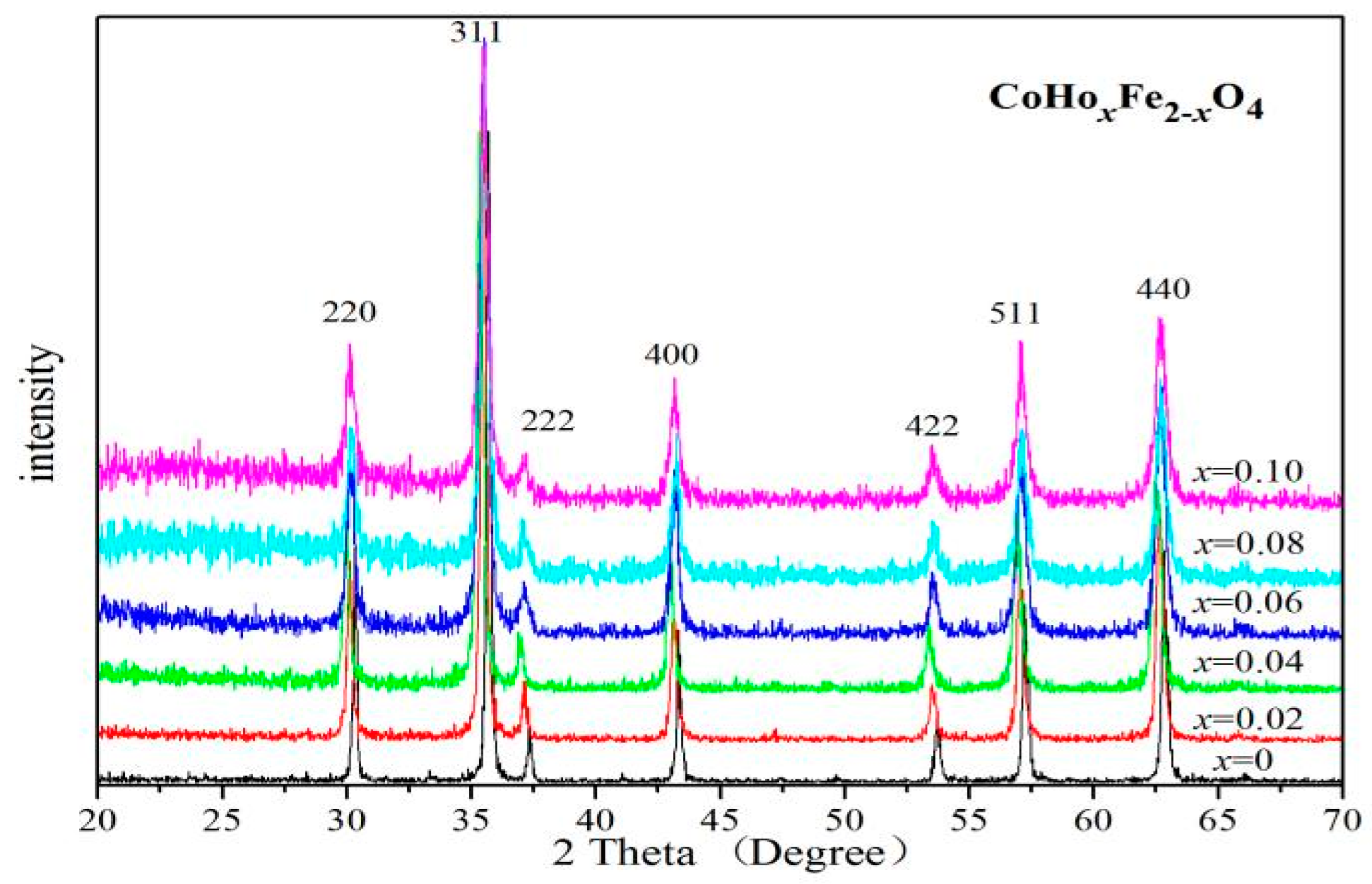
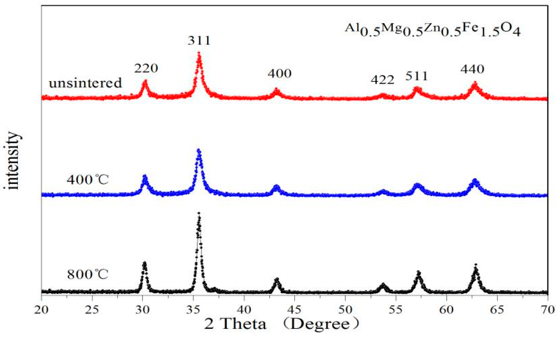
2.1. X-ray Diffraction Characterisation

Figure 1 shows the X-ray diffraction (XRD) patterns of ferrites CoHoxFe2−xO4 (x = 0–0.10) calcinated at 800 °C. The XRD patterns show that the CoHoxFe2−xO4 samples are single spinel-structure ferrites (JCPDS card no. 22-1086). No impurity peaks are observed in these XRD patterns. Owing to the ionic radius of Fe3+ ions (0.645 Å) being smaller than Ho3+ ions (0.901 Å) [9,11,12], when x ≤ 0.04, the lattice constants of CoHoxFe2−xO4 ferrite are larger than CoFe2O4 ferrite, as shown in Table 1. When x ≥ 0.06, the lattice parameter of CoHoxFe2−xO4 ferrite is no longer increasing, possibly owing to the solubility limit of Ho3+ ions [9]. The average crystallite size of the CoHoxFe2−xO4 samples estimated using the Debye–Scherrer formula [5,10,12] is between 21 and 55.6 nm. With Ho3+ doping, the decrease in the average crystallite size can be explained using the studies by others [14,15,16]. The RE3+ ions have an empty, half or filled 4f electron shell, such that rare earth Ho3+ ions substituted ferrites need more energy to grow grains and complete crystallisation.
The X-ray density of the sample was calculated by the following relationship [8,12,13]:
ρ x = 8 M N a 3
where a is the lattice parameter, N is Avogadro’s constant, and M is the relative molecular mass.
Table 1 shows that the lattice parameter also tends to increase when the relative molecular mass increases with Ho3+ substitution. So, according to Formula (1), the X-ray density tends to increase with Ho3+ substitution because of the increase in the relative molecular mass.
The XRD patterns of the un-sintered and sintered CoHo0.02Fe2−xO4 samples are single spinel-structured ferrites, as shown in Figure 2. No impurity peaks were found in these XRD patterns. The breadth of XRD lines decreases with increasing heat treatment temperatures. As shown in Table 2, the X-ray density and the lattice parameter have no noticeable changes, and the average crystallite size of CoHo0.02Fe1.98O4 increases with increasing calcination temperature. Unlike in this study, the reported XRD diffraction peaks of CoFe2O4 calcined at a low temperature are not very sharp [4,14]. This indicates that the sample has good crystallinity without calcination.
Figure 3 and Figure 4 show the X-ray diffraction (XRD) characterization of ferrites GdxMg0.5Zn0.5Fe2−xO4 and AlxMg0.5Zn0.5Fe2−xO4 (x = 0~0.1). No impurity peaks were found in these XRD patterns. Table 3 indicates that the Gd0.075Mg0.5Zn0.5Fe1.925O4 ferrite has the maximum lattice constant value, due to the radius of Fe3+ ions being smaller than the radius of Gd3+ ions [30,33]. With the doping of rare earth Gd3+ ions, the lattice constant does not increase monotonously, due to the larger rare earth ionic radius leading to the lattice distortion of Mg-Zn ferrite [31,32]. Due to the radius of Al3+ ions being smaller than Fe3+ ions [34], the lattice constant of AlxMg0.5Zn0.5Fe2−xO4 ferrite is smaller than Mg0.5Zn0.5Fe2O4 ferrite, and are shown in Table 4.
With the investigated samples GdxMg0.5Zn0.5Fe2−xO4 (x = 0~0.1), the average crystallite size estimated by the Debye-Scherrer formula is between 21.4 nm and 37.7 nm [29,30]. Additionally, the average crystallite size of AlxMg0.5Zn0.5Fe2−xO4 (x = 0~0.1) is between 15.8 nm and 37.7 nm. The XRD density increases with Gd3+ substitution from Table 3. The doping of rare earth Gd ions will cause the relative molecular mass to increase, and the lattice constant of Mg-Zn ferrite has no significant change. Therefore, the increases in XRD density of GdxMg0.5Zn0.5Fe2−xO4 is attributed to the relative molecular mass increase. Moreover, the decreases in XRD density of AlxMg0.5Zn0.5Fe2−xO4 are attributed to the fact that the relative molecular mass decreases.
Figure 5 and Figure 6 are the XRD patterns of Gd0.05Mg0.5Zn0.5Fe1.95O4 and Al0.5Mg0.5Zn0.5Fe1.5O4 sintered at different temperatures. No impurity peaks were found in these XRD patterns. The breadth of XRD lines trends to decrease with increasing heat treatment temperatures. From Table 5 we know that the lattice constant and average crystallite size increases, and the XRD Density decreases with increasing the sintered temperature for the Gd0.05Mg0.5Zn0.5Fe1.95O4. For the Al0.5Mg0.5Zn0.5Fe1.5O4, the lattice parameter decreases and the X-ray density increases, and average crystallite size is trend to increase with increasing the sintering temperature from Table 6. In other literature studies [35], the diffraction peaks of XRD is not very sharpness for CoGd0.1Fe1.9O4 calcined at low temperature, the diffraction peaks of XRD is very sharpness for Gd0.05Mg0.5Zn0.5Fe1.95O4 and Al0.5Mg0.5Zn0.5Fe1.5O4 without burning. This indicates that the sample has good crystallinity without sintering.

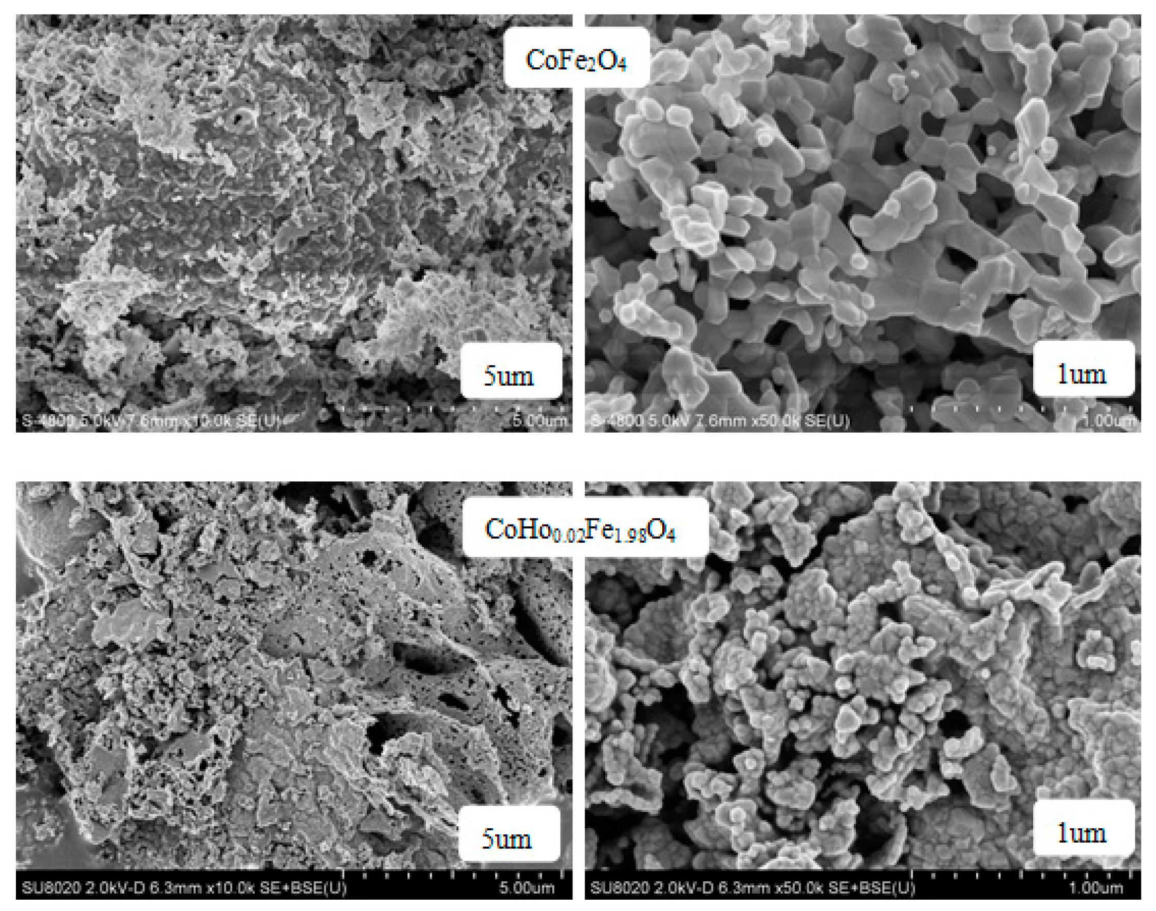
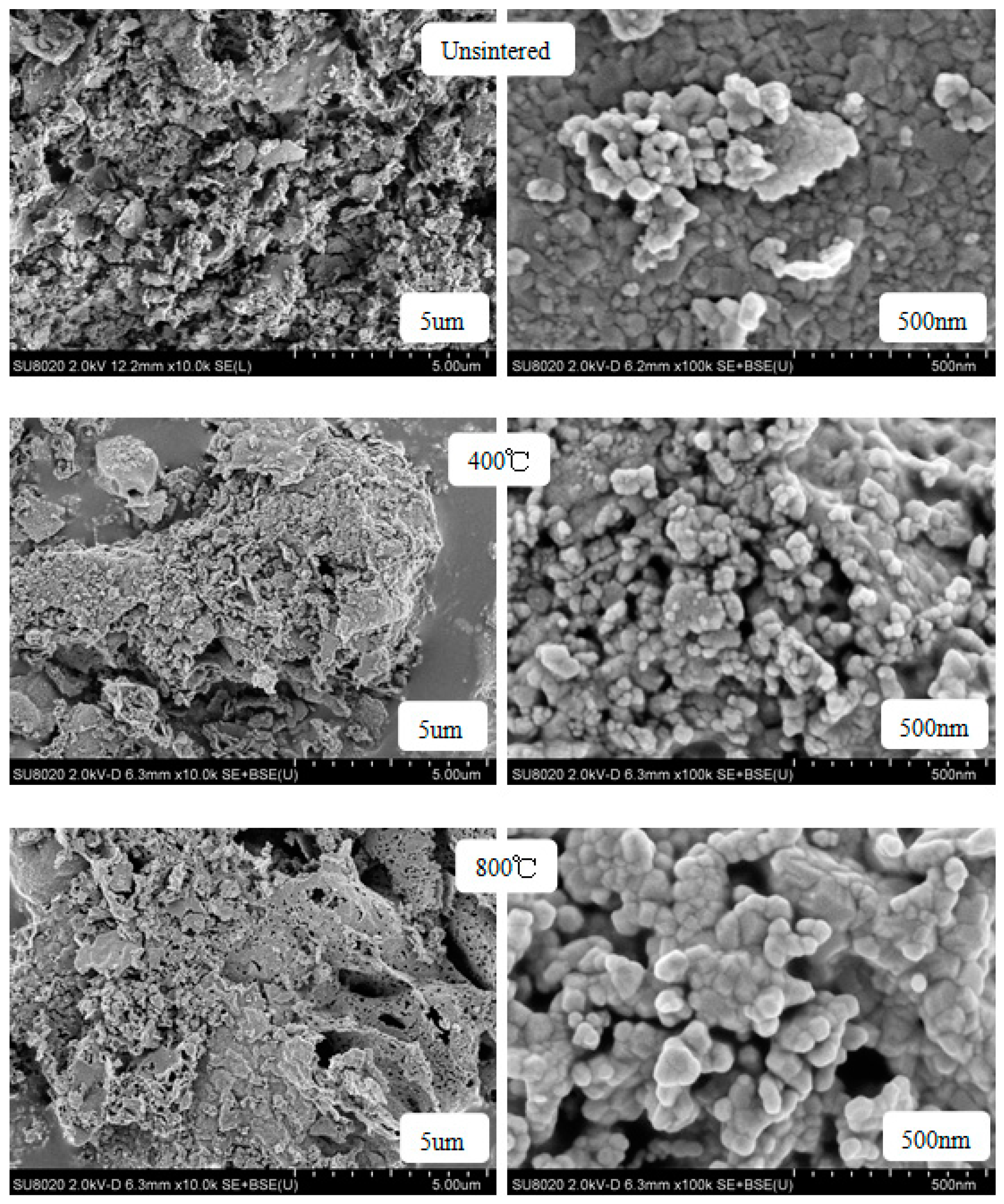
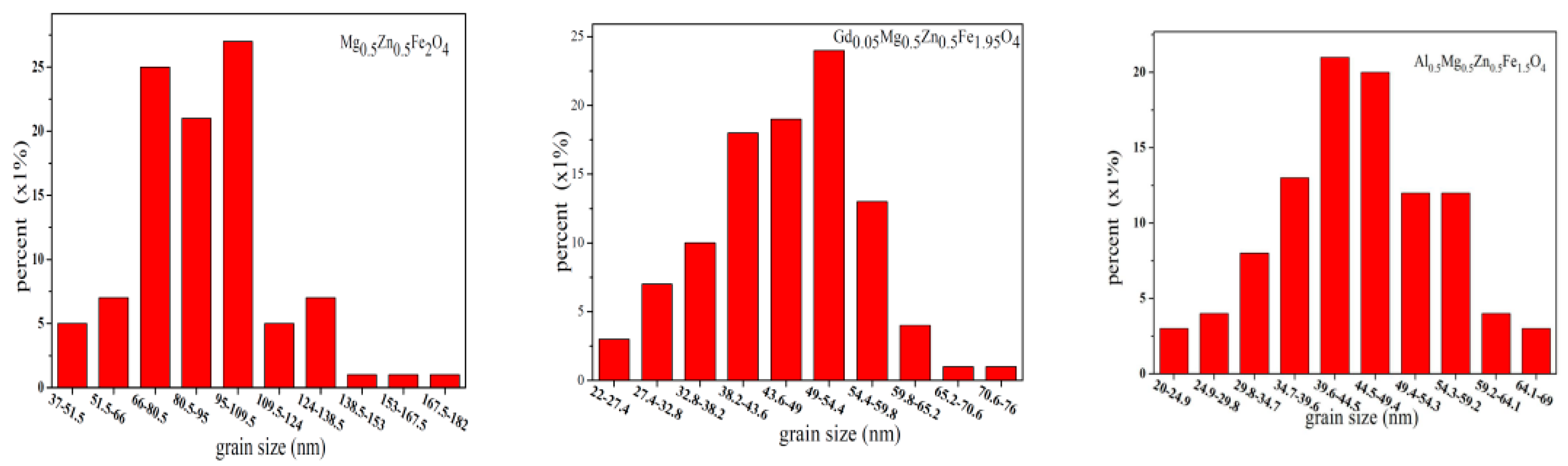
2.2. Scanning Electron Microscopy (SEM)

Figure 7 shows the SEM images of CoHoxFe2−xO4 (x = 0, 0.02) annealed at 800 °C. As shown in Figure 3, the sample is well-crystallised, and the grain size is almost uniform. With Ho3+ substitution, some cobalt ferrite particles are agglomerated, possibly owing to the magnetic interactions between CoHoxFe2−xO4 particles [15].
Figure 8 is the grain size distribution of CoFe2O4 and CoHo0.02Fe1.98O4. Using a statistical method, the calculated average grain size of CoFe2O4 and CoHo0.02Fe1.98O4 are ~96.26 and ~47.15 nm, respectively. The grain size of ferrite samples is less than 100 nm, indicating that they are nanoparticles. It is slightly larger than the average crystallite size determined using an X-ray, suggesting that each ferrite particle comprises several crystallites [16,17].
The SEM images of CoHo0.02Fe1.98O4 un-sintered and sintered at 400 °C and 800 °C are shown in Figure 9. As the heat treatment temperature increased, the crystallinity of the sample was improved, and the grain size increased. But the ferrite powders have a good crystallinity without heat treatment, consistent with the XRD analysis. Some cobalt ferrite particles are agglomerated with the increasing heat treatment temperature, owing to the magnetic interactions between CoHo0.02Fe1.98O4 particles [15], but the degree of agglomeration is small.
Figure 10 are the SEM images of Gd0.05Mg0.5Zn0.5Fe1.95O4 and Al0.5Mg0.5Zn0.5Fe1.5O4 sintered 800 °C. The sample is well crystallized, and the grain size is almost uniform. With the Gd3+ ions and Al3+ ions substituting, some particles of cobalt ferrite are agglomerated [21]. Figure 11 are the grain size histogram of Mg0.5Zn0.5Fe2O4, Gd0.05Mg0.5Zn0.5Fe1.95O4 and Al0.5Mg0.5Zn0.5Fe1.5O4. The average grain size is approximately 90.74nm, 46.11 nm and 44.84nm, respectively, and by a statistical method, the aluminum ion doping can make the grain size of the sample smaller. The grain size of ferrite samples is less than 100 nanometers, indicating that they are nanoparticles. However, they are slightly bigger than average crystallite size for an X-ray. Therefore, each particle is made up of several crystallites [17].

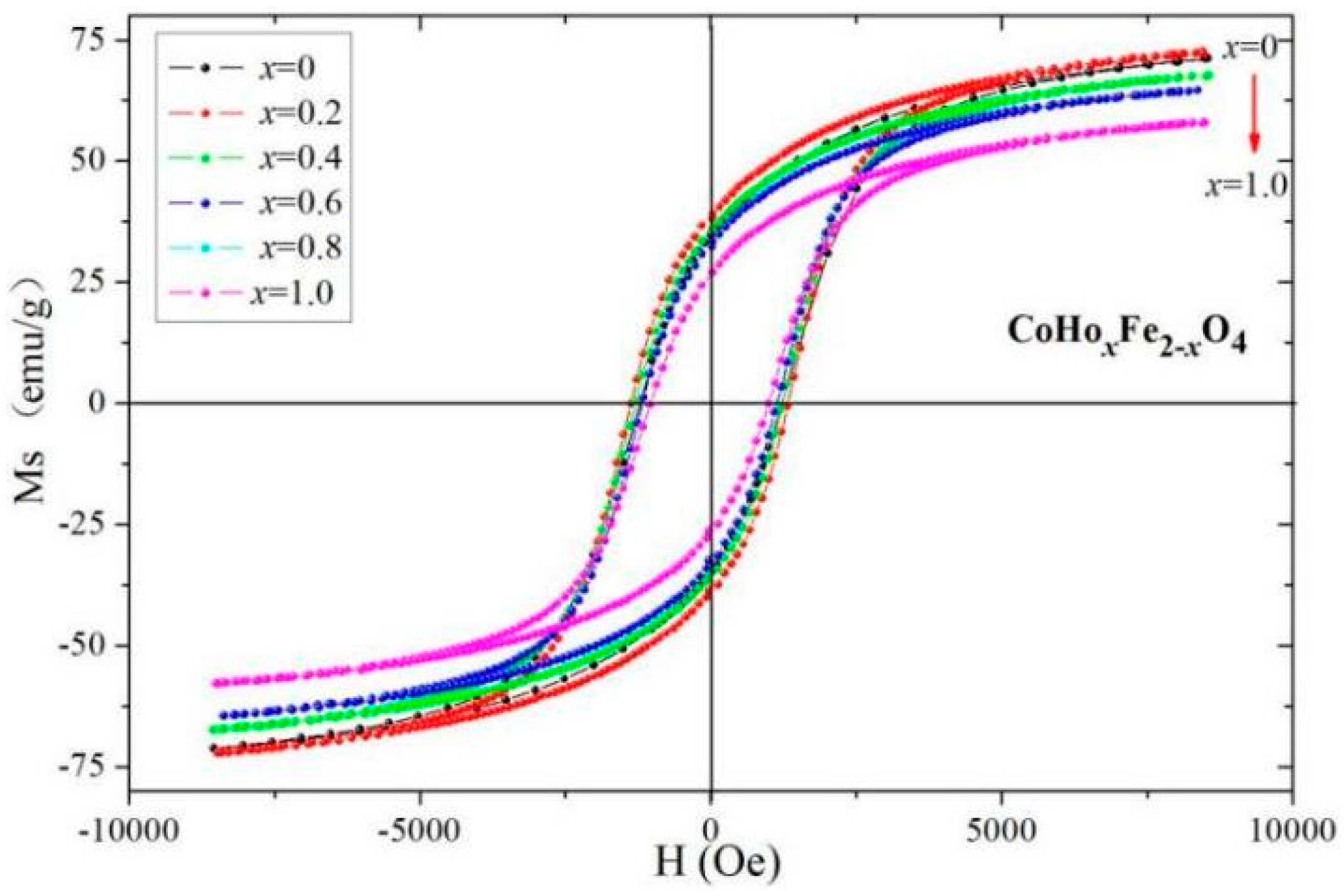
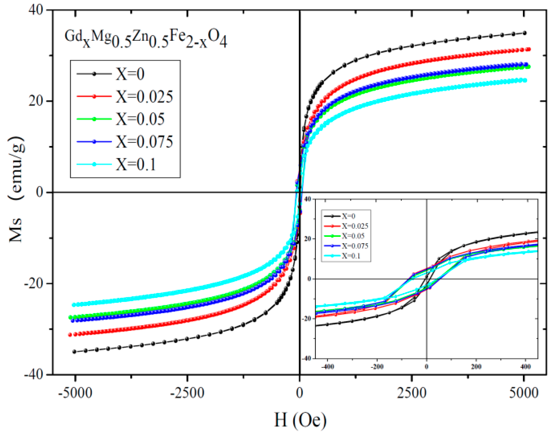
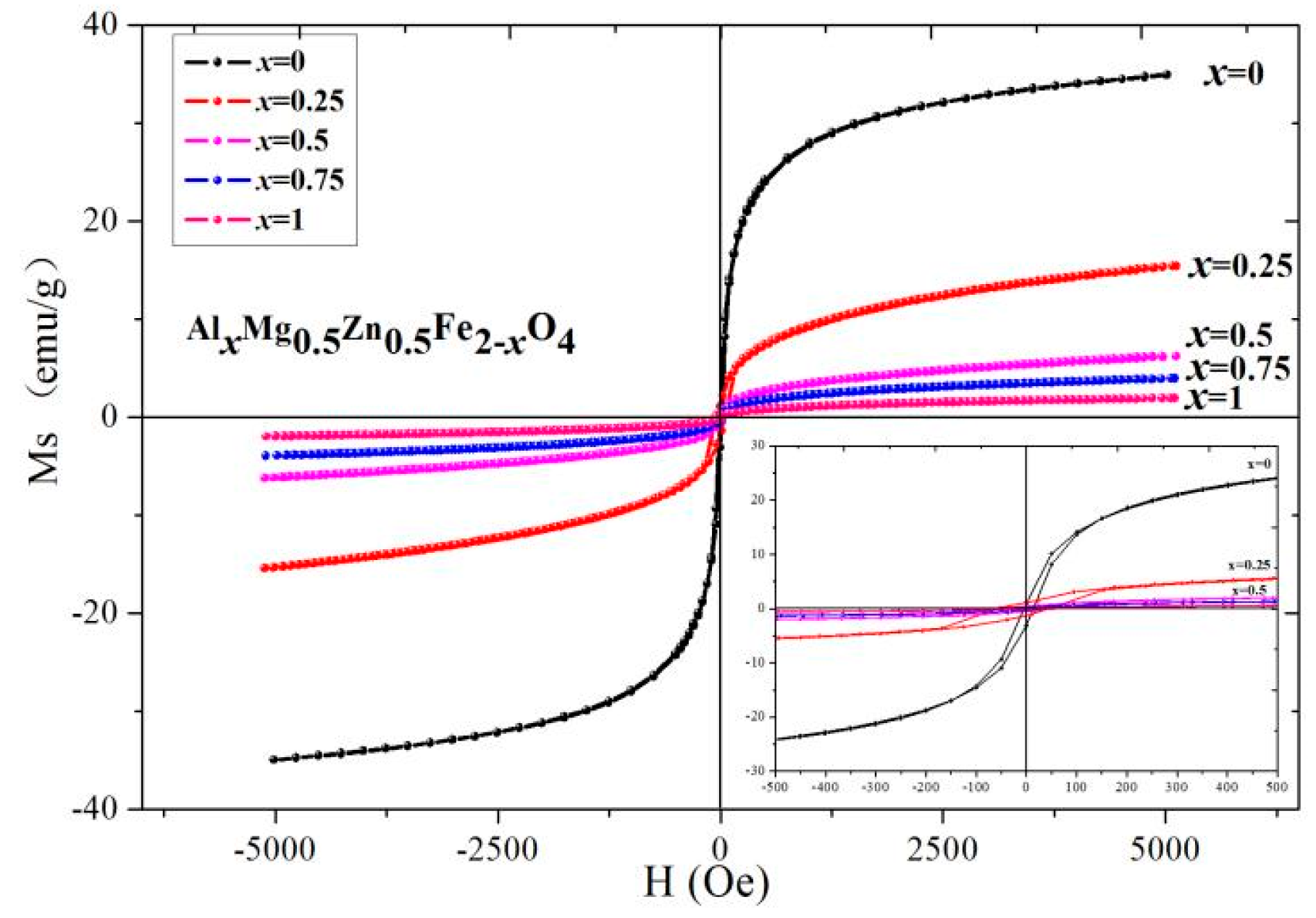
2.3. Magnetic Analysis

Figure 12 shows the RT (at 295 K) magnetic hysteresis curve of CoHoxFe2−xO4 measured at 295 K. The magnetisation of CoHoxFe2−xO4 (x = 0–0.10) nearly reaches saturation at 10,000 Oe. The saturation magnetisation (MS) decreases with Ho3+ substitution, as shown in Table 7, which could be calculated according to the following relation [8,18]:
M S = 5585 × n B M
where nB is the Bohr magneton and M is the molecular mass. The relative molecular mass of CoHoxFe2−xO4 increases as Ho3+ is replaced. The Ho3+, Co2+ and Fe3+ ions are 10.6, 3 and 5 μB [8], respectively. Ho exhibits para-magnetism at 295 K [9,10]. Ho3+ ions preferred to occupy B sites owing to the large Ho3+ ionic radius. Thus, the magnetic moment of the octahedral B site will decrease as the iron is replaced by holmium; this combined with the Néel theory, the total magnetic moment (nB) decreases. Owing to the increased M and the decreased nB with the substitution of Ho3+ ions, MS decreases according to the Formula (2). These results are consistent with other literature reports [9,20].
However, Table 7 shows that MS increases with Ho3+ concentration for x ≤ 0.02 because of a small amount of paramagnetic Ho3+ ions occupying A sites. The coercivity (HC) variation of cobalt ferrite with the substitution of Ho3+ ions can be explained as follows. The rare earth Ho3+ ions have substantial magneto-crystalline anisotropy [8,19,20]. HC does not increase monotonously with the doping of rare earth Ho3+ ions because HC is also related to the factors such as crystallinity and crystallite size [21].
Magnetic hysteresis loops of CoHo0.02Fe1.98O4 and CoHo0.10Fe1.90O4 at different sintering temperatures are shown in Figure 13 and Figure 14, respectively. After annealing at 900 °C, CoHo0.02Fe1.98O4 and CoHo0.10Fe1.90O4 have the maximum MS value because particle size increases with the sintering temperature [22].
The MS of CoHo0.02Fe1.98O4 and CoHo0.10Fe1.90O4 decrease after annealing at 400 °C because the un-sintered sample may have a good crystallinity, as determined by XRD and SEM. Table 8 and Table 9 show that MS increases with Ho3+ concentration for x = 0.02 and x = 0.10. The MS of CoHo0.02Fe1.98O4 is larger than that of CoHo0.10Fe1.90O4, owing to the small amount of paramagnetic Ho3+ occupying A sites and the decreasing of the magnetic moment at the tetrahedral A site as the iron is replaced by holmium. According to Néel theory, the paramagnetic Ho3+ occupying A sites will increase MS. The HC of CoHo0.02Fe1.98O4 and CoHo0.10Fe1.90O4 initially increases, decreasing as the sintering temperature increases. HC is related to grain size (D), and their relationship is HC = gh/D2 (single domain region) and HC = a + b/D (multi-domain region). According to the above-mentioned SEM analysis, the particle size increases with increasing calcination temperature, so the HC increases initially and then decreases as the annealing temperature increases.
Figure 15 and Figure 16 show the magnetic hysteresis curve of GdxMg0.5Zn0.5Fe2−xO4 measured at 295 K. The magnetization nearly reaches saturation at 5000 Oe. The saturation magnetization decreases with the substitution of Gd3+ ions and Al3+ ions from Table 10 and Table 11. As Gd3+ replaces, the relative molecular mass of GdxMg0.5Zn0.5Fe2−xO4 increases. The magnetic moments of rare earth ions generally come from localized 4f electrons in solids, and the magnetic ordering temperature is low, and the Gd rare-earth element has the Curie temperature of 293.2 K, close to RT(295 K) [36,37]. The magnetic dipole orientation is disordered at 295 K, and the rare-earth ion doping has no contribution to magnetization. Therefore, the Gd3+ ions are considered as non-magnetic ions at 295 K [38]. Moreover, the magnetic moment of Fe3+ ions is 5 μB [21].
Due to the relative molecular mass increase and the magnetic moment nB decrease with the doping of Gd3+ ions, the saturation magnetization decreases. Therefore, the magnetic moment of the octahedral B site will decrease as the iron is replaced by gadolinium and according to Neel theory the total magnetic moment nB will decrease. For the AlxMg0.5Zn0.5Fe2−xO4, as the Al3+ are replaced the relative molecular mass and the total magnetic moment nB decreases, and the influence of the magnetic moment is greater than the relative molecular mass, so the saturation magnetization decreases. The coercivity of GdxMg0.5Zn0.5Fe2−xO4 is larger than that of the Mg0.5Zn0.5Fe2O4 from Table 10. The variation of coercivity with Gd3+ ions substituted MgZn ferrite can be explained as follows. Rare earth ions (Gd3+) have stronger magnetocrystalline anisotropy [33,39].
Magnetic hysteresis loops of Gd0.05Mg0.5Zn0.5Fe1.95O4 at different sintering temperatures are shown in Figure 17. Table 12 indicates that Gd0.05Mg0.5Zn0.5Fe1.95O4 after annealing at 800 °C has the maximum value for saturation magnetization, because particle size increases with an increase in sintering temperature [29]. The coercivity of Gd0.05Mg0.5Zn0.5Fe1.95O4 decreases as sintering temperature increases. The coercivity (HC) is related to grain size (D), and their relationship is HC = gh/D2 (single domain region) and HC = a + b/D (multidomain region) [30]. The coercivity increases with grain diameter increases in the single domain region, and the coercivity decreases as the grain size increases in the multidomain region. The grain size of Gd0.05Mg0.5Zn0.5Fe1.95O4 sintered at different temperatures is the multidomain region, so the coercivity decreases as annealing temperature increases.

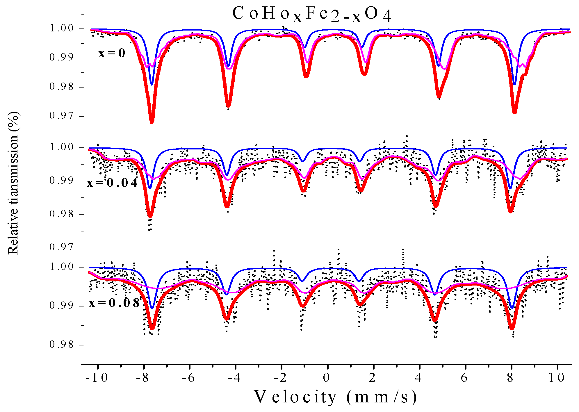
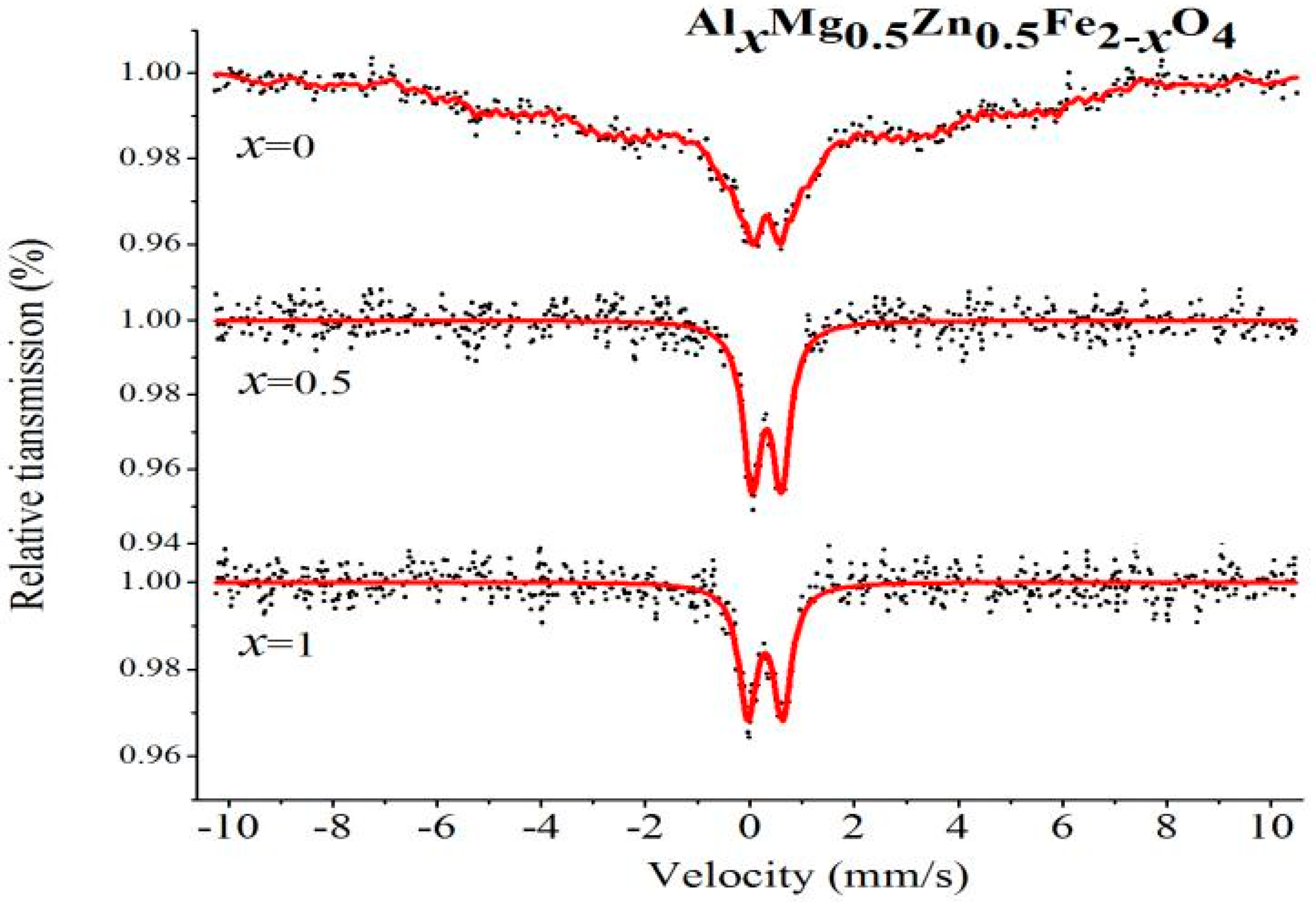
2.4. Mössbauer Spectroscopy

Figure 18 shows the Mössbauer spectra of CoHoxFe2−xO4 (x = 0, 0.04 and 0.08) polycrystalline ferrite powders measured at room temperature. All Mössbauer spectra were fitted using the Mösswinn 3.0 programme, and the spectra consist of two split Zeeman sextets, which indicates that the samples are ferrimagnetic. The six-line magnetic pattern with a larger isomer shift (I.S.) corresponds to the iron ion at position B, while the other six-line peak corresponds to the iron ion at position A [23,24]. As shown in Table 13, the changes in I.S. values are relatively small with the effects of Ho3+ substitution, and Ho3+ doping has no apparent impact on the s-electron charge distribution of Fe nuclei [24]. According to other reports, the I.S. values of Fe3+ ions are 0.1–0.5 mm/s [25,26]. From Table 13, the I.S. values indicate that the iron in the samples of this study is in the form Fe3+. Table 13 shows that the magnetic hyperfine field (H) tends to decrease by Ho3+ substitution, owing to the decrease of the A–B super-exchange by the paramagnetic rare-earth Ho3+ ions [27,28]. The value of the quadrupole shift is very small in the specimens of CoHoxFe2−xO4, indicating that the symmetry of the electric field around the nucleus is good in the cobalt ferrites. The absorption area of Mössbauer spectra for CoHoxFe2−xO4 has changed with increasing Ho substitution, which indicates that Ho3+ substitution influences the Fe3+ fraction at tetrahedral A and octahedral B sites.
Figure 19 and Figure 20 show the Mössbauer spectroscopy curve for GdxMg0.5Zn0.5Fe2−xO4 and AlxMg0.5Zn0.5Fe2−xO4 polycrystalline ferrite powders measured at room temperature. For the Mg0.5Zn0.5Fe2O4, the spectra analyzed were a Zeeman-sextets-split, which shows features of relaxation effects. For the GdxMg0.5Zn0.5Fe2−xO4, when x = 0.05, 0.1, spectra of the samples fit a Zeeman-sextets-split and a central paramagnetic doublet [24,25,40]. According to other reports, the isomer shifts (I.S.) values of Fe3+ ions are 0.1–0.5 mm/s [27,36]. From Table 14 and Table 15, the isomer shifts values indicate that iron is in the form of Fe3+ ions. The value of the quadrupole shift is relatively small with effects of Gd3+ and Al3+ ions substitution, and Gd3+ doping has no obvious effect on the s-electron charge distribution of Fe nuclei [25,27]. The magnetic hyperfine field of the Zeeman-split sextet spectra decreases with rare earth Gd3+ ions and Al3+ ions substitution.

3. Experimental

AyB1−yCxFe2−xO4 (C=Ho,Gd,Al) ferrite powders were synthesised via the sol-gel- combustion route. The synthetic raw materials of the sample are analytically pure nitrates (Co(NO3)2·6H2O, Fe(NO3)3·9H2O,Mg(NO3)2·6H2O, Zn(NO3)2·6H2O, Gd(N O3)3·9H2O, Al(NO3)3·9H2O and Ho(NO3)3·6H2O), ammonia (NH3·H2O) and citric acid (C6H8O7·H2O). Deionised water was added to form separate solutions of citric acid and metal nitrates. Then, citric acid and metal nitrates were mixed while adjusting the pH value at ~7 with ammonia. This solution mixture was kept on a thermostat water bath at 80 °C while electrically stirring the solution until it became a dried gel. The gel was further dried in the drying oven at 120 °C and ignited in the air with a small amount of alcohol as an oxidant. After spontaneous combustion, the material was annealed in the muffle furnace according to the specific temperature (400–900 °C). The crystalline structure was investigated by X-ray diffraction (D/max-2500V/PC, Rigaku, Tokyo, Japan). The micrographs were obtained by scanning electron microscopy (NoVaTM Nano SEM 430, Diamond Bar, CA, USA). The Mössbauer spectrum was performed at room temperature, using a conventional Mössbauer spectrometer (Fast Com Tec PC-mossII, Oberhaching, Germany). Magnetization measurements were carried out with vibrating sample magnetometer (JDAW-2000D, Changchun INPRO Magneto-electric Technology Co., Ltd., Changchun, China) at room temperature.

4. Conclusions

AyB1−yCxFe2−xO4 (C=Ho,Gd,Al) ferrite powders were synthesised via the sol-gel- combustion route. The XRD results show that CoHoxFe2−xO4 are single spinel-structured ferrites, and the CoHo0.02Fe1.98O4 samples calcined at different temperatures have good crystallinity. The SEM images further confirmed that the sample was well-crystallised, the grain was distributed homogeneously, and the sample comprised nanoparticles. The RT Mössbauer spectroscopy of CoHoxFe2−xO4 displayed ferromagnetic behaviour. Mössbauer spectra of CoHo0.02Fe1.98O4 showed that the calcination temperature affected the magnetic properties. The magnetisation results showed that rare-earth ion doping affects magnetic parameters, such as coercivity and saturation magnetisation, so the magnetic properties of samples can be regulated by rare-earth ion doping. Mg0.5Zn0.5CxFe2−xO4(C=Gd,Al) ferrite sintered at different temperatures have good crystallinity. The rare-earth ion and aluminum ion doping, and sintering process had an effect on magnetic parameters such as coercivity and saturation magnetization. The Mössbauer spectra showed that the sample exhibited ferrimagnetic and paramagnetic character with the replaced Gd3+ ions; that the sample exhibited paramagnetic character with the replaced Al3+ ions; and that the isomer shift values indicated that iron is in the form of Fe3+ ions.

Author Contributions

Conceptualization, Q.L. and J.L.; validation, F.Y., Q.Z. and Q.L.; formal analysis, K.S., H.X., F.Y. and Q.Z.; investigation, K.S., H.X., Y.H. and Q.L.; writing—original draft preparation, J.L., F.Y., Y.H. and Q.L.; writing—review and editing, J.L., Y.H., H.X. and Q.L.; supervision, Q.L. and J.L. All authors have read and agreed to the published version of the manuscript.

Funding

This work was financially supported by the National Natural Science Foundation of China (No. 12164006, 11364004) and Hainan Provincial Natural Science Foundation of China (No. 323QN241), Hainan Medical College’s research start-up fund.

Institutional Review Board Statement

Not applicable.

Informed Consent Statement

Not applicable.

Data Availability Statement

Not applicable.

Acknowledgments

Q.L., F.Y. and Q.Z. contributed equally to this work. All authors discussed the results and commented on the manuscript. H.X. and J.L. are co-corresponding authors contributed equally to this work.

Conflicts of Interest

The authors declare no conflict of interest.

Sample Availability

Not applicable.

References

  1. Mohamed, R.M.; Rashada, M.M.; Haraz, F.A.; Sigmund, W. Structure and magnetic properties of nanocrystalline cobalt ferrite powders synthesized using organic acid precursor method. J. Magn. Magn. Mater. 2010, 322, 2058–2064. [Google Scholar] [CrossRef]
  2. Amiri, S.; Shokrollahi, H. The role of cobalt ferrite magnetic nanoparticles in medical science. Mater. Sci. Eng. C 2013, 33, 1–8. [Google Scholar] [CrossRef] [PubMed]
  3. Sanpo, N.; Berndt, C.C.; Wen, C.; Wang, J. Transition metal-substituted cobalt ferrite nanoparticles for biomedical applications. Acta Biomater. 2013, 9, 5830–5837. [Google Scholar] [CrossRef] [PubMed]
  4. Lo, C.C.H. Experimental and modeling studies of the magnetomechanical effect in substituted cobalt ferrites for magnetoelastic stress sensors. J. Appl. Phys. 2010, 107, 5798. [Google Scholar] [CrossRef]
  5. Zhao, L.; Yang, H.; Zhao, X.; Yu, L.; Cui, Y.; Feng, S. Magnetic properties of CoFe2O4 ferrite doped with rare earth ion. Mater. Lett. 2006, 60, 1–6. [Google Scholar] [CrossRef]
  6. Meng, X.; Li, H.; Chen, J.; Mei, L.; Wang, K.; Li, X. Mössbauer study of cobalt ferrite nanocrystals substituted with rare-earth Y3+ ions. J. Magn. Magn. Mater. 2009, 321, 1155–1158. [Google Scholar] [CrossRef]
  7. Zhang, Y.; Wen, D. Infrared emission properties of RE (RE = La, Ce, Pr, Nd, Sm, Eu, Gd, Tb, and Dy) and Mn co-doped Co0.6Zn0.4Fe2O4 ferrites. Mater. Chem. Phys. 2012, 131, 575–580. [Google Scholar] [CrossRef]
  8. Lohar, K.S.; Pachpinde, A.M.; Langade, M.M.; Kadam, R.H.; Shirsath, S.E. Self-propagating high temperature synthesis, structural morphology and magnetic interactions in rare earth Ho3+ doped CoFe2O4 nanoparticles. J. Alloys Compd. 2014, 604, 204–210. [Google Scholar] [CrossRef]
  9. Ali, I.; Islam, M.U.; Ishaque, M.; Khan, H.M.; Ashiq, M.N.; Rana, M.U. Structural and magnetic properties of holmium substituted cobalt ferrites synthesized by chemical co-precipitation method. J. Magn. Magn. Mater. 2012, 324, 3773–3777. [Google Scholar] [CrossRef]
  10. Pervaiz, E.; Gul, I.H. High frequency AC response, DC resistivity and magnetic studies of holmium substituted Ni-ferrite: A novel electromagnetic material. J. Magn. Magn. Mater. 2014, 349, 27–34. [Google Scholar] [CrossRef]
  11. Muthuselvam, I.P.; Bhowmik, R.N. Mechanical alloyed Ho3+ doping in CoFe2O4 spinel ferrite and understanding of magnetic nanodomains. J. Magn. Magn. Mater. 2010, 322, 767–776. [Google Scholar] [CrossRef]
  12. Amiri, S.; Shokrollahi, H. Magnetic and structural properties of RE doped Co-ferrite (RE = Nd, Eu, and Gd) nano-particles synthesized by co-precipitation. J. Magn. Magn. Mater. 2013, 345, 18–23. [Google Scholar] [CrossRef]
  13. Shinde, T.J.; Gadkari, A.B.; Vasambeka, P.N. Effect of Nd3+ substitution on structural and electrical properties of nanocrystalline zinc ferrite. J. Magn. Magn. Mater. 2010, 322, 2777–2781. [Google Scholar] [CrossRef]
  14. Panda, R.N.; Shih, J.C.; Chin, T.S. Magnetic properties of nano-crystalline Gd- or Pr-substituted CoFe2O4 synthesized by the citrate precursor technique. J. Magn. Magn. Mater. 2003, 257, 79–86. [Google Scholar] [CrossRef]
  15. Ruiz, M.M.; Mietta, J.L.; Antonel, P.S.; Pérez, O.E.; Negri, R.M.; Jorge, G. Structural and magnetic properties of Fe2-xCoSmxO4-nanoparticles and Fe2-xCoSmxO4-PDMS magnetoelastomers as a function of Sm content. J. Magn. Magn. Mater. 2013, 327, 11–19. [Google Scholar] [CrossRef]
  16. Tahar, L.B.; Smiri, L.S.; Artus, M.; Joudrier, A.-L.; Herbst, F.; Aulay, M.J.; Ammar, S.; Fiévet, F. Characterization and magnetic properties of Sm- and Gd-substituted CoFe2O4 nanoparticles prepared by forced hydrolysis in polyol. Mater. Res. Bull. 2007, 42, 1888–1896. [Google Scholar] [CrossRef]
  17. Borhan, A.I.; Slatineanu, T.; Iordan, A.R.; Palamaru, M.N. Influence of chromium ion substitution on the structure and properties of zinc ferrite synthesized by the sol-gel auto-combustion method. Polyhedron 2013, 56, 82–89. [Google Scholar] [CrossRef]
  18. Karimi, Z.; Mohammadifar, Y.; Shokrollahi, H.; Asl, S.K.; Yousefi, G.; Karimi, L. Magnetic and structural properties of nano sized Dy-doped cobalt ferrite synthesized by co-precipitation. J. Magn. Magn. Mater. 2014, 361, 150–156. [Google Scholar] [CrossRef]
  19. Zhao, L.; Yang, H.; Yu, L.; Cui, Y.; Zhao, X.; Feng, S. Study on magnetic properties of nanocrystalline La-, Nd-, or Gd-substituted Ni-Mn ferrite at low temperatures. J. Magn. Magn. Mater. 2006, 305, 91–94. [Google Scholar] [CrossRef]
  20. Nikumbh, A.K.; Pawar, R.A.; Nighot, D.V.; Gugale, G.S.; Sangale, M.D.; Khanvilkar, M.B.; Nagawade, A.V. Structural, electrical, magnetic and dielectric properties of rare-earth substituted cobalt ferrites nanoparticles synthesized by the co-precipitation method. J. Magn. Magn. Mater. 2014, 355, 201–209. [Google Scholar] [CrossRef]
  21. He, Y.; Yang, X.; Lin, J.; Lin, Q.; Dong, J. Mössbauer spectroscopy, Structural and magnetic studies of Zn2+ substituted magnesium ferrite nanomaterials prepared by Sol-Gel method. J. Nanomater. 2015, 2015, 854840. [Google Scholar] [CrossRef]
  22. Singhal, S.; Barthwal, S.K.; Chandra, K. XRD, magnetic and Mössbauer spectral studies of nano size aluminum substituted cobalt ferrites (CoAlxFe2-xO4). J. Magn. Magn. Mater. 2006, 306, 233–240. [Google Scholar] [CrossRef]
  23. Zhao, L.; Han, Z.; Yang, H.; Yu, L.; Cui, Y.; Jin, W.; Feng, S. Magnetic properties of nanocrystalline Ni0.7Mn0.3Gd0.1Fe1.9O4 ferrite at low temperatures. J. Magn. Magn. Mater. 2007, 309, 11–14. [Google Scholar] [CrossRef]
  24. Inbanathan, S.S.R.; Vaithyanathan, V.; Chelvane, J.A.; Markandeyulu, G.; Bharathi, K.K. Mössbauer studies and enhanced electrical properties of R (R = Sm, Gd and Dy) doped Ni ferrite. J. Magn. Magn. Mater. 2014, 353, 41–46. [Google Scholar] [CrossRef]
  25. He, Y.; Lei, C.; Lin, Q.; Dong, J.; Yu, Y.; Wang, L. Mössbauer and Structural properties of La-substituted Ni0.4Cu0.2Zn0.4Fe2O4 nanocrystalline ferrite. Sci. Adv. Mater. 2015, 7, 1809–1815. [Google Scholar] [CrossRef]
  26. Kumar, S.; Farea, A.M.M.; Batoo, K.M.; Lee, C.G.; Koo, B.H.; Yousef, A. Mössbauer studies of Co0.5CdxFe2.5-xO4 (0.0–0.5) ferrite. Phys. B Condens. Matter 2008, 403, 3604–3607. [Google Scholar] [CrossRef]
  27. Lin, J.; He, Y.; Lin, Q.; Wang, R.; Chen, H. Microstructural and Mössbauer spectroscopy Studies of Mg1-xZnxFe2O4(x = 0.5, 0.7) nanoparticles. J. Spectrosc. 2014, 2014, 540319. [Google Scholar] [CrossRef]
  28. Al-Maashani, M.; Gismelseed, A.M.; Khalaf, K.A.M.; Yousif, A.A.; Al-Rawas, A.D.; Widatallah, H.M.; Elzain, M.E. Structural and Mössbauer study of nanoparticles CoFe2O4, prepared by sol-gel auto-combustion and subsequent sintering. Hyperfine Interact. 2018, 239, 15. [Google Scholar] [CrossRef]
  29. Choodamani, C.; Nagabhushana, G.P.; Ashoka, S.; Prasad, B.D.; Rudraswamy, B.; Chandrappa, G.T. Structural and magnetic studies of Mg(1-x)ZnxFe2O4 nanoparticles prepared by a solution combustion method. J. Alloys Compd. 2013, 578, 103–109. [Google Scholar] [CrossRef]
  30. Mohseni, H.; Shokrollahi, H.; Sharifi, I.; Gheisari, K. Magnetic and structural studies of the Mn-doped Mg-Zn fer rite nanoparticles synthesized by the glycine nitrate process. J. Magn. Magn. Mater. 2012, 324, 3741–3747. [Google Scholar] [CrossRef]
  31. Haralkar, S.J.; Kadam, R.H.; More, S.S.; Shirsath, S.E.; Mane, M.L.; Patil, S.; Mane, D.R. Substitutional effect of Cr3+ ions on the properties of Mg-Zn ferrite nanoparticles. Phys. B Condens. Matter 2012, 407, 4338–4346. [Google Scholar] [CrossRef]
  32. Dascalu, G.; Popescu, T.; Feder, M.; Caltun, O.F. Structural, electric and magnetic properties of CoFe1.8RE0.2O4 (RE = Dy, Gd, La) bulk materials. J. Magn. Magn. Mater. 2013, 333, 69–74. [Google Scholar] [CrossRef]
  33. Chand, J.; Kumar, G.; Kumar, P.; Sharma, S.K.; Knobel, M.; Singh, M. Effect of Gd3+ doping on magnetic, electric and dielectric properties of MgGdxFe2-xO4 ferrites processed by solid state reaction technique. J. Alloys Compd. 2011, 509, 9638–9644. [Google Scholar] [CrossRef]
  34. Chae, K.P.; Lee, J.-G.; Kweon, H.S.; Lee, Y.B. Synthesis and magnetic properties of AlxCoFe2-xO4 ferrite powders. Phys. Status Solidi 2004, 201, 1883–1888. [Google Scholar] [CrossRef]
  35. Chandradass, J.; Jadhav, A.H.; Kim, K.H.; Kim, H. Influence of processing methodology on the structural and magnetic behavior of MgFe2O4 nanopowders. J. Alloys Compd. 2012, 517, 164–169. [Google Scholar] [CrossRef]
  36. Singhal, S.; Barthwal, S.K.; Chandra, K. Cation distribution in the nano size aluminium substituted cobalt ferrites using XRD, magnetic and Mössbauer spectral studies. Indian J. Pure Appl. Phys. 2007, 45, 821–825. [Google Scholar]
  37. Chandradass, J.; Jadhav, A.H.; Kim, H. Surfactant modified MgFe2O4 nanopowders by reverse micelle processing: Effect of water to surfactant ratio (R) on the particle size and magnetic property. Appl. Surf. Sci. 2012, 258, 3315–3320. [Google Scholar] [CrossRef]
  38. Peng, J.; Hojamberdiev, M.; Xu, Y.; Cao, B.; Wang, J.; Wu, H. Hydrothermal synthesis and magnetic properties of gadolinium-doped CoFe2O4 nanoparticles. J. Magn. Magn. Mater. 2011, 323, 133–138. [Google Scholar] [CrossRef]
  39. Gabal, M.A.; Abdel-Daiem, A.M.; Al Angari, Y.M.; Ismaeel, I.M. Influence of Al-substitution on structural, electrical and magnetic properties of Mn-Zn ferrites nanopowders prepared via the sol-gel auto-combustion method. Polyhedron 2013, 57, 105–111. [Google Scholar] [CrossRef]
  40. Lin, Q.; Lei, C.; He, Y.; Xu, J.; Wang, R. Mössbauer and XRD studies of Ni0.6Cu0.2Zn0.2CexFe2−xO4 ferrites By Sol-Gel auto-combustion. J. Nanosci. Nanotechnol. 2015, 15, 2997–3003. [Google Scholar] [CrossRef]
Figure 1. X-ray diffraction (XRD) patterns of CoHoxFe2−xO4 calcined at 800 °C.
Molecules 28 04226 g001
Figure 2. XRD patterns of CoHo0.02Fe1.98O4 un-sintered and sintered at 400 °C and 800 °C.
Molecules 28 04226 g002
Figure 3. XRD patterns of GdxMg0.5Zn0.5Fe2−xO4 sintered at 800 °C.
Molecules 28 04226 g003
Figure 4. XRD patterns of AlxMg0.5Zn0.5Fe2−xO4 sintered at 800 °C.
Molecules 28 04226 g004
Figure 5. XRD patterns of Gd0.05Mg0.5Zn0.5Fe1.95O4 sintered at different temperatures.
Molecules 28 04226 g005
Figure 6. XRD patterns of Al0.5Mg0.5Zn0.5Fe1.5O4 sintered at different temperatures.
Molecules 28 04226 g006
Figure 7. SEM images of CoFe2O4 and CoHo0.02Fe1.98O4 sintered at 800 °C.
Molecules 28 04226 g007
Figure 8. Grain size distribution of CoFe2O4 and CoHo0.02Fe1.98O4 calcined at 800 °C.
Molecules 28 04226 g008
Figure 9. SEM images of CoHo0.02Fe1.98O4 un-sintered and sintered at 400 °C and 800 °C.
Molecules 28 04226 g009
Figure 10. SEM micrographs of Mg0.5Zn0.5Fe2O4, Gd0.05Mg0.5Zn0.5Fe1.95O4 and Al0.5Mg0.5Zn0.5Fe1.5O4 sintered at 800 °C.
Molecules 28 04226 g010
Figure 11. Histogram of grain size distribution of Mg0.5Zn0.5Fe2O4, Gd0.05Mg0.5Zn0.5Fe1.95O4 and Al0.5Mg0.5Zn0.5Fe1.5O4 sintered at 800 °C.
Molecules 28 04226 g011
Figure 12. Hysteresis loops of CoHoxFe2−xO4 calcined at 800 °C.
Molecules 28 04226 g012
Figure 13. Hysteresis loops of CoHo0.02Fe1.98O4 at different sintering temperatures.
Molecules 28 04226 g013
Figure 14. Hysteresis loops of CoHo0.10Fe1.90O4 at different sintering temperatures.
Molecules 28 04226 g014
Figure 15. Hysteresis loops of GdxMg0.5Zn0.5Fe2−xO4 sintered at 800 °C.
Molecules 28 04226 g015
Figure 16. Hysteresis loops of AlxMg0.5Zn0.5Fe2−xO4 sintered at 800 °C.
Molecules 28 04226 g016
Figure 17. Hysteresis loops of Gd0.05Mg0.5Zn0.5Fe1.95O4 sintered at different temperatures.
Molecules 28 04226 g017
Figure 18. Mössbauer spectra of CoHoxFe2−xO4 calcined at 800 °C.
Molecules 28 04226 g018
Figure 19. Mössbauer spectra of GdxMg0.5Zn0.5Fe2−xO4 sintered at 800 °C.
Molecules 28 04226 g019
Figure 20. Mössbauer spectra of AlxMg0.5Zn0.5Fe2−xO4 sintered at 800 °C.
Molecules 28 04226 g020
Table 1. XRD date of CoHoxFe2−xO4 calcined at 800 °C.
Content (x)Lattice Parameter (Å)Average Crystallite Size (Å)Density (g/cm3)
08.354975565.3468
0.028.386013505.3342
0.048.406813085.3434
0.068.381322295.4415
0.088.377192495.4989
0.108.389282215.5243
Table 2. XRD date of CoHo0.02Fe1.98O4 un-sintered and sintered at 400 °C, 800 °C.
Temperature (°C)Lattice Parameter (Å)Average Crystallite Size (Å)Density (g/cm3)
Un-sintered8.383792495.3384
4008.392792935.3212
8008.386013505.3342
Table 3. XRD date of GdxMg0.5Zn0.5Fe2−xO4 sintered at 800 °C.
Content (x)Lattice Constant (Å)Average Crystallite Size (Å)Density (g/cm3)
08.431333774.8879
0.0258.420943444.9624
0.058.430473015.0018
0.0758.445932905.0303
0.18.421422145.1307
Table 4. XRD date of AlxMg0.5Zn0.5Fe2−xO4 sintered at 800 °C.
Content (x)Lattice Parameter (Å)Average Crystallite Size (Å)Density (g/cm3)
08.431333774.8879
0.258.391102904.7963
0.58.364802024.6779
0.758.313151584.5988
1.08.258741794.5201
Table 5. XRD date of Gd0.05Mg0.5Zn0.5Fe1.95O4 sintered at different temperatures.
Temperature (°C)Lattice Constant (Å)Average Crystallite Size (Å)Density (g/cm3)
Un-sintered8.415172285.0292
400 °C8.418292335.0236
800 °C8.430473015.0018
Table 6. XRD date of Al0.5Mg0.5Zn0.5Fe1.5O4 sintered at different temperatures.
Temperature (°C)Lattice Parameter (Å)Average Crystallite Size (Å)Density (g/cm3)
Un-sintered8.382261654.6487
400 °C8.369851374.6694
800 °C8.364802024.6779
Table 7. Magnetic data for CoHoxFe2−xO4 calcined at 800 °C.
Content (x)MS (emu/g)HC (Oe)Mr (emu/g)
070.58100534.71
0.0272.54135137.77
0.0467.58126635.06
0.0664.64115831.90
0.0861.82109430.24
0.1057.96101626.28
Table 8. Magnetic data of CoHo0.02Fe1.98O4 at different sintering temperatures.
Temperature (°C) MS (emu/g)HC (Oe)Mr (emu/g)
Un-sintered60.01130129.98
40055.80223429.62
50061.10194432.37
60063.52165533.56
70069.21142636.91
80072.54135137.77
90076.16115240.99
Table 9. Magnetic data of CoHo0.10Fe1.90O4 at different sintering temperatures.
Temperature (°C)MS (emu/g)HC (Oe)Mr (emu/g)
Un-sintered47.89129020.68
40039.75205218.83
50045.72153320.42
60050.30117421.50
70057.05101425.13
80057.96101626.28
90066.1291433.8
Table 10. Magnetic data for GdxMg0.5Zn0.5Fe2−xO4 sintered at 800 °C.
Content (x)MS (emu/g)HC (Oe)Mr (emu/g)
034.9813.641.10
0.02531.3862.524.67
0.0527.5455.793.79
0.07528.1762.994.36
0.124.6750.522.61
Table 11. Magnetic data for AlxMg0.5Zn0.5Fe2−xO4 calcined at 800 °C.
Content (x)MS (emu/g)HC (Oe)Mr (emu/g)
034.9813.641.10
0.2515.3950.371.38
0.56.2653.560.54
0.753.9650.370.33
11.9459.060.16
Table 12. Magnetic data for Gd0.05Mg0.5Zn0.5Fe1.95O4 sintered at different temperatures.
Temperature (°C)MS (emu/g)HC (Oe)Mr (emu/g)
Un-sintered25.2971.924.39
400 °C22.9369.583.73
800 °C27.5455.793.79
Table 13. Mössbauer parameters of CoHoxFe2−xO4 calcined at 800 °C.
Content (x)ComponentI.S. (mm/s)Q.S. (mm/s)H (T)Γ (mm/s)A0 (%)
0Sextet (A)0.237−0.00448.9460.36032.4
Sextet (B)0.375−0.02445.6950.32267.6
0.04Sextet (A)0.126−0.05048.6130.38722.2
Sextet (B)0.3160.15144.1560.42977.8
0.08Sextet (A)0.1570.06148.5460.46324.5
Sextet (B)0.224−0.13743.6410.74075.5
Table 14. Mössbauer parameters of GdxMg0.5Zn0.5Fe2−xO4 sintered at 800 °C.
Content (x)ComponentI.S. (mm/s)Q.S. (mm/s)H (T)Γ (mm/s)A0 (mm/s)
0Sextet (B)0.3180.00524.2340.268100
0.05Sextet (B)0.302−0.17330.3500.33493.1
Doublet0.3010.409-0.3276.9
0.1Sextet (B)0.116−0.02938.6790.52879.4
Doublet0.3480.627-0.53720.6
Table 15. Mössbauer parameters of AlxMg0.5Zn0.5Fe2−xO4 sintered at 800 °C.
Content (x)ComponentI.S. (mm/s)Q.S. (mm/s)H (T)Γ (mm/s)A0 (mm/s)
0Sextet (B)0.3180.00524.2340.268100
0.5Double0.3220.568-0.417100
1Double0.2920.672-0.414100
Disclaimer/Publisher’s Note: The statements, opinions and data contained in all publications are solely those of the individual author(s) and contributor(s) and not of MDPI and/or the editor(s). MDPI and/or the editor(s) disclaim responsibility for any injury to people or property resulting from any ideas, methods, instructions or products referred to in the content.

