
The Use of UV-Visible Diffuse Reflectance Spectrophotometry for a Fast, Preliminary Authentication of Gemstones

 , , , and
Abstract
:1. Introduction
2. Results
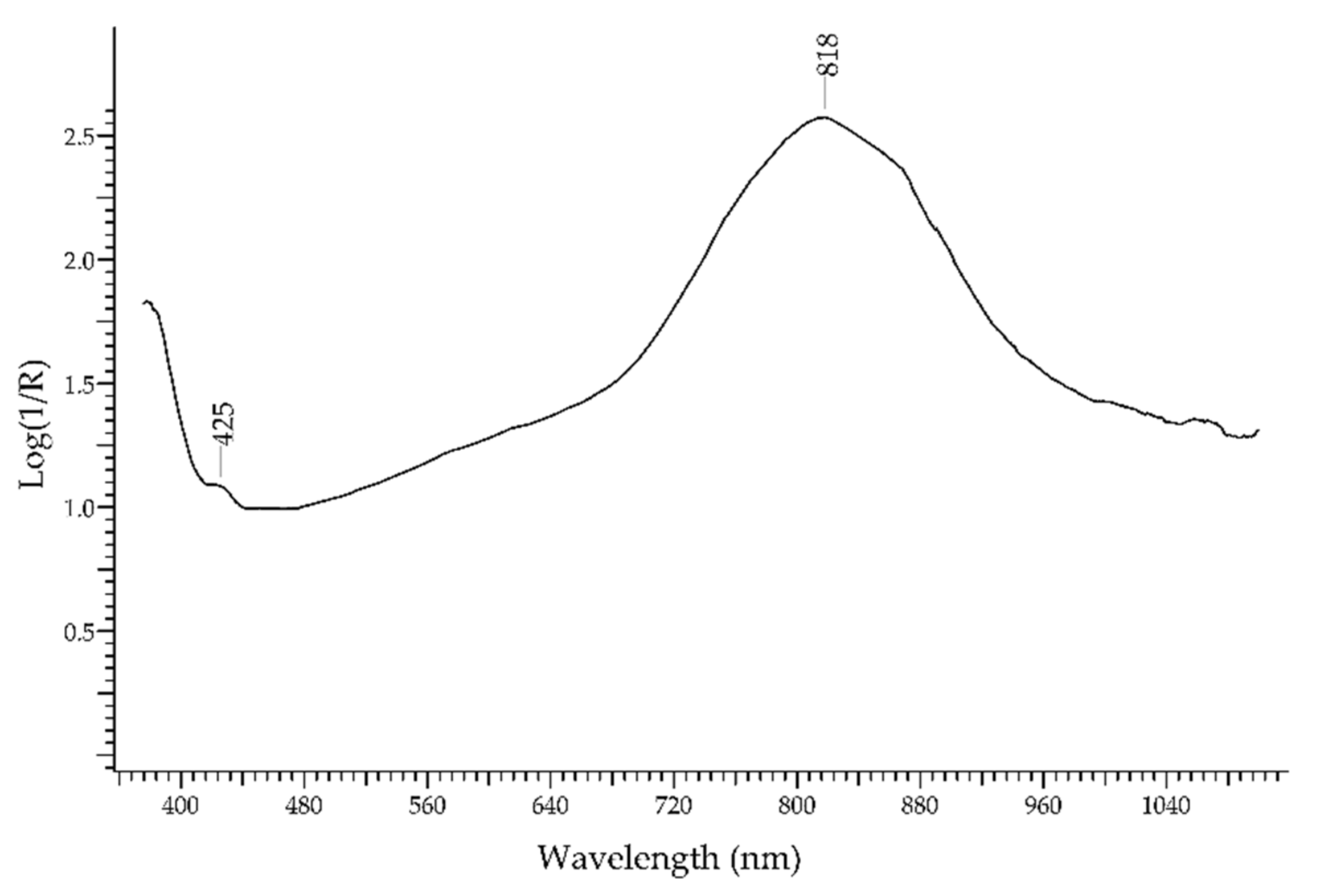
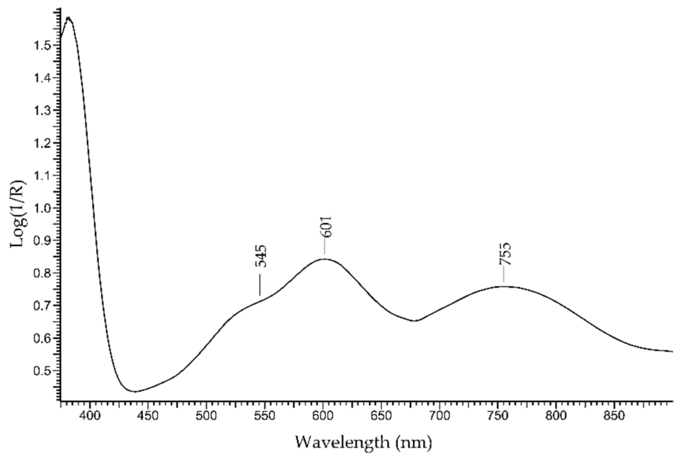
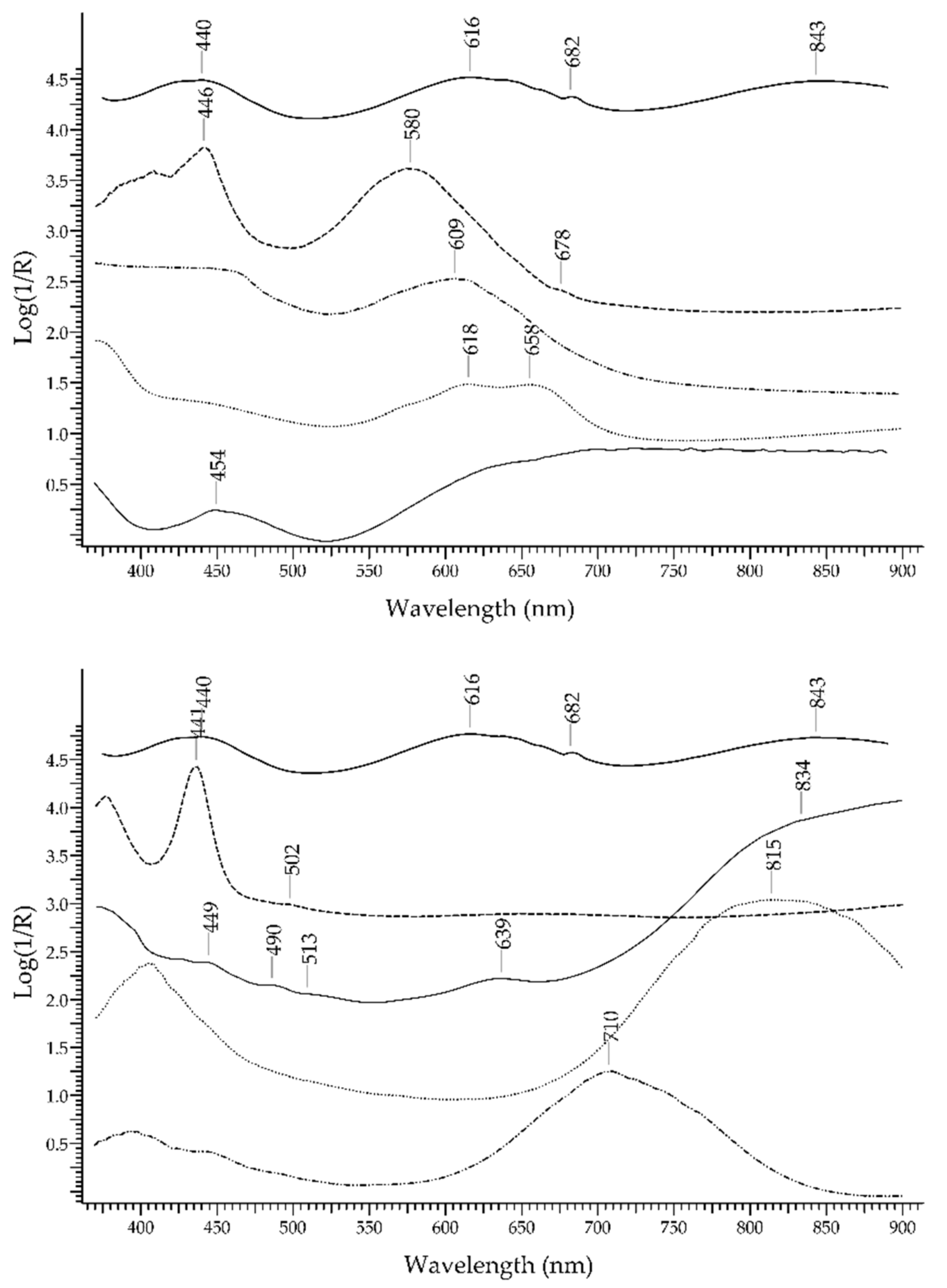
2.1. Blue Gemstones
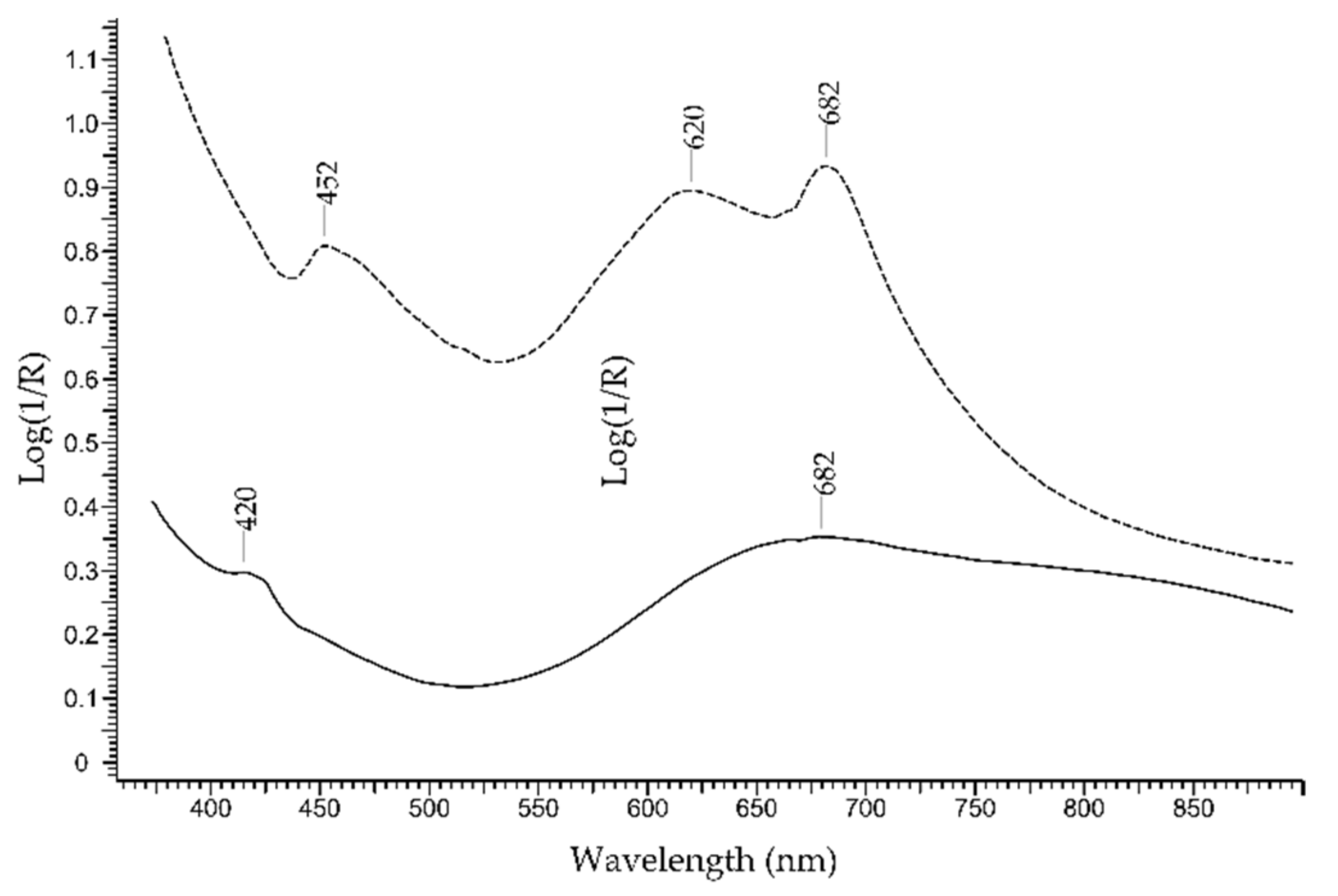
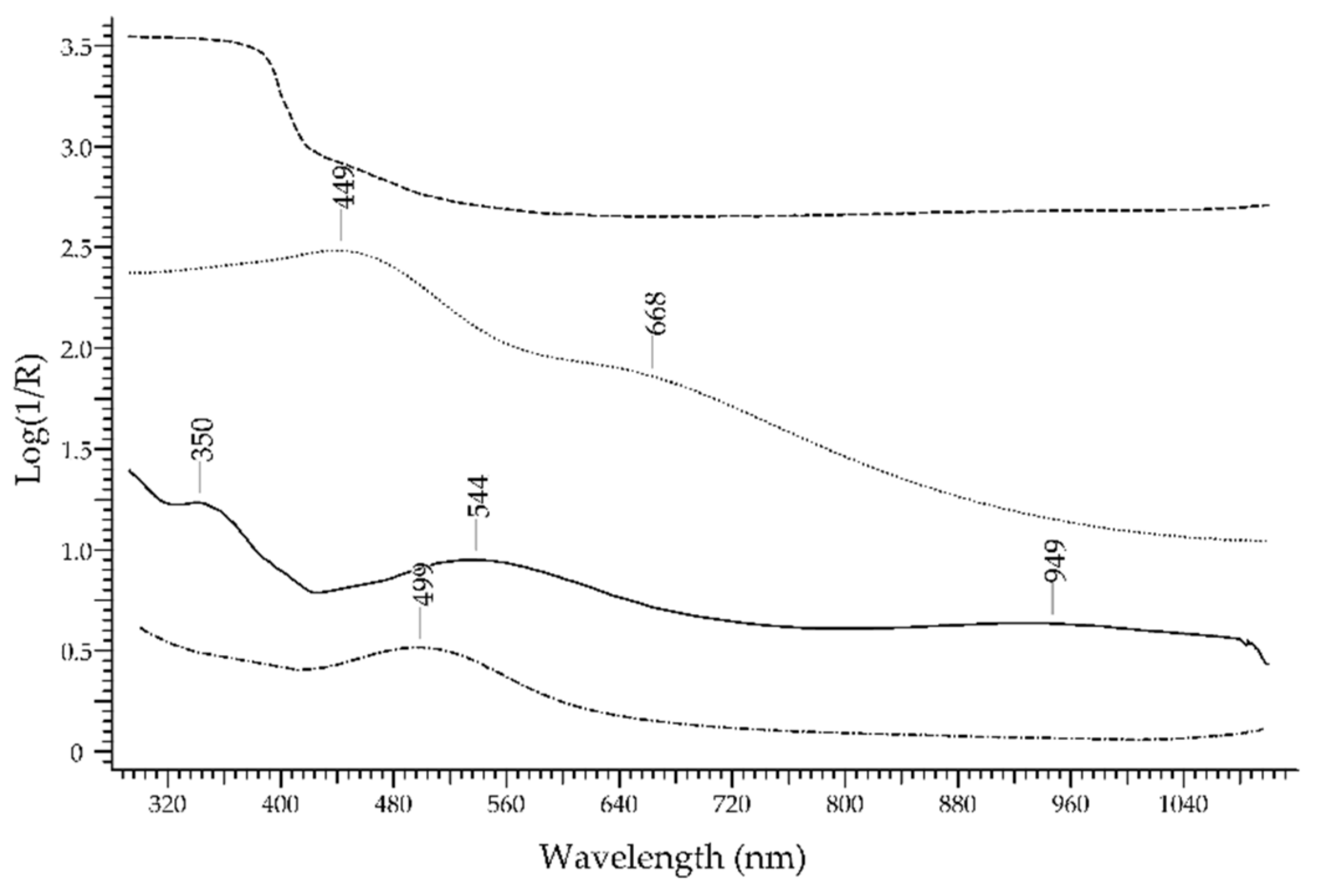
2.2. Green Gemstones
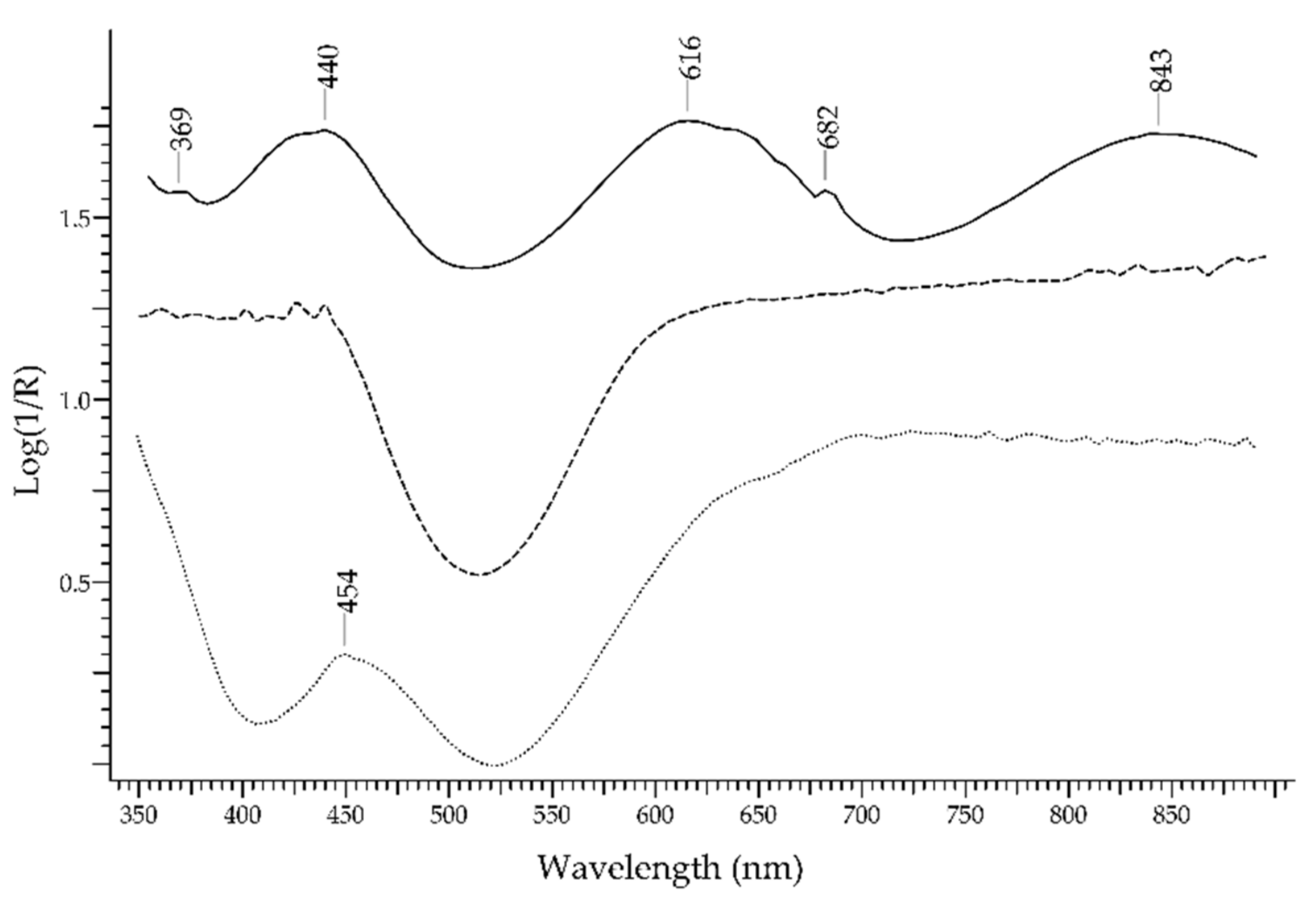
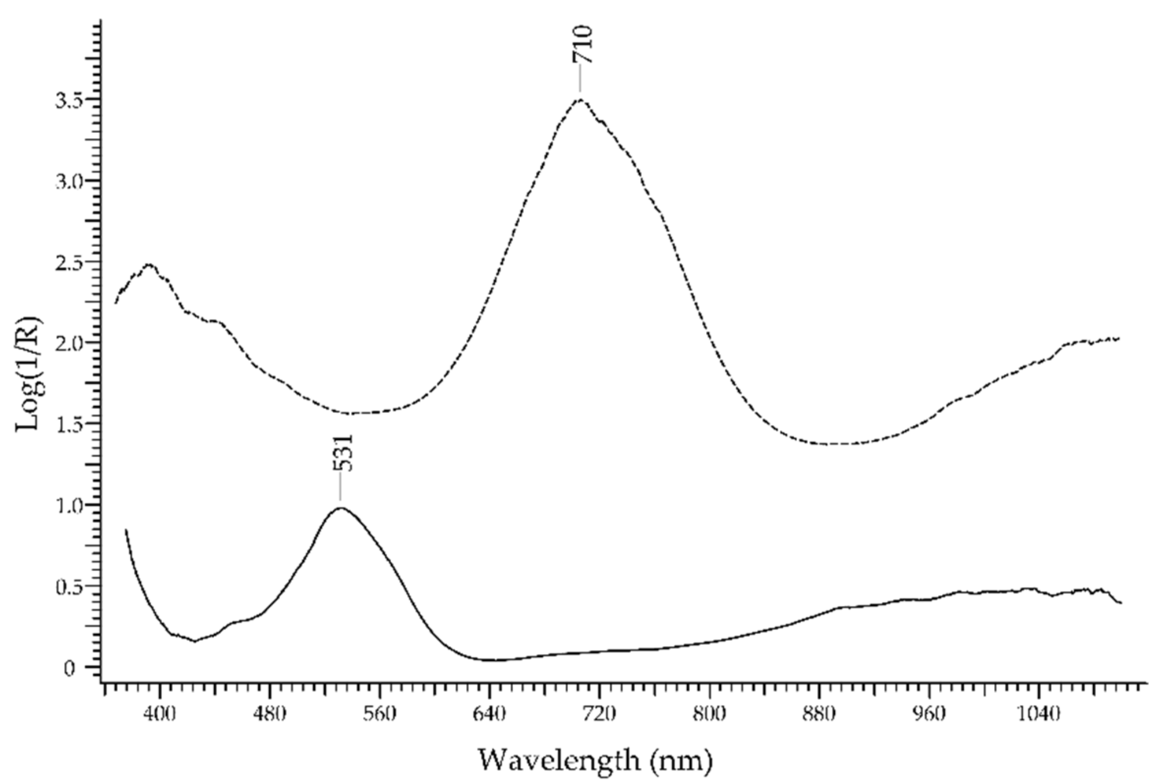
2.3. Pink Gemstones
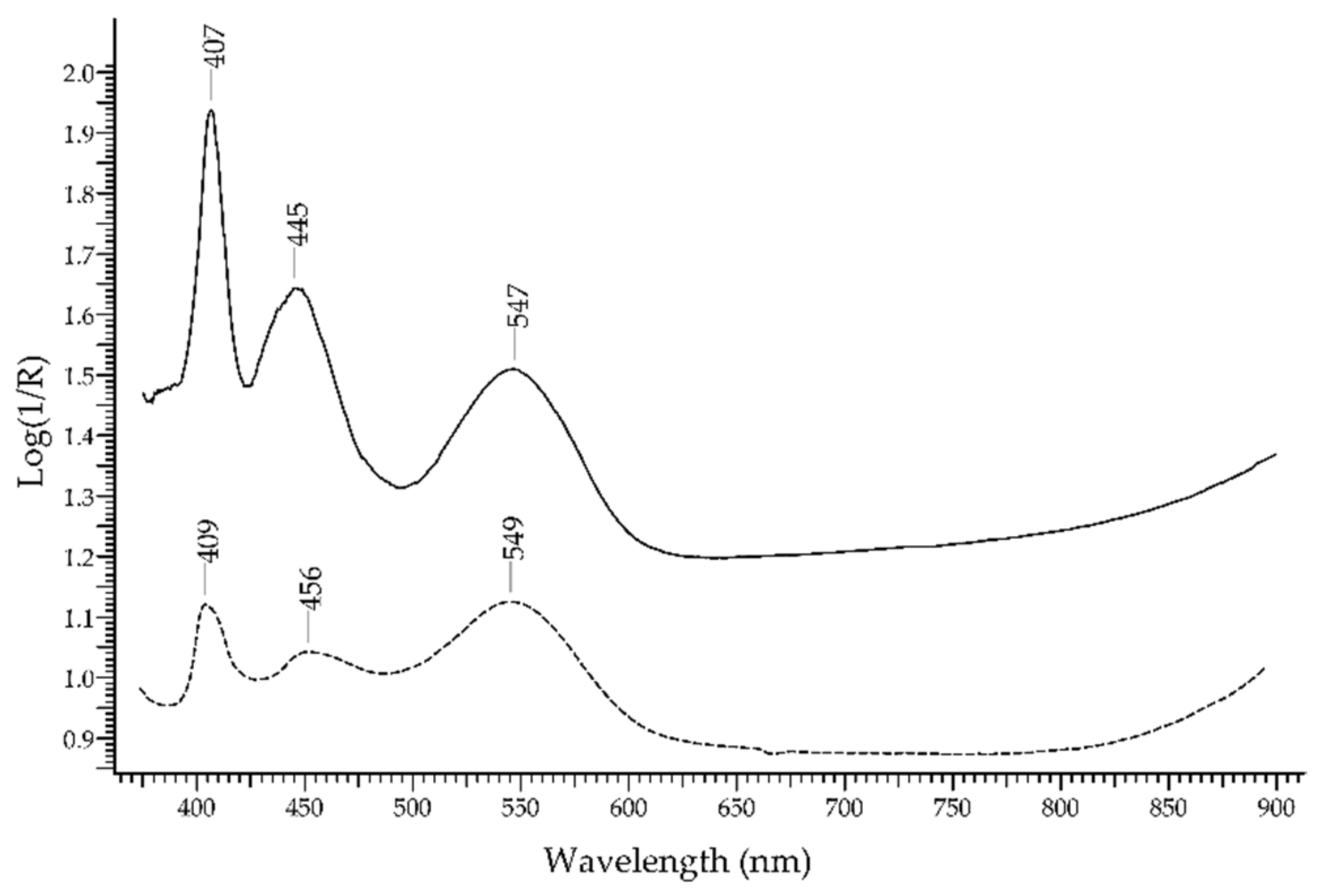
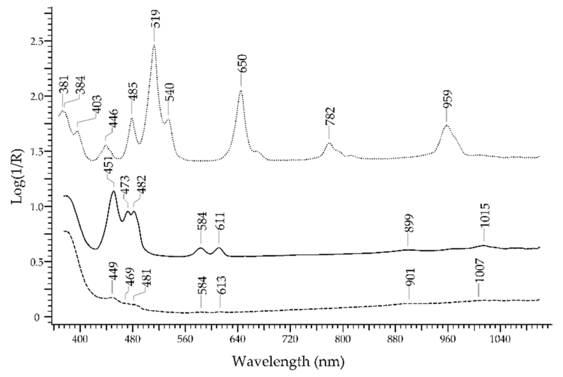
2.4. Red and Purple Gemstones
2.5. Violet Gemstones
2.6. Yellow Gemstones
2.7. Multicoloured Gemstones
2.8. Uncoloured Gemstones
2.9. Glassy Materials
2.10. Comparison of FORS with other Techniques
2.11. Chemometric Treatment of Data
- 26 emeralds (em);
- 10 emerald-like glasses coloured with Cu2+ (gg em);
- 6 green glasses coloured with Ni2+ (gg);
- 2 chrome-chalcedony gemstones (cc).
3. Discussion
3.1. Ruby vs. Red Gemstones
3.2. Sapphire vs. Blue Gemstones
3.3. Emerald vs. Green Gemstones
3.4. Final Considerations
4. Materials and Methods
4.1. Samples of Gemstones
4.2. UV-Visible Diffuse Reflectance Spectrophotometry with Optic Fibres (FORS)
4.3. Raman Spectroscopy
Author Contributions
Funding
Institutional Review Board Statement
Informed Consent Statement
Data Availability Statement
Acknowledgments
Conflicts of Interest
Sample Availability
References
- O’Donoghue, M.; Joyner, L. Identification of Gemstones; Butterworth-Heinemann: Oxford, UK, 2003; ISBN 9780750655125. [Google Scholar]
- Riccardi, M.P.; Prosperi, L.; Tarantino, S.C.; Zema, M. Gemmology in the Service of Archaeometry. In EMU Notes in Mineralogy; Artioli, G., Oberti, R., Eds.; Mineralogical Society of Great Britain & Ireland: Twickenham, UK, 2019; Volume 20, pp. 345–366. ISBN 978-0903056-61-8. [Google Scholar]
- Bersani, D.; Lottici, P.P. Applications of Raman Spectroscopy to Gemology. Anal. Bioanal. Chem. 2010, 397, 2631–2646. [Google Scholar] [CrossRef] [PubMed]
- Kiefert, L.; Karampelas, S. Use of the Raman Spectrometer in Gemmological Laboratories: Review. Spectrochim. Acta Part A Mol. Biomol. Spectrosc. 2011, 80, 119–124. [Google Scholar] [CrossRef]
- Jehlička, J.; Culka, A.; Baštová, M.; Bašta, P.; Kuntos, J. The Ring Monstrance from the Loreto Treasury in Prague: Handheld Raman Spectrometer for Identification of Gemstones. Philos. Trans. R. Soc. A Math. Phys. Eng. Sci. 2016, 374, 20160042. [Google Scholar] [CrossRef]
- Osterrothová, K.; Minaříková, L.; Culka, A.; Kuntoš, J.; Jehlička, J. In Situ Study of Stones Adorning a Silver Torah Shield Using Portable Raman Spectrometers. J. Raman Spectrosc. 2014, 45, 830–837. [Google Scholar] [CrossRef]
- Barone, G.; Bersani, D.; Crupi, V.; Longo, F.; Longobardo, U.; Lottici, P.P.; Aliatis, I.; Majolino, D.; Mazzoleni, P.; Raneri, S.; et al. A Portable versus Micro-Raman Equipment Comparison for Gemmological Purposes: The Case of Sapphires and Their Imitations. J. Raman Spectrosc. 2014, 45, 1309–1317. [Google Scholar] [CrossRef]
- Barone, G.; Bersani, D.; Jehlička, J.; Lottici, P.P.; Mazzoleni, P.; Raneri, S.; Vandenabeele, P.; Di Giacomo, C.; Larinà, G. Nondestructive Investigation on the 17–18th Centuries Sicilian Jewelry Collection at the Messina Regional Museum Using Mobile Raman Equipment. J. Raman Spectrosc. 2015, 46, 989–995. [Google Scholar] [CrossRef] [Green Version]
- Hainschwang, T. Gemstone Analysis by Spectroscopy. In Encyclopedia of Spectroscopy and Spectrometry; Elsevier: Amsterdam, The Netherlands, 2017; pp. 18–24. [Google Scholar]
- Fritsch, E.; Rossman, G.R. An Update on Color in Gems. Part 1: Introduction and Colors Caused by Dispersed Metal Ions. Gems Gemol. 1987, 23, 126–139. [Google Scholar] [CrossRef] [Green Version]
- Nassau, K. The Origin of Color in Minerals. Am. Mineral. 1978, 63, 219–229. [Google Scholar]
- Nassau, K. The Physics and Chemistry of Color: The Fifteen Causes of Color; John Wiley & Sons Ltd.: New York City, NY, USA, 1983. [Google Scholar]
- Fritsch, E.; Rossman, G.R. An Update on Color in Gems. Part 2: Colors Involving Multiple Atoms and Color Centers. Gems Gemol. 1988, 24, 3–15. [Google Scholar] [CrossRef] [Green Version]
- Fritsch, E.; Rossman, G.R. An Update on Color in Gems. Part 3: Colors Caused by Band Gaps and Physical Phenomena. Gems Gemol. 1988, 24, 81–102. [Google Scholar] [CrossRef] [Green Version]
- Takahashi, H.; Perera, P.N. Ultraviolet-Visible Absorption Spectroscopy for Gemstone Identification. U.S. Patent 17/382,317, 3 February 2022. [Google Scholar]
- Dubinsky, E.V.; Stone-Sundberg, J.; Emmett, J.L. A Quantitative Description of the Causes of Color in Corundum. Gems Gemol. 2020, 56, 2–28. [Google Scholar] [CrossRef]
- Bristow, J.K.; Parker, S.C.; Catlow, C.R.A.; Woodley, S.M.; Walsh, A. Microscopic Origin of the Optical Processes in Blue Sapphire. Chem. Commun. 2013, 49, 5259–5261. [Google Scholar] [CrossRef] [PubMed] [Green Version]
- Fontana, I.; Le Donne, A.; Palanza, V.; Binetti, S.; Spinolo, G. Optical Spectroscopy Study of Type 1 Natural and Synthetic Sapphires. J. Phys. Condens. Matter 2008, 20, 125228. [Google Scholar] [CrossRef]
- Palanza, V.; Chiodini, N.; Galli, A.; Lorenzi, R.; Moretti, F.; Paleari, A.; Spinolo, G. Updating of the Interpretation of the Optical Absorption and Emission of Verneuil Synthetic and Natural Metamorphic Blue Sapphire: The Role of V2+, V3+ and Cr2+. IOP Conf. Ser. Mater. Sci. Eng. 2010, 15, 012087. [Google Scholar] [CrossRef]
- Marfunin, A.S. Luminescence. In Spectroscopy, Luminescence and Radiation Centers in Minerals; Springer: Berlin/Heidelberg, Germany, 1979; pp. 141–222. [Google Scholar]
- Gaft, M.; Reisfeld, R.; Panczer, G. Interpretation of Luminescence Centers. In Modern Luminescence Spectroscopy of Minerals and Materials; Springer: Berlin/Heidelberg, Germany, 2005; pp. 119–251. [Google Scholar]
- Jaliya, R.G.C.; Dharmaratne, P.G.R.; Wijesekara, K.B. Characterization of Heat Treated Geuda Gemstones for Different Furnace Conditions Using FTIR, XRD and UV–Visible Spectroscopy Methods. Solid Earth Sci. 2020, 5, 282–289. [Google Scholar] [CrossRef]
- Huang, R.R.; Yin, Z.W. Spectroscopy Identification of Untreated and Heated Corundum. Spectrosc. Spectr. Anal. 2017, 37, 80–84. [Google Scholar]
- Calvo Del Castillo, H.; Deprez, N.; Dupuis, T.; Mathis, F.; Deneckere, A.; Vandenabeele, P.; Calderón, T.; Strivay, D. Towards the Differentiation of Non-Treated and Treated Corundum Minerals by Ion-Beam-Induced Luminescence and Other Complementary Techniques. Anal. Bioanal. Chem. 2009, 394, 1043–1058. [Google Scholar] [CrossRef]
- Bunnag, N.; Kasri, B.; Setwong, W.; Sirisurawong, E.; Chotsawat, M.; Chirawatkul, P.; Saiyasombat, C. Study of Fe Ions in Aquamarine and the Effect of Dichroism as Seen Using UV–Vis, NIR and X-ray. Radiat. Phys. Chem. 2020, 177, 109107. [Google Scholar] [CrossRef]
- Bacci, M.; Cucci, C.; Del Federico, E.; Ienco, A.; Jerschow, A.; Newman, J.M.; Picollo, M. An Integrated Spectroscopic Approach for the Identification of What Distinguishes Afghan Lapis Lazuli from Others. Vib. Spectrosc. 2009, 49, 80–83. [Google Scholar] [CrossRef]
- Qiu, J.-T.; Qi, H.; Duan, J.-L. Reflectance Spectroscopy Characteristics of Turquoise. Minerals 2016, 7, 3. [Google Scholar] [CrossRef] [Green Version]
- Han, W.; Lu, T.; Dai, H.; Su, J.; Dai, H. Impregnated and Dyed Turquoise. Gems Gemol. 2015, 51, 3. [Google Scholar]
- Wood, D.L.; Nassau, K. The Characterization of Beryl and Emerald by Visible and Infrared Absorption Spectroscopy. Am. Mineral. 1968, 53, 777–800. [Google Scholar]
- Schmetzer, K.; Hyršl, J.; Bernhardt, H.J.; Hainschwang, T. Purple to Reddish Purple Chrysoberyl from Brazil. J. Gemmol. 2014, 34, 32–40. [Google Scholar] [CrossRef]
- Butini, E.; Aliprandi, R. Le Gemme Di Oplontis: Aspetto Gemmologico. In Gli ori di Oplontis Gioielli Romani dal Suburbio Pompeiano; D’Ambrosio, A., Ed.; Bibliopolis—Edizioni di Filosofia e Scienze, Soprintendenza Archeologica di Pompei: Napoli, Italy, 1987. [Google Scholar]
- De Michele, V.; Aceto, M.; Agostino, A.; Fenoglio, G. La Gemmatura Nella Pace Di Chiavenna. Considerazioni Gemmologiche. Indagini Archeometriche Non Invasive. Arte Lomb. 2019, 185, 72–80. [Google Scholar] [CrossRef]
- Agostino, A.; Aceto, M.; Fenoglio, G.; Operti, L. Caratterizzazione Chimica Della Coperta Del Codice, C. In Tabula Ornata Lapidibus Diversorum Colorum. la Legatura Preziosa del Codice C nel Museo del Tesoro del Duomo di Vercelli; Lomartire, S., Ed.; Viella: Roma, Italy, 2015; pp. 125–162. [Google Scholar]
- Aceto, M.; Agostino, A.; Fenoglio, G.; Idone, A.; Gulmini, M.; Picollo, M.; Ricciardi, P.; Delaney, J.K. Characterisation of Colourants on Illuminated Manuscripts by Portable Fibre Optic UV-Visible-NIR Reflectance Spectrophotometry. Anal. Methods 2014, 6, 1488. [Google Scholar] [CrossRef]
- Ravikumar, R.V.S.S.N.; Madhu, N.; Chandrasekhar, A.V.; Reddy, B.J.; Reddy, Y.P.; Rao, P.S. Cu(II), Mn(II) in Tetragonal Site in Chrysocolla. Radiat. Eff. Defects Solids 1998, 143, 263–272. [Google Scholar] [CrossRef]
- Adamo, I.; Bocchio, R.; Pavese, A.; Prosperi, L. Characterization of Peridot from Sardinia, Italy. Gems Gemol. 2009, 45, 130–133. [Google Scholar] [CrossRef] [Green Version]
- Tilley, R.J.D. Colour and the Optical Properties of Materials: An Exploration of the Relationship Between Light, the Optical Properties of Materials and Colour; John Wiley & Sons: Hoboken, NJ, USA, 2011; ISBN 9780470746967. [Google Scholar]
- Deer, W.A.; Howie, R.A.; Zussman, J. An Introduction to the Rock-Forming Minerals, 2nd ed.; Longman: London, UK, 1992. [Google Scholar]
- Manning, P.G. The Optical Absorption Spectra of the Garnets; Almandine-Pyrope, Pyrope and Spessartine and Some Structural Interpretations of Mineralogical Significance. Can. Mineral. 1967, 9, 237–251. [Google Scholar]
- Izawa, M.R.M.; Cloutis, E.A.; Rhind, T.; Mertzman, S.A.; Poitras, J.; Applin, D.M.; Mann, P. Spectral Reflectance (0.35–2.5 µm) Properties of Garnets: Implications for Remote Sensing Detection and Characterization. Icarus 2018, 300, 392–410. [Google Scholar] [CrossRef]
- Smith, C.P.; McClure, S.F.; Eaton-Magaña, S.; Kondo, D.M. Pink-to-Red Coral: A Guide to Determining Origin of Color. Gems Gemol. 2007, 43, 4–15. [Google Scholar] [CrossRef]
- Koziarska, B.; Godlewski, M.; Suchocki, A.; Czaja, M.; Mazurak, Z. Optical Properties of Zoisite. Phys. Rev. B 1994, 50, 12297–12300. [Google Scholar] [CrossRef] [PubMed]
- Andersson, L.O. The Yellow Color Center and Trapped Electrons in Beryl. Can. Mineral. 2013, 51, 15–25. [Google Scholar] [CrossRef]
- Wang, G.-Y.; Yu, X.-Y.; Liu, F. Genesis of Color Zonation and Chemical Composition of Penglai Sapphire in Hainan Province, China. Minerals 2022, 12, 832. [Google Scholar] [CrossRef]
- Rossman, G.R. Colored Varieties of the Silica Minerals. Rev. Mineral. Geochem. 1994, 29, 433–463. [Google Scholar]
- Jovanovski, G.; Šijakova-Ivanova, T.; Boev, I.; Boev, B.; Makreski, P. Intriguing Minerals: Quartz and Its Polymorphic Modifications. ChemTexts 2022, 8, 14. [Google Scholar] [CrossRef]
- Henn, U.; Schultz-Güttler, R. Review of Some Current Coloured Quartz Varieties. J. Gemmol. 2012, 33, 29–43. [Google Scholar] [CrossRef]
- Lehmann, G. On the Color Centers of Iron in Amethyst and Synthetic Quartz: A Discussion. J. Earth Planet. Mater. 1975, 60, 335–337. [Google Scholar]
- Kibar, R.; Garcia-Guinea, J.; Çetin, A.; Selvi, S.; Karal, T.; Can, N. Luminescent, Optical and Color Properties of Natural Rose Quartz. Radiat. Meas. 2007, 42, 1610–1617. [Google Scholar] [CrossRef]
- Renfro, N.; McClure, S.F. Dyed Purple Hydrophane Opal. Gems Gemol. 2011, 47, 260–270. [Google Scholar] [CrossRef]
- Wu, J.; Ma, H.; Ma, Y.; Ning, P.; Tang, N.; Li, H. Comparison of Natural and Dyed Fire Opal. Crystals 2022, 12, 322. [Google Scholar] [CrossRef]
- Andreozzi, G.B.; D’Ippolito, V.; Skogby, H.; Hålenius, U.; Bosi, F. Color Mechanisms in Spinel: A Multi-Analytical Investigation of Natural Crystals with a Wide Range of Coloration. Phys. Chem. Miner. 2019, 46, 343–360. [Google Scholar] [CrossRef]
- Krambrock, K.; Ribeiro, L.G.M.; Pinheiro, M.V.B.; Leal, A.S.; Menezes, M.D.B.; Spaeth, J.M. Color Centers in Topaz: Comparison between Neutron and Gamma Irradiation. Phys. Chem. Miner. 2007, 34, 437–444. [Google Scholar] [CrossRef]
- Skvortsova, V.; Mironova-Ulmane, N.; Trinkler, L.; Chikvaidze, G. Optical Properties of Natural Topaz. IOP Conf. Ser. Mater. Sci. Eng. 2013, 49, 012051. [Google Scholar] [CrossRef] [Green Version]
- Phichaikamjornwut, B.; Pongkrapan, S.; Intarasiri, S.; Bootkul, D. Conclusive Comparison of Gamma Irradiation and Heat Treatment for Color Enhancement of Rubellite from Mozambique. Vib. Spectrosc. 2019, 103, 102926. [Google Scholar] [CrossRef]
- Kempe, U.; Trinkler, M.; Pöppl, A.; Himcinschi, C. Coloration of Natural Zircon. Can. Mineral. 2016, 54, 635–660. [Google Scholar] [CrossRef]
- Diamonds—Characterized by FT-IR Spectroscopy; Application Note no. AN#81; Bruker: Ettlingen, Germany, 2010; pp. 1–3. Available online: https://www.optikinstruments.hr/runtime/cache/an-m81-diamonds-en-daa1b24eb7b29aa45a60e6d8355a40d7.pdf (accessed on 21 June 2022).
- Lipatov, E.I.; Burachenko, A.G.; Avdeev, S.M.; Tarasenko, V.F.; Bublik, M.A. Identification of Natural and Synthetic Diamonds from Their Optical Absorption and Cathodoluminescence Spectra. Russ. Phys. J. 2018, 61, 469–483. [Google Scholar] [CrossRef]
- Aceto, M.; Fenoglio, G.; Labate, M.; Picollo, M.; Bacci, M.; Agostino, A. A Fast Non-Invasive Method for Preliminary Authentication of Mediaeval Glass Enamels Using UV–Visible–NIR Diffuse Reflectance Spectrophotometry. J. Cult. Herit. 2020, 45, 33–40. [Google Scholar] [CrossRef]
- Superchi, M. Le Gemme Dell’Evangeliario Di Ariberto. In Evangeliario di Ariberto; Tomei, A., Ed.; Silvana Editore: Milano, Italy, 1999; pp. 149–157. [Google Scholar]
- California Institute of Technology Mineral Spectroscopy Server. Available online: http://minerals.gps.caltech.edu/FILES/Visible/Index.html (accessed on 21 June 2022).


| Gemstones | Pace di Ariberto | Pace di Chiavenna | Legatura di Vercelli | |||
|---|---|---|---|---|---|---|
| FORS | Raman | FORS | Ref | FORS | Raman | |
| agate | - | 1 | ||||
| amethyst | 24 | 24 | 6 | 6 | 5 | 5 |
| carnelian | - | 1 | ||||
| chalcedony | - | 2 | ||||
| doublet | - | 4 | ||||
| emerald | 16 | 16 | 7 | 7 | 3 | 3 |
| garnet | 11 | 11 | 55 | 56 | 14 | 14 |
| glass/vitreous paste | - | 23 | - | 2 | - | 31 |
| mtorolite | - | 1 | - | 1 | ||
| pearl | - | 21 | - | 93 | - | 23 |
| rock crystal | - | 18 | - | 2 | ||
| sapphire | 10 | 10 | 19 | 19 | 5 | 5 |
| turquoise | 2 | 2 | ||||
| other stones | 1 | 6 | ||||
| total identified | 63 | 134 | 87 | 190 | 27 | 84 |
| unidentified by FORS | 71 | 103 | 57 | |||
| total excluding pearls, rock crystals and glassy materials | 63 | 72 | 87 | 95 | 27 | 28 |
| unidentified by FORS excluding pearls, rock crystals and glassy materials | 9 | 8 | 1 | |||
| Gemstone | Provenance | Colour | Notes |
|---|---|---|---|
| alexandrite | Brazil | green | |
| amethyst | Brazil | violet | |
| aquamarine | Brazil | blue | |
| chrome-chalcedony | unknown | green | 1 |
| chrysoberyl | Brazil | yellow | |
| citrine quartz | Brazil | yellow | |
| coral | Italy | red | |
| emerald | Colombia | green | |
| garnet | India | purple | Pyrope–almandine |
| garnet | Kenya | orange | spessartite |
| glass with Cr3+ | green | Artificial 2 | |
| glass with Se | red | Artificial 2 | |
| heliodor | Brazil | green-yellow | |
| lapis lazuli | Afghanistan | blue | |
| opal | Mexico | various | |
| opal | Ethiopia | various | |
| peridot | Sri Lanka | green | |
| rhodochrosite | Romania | pink | |
| rhodonite | Tanzania | pink | |
| rose quartz | Brazil | pink | |
| ruby | Myanmar | red | |
| sapphire | Cambodia | blue | |
| sapphire | yellow | artificial | |
| smoky quartz | Brazil | grey | |
| spinel | Russia | blue | |
| spinel | Myanmar | red | |
| tanzanite | Tanzania | violet | |
| topaz | Brazil | blue | |
| topaz | green | artificial | |
| topaz | Brazil | pink | |
| tourmaline | Brazil | green | |
| tourmaline | Brazil | red | rubellite |
| turquoise | China | turquoise | |
| turquoise | China | turquoise | dyed |
| zircon | Myanmar | blue | |
| zircon | green | artificial | |
| zircon | pink | artificial | |
| zircon | yellow | artificial |
Publisher’s Note: MDPI stays neutral with regard to jurisdictional claims in published maps and institutional affiliations. |
© 2022 by the authors. Licensee MDPI, Basel, Switzerland. This article is an open access article distributed under the terms and conditions of the Creative Commons Attribution (CC BY) license (https://creativecommons.org/licenses/by/4.0/).
Share and Cite
Aceto, M.; Calà, E.; Gulino, F.; Gullo, F.; Labate, M.; Agostino, A.; Picollo, M. The Use of UV-Visible Diffuse Reflectance Spectrophotometry for a Fast, Preliminary Authentication of Gemstones. Molecules 2022, 27, 4716. https://doi.org/10.3390/molecules27154716
Aceto M, Calà E, Gulino F, Gullo F, Labate M, Agostino A, Picollo M. The Use of UV-Visible Diffuse Reflectance Spectrophotometry for a Fast, Preliminary Authentication of Gemstones. Molecules. 2022; 27(15):4716. https://doi.org/10.3390/molecules27154716
Chicago/Turabian StyleAceto, Maurizio, Elisa Calà, Federica Gulino, Francesca Gullo, Maria Labate, Angelo Agostino, and Marcello Picollo. 2022. "The Use of UV-Visible Diffuse Reflectance Spectrophotometry for a Fast, Preliminary Authentication of Gemstones" Molecules 27, no. 15: 4716. https://doi.org/10.3390/molecules27154716