Article

Biocompatible Organic Coatings Based on Bisphosphonic Acid RGD-Derivatives for PEO-Modified Titanium Implants

by
Lyudmila V. Parfenova
1,*,
Elena S. Lukina
1,
Zulfia R. Galimshina
1,
Guzel U. Gil’fanova
1,
Veta R. Mukaeva
2,
Ruzil G. Farrakhov
2,
Ksenia V. Danilko
3,
Grigory S. Dyakonov
4 and
Evgeny V. Parfenov
2
1
Institute of Petrochemistry and Catalysis of Russian Academy of Sciences, 141, Prospekt Oktyabrya, 450075 Ufa, Russia
2
Department of Theoretical Basis of Electrical Engineering, Ufa State Aviation Technical University, 12 Karl Marx Street, 450008 Ufa, Russia
3
Bashkir State Medical University, 3 Lenin Street, 450000 Ufa, Russia
4
Institute of Physics of Advanced Materials, Ufa State Aviation Technical University, 12 Karl Marx Street, 450008 Ufa, Russia
*
Author to whom correspondence should be addressed.
Molecules 2020, 25(1), 229; https://doi.org/10.3390/molecules25010229
Submission received: 26 November 2019 / Revised: 25 December 2019 / Accepted: 1 January 2020 / Published: 6 January 2020
(This article belongs to the Section Materials Chemistry)

Abstract

:
Currently, significant attention is attracted to the problem of the development of the specific architecture and composition of the surface layer in order to control the biocompatibility of implants made of titanium and its alloys. The titanium surface properties can be tuned both by creating an inorganic sublayer with the desired morphology and by organic top coating contributing to bioactivity. In this work, we developed a composite biologically active coatings based on hybrid molecules obtained by chemical cross-linking of amino acid bisphosphonates with a linear tripeptide RGD, in combination with inorganic porous sublayer created on titanium by plasma electrolytic oxidation (PEO). After the addition of organic molecules, the PEO coated surface gets nobler, but corrosion currents increase. In vitro studies on proliferation and viability of fibroblasts, mesenchymal stem cells and osteoblast-like cells showed the significant dependence of the molecule bioactivity on the structure of bisphosphonate anchor and the linker. Several RGD-modified bisphosphonates of β-alanine, γ-aminobutyric and ε-aminocaproic acids with BMPS or SMCC linkers can be recommended as promising candidates for further in vivo research.

1. Introduction

Traumatology and orthopedics generate high demand in implants for osseosynthesis, and, according to market forecasts, this demand will increase due to the spread of extreme activities and the aging of the population. Currently, various biomimetic approaches that ensure the biocompatibility of the implants are developed [1]. As a rule, they suggest changes in the architecture and composition of the surface layer so that the devices gain the properties of the bone tissue and cell membranes. In current medical practice, preference is given to implants made of titanium and its alloys, due to their bioinert properties and high corrosion resistance [2]. The modification of the titanium surface is achieved both through inorganic coatings that approximate the phase composition and morphology of the human bone and by introduction of organic molecules containing functional fragments actively interacting with the proteins of the extracellular matrix (ECM). These approaches should provide both biomechanical and biochemical compatibility of the implants, their corrosion resistance, and, therefore, should successfully initiate the metal device osseointegration into the bone tissue.
Currently, significant attention is attracted to the biomimetic coatings based on the method of plasma electrolytic oxidation (PEO) [3,4]. The inorganic parts of the PEO coatings contain stable titania (rutile and anatase) tightly attached to the surface [5]. PEO helps to incorporate the anions and cations of the electrolyte into the coating; this provides Ca-, P- containing bioactive crystalline phases within the coating: hydroxyapatite, tricalciumphosphate, tetracalciumphosphate, perovskite [6]. Nevertheless, medical implants, once introduced into a biological system, get covered by nonspecific proteins within a few seconds. The adsorbed layer initiates a foreign body reaction of the organism; this causes the formation of a vascular fibrous capsule isolating the implant from the target tissue [7]. Consequently, current state-of-the-art includes the development of biocompatible bifunctional organic top coatings, which, on one hand would mask the implant, on the other hand, would initiate the implant osseointegration. These modern materials include: non-fouling coatings based on polysaccharides; hydrophilic self-assembled monolayers or polyethylene glycol, also in combination with oligopeptides (OP); bioactive coatings based on the extracellular matrix proteins (e.g., fibronectin, collagen, laminin, and osteopontin), having both long and short sequences of the amino acids [8,9,10,11,12]. The combination of the PEO inorganic sublayer and the organic top coating appears to be a promising direction for the development of implants with enhanced osseointegration [13,14,15].
Among relatively short bioactive peptide sequences, special attention is paid to RGD (arginine-glycine-aspartic acid), which is a tripeptide fragment appearing in numerous extracellular matrix proteins, and to which the cells attach using specific receptors on their surface [16]. Its discovery led to the identification of a large family of the cell surface adhesion receptors, called integrins, which recognize RGD-specific sequences in certain proteins of the extracellular matrix. The literature survey shows significant research efforts put into the application of the RGD-containing oligopeptides for the titanium surface modification. Various methods of the deposition have been developed: silanization of the surface by aminosilanes (including APTES) followed by the binding with RGD-containing oligopeptide via various linkers [17,18,19,20,21,22,23,24,25,26,27,28,29,30]; oligopeptide- functionalized synthetic polymers [31,32,33,34] and natural biopolymers [35,36,37,38,39]; oligopeptides attachment to the titanium via CDI [40,41] or tresyl chloride [42] surface activation; immobilization of OP on Ti via electrodeposited poly(ethyleneglycol) (PEG) [43,44,45]; physicochemical adsorption of oligopeptides as well as SH-ended derivatives [23,46,47,48,49,50,51] and 3,4-dihydroxyphenylalanine (DOPA) functionalized OP [52] from solutions; physicochemical adsorption of OP with a phosphonate anchor [22,23,36,51,53,54,55,56,57,58]. The strategy of the phosphonate anchors appears to be very attractive when bonding the inorganic oxide sublayer with the organic top coating. As shown elsewhere [59,60,61], phosphonate groups have high affinity to metal ions and their oxides, and they are almost not prone to hydrolytic cleavage, unlike siloxanes [62,63]. The introduction of gem-bisphosphonic group into the molecules increases their affinity to the oxidized surfaces compared to monophosphonates, and this increases their aqueous solubility due to the changing of the molecule polarity [64,65].
Plasma electrolytic oxidation currently is a well-developed electrochemical technique used to produce ceramic coatings with improved adhesion over the surface of valve metals such as titanium, magnesium, aluminum and zirconium [66,67,68]. Due to the unique surface morphology of the porous coating, PEO provides better biocompatibility of the surface and for the cell proliferation [69]. The PEO process is an expansion of the traditional anodizing into high voltages of up to 600 V; these voltages promote microdischarges within the coating; this results in its resoldifying and intensive growth [70]. The process mechanism includes electrochemical oxidation and numerous melting and crystallizing events at the microdischarge sites. This coating formation mechanism helps to develop coatings with regulated porosity, with the pore size from 0.1 to 10 µm and roughness Ra from 1 to 10 µm; the wide network of the pores forms a fractal-like structure, with the pore size increasing to the top of the coating [71]. The high surface area of the PEO coating allows deposition of various functional organic components, and the PEO coating acts as a sublayer increasing adhesion, and as the organic substance carrier [72,73,74,75,76].
Our recent study [15] showed that the combination of the PEO coating and RGD-modified bisphosphonic acid on nano-Ti gives a 45% increase in the number of proliferated cells compared to uncoated nano-Ti, and 66% compared to coarse grain Ti.
In the presented study, we expanded the number of hybrid RGD-functionalyzed molecules in order to uderstand how the structure of the bisphosphonate anchors and linkers (BMPS, EMCS, SMCC) affects the biological activity of the composite coatings based on inorganic porous PEO sublayer on titanium; moreover, the biological effect of the synthesized organic molecules was studied in vitro on a larger set of cell lines (mesenchymal stem cells, fibroblasts and osteoblast-like cells). Therefore, the aim of this work is to synthesize the hybrid molecules by chemical cross-linking of amino acid bisphosphonates with a linear RGD tripeptide through linkers that differ in length and structure for effective use in combination with inorganic porous PEO sublayer on titanium, and to identify the most promising options for the design of the proposed biomimetic coatings.

2. Results and Discussion

The experimental design concerns the variation of the anchors and linkers to attach the RGD peptide to the titanium implant. For the sake of the clarity, the following sample identification is used: Ti—for the metal substrate; Ti-PEO—for the inorganic PEO coating over the substrate; Ti-PEO-RGD—for the PEO coating soaked in the RGD aqueous solution; Ti-PEO-(number)-RGD—for the PEO coating soaked in the aqueous solution of the RGD-modified bisphosphonic acids, where the (number) indicates the compound in accordance with the reaction Scheme 1 and the corresponding description in Section 3.2. As a result, this sample identification shows the structures of the proposed composite coatings.

2.1. Synthesis of Bioactive Bifunctional Molecules

As a base for the design of the RGD-containing self- assembled monolayers on the PEO-modified meal surface, we synthesized aminoacid bisphosphonates 1–3 via the reaction of the corresponding amino acids (β-alanine, γ-aminobutyric and ε-aminocaproic acids) with PCl3 in methanesulfonic acid (Scheme 1). We found that carrying out the synthesis at elevated temperature (85–90 °C) decreases the reaction time to 4–5 h, and, along with the separation of the products at pH = 6, increases the resulting yield of the amino bisphosphonates up to 85–89%. The N-maleimidosuccinimide linkers (BMPS, EMCS and SMCC) 46 were obtained in yields of 60–89% via the reaction of maleic anhydride, N–hydroxysuccinimide and aminocarboxylic acids in DMF [77,78]. The N-maleimido derivatives 714 for further modification by the oligopeptide were obtained by interaction of amino bisphosphonates with N-maleimidosuccinimide linkers. The application of acetone instead of dioxane as the solvent used elsewhere [79] helped to avoid the formation of relatively heavy side products whose appearance is an inevitable consequence of the dioxane cycle modification. Target RGD-modified amino acid bisphosphonates 1522 were obtained by the reaction of the corresponding derivatives 714 with RGDC in an aqueous medium at pH = 7 by Michael reaction.
The structure of the RGD-modified aminoacid bisphosphonates was confirmed by mass-spectrometry MALDI TOF/TOF, 1D (1H, 13C, 31P) and 2D NMR spectroscopy. In the MALDI TOF/TOF mass spectra of the hybrid molecules, the peaks with m/z corresponding to the molecular ions were detected. The 31P-NMR spectra exhibited single resonance lines at δP 17–18 ppm, characteristic to bisphosphonate groups. Signals corresponding to fragments of amino bisphosphonate, linker, and RGDC were observed in 1H- and 13C-NMR spectra. The formation of compounds 1522 was accompanied by the disappearance of the signal of double bond protons of the N-maleimido fragment in derivatives 714 at δH 6–7 ppm in the 1H-NMR spectrum.

2.2. PEO Coating Characterization

Figure 1 shows the top view and cross-section of the PEO coating. The coating exhibits a porous morphology, with porosity evaluated as 18.2 ± 2.5%. The coating thickness is 19.3 ± 1.5 µm, surface roughness 1.5 ± 0.2 µm. This type of morphology contributes towards cell proliferation, and it supplies a well-developed surface for the introduction of the organic compounds with bisphosphonate anchors into the resulting composite coating [15]. The coating surface porosity is 9.5 ± 0.8%; the real 3D structure of this well-developed network of pores can be seen elsewhere for a similar PEO coating [80].
As follows from Figure 2, the PEO coating consists of crystalline titania in two phase modifications—rutile and anatase (49 and 51% respectively). Also, the XRD pattern shows the peaks belonging to α-Ti substrate. This coating composition and morphology provides sufficient surface passivation for the implant prolonged operation in the corrosive fluids of a human body [81]. Also, this type of coating exhibits exceptional adhesion to the titanium surface as summarized elsewhere [5].

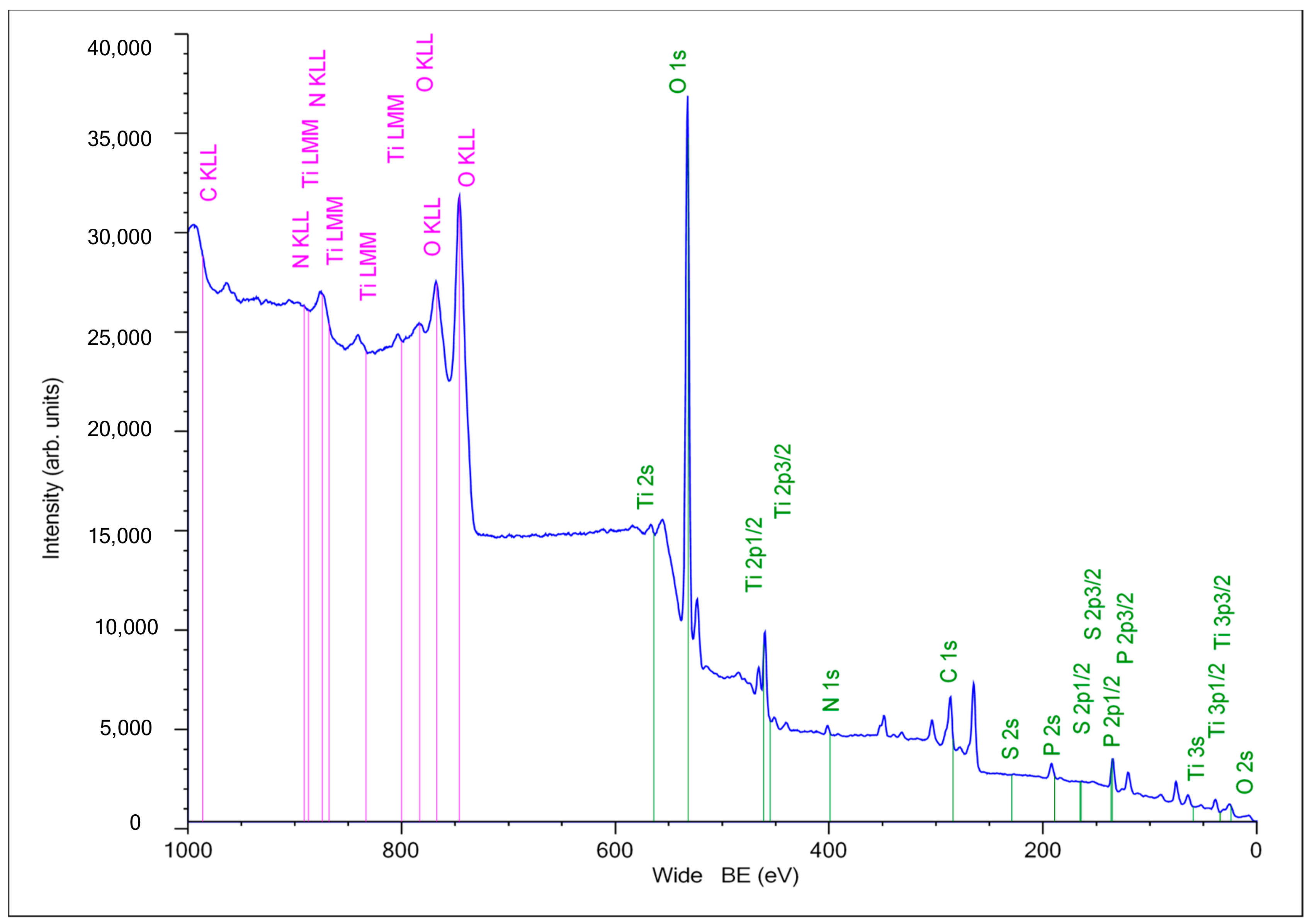
2.3. XPS Analysis of the Composite PEO Coating Functionalized with RGD Derivatives

Since the introduction of the RGD derivatives into the inorganic PEO coating was performed at a low concentration of 10−3 M, it falls below the capabilities of the SEM and XRD techniques to detect organic compounds on the titania surface. Therefore, XPS studies were employed in order to verify the presence of the RGD derivatives within the coating; a typical survey spectrum is shown in Figure 3. In details the XPS spectra are presented in the Supplementary Materials. The survey XPS spectra collected from the PEO-coated titanium samples indicate typical Ti 2p, Ti 3s O 1s, C 1s, P 2p, N 1s, O KLL and Ti LMM Auger peaks. The spectra of the RGD-modified samples confirm changes in the surface layer chemistry compared to the unmodified PEO sample.
As follows from the high-resolution XPS spectra, the organic molecules attached to the surface affect the relative content of nitrogen, phosphorus, carbon, oxygen, and sulfur (Table 1). The most notable differences appear for the Ti2p, C1s and N1s peaks. The Ti2p signal decreased after the RGD modification, and the N1s and C1s signals increased. Also, the RGD-modified surface shows the presence of S2p signals, whereas for the Ti-PEO surface this signal does not appear. The changes in the atomic ratio (Ti2p/C1s) and (Ti2p/P2p) show that the titanium signal decreases for all the RGD modified types of the surface. This is consistent with the observations made elsewhere [27,82,83].

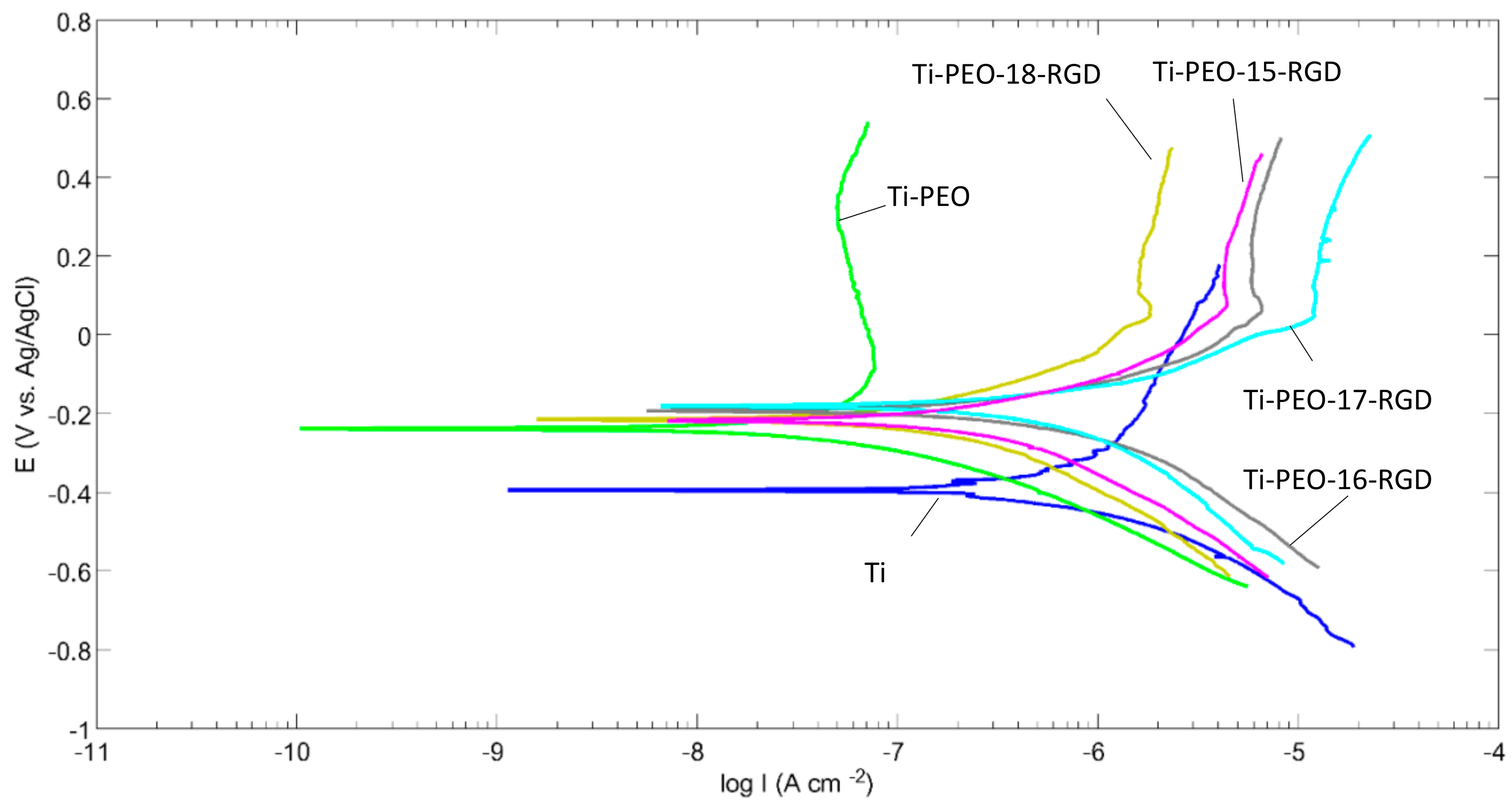
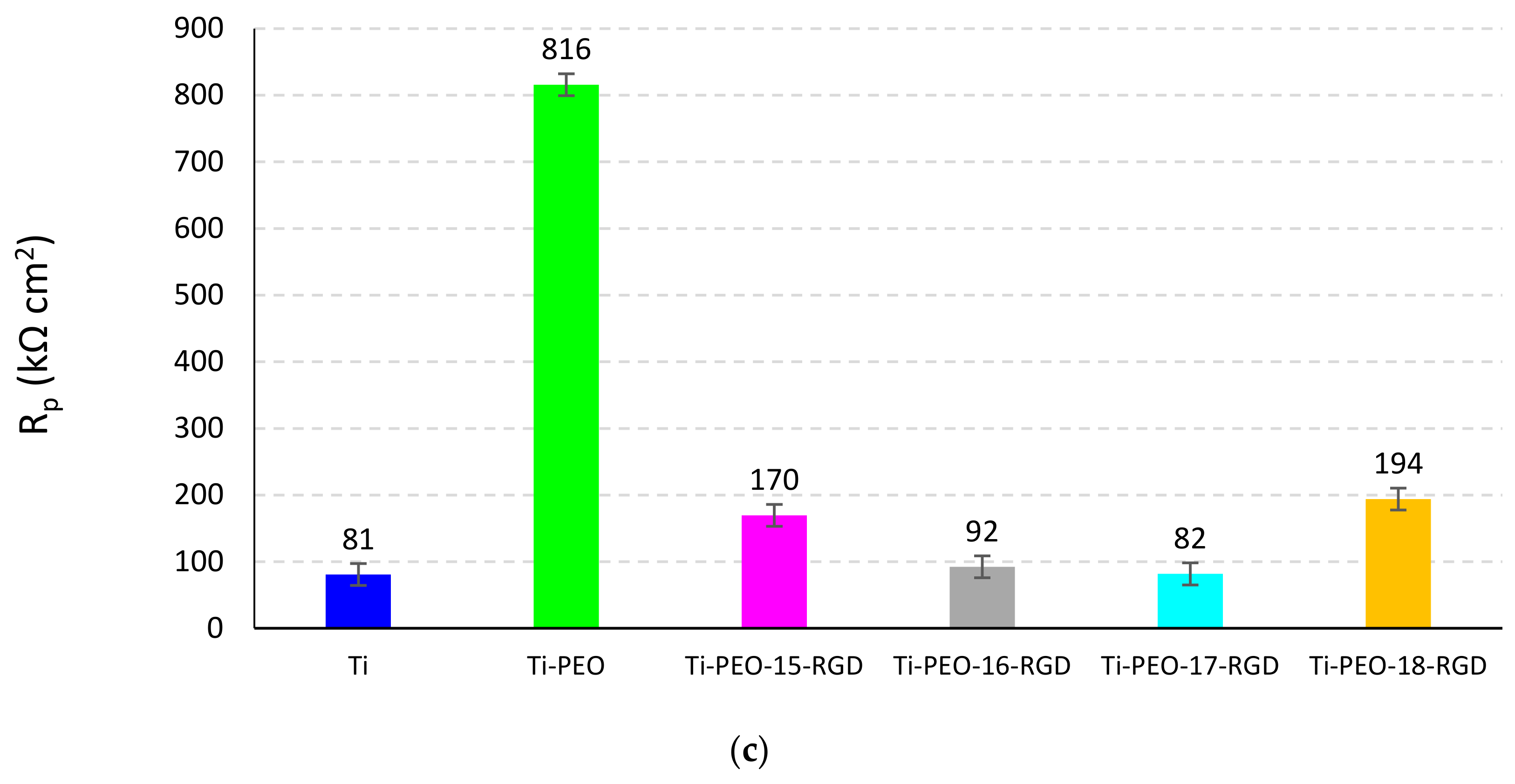
2.4. Electrochemical Behavior of the Composite PEO Coating Functionalized with RGD Derivatives

The electrochemical behavior of the composite PEO coating functionalized with RGD derivatives was studied using compounds 1518 as examples. The polarization curves (Figure 4) show notable differences among the surface having PEO and various RGD treatments. As expected, after the PEO, the surface passivates, and the Ecorr becomes nobler; the corrosion current icorr decreases, which is followed by an order of magnitude increase in the polarization resistance Rp (Figure 5). All the RGD functionalized coatings show the surface, nobler than that of the Ti-PEO sample. The cathodic parts of the polarization curves do not show significant differences compared to Ti and Ti-PEO samples. This indicates that cathodic corrosion processes are not influenced by this type of coating since the cathodic Tafel parts show almost similar slopes. However, the anodic behavior is significantly influenced by the coating. Ti-PEO sample shows a passivation region above −0.1 V(AgCl). The introduction of the RGD- modified bisphosphonates depassivates the surface; the corrosion current increases compared to that of Ti-PEO and Ti samples (Figure 5). The depassivation region belongs to the range −0.1 to 0.1 V (AgCl) and ends with a pitting current tip followed by a passivation region above 0.1 V (AgCl). The changes in the anodic kinetics rise the Ecorr, and this is followed by the increase of the icorr. This indicates that the RGD functionalization increases the exchange currents, and this icorr behavior is supported by the decrease of Rp.

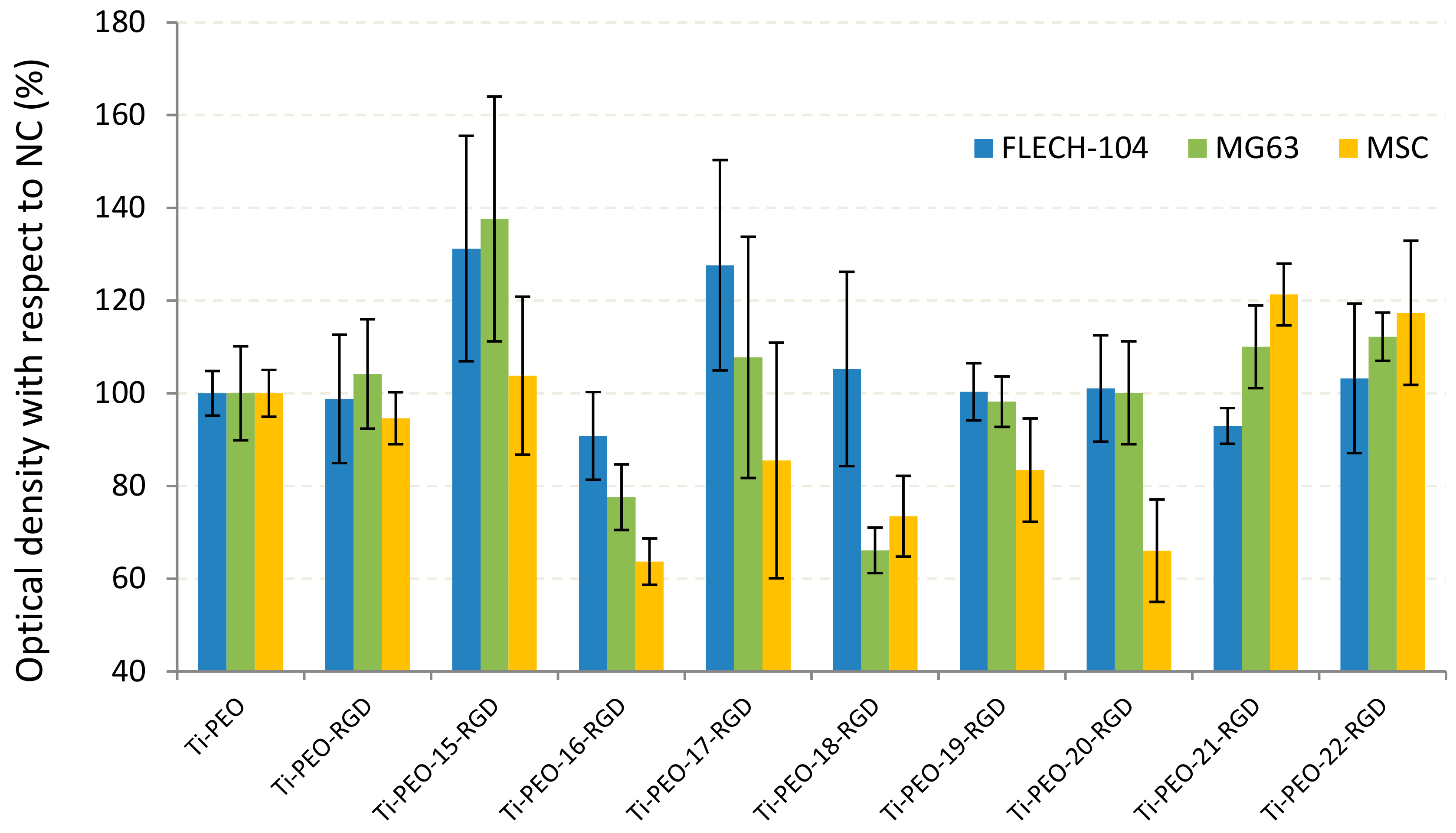
2.5. In Vitro Test Results Supporting The Efficiency of Proposed PEO Coating Functionalized with RGD Derivatives

As follows from the literature analysis, the application of RGD-containing bisphosphonates to the titanium surface leads to the increase in cell adhesion, proliferation and mineralization [22,23,51,53]. The cell adhesion appeared to be sensitive to the anchor used [57,59].
The in vitro results presented in Figure 6 show that the different cell lines give dissimilar reactions to the same type of coating. The biological response also depends on the structure of the source amino bisphosphonate and the linker. It was shown that RGDC-oligopeptide itself introduced into the PEO coating without the anchor does not affect the cell life activity on the surface.
A significant (up to 30–40%) increase in the fibroblast proliferation was observed when using the organic molecules with relatively short linkers and aminobisphosphonate fragments BMPS-β (Ti-PEO-15-RGD) and BMPS-γ (Ti-PEO-17-RGD). Moreover, the sample Ti-PEO-15-RGD based on β-alanine bisphosphonate with the short BMPS linker and RGD peptide has shown the best adhesion for the osteoblast-like cells MG-63 compared to the other samples.
The derivatives containing EMCS linker (Ti-PEO-16-RGD and Ti-PEO-18-RGD) lead to a decrease in the proliferation of MG-63 on the PEO-modified surface. The same tendency was found in the case of mesenchymal stem cells then the long-chained molecules containing EMCS linker (Ti-PEO-16-RGD, Ti-PEO-18-RGD, and Ti-PEO-20-RGD) or derivative of ε-aminocaproic acid (Ti-PEO-19-RGD) were applied. In contrast, SMCC-linked compounds (Ti-PEO-21-RGD and Ti-PEO-22-RGD) provide the increasing of the cell growth up to 20% on the surface.
Summarizing the in vitro studies, we can conclude that RGD functionalized hybrid molecules 15, 17, 21, 22 appear to be promising organic top coatings for modulation of the biological activity of the PEO modified surface of titanium implants. The differences in the cell behavior depending on the compound structure can be caused by their cytotoxicity and orientation on the PEO surface during adsorption; this requires further studies that are currently conducted in our research group.

3. Materials and Methods

3.1. General Information

The following reagents were used for the synthesis: methanesulfonic acid (98%, Acros Organics, Geel, Belgium), PCl3 (98%, Acros Organics), β-alanine (97%, Acros Organics), γ-aminobutanoic (99+%, Acros Organics) and ε-aminocaproic acid (≥98.5%, Merk, Darmstadt, Germany), maleic anhydride (98+%, Acros Organics), N-hydroxysuccinimide (98+%, Acros Organics), dicyclohexylcarbodiimide (DCC, 99%, Acros Organics) and RGDC oligopeptide (Arg-Gly-Asp-Cys trifluoroacetate, Bachem, Bubendorf, Switzerland).
Spectroscopic studies were performed by 1H, 13C and 31P-NMR on an AVANCE-500 spectrometer (Bruker, Rheinstetten, Germany; operating frequency 500.17 MHz (1H), 125.78 MHz (13C) and 202.48 MHz (31P)). D2O and CDCl3 were used as internal standards and solvents. 31P-NMR chemical shifts are given relative to the standard, an 85% solution of H3PO4 in H2O (δP 0 ppm). Samples were prepared in a standard tube with a diameter of 5 mm. The chemical shifts of carbon and hydrogen atoms are given on the scale δ (ppm) with respect to TMS, the KCCB values (J) are given in Hz. One and two-dimensional NMR spectra (COSY HH, HSQC, HMBC, NOESY) were recorded using standard pulse sequences. Mass spectra were obtained on MALDI-TOF/TOF Autoflex III system (Bruker) using 2,5-dihydroxybenzoic acid (2,5-DHB) or α-cyano-4-hydroxycinnamic acid (CHCA) as matrices.

3.2. Synthesis of RGD-Functionalized Derivatives

3.2.1. Synthesis of Amino-1-hydroxyalkan-1,1-diyl-bisphosphonic acids 13

The synthesis of aminobisphosphonic acids was basd on a literature method [84] somewhat modified by us. A portion of the amino acid (0.025 mol) (βalanine, γ–butanoic acid, εaminocaproic acid) is dissolved in 10 mL of methanesulfonic acid (MSA) in a flask with intensive stirring. Then, 7.0 mL (0.080 mol) of phosphorus (III) chloride is slowly added dropwise to the solution over 15 min. The contents of the flask are stirred under heating (80–85 °C) for 4–6 h, then cooled to room temperature. After cooling, 12 mL (0.67 mol) of water are added, and the reaction mixture is hydrolyzed at 105 °C for 4 h. After cooling, the pH of the solution is adjusted to 6–7 by adding to the mixture 12 mL of 50% aqueous sodium hydroxide solution. Then the reaction mass precipitated with 40 mL of MeOH. The precipitate is separated on the Schott filter. The crude product is dissolved in 20 mL of water, stirred at 50–70 °C for 30 min, then cooled and precipitated with 40 mL of MeOH. The solid phase is separated using a Schott filter.
(3-Amino-1-hydroxypropane-1,1-diyl)-bisphosphonic acid (1). Yield 5.3 g (85%). IR (ν, cm−1): 3448, 1613 (OH), 1526, 916 (NH2), 1377, 1171 (P=O), 2726, 2675, 1281 (CH2). 1H-NMR (D2O) δ: 2.18 (sept, 3J = 6.1 Hz, 2H, C2H2), 3.24 (t, 3J = 6.5 Hz, 2H, C3H2). 13C-NMR (D2O) δ: 30.52 (C2), 36.12 (C3), 72.64 (t, 1JC-P = 133.8 Hz, C1). 31P-NMR (D2O) δ: 17.04. MALDI-TOF/TOF m/z 234.018 [M − H]+, calc. for C3H10NO7P2 235.069.
(4-Amino-1-hydroxybutane-1,1-diyl)-bisphosphonic acid (2). Yield 5.5 g (88%). IR (ν, cm−1): 3460, 1651 (OH), 1538, 907 (NH2), 1376, 1149 (P=O), 2526, 2435, 1272 (CH2). 1H-NMR (D2O) δ: 1.77–1.94 (m, 4H, C2H2, C3H2), 2.82 (t, 3J = 6.1 Hz, 2H, C4H2). 13C-NMR (D2O) δ: 31.90 (C2), 23.93 (t, 3JC-P = 5.5 Hz, C3), 40.56 (C4), 73.3 (t, 1JC-P = 134.1 Hz, C1). 31P-NMR (D2O) δ: 17.93. MALDI TOF/TOF m/z: 247.086 [M − 2H]+, 269.069 [M − 2H + Na]+, 285.036 [M − 2H + K]+, calc. for C4H13NO7P2 249.016.
(6-Amino-1-hydroxyhexane-1,1-diyl)-bisphosphonic acid (3). Yield 6.2 g (89%). IR (ν, cm−1): 3433, 1636 (OH), 1547, 910 (NH2), 1376, 1167 (P=O), 2723, 2671, 1282 (CH2). 1H-NMR (D2O) δ: 1.76–1.89 (m, 2H, C2H2), 1.63 (quint., 3J = 7.3 Hz, 2H, C5H2), 1.33 (quint., 3J = 7.3 Hz, 2H, C4H2), 1.47–1.58 (m, 2H, C3H2), 2.94 (t, 3J = 7.3 Hz, 2H, C6H2). 13C-NMR (D2O) δ: 22.87 (t, 3JC-P = 5.6 Hz, C3), 26.17 (C4), 26.33 (C5), 33.35 (C2), 39.27 (C6), 74.07 (t, 1JC-P = 134.7 Hz, C1). 31P-NMR (D2O) δ: 18.48. MALDI TOF/TOF, m/z: 299.193 [M—H + Na]+, calc. for C6H17NO7P2 277.048.

3.2.2. Synthesis of (N-Maleimidoalkyl)succinimide Esters (46)

The synthesis of compounds was carried out according to the method described in [77,78]. A portion of 3 mmol of an amino acid is added to a solution of maleic anhydride (0.3 g, 3 mmol) in 4 mL of DMF, stirred for 2 h at room temperature. After complete dissolution, at a temperature of 0 °C, 0.43 g (3.75 mmol) of N-hydroxysuccinimide (NHS) and 1.24 g (6 mmol) of dicyclohexylcarbodiimide (DCC) are added to the solution. The reaction mass is stirred in an ice bath 5–10 min and then incubated at room temperature for a day. The formation of a white precipitate of dicyclohexylurea is observed. The precipitate is washed with 20 mL of water and 20 mL of methylene chloride. The organic layer is treated with 10 mL of water, extracted with methylene chloride, then 10 mL of 5% NaHCO3 are added and extracted with methylene chloride (the procedure is carried out three times). The organic layer was dried over Na2SO4, the solution was filtered and the solvent was evaporated at the reduced pressure. The residue was purified by column chromatography on SiO2 with CHCl3/MeOH (from 1:0 to 100:1) as an eluent to give compounds 46.
(2,5-Dioxopyrrolidin-1-yl)-3-(2,5-dioxopyrrol-1-yl)propanoate (BMPS) (4). Yield 0.47 g (60%). 1H-NMR spectrum matches the one reported in [85]. 1H-NMR (CDCl3) δ: 2.84 (s, 4H, NHS), 3.04 (t, J = 6.5 Hz, 2H, CH2C=O), 3.85 (t, J = 6.5 Hz, 2H, CH2N), 6.79 (s, 2H, CH=CH).
(2,5-Dioxopyrrolidin-1-yl)-6-(2,5-dioxopyrrol-1-yl)hexanoate (EMCS) (5). Yield 0.55 g (60%). The 1H-NMR spectrum corresponds to the one reported in [86] 1H-NMR (CDCl3) δ: 1.29 (quint, J = 7.6 Hz, 2H, CH2), 1.54 (quint, J = 7.6 Hz, 2H, CH2), 1.67 (quint, J = 7.2 Hz, 2H, CH2), 2.63 (t, J = 7.2 Hz, 2H, CH2C=O), 2.86 (s, 4H, NHS), 3.45 (d, J = 6.8 Hz, 2H, CH2N), 6.75 (s, 2H, CH=CH).
(2,5-Dioxopyrrolidin-1-yl)-4-[(2,5-dioxopyrrol-1-yl)methyl]cyclohexanecarboxylate (SMCC) (6). Yield 0.89 g (89%). 1H-NMR spectrum corresponds to the literature data [85,87]. 1H-NMR (CDCl3) δ: 1.01–1.14 (m, 2H, cyclohexyl CH2ax), 1.47–1.61 (m, 2H, cyclohexyl COCHCH2ax), 1.60–1.75 (m, 1H, cyclohexyl CH), 1.76–1.85 (m, 2H, cyclohexyl CH2eq), 2.11–2.22 (m, 2H, cyclohexyl COCHCH2eq), 2.55–2.64 (m, 1H, cyclohexyl COCH), 2.83 (s, 4H, NHS), 3.40 (d, J = 7.3 Hz, 2H, CH2N), 6.72 (s, 2H, CH=CH).

3.2.3. Synthesis of Conjugates of Aminobisphosphonic Acids with Maleimidosuccinimide Linkers 714

The synthesis described in [79] was modified. Bisphosphonates 13 (0.04 mmol) are dissolved in 0.6 mL of water, the pH of the solution is adjusted to 8–9 with 0.1 N NaOH solution (~150 μL). With good stirring, an equimolar amount of linker 46 (0.04 mmol) dissolved in 0.6 mL of acetone is added to the resulting solution. The reaction mass is stirred at room temperature for 15–30 min, then neutralized to pH = 7 with a 0.1 N HCl solution and concentrated under reduced pressure. Compounds 714 are obtained as white powders.
(3-{[3-(2,5-Dioxo-2,5-dihydro-1H-pyrrol-1-yl)propanoyl]amino}-1-hydroxypropane-1,1-diyl)-bis-(phosphonic acid) (BMPS-β) (7). Yield 0.009 g (60%). 1H-NMR (D2O): 1.93–2.09 (m, 2H, C2H2), 2.42 (t, 3J = 6.5 Hz, 2H, C5H2), 3.35 (t, 3J = 7.8 Hz, 2H, C3H2), 3.71 (t, 3J = 6.0 Hz, 2H, C6H2), 6.78 (s, HC=CH). 13C-NMR (D2O): 32.61 (C2), 34.37 (C6), 34.77 (C5), 35.57 (t, 3JC-P = 7.8 Hz, C6), 72.87 (t, 1JC-P = 133.7 Hz, C1), 134.43 (C8), 172.65 (C4), 173.01 (C7). 31P-NMR (D2O): 17.68. MALDI-TOF/TOF m/z 446.093 [M + Na + K]+, 468.063 [M + 2Na + K]+, calc. for C10H16N2O10P2 386.189.
(3-{[6-(2,5-Dioxo-2,5-dihydro-1H-pyrrol-1-yl)hexanoyl]amino}-1-hydroxypropane-1,1-diyl)-bis-(phosphonic acid) (EMCS-β) (8). (4-{[3-(2,5-Dioxo-2,5-dihydro-1H-pyrrol-1-yl)propanoyl]amino}-1-hydroxybutane-1,1-diyl)bis(phosphonic acid) (BMPS-γ) (9). Yield 0.014 g (88%). 1H-NMR (D2O): 1.63–1.76 (m, 2H, C3H2), 1.77–1.89 (m, 2H, C2H2), 2.42 (t, 3J = 6.4 Hz, 2H, C6H2), 3.06 (t, 3J = 6.8 Hz, 2H, C4H2), 3.71 (t, 3J = 6.6 Hz, 2H, C7H2), 6.78 (s, HC=CH). 13C-NMR (D2O): 23.18 (t, 3JC-P = 6.8 Hz, C3), 31.03 (C2), 34.46 (C6), 34.70 (C7), 40.14 (C4), 73.78 (t, 1JC-P = 134.8 Hz, C1), 134.47 (C9), 172.66 (C8), 173.37 (C5). 31P-NMR (D2O): 18.20. MALDI-TOF/TOF m/z 400.080 [M]+, calc. for C11H18N2O10P2 400.215.
(4-{[6-(2,5-Dioxo-2,5-dihydro-1H-pyrrol-1-yl)hexanoyl]amino}-1-hydroxybutane-1,1-diyl)bis(phosphonic acid) (EMCS-γ) (10). Yield 0.014 g (79%). 1H-NMR (D2O): 1.12–1.28 (m, 2H, C8H2), 1.41–1.58 (m, 4H, C7H2, C9H2), 1.66–1.80 (m, 2H, C3H2), 1.80–1.95 (m, 2H, C2H2), 2.14 (t, 3J = 7.5 Hz, 2H, C6H2), 3.11 (t, 3J = 6.8 Hz, 2H, C4H2), 3.42 (t, 3J = 6.9 Hz, 2H, C10H2), 6.76 (s, 2H, HC=CH). 13C-NMR (D2O): 23.36 (C3), 24.87 (C7), 25.26 (C8), 27.43 (C9), 31.06 (C2), 35.61 (C6), 37.34 (C10), 39.98 (C4), 73.81 (t, 1JC-P = 134.1 Hz, C1), 134.26 (C12), 173.41 (C11), 176.80 (C5). 31P-NMR (D2O): 18.21. MALDI-TOF/TOF m/z 440.322 [M − 2H]+, calc. for C14H24N2O10P2 442.295.
(6-{[3-(2,5-Dioxo-2,5-dihydro-1H-pyrrol-1-yl)propanoyl]amino}-1-hydroxyhexane-1,1-diyl)bis(phosphonic acid) (BMPS-ε) (11). Yield 0.013g (79%). 1H-NMR (D2O): 1.11–1.22 (m, 2H, C4H2), 1.32–1.43 (m, 2H, C5H2), 1.42–1.56 (m, 2H, C3H2), 1.76–1.91 (m, 2H, C2H2), 2.41 (t, 3J = 6.0 Hz, 2H, C8H2), 3.04 (t, 3J = 6.8 Hz, 2H, C6H2), 3.71 (t, 3J = 6.4 Hz, 2H, C9H2), 6.82 (s, 2H, C11H2), 13C-NMR (D2O): 23.23 (t, 3JC-P = 5.8 Hz, C3), 27.00 (C4), 27.97 (C5), 33.56 (C2), 34.55 (C9), 34.83 (C8), 39.41 (C6), 74.18 (s, 1JC-P = 134.5 Hz, C1), 134.53 (C11), 172.60 (C10), 173.21 (C7). 31P-NMR (D2O): 18.50. MALDI-TOF/TOF m/z 428.086 [M]+, calc. for C13H22N2O10P2 428.270.
(6-{[6-(2,5-Dioxo-2,5-dihydro-1H-pyrrol-1-yl)hexanoyl]amino}-1-hydroxyhexane-1,1-diyl)bis(phosphonic acid) (EMCS-ε) (12). Yield 0.014 g (75%). 1H-NMR (D2O): 1.13–1.21 (m, 2H, C10H2), 1.20–1.31 (m, 2H, C4H2), 1.38–1.52 (m, 2H, C5H2), 1.44–1.57 (m, 6H, C3H2, C9H2, C11H2), 1.78–1.92 (m, 2H, C2H2), 2.13 (t, 3J = 7.2 Hz, 2H, C8H2), 3.09 (t, 3J = 6.7 Hz, 2H, C6H2), 3.42 (t, 3J = 6.8 Hz, 2H, C12H2), 6.76 (s, 2H, C14H2). 13C-NMR (D2O): 23.23 (t, 3JC-P = 6.0 Hz, C3), 24.90 (C9), 25.33 (C10), 26.92 (C4), 27.26 (C11), 28.12 (C5), 33.55 (C2), 35.57 (C8), 37.46 (C12), 39.29 (C6), 74.21 (t, 1JC-P = 133.6 Hz, C1), 134.27 (C14), 173.41 (C13), 176.66 (C7). 31P- NMR (D2O): 18.52. MALDI-TOF/TOF m/z: 470.391 [M]+, calc. for C16H28N2O10P2 470.348.
{4-[({4-[(2,5-Dioxo-2,5-dihydro-1H-pyrrol-1-yl)methyl]cyclohexyl}carbonyl)amino]-1-hydroxybutane-1,1-diyl}bis(phosphonic acid) (SMCC-γ) (13). Yield 0.017 g (93%). 1H-NMR (D2O): 0.86–1.00, 1.60–1.71 (m, 4H, C8H2), 1.16–1.34, 1.71–1.81 (m, 4H, C7H2), 1.48–1.65 (m, 1H, C9H), 1.64–1.78 (m, 2H, C3H2), 1.80–1.92 (m, 2H, C2H2), 2.06–2.16 (m, 1H, C6H), 3.10 (t, 3J = 6.7 Hz, 2H, C4H2), 3.28 (d, 3J = 7.0 Hz, 2H, C10H2), 6.75 (s, 2H, CH=CH). 13C-NMR (D2O): 23.46 (t, 3JC-P = 6.2 Hz, C3), 28.33 (C7), 29.14 (C8), 31.15 (C2), 36.05 (C9), 39.95 (C4), 43.47 (C10), 44.86 (C6), 73.83 (t, 1JC-P = 132.3, C1), 134.17 (C12), 173.58 (C11), 179.68 (C5). 31P-NMR (D2O): 18.25.
{6-[({4-[(2,5-Dioxo-2,5-dihydro-1H-pyrrol-1-yl)methyl]cyclohexyl}carbonyl)amino]-1-hydroxyhexane-1,1-diyl}bis(phosphonic acid) (SMCC-ε) (14). Yield 0.018 g (90%). 1H-NMR (D2O): 0.86–1.00, 1.61–1.70 (m, 4H, C10H2), 1.20–1.30 (m, 2H, C4H2), 1.23–1.34, 1.70–1.80 (m, 2H, C9H2), 1.38–1.52 (m, 2H, C5H2), 1.44–1.56 (m, 2H, C3H2), 1.48–1.64 (m, 1H, C8H), 1.77–1.90 (m, 2H, C2H2), 2.04–2.16 (m, 1H, C8H), 3.09 (t, 3J = 6.8 Hz, 2H, C6H2), 3.28 (d, 3J = 7.3 Hz, 2H, C12H2), 6.75 (s, 2H, CH=CH). 13C-NMR (D2O): 23.40 (t, 3JC-P = 5.8 Hz, C3), 26.90 (C4), 28.20 (C9), 28.34 (C5), 29.14 (C10), 33.73 (C2), 36.04 (C11), 39.18 (C6), 43.46 (C12), 44.78 (C8), 74.26 (t, 1JC-P = 131.3 Hz, C1), 134.18 (C14), 173.58 (C13), 179.56 (C7). 31P-NMR (D2O) 18.60. MALDI-TOF/TOF m/z: 499.066 [M + 3H]+, calc. for C18H30N2O10P2 496.137.

3.2.4. Synthesis of RGDC Derivatives 1522

According to [79], 5 mg (0.01 mmol) of RGDC was dissolved in 1.45 mL of bidistilled water, the pH was adjusted to 7 by addition of 0.1 N NaOH (~120 μL). An equivalent amount of compounds 714 (0.01 mmol) was added to the solution. The reaction mixture was stirred for 1–2 h at 38–40 °C until a pinkish-violet tint appeared, and then the solvent was removed under reduced pressure. Compounds 1522 were obtained in quantitative yield as pink-white powders.
RGD-BMPS-β (15). 1H-NMR (D2O) δ: 1.55–1.68 (m, 2H, C23H2), 1.74–1.84 (m, 2H, C22H2), 1.96–2.10 (m, 2H, C2H2), 2.38–2.45 (m, 2H, C5H2), 2.52 (dd, 3J = 8.4 Hz, 2J = 16.0 Hz, 1H, C16HH), 2.66 (dd, 3J = 4.9 Hz, 2J = 16.0 Hz, 1H, C16HH), 2.94 (dd, 3J = 8.5 Hz, 2J = 14.0 Hz, 2H, C11H2), 3.11–3.20 (m, 3H, C24H2, C11HH), 3.31–3.43 (m, 2H, C3H2), 3.70 (t, 3J = 7.0 Hz, 2H, C6H2), 3.70–3.80 (m, 1H, C21H), 3.87 (d, 1H, 2J = 16.9 Hz, C19HH), 4.02 (d, 2J = 16.9 Hz, 1H, C19HH), 4.40 (dd, 3J = 4.1 Hz, 3J = 8.5 Hz, 1H, C12H), 4.58–4.70 (m, 1H, C15H). 13C-NMR (D2O): 23.77 (C23), 29.49 (C22), 32.97 (C2), 34.79 (C5), 35.84 (C6), 36.07 (C3), 38.21 (C16), 39.95 (C11), 40.59 (C24), 42.48 (19), 51.39 (C15), 53.20 (C21), 54.39 (C12), 156.76 (C25), 170.81 (C18), 172.54 (C14), 172.64 (C4), 173.82 (C20), 176.43 (C13), 178.03 (C17), 178.91 (C7, C10). 31P-NMR (D2O): 17.93. MALDI-TOF/TOF m/z: 836.169 [M + H]+, calc. for C25H43N9O17P2S 835.671.
RGD-EMCS-β (16). NMR and mass spectrometry data correspond to those reported in [15].
RGD-BMPS-γ (17). 1H-NMR (D2O) δ: 1.58–1.70 (m, 2H, C24H2), 1.66–1.77 (m, 2H, C3H2), 1.80–1.91 (m, 2H, C2H2), 1.85–1.94 (m, 2H, C23H2), 2.40–2.46 (m, 2H, C6H2), 2.62–2.84 (m, 2H, C17H2), 2.98 (dd, 3J = 8.3 Hz, 2J = 14.0 Hz, 1H, C12HH), 3.01–3.12 (m, 2H, C4H2), 3.10–3.20 (m, 1H, C12HH ), 3.14–3.20 (m, 2H, C25H2), 3.68–3.75 (m, 2H, C7H2), 3.85–3.97 (m, 1H, C20HH), 3.98–4.08 (m, 2H, C20HH), 3.99–4.09 (m, 1H, C22H), 4.39–4.46 (m, 1H, C13H), 4.66–4.73 (m, 1H, C16H). 13C-NMR (D2O): 23.22 (C3), 23.46 (C24), 27.84 (C23), 31.01 (C2), 33.62 (C6), 35.82 (C7), 36.60 (C17), 40.14 (C12), 40.32 (C4, C25), 42.31 (C20), 50.53 (C16), 52.82 (C22), 54.21 (C13), 156.76 (C26), 169.98 (C19), 172.01 (C15, C21), 172.89 (C5), 175.97 (C14), 176.14 (C18), 178.31 (C8, C11). 31P-NMR (D2O): 18.17. MALDI-TOF/TOF m/z: 849.15 [M]+, calc. for C26H45N9O17P2S 849.698.
RGD-EMCS-γ (18). 1H NMR (D2O) δ: 1.16–1.31 (m, 2H, C8H2), 1.38–1.55 (m, 4H, C7H2, C9H2), 1.53–1.67 (m, 2H, C27H2), 1.61–1.77 (m, 2H, C26H2), 1.66–1.77 (m, 2H, C3H2), 1.77–1.90 (m, 2H, C2H2), 2.05–2.20 (m, 2H, C6H2), 2.52 (dd, 3J = 8.4 Hz, 2J = 16.2 Hz, 1H, C20HH), 2.65 (dd, 3J = 4.8 Hz, 2J = 16.2 Hz, 1H, C20HH), 2.93 (dd, 3J = 8.5 Hz, 2J = 14.0 Hz, 1H, C15HH), 3.16 (dd, 3J = 4.0 Hz, 2J = 14.0 Hz, 1H, C15HH), 3.08–3.15 (m, 2H, C4H2), 3.10–3.18 (m, 2H, C28H2), 3.42 (t, 3J = 7.0 Hz, 2H, C10H2), 3.54 (t, 3J = 6.1 Hz, 1H, C25H), 3.86 (d, 3J = 16.8 Hz, 1H, C23HH), 4.01 (d, 3J = 16.8 Hz, 1H, C23HH), 4.40 (dd, 3J = 4.0 Hz, 3J = 8.5 Hz, 1H, C16H), 4.59–4.68 (m, 1H, C19H). 13C-NMR (D2O): 23.73 (C3), 23.98 (C27), 25.45 (C8), 25.67 (C7), 27.73 (C9), 30.53 (C26), 31.48 (C2), 35.70 (C6), 38.20 (C20), 38.72 (C10), 39.75 (C4), 39.82 (C15), 40.71 (C28), 42.45 (C23), 51.37 (C19), 53.79 (C25), 54.32 (C16), 156.75 (C29), 170.95 (C22), 172.61 (C18), 175.85 (C17), 176.44 (C24), 176.96 (C5), 178.01 (C21). 31P-NMR (D2O): 18.24. MALDI-TOF/TOF m/z: 910.533 [M − 4H + Na]+, 926.498 [M − 4H + K]+, calc. for C29H51N9O17P2S 891.778.
RGD-BMPS-ε (19). 1H-NMR (D2O) δ: 1.18–1.32 (m, 2H, C4H2), 1.38–1.46 (m, 2H, C5H2),1.46–1.58 (m, 2H, C3H2), 1.57–1.72 (m, 2H, C26H2), 1.80–1.93 (m, 2H, C2H2), 1.84–1.96 (m, 2H, C25H2), 2.42 (t, 3J = 6.7 Hz, 2H, C8H2), 2.72–2.89 (m, 2H, C19H2), 2.94–3.02 (m, 1H, C14HH), 3.00–3.10 (m, 2H, C6H2), 3.10–3.20 (m, 1H, C14HH), 3.14–3.20 (m, 2H, C27H2), 3.67–3.75 (m, 2H, C9H2), 3.87–4.09 (m, 2H, C22H2), 3.98–4.08 (m, 1H, C24H), 4.40–4.51 (m, 1H, C15H), 4.65–4.77 (m, 1H, C18H). 13C-NMR (D2O): 23.19 (C3), 23.42 (C26), 26.99 (C4), 27.89 (C5, C25), 33.58 (C2), 33.71 (C8), 35.75 (C9), 35.93 (C19), 39.50 (C6, C14), 40.35 (C27), 42.29 (C22), 50.32 (C18), 52.81 (C24), 53.88 (C15), 156.76 (C28), 170.08 (C23), 170.74 (C21), 171.93 (C17), 172.75 (C7), 174.32 (C16), 174.80 (C20), 177.71 (C10, C13). 31P-NMR (D2O): 18.65. MALDI-TOF/TOF m/z: 879.242 [M + 2H]+, calc. for C28H49N9O17P2S 877.751.
RGD-EMCS-ε (20). 1H-NMR (D2O) δ: 1.15–1.30 (m, 4H, C4H2, C10H2), 1.41–1.53 (m, 2H, C5H2), 1.42–1.50 (m, 2H, C3H2), 1.45–1.61 (m, 4H, C9H2, C11H2), 1.54–1.65 (m, 2H, C29H2), 1.64–1.74 (m, 2H, C28H2), 1.74–1.90 (m, 2H, C2H2), 2.13 (t, 3J = 7.4 Hz, 2H, C8H2), 2.48–2.72 (m, 2H, C22H2), 2.88–2.99 (m, 1H, C17HH), 3.12–3.29 (m, 1H, C17HH), 3.06–3.17 (m, 2H, C6H2), 3.10–3.18 (m, 2H, C30H2), 3.42 (t, 3J = 7.10 Hz, 2H, C12H2), 3.45–3.52 (m, 1H, C27H), 3.82–3.92(m, 1H, C25HH), 3.93–4.04 (m, 1H, C25HH), 4.40 (dd, 3J = 4.0 Hz, 3J = 8.6 Hz, 1H, C18H), 4.56–4.72 (m, 1H, C21H). 13C-NMR (D2O): 23.55 (t, 3JC-P = 6.0 Hz, C3), 24.03 (C29), 25.48 (C9), 26.15 (C10), 26.36 (C4, C11), 30.89 (C28), 33.97 (C2), 35.61 (C8), 38.23 (C22), 38.94 (C12), 39.40 (C6, C17), 40.72 (C30), 42.40 (C25), 51.43 (C21), 53.93 (C27), 54.64 (C18), 74.37 (C1), 156.74 (C31), 171.03 (C24), 172.61 (C20), 177.92 (C23), 176.46 (C19), 176.56 (C7, C26), 180.08 (C13, C16). 31P-NMR (D2O): 18.71. MALDI-TOF/TOF m/z: 920.398 [M + H]+, calc. for C31H55N9O17P2S 919.831.
RGD-SMCC-γ (21). 1H-NMR (D2O) δ: 0.83–0.99 (m, 2H, C8HH), 1.19–1.30 (m, 2H, C7HH), 1.37–1.50 (m, 1H, C9H), 1.52–1.65 (m, 2H, C27H2), 1.55–1.72 (m, 2H, C26H2), 1.67–1.77 (m, 2H, C3H2), 1.68–1.77 (m, 2H, C8HH), 1.77–1.85 (m, 2H, C7HH), 1.78–1.87 (m, 2H, C2H2), 1.96–2.06 (m, 1H, C6H), 2.53 (dd, 3J = 8.2 Hz, 2J = 16.0 Hz, 1H, C22HH), 2.66 (dd, 3J = 4.8 Hz, 2J = 16.0 Hz, 1H, C22HH), 2.99–3.05 (m, 2H, C10H2), 2.93 (dd, 3J = 8.6 Hz, 2J = 14.0 Hz, 1H, C15HH), 3.17 (dd, 3J = 4.1 Hz, 2J = 14.0 Hz, 1H, C15HH), 3.08–3.12 (m, 2H, C4H2), 3.11–3.16 (m, 2H, C28H2), 3.41 (t, 3J = 6.0 Hz, 1H, C25H), 3.85 (d, 2J = 16.8 Hz, 1H, C23HH), 4.00 (d, 2J = 16.8 Hz, 1H, C23HH), 4.40 (dd, 3J = 4.1 Hz, 3J = 8.6 Hz, 1H, C16H), 4.63 (dd, 3J = 4.8 Hz, 3J = 8.2 Hz, 1H, C19H). 13C-NMR (D2O): 23.86 (C3), 24.13 (C27), 29.25 (C7), 29.53 (C8), 29.56 (C2), 31.25 (C26), 36.71 (C9), 38.27 (C20), 39.71 (C15), 40.17 (C4), 40.83 (C28), 42.45 (C23), 45.41 (C10), 46.98 (C6), 51.36 (C19), 54.06 (C25), 54.32 (C16), 156.74 (C29), 171.11 (C22), 172.63 (C18), 176.49 (C17), 177.91 (C21), 178.01 (C24). MALDI-TOF/TOF m/z: 941.786 [M + H + Na]+, calc. for C31H53N9O17P2S 917.275.
RGD-SMCC-ε (22). 1H-NMR (D2O) δ: 0.84–1.00 (m, 2H, C10HH), 1.18–1.32 (m, 2H, C9HH), 1.20–1.30 (m, 2H, C4H2), 1.37–1.52 (m, 1H, C11H), 1.41–1.52 (m, 2H, C5H2), 1.48–1.56 (m, 2H, C3H2), 1.55–1.66 (m, 2H, C29H2), 1.66–1.80 (m, 2H, C10HH), 1.66–1.82 (m, 2H, C28H2), 1.79–1.85 (m, 2H, C9HH), 1.77–1.87 (m, 2H, C2H2), 1.96–2.06 (m, 1H, C8H), 2.53 (dd, 3J = 8.4 Hz, 2J = 16.0 Hz, 1H, C22HH), 2.67 (dd, 3J = 5.0 Hz, 2J = 16.0 Hz, 1H, C22HH), 2.99–3.05 (m, 2H, C12H2), 2.94 (dd, 3J = 8.5 Hz, 2J = 14.0 Hz, 1H, C17HH), 3.18 (dd, 3J = 4.1 Hz, 2J = 14.0 Hz, 1H, C17HH), 3.10 (t, 3J = 6.7 Hz, 2H, C6H2), 3.13–3.19 (m, 2H, C30H2), 3.58–3.66 (m, 1H, C27H), 3.87 (d, 2J = 16.9 Hz, 1H, C25HH), 4.02 (d, 2J = 16.9 Hz, 1H, C25HH), 4.41 (dd, 3J = 4.1 Hz, 3J = 8.5 Hz, 1H, C18H), 4.64 (dd, 3J = 50 Hz, 3J = 8.4 Hz, 1H, C21H). 13C-NMR (D2O): 23.61 (C3), 23.92 (C29), 27.01 (C4), 28.29 (C5), 29.26 (C9), 29.54 (C10), 30.19 (C28), 34.22 (C2), 36.77 (C11), 38.25 (C22), 39.26 (C6), 39.86 (C17), 40.69 (C30), 42.48 (C25), 45.45 (C12), 46.92 (C8), 51.38 (C21), 53.67 (C27), 54.36 (C18), 156.75 (C31), 172.59 (C20), 175.44 (C26), 176.46 (C19), 178.03 (C23), 179.85 (C7). 31P-NMR (D2O): 18.77.

3.3. Metal Sample Preparation and PEO Coating

The metal samples 10 mm in diameter were cut using a wire-EDM out of a commercially pure Cp-Ti sheet, 0.5 mm thick. Further, the disk samples were polished on a SiC grit to achieve the surface roughness Ra < 0.1 µm. Before the plasma electrolytic oxidation, the samples were washed with deionized water and ultrasonically cleaned in isopropanol for 5 min. To achieve a good contact both the for the PEO and consequent electrochemical tests, a copper wire was attached into a small hole drilled at the sample edge. The contact place and the copper wire were coated with an epoxy resin which could withstand the electrochemical treatments. In order to produce the samples for the in vitro tests, a titanium screw holder was used to supply current to the sample. The holder was PEO coated to form a dielectric layer over its surface except the contact points prior to the treatment of the samples.
The plasma electrolytic oxidation was processed in a 10-L glass vessel equipped with a stainless steel heat exchanger arranged around its walls inside the electrolyte which comprised 20 g/L aqueous solution of Na3PO4·12H2O of puris grade. The electrolyte temperature was maintained at 20 ± 1 °C using microcontroller regulation. The PEO process was run in a pulsed bipolar mode under the voltage regulation; the positive pulse was 470 V, negative 40 V; the frequency was 300 Hz. The positive pulse duration was 1.7 ms, the negative pulse was 0.87 ms; both separated with pauses of 0.38 ms. The PEO treatment duration was 5 min.

3.4. Surface Characterization

The PEO coating was studied using a JSM-6490LV scanning electron microscope (JEOL, Tokyo, Japan) at accelerating voltage of 10 kV. Both top view and cross-sections were examined. The coating thickness was measured using a Positector 6000 eddy current gauge (Defelsko, Ogdensburg, NY, USA) of N-type. The surface roughness was measured with TR-220 profilometer. In order to evaluate the coating porosity, the SEM images were processed using ImageJ software using the technique described elsewhere [88]. By adjusting the threshold in the grayscale images, the relative surface area taken by the objects of interest being darker than the neighboring places was calculated by the program in percents. The phase composition of the surface layer was characterised by an Ultima IV X-ray diffractometer (Rigaku, Tokyo, Japan) in Cu Kα radiation at 40 kV and 40 mA using 0.02 deg. step scan with 2 s exposure, from 20 to 80 degrees 2θ. Further, the XRD spectra were processed using the X’Pert Highscore Plus software (Philips) with PDF2 pattern database and a built-in SemiQuant algorithm was employed to quantify the amounts of the crystalline phases in the coating.
XPS spectra were obtained using a JEOL JPS 9010MX spectrometer equipped with an (Mg Kα) X-ray source. The pressure in the analytical chamber during the spectral acquisition was less than 7 × 10⁻⁸ Pa. The survey spectra were collected from 0 to 1000 eV with a pass energy of 50 eV, and high-resolution spectra were collected for each detected element of interest (C, N, O, P, S, and Ti) with a pass energy of 10 eV. The JEOL SpecSurf Program V. 1.9.2 was used to identify the characteristic peaks, to calculate the elemental compositions, and to fit the peaks of the high-resolution spectra.

3.5. Electrochemical Tests

The electrochemical tests were carried out in Ringer’s solution (0.86 wt% NaCl, 0.03 wt% KCl, 0.033 wt% CaCl2, pH 7.4) at temperature 37.0 ± 0.2 °C using a P-5X electrochemical system (Elins, Moscow, Russia). After the open circuit potential (OCP) was settled within ±20 mV for at least 30 min, the potentiodynamic polarization (PDP) test was run from −350 mV to +350 mV with respect to the settled OCP at a rate of 0.25 mV/s. The reference electrode was a silver chloride electrode filled with 3.5M KCl. The counter electrode was a platinum rod. The PDP results were processed using Tafel analysis [89]. For the PDP curves having linear Tafel regions at ±200 mV overpotentials, both anodic and cathodic slopes were used; for those having only cathodic linear Tafel regions, only cathodic slopes were used. The corrosion current icorr was evaluated at the intersection of cathodic and anodic (if available) tangent lines with the level of Ecorr. The polarization resistance Rp was evaluated as a slope of the polarization curve at±10 mV overpotentials for all the samples [90].

3.6. In Vitro Assessment

The PEO coated Ti samples were ultrasonically cleaned for 10 min in 95% ethanol and finally washed with deionized water, dried on air and sterilized by autoclaving at 134 °C. This temperature does not affect the PEO coating. In order to deposit the organic pore filler, the PEO coated Ti samples were put into a Petri dish with 10−3 M solutions of the RGD-derivatives, which were preliminarily sterilized by filtration with CA 0.22 µm filter. In 1 h the samples were dried on air in a laminar box. Then, all the samples were put into a plastic 24-well tissue culture plate.
Human embryonic lung fibroblasts (FLECH-104) and mesenchymal stem cells of human fat tissue (MSC) were purchased from BIOLOT (Saint Petersburg, Russia), human osteosarcoma cells (MG-63) were obtained from Russian cellular collection, Institute of Cytology RAS (Saint Petersburg, Russia). The cells were cultured in Dulbecco’s modified Eagle’s medium (DMEM) (Sigma) containing 10% fetal bovine serum (FBS) (BioWest) and gentamicin, in 25-cm2 culture flasks (SPL life Sciences, Pocheon-si, South Korea) in a humidified 5% CO2 atmosphere. The medium was changed twice a week. After reaching a monolayer, the cells were detached using 0.25% trypsine solution (PanEco, Moscow, Russia) and counted using automated cell counter TC20 (BioRad, Hercules, CA, USA).
The FLECH-104, MSC or MG-63 cells suspension was placed into each well of the plate with the samples (0.8 mL containing 20·103 cells). The cells in the wells with uncoated Ti+PEO samples were treated as a control. The culture plates were incubated for 7 days in the standard conditions (37 °C, 5 vol% CO2). The culture plate itself (polystyrene) was used as a blank.
The cell proliferation was determined by EZ4U assay (Biomedica, Vienna, Austria), a modification of MTT test, which evaluates the cell metabolic activity is proportional to the number of adherent cells. Three samples of each type were transferred after the incubation into another 24-well plate with 0.8 mL of fresh DMEM medium. Then 80 µL of activated EZ4U solution was added to every well and incubated at 37 °C, 5 vol% CO2 for 3.5 h. Optical absorbance was measured using microplate reader (Spark10M, Tecan, Männedorf, Switzerland) at 450 nm with a reference wavelength of 620 nm. The optical density per mm2 was calculated as:
OD % = a a b l a n k A a b l a n k · 100 %
where a —absorbance of the test sample at 450 nm minus absorbance at 620 nm; A —absorbance of the control sample at 450 nm minus absorbance at 620 nm; a b l a n k —the absorbance of the blank solution of DMEM with no cells at 450 nm and at 620 nm.
The mean value and the standard deviation for four measurements of the optical density were calculated with respect to the control.

4. Conclusions

We have synthesized a set of RGD-derivatives of amino bisphosphonates, obtained from β-alanine, γ-aminobutyric and ε-aminocaproic acids, containing various linkers (BMPS, EMSC, SMCC) and used them as organic top coatings on porous PEO layer on titanium. The compounds can be introduced into the PEO coating pores due to the presence of bisphosphonate groups that facilitate physicochemical adsorption. Electrochemical studies showed that after RGD functionalization, the PEO surface gets slightly nobler, but the corrosion currents notably increase; also, the anodic PDP curves of the PEO surface lose the passivation region after the introduction of the RGD-derivatives, so their presence can be assessed by indirect electrochemical tests. The presence of organic molecules in the coating was confirmed by XPS spectroscopy. The appearance of compounds 1520 on the surface provided a decrease in the Ti 2p line intensity and an increase of N1s and C1s line intensities; in addition, a significant decrease in the ratio of Ti 2p/C 1s and Ti 2p/P 2p was observed.
In vitro studies on proliferation and viability of fibroblasts, mesenchymal stem cells and osteoblast-like cells showed the dependence of the molecule bioactivity on the structure of the anchor and linker. In particular, RGD derivatives with relatively short bisphosphonate anchors and BMPS linker, as well as molecules containing a linker with a cyclohexyl fragment, increase cell proliferation on the surface of PEO-modified titanium. RGDC without anchor on Ti-PEO does not affect the cell proliferation. RGD-functionalyzed β-BMPS, γ-BMPS, γ-SMCC, ε-SMCC can be recommended as promising candidates for further in vivo research.

Supplementary Materials

The following are available online. Figures S1–S27: NMR and mass-spectra of compounds 122, Figure S28: XPS spectra of Ti-PEO modified by compounds 1520.

Author Contributions

Conceptualization, L.V.P., and E.V.P.; investigation, E.S.L., Z.R.G., G.U.G., V.R.M., R.G.F., K.V.D., and G.S.D.; writing—original draft preparation, L.V.P.; writing—review and editing, E.V.P.; supervision, L.V.P. and E.V.P.; project administration, E.S.L. All authors have read and agreed to the published version of the manuscript.

Funding

The authors are grateful to the financial support received through RFBR project No. 17-03-01042. The part of the research was carried out in accordance with federal program № AAAA-A19-119022290012-3.

Acknowledgments

The structural studies of compounds (1)–(22) were carried out at the Center for Collective Use “Agidel” at the Institute of Petrochemistry and Catalysis, Russian Academy of Sciences. The authors also thank Nanotech Centre at Ufa State Aviation Technical University for the surface characterisation.

Conflicts of Interest

The authors declare no conflict of interest. The funders had no role in the design of the study; in the collection, analyses, or interpretation of data; in the writing of the manuscript, or in the decision to publish the results.

References

  1. Wu, S.; Liu, X.; Yeung, K.W.K.; Liu, C.; Yang, X. Biomimetic porous scaffolds for bone tissue engineering. Mater. Sci. Eng. R Rep. 2014, 80, 1–36. [Google Scholar] [CrossRef]
  2. Geetha, M.; Singh, A.K.; Asokamani, R.; Gogia, A.K. Ti based biomaterials, the ultimate choice for orthopaedic implants—A review. Prog. Mater. Sci. 2009, 54, 397–425. [Google Scholar] [CrossRef]
  3. Mohedano, M.; Guzman, R.; Arrabal, R.; Lõpez Lacomba, J.L.; Matykina, E. Bioactive plasma electrolytic oxidation coatings—The role of the composition, microstructure, and electrochemical stability. J. Biomed. Mater. Res. Part B Appl. Biomater. 2013, 101, 1524–1537. [Google Scholar] [CrossRef] [PubMed]
  4. Cordeiro, J.M.; Nagay, B.E.; Ribeiro, A.L.R.; da Cruz, N.C.; Rangel, E.C.; Fais, L.M.G.; Vaz, L.G.; Barão, V.A.R. Functionalization of an experimental Ti-Nb-Zr-Ta alloy with a biomimetic coating produced by plasma electrolytic oxidation. J. Alloy. Compd. 2019, 770, 1038–1048. [Google Scholar] [CrossRef]
  5. Sharifi, H.; Aliofkhazraei, M.; Darband, G.B.; Shrestha, S. A review on adhesion strength of peo coatings by scratch test method. Surf. Rev. Lett. 2018, 25. [Google Scholar] [CrossRef]
  6. Qadir, M.; Li, Y.; Munir, K.; Wen, C. Calcium phosphate-based composite coating by micro-arc oxidation (MAO) for biomedical application: A review. Crit. Rev. Solid State Mater. Sci. 2018, 43, 392–416. [Google Scholar] [CrossRef]
  7. Anderson, J.M. Biological responses to materials. Ann. Rev. Mater. Sci. 2001, 31, 81–110. [Google Scholar] [CrossRef]
  8. Shekaran, A.; García, A.J. Extracellular matrix-mimetic adhesive biomaterials for bone repair. J. Biomed. Mater. Res. Part A 2011, 96A, 261–272. [Google Scholar] [CrossRef] [Green Version]
  9. Meyers, S.R.; Grinstaff, M.W. Biocompatible and bioactive surface modifications for prolonged in vivo efficacy. Chem. Rev. 2012, 112, 1615–1632. [Google Scholar] [CrossRef] [Green Version]
  10. Tejero, R.; Anitua, E.; Orive, G. Toward the biomimetic implant surface: Biopolymers on titanium-based implants for bone regeneration. Prog. Polym. Sci. 2014, 39, 1406–1447. [Google Scholar] [CrossRef]
  11. Mas-Moruno, C. 3-Surface functionalization of biomaterials for bone tissue regeneration and repair. In Peptides and Proteins as Biomaterials for Tissue Regeneration and Repair; Barbosa, M.A., Martins, M.C.L., Eds.; Woodhead Publishing: Amsterdam, The Netherlands, 2018. [Google Scholar]
  12. Park, J.M.; Koak, J.Y.; Jang, J.H.; Han, C.H.; Kim, S.K.; Heo, S.J. Osseointegration of anodized titanium implants coated with fibroblast growth factor-fibronectin (FGF-FN) fusion protein. Int. J. Oral Maxillofac. Implant. 2006, 21, 859–866. [Google Scholar]
  13. Kazek-Kęsik, A.; Jaworska, J.; Krok-Borkowicz, M.; Gołda-Cępa, M.; Pastusiak, M.; Brzychczy-Włoch, M.; Pamuła, E.; Kotarba, A.; Simka, W. Hybrid oxide-polymer layer formed on Ti-15Mo alloy surface enhancing antibacterial and osseointegration functions. Surf. Coat. Technol. 2016, 302, 158–165. [Google Scholar] [CrossRef]
  14. Zaporozhets, T.S.; Puz’, A.V.; Sinebryukhov, S.L.; Gnedenkov, S.V.; Smolina, T.P.; Besednova, N.N. Biocompatibility of modified osteoinductive calcium-phosphate coatings of metal implants. Bull. Exp. Biol. Med. 2017, 162, 366–369. [Google Scholar] [CrossRef] [PubMed]
  15. Parfenov, E.V.; Parfenova, L.V.; Dyakonov, G.S.; Danilko, K.V.; Mukaeva, V.R.; Farrakhov, R.G.; Lukina, E.S.; Valiev, R.Z. Surface functionalization via PEO coating and RGD peptide for nanostructured titanium implants and their in vitro assessment. Surf. Coat. Technol. 2019, 357, 669–683. [Google Scholar] [CrossRef]
  16. Ruoslahti, E. RGD and other recognition sequences for integrins. Annu. Rev. Cell Dev. Biol. 1996, 12, 697–715. [Google Scholar] [CrossRef] [PubMed]
  17. Xiao, S.J.; Textor, M.; Spencer, N.D.; Wieland, M.; Keller, B.; Sigrist, H. Immobilization of the cell-adhesive peptide Arg–Gly–Asp–Cys (RGDC) on titanium surfaces by covalent chemical attachment. J. Mater. Sci. Mater. Med. 1997, 8, 867–872. [Google Scholar] [CrossRef]
  18. Rezania, A.; Johnson, R.; Lefkow, A.R.; Healy, K.E. Bioactivation of metal oxide surfaces. 1. surface characterization and cell response. Langmuir 1999, 15, 6931–6939. [Google Scholar] [CrossRef]
  19. Zreiqat, H.; Akin, F.; Howlett, C.; Markovic, B.; Haynes, D.; Lateef, S.; Hanley, L. Differentiation of human bone-derived cells grown on GRGDSP-peptide bound titanium surfaces. J. Biomed. Mater. Res. 2003, 64, 105–113. [Google Scholar] [CrossRef]
  20. Barber, T.A.; Gamble, L.J.; Castner, D.G.; Healy, K.E. In vitro characterisation of peptide-modified p(AAm-co-EG/AAc) IPN-coated titanium implants. J. Orthop. Res. 2006, 24, 1366–1376. [Google Scholar] [CrossRef]
  21. Barber, T.A.; Golledge, S.L.; Castner, D.G.; Healy, K.E. Peptide-modified p(AAm-co-EG/AAc) IPNs grafted to bulk titanium modulate osteoblast behavior in vitro. J. Biomed. Mater. Res. Part A 2003, 64A, 38–47. [Google Scholar] [CrossRef]
  22. Gawalt, E.S.; Avaltroni, M.J.; Danahy, M.P.; Silverman, B.M.; Hanson, E.L.; Midwood, K.S.; Schwarzbauer, J.E.; Schwartz, J. Bonding organics to Ti alloys:  Facilitating human osteoblast attachment and spreading on surgical implant materials. Langmuir 2003, 19, 200–204. [Google Scholar] [CrossRef]
  23. Pallu, S.; Bourget, C.; Bareille, R.; Labrugère, C.; Dard, M.; Sewing, A.; Jonczyk, A.; Vernizeau, M.; Christine Durrieu, M.; Amédée-Vilamitjana, J. The effect of cyclo-DfKRG peptide immobilization on titanium on the adhesion and differentiation of human osteoprogenitor cells. Biomaterials 2005, 26, 6932–6940. [Google Scholar] [CrossRef] [PubMed]
  24. Bagno, A.; Piovan, A.; Dettin, M.; Chiarion, A.; Brun, P.; Gambaretto, R.; Fontana, G.; Di Bello, C.; Palu, G.; Castagliuolo, I. Human osteoblast-like cell adhesion on titanium substrates covalently functionalized with synthetic peptides. Bone 2007, 40, 693–699. [Google Scholar] [CrossRef] [PubMed]
  25. Secchi, A.G.; Grigoriou, V.; Shapiro, I.M.; Cavalcanti-Adam, E.A.; Composto, R.J.; Ducheyne, P.; Adams, C.S. RGDS peptides immobilized on titanium alloy stimulate bone cell attachment, differentiation and confer resistance to apoptosis. J. Biomed. Mater. Res. Part A 2007, 83, 577–584. [Google Scholar] [CrossRef] [Green Version]
  26. Kaemmerer, P.; Heller, M.; Brieger, J.; Klein, M.; Al-Nawas, B.; Gabriel, M. Immobilisation of linear and cyclic RGD-peptides on titanium surfaces and their impact on endothelial cell adhesion and proliferation. Eur. Cell Mater. 2011, 21, 364–372. [Google Scholar] [CrossRef]
  27. Ryu, J.-J.; Park, K.; Kim, H.-S.; Jeong, C.-M.; Huh, J.-B. Effects of anodized titanium with Arg-Gly-Asp (RGD) peptide immobilized via chemical grafting or physical adsorption on bone cell adhesion and differentiation. Int. J. Oral Maxillofac Implant. 2013, 28, 963–972. [Google Scholar] [CrossRef] [Green Version]
  28. Lin, W.; Junjian, C.; Chengzhi, C.; Lin, S.; Sa, L.; Li, R.; Yingjun, W. Multi-biofunctionalization of a titanium surface with a mixture of peptides to achieve excellent antimicrobial activity and biocompatibility. J. Mater. Chem. B 2015, 3, 30–33. [Google Scholar] [CrossRef]
  29. Heller, M.; Kumar, V.; Pabst, A.; Brieger, J.; Al-Nawas, B.; Kaemmerer, P. Osseous response on linear and cyclic RGD-peptides immobilized on titanium surfaces in vitro and in vivo: RGD-peptides influence osseous healing. J. Biomed. Mater. Res. Part A 2018, 106, 419–427. [Google Scholar] [CrossRef]
  30. Ortiz-Hernandez, M.; Rappe, S.K.; Molmeneu, M.; Mas-Moruno, C.; Guillem-Marti, J.; Punset, M.; Caparros, C.; Calero, J.; Franch, J.; Fernandez-Fairen, M.; et al. two different strategies to enhance osseointegration in porous titanium: Inorganic thermo-chemical treatment versus organic coating by peptide adsorption. Int. J. Mol. Sci. 2018, 19, 2574. [Google Scholar] [CrossRef] [Green Version]
  31. Tosatti, S.; Schwartz, Z.; Campbell, C.T.; Cochran, D.L.; VandeVondele, S.; Hubbell, J.A.; Denzer, A.; Simpson, J.; Wieland, M.; Lohmann, C.; et al. RGD-containing peptide GCRGYGRGDSPG reduces enhancement of osteoblast differentiation by poly(l-lysine)-graft-poly(ethylene glycol)-coated titanium surfaces. J. Biomed. Mater. Res. 2004, 68A, 458–472. [Google Scholar] [CrossRef]
  32. Maddikeri, R.R.; Tosatti, S.; Schuler, M.; Chessari, S.; Textor, M.; Richards, R.G.; Harris, L.G. Reduced medical infection related bacterial strains adhesion on bioactive RGD modified titanium surfaces: A first step toward cell selective surfaces. J. Biomed. Mater. Res. Part A 2008, 84A, 425–435. [Google Scholar] [CrossRef] [PubMed]
  33. Petrie, T.; Raynor, J.E.; Reyes, C.D.; Burns, K.L.; Collard, D.M.; García, A. The effect of integrin-specific bioactive coatings on tissue healing and implant osseointegration. Biomaterials 2008, 29, 2849–2857. [Google Scholar] [CrossRef] [PubMed] [Green Version]
  34. Bell, B.F.; Schuler, M.; Tosatti, S.; Textor, M.; Schwartz, Z.; Boyan, B. Osteoblast response to titanium surfaces functionalized with extracellular matrix peptide biomimetics. Clin. Oral Implant. Res. 2011, 22, 865–872. [Google Scholar] [CrossRef] [PubMed] [Green Version]
  35. Schliephake, H.; Scharnweber, D.; Dard, M.; Roessler, S.; Sewing, A.; Meyer, J.; Hoogestraat, D. Effect of RGD peptide coating of titanium implants on periimplant bone formation in the alveolar crest. Clin. Oral Implant. Res. 2002, 13, 312–319. [Google Scholar] [CrossRef] [PubMed]
  36. Rammelt, S.; Illert, T.; Bierbaum, S.; Scharnweber, D.; Zwipp, H.; Schneiders, W. Coating of titanium implants with collagen, RGD peptide and chondroitin sulfate. Biomaterials 2006, 27, 5561–5571. [Google Scholar] [CrossRef] [PubMed]
  37. Chua, P.-H.; Neoh, K.-G.; Kang, E.-T.; Wang, W. Surface functionalization of titanium with hyaluronic acid/chitosan polyelectrolyte multilayers and RGD for promoting osteoblast functions and inhibiting bacterial adhesion. Biomaterials 2008, 29, 1412–1421. [Google Scholar] [CrossRef]
  38. Huang, Y.; Luo, Q.; Li, X.; Zhang, F.; Zhao, S. Fabrication and in vitro evaluation of the collagen/hyaluronic acid PEM coating crosslinked with functionalized RGD peptide on titanium. Acta Biomater. 2012, 8, 866–877. [Google Scholar] [CrossRef]
  39. Jin, C.; Ren, L.-F.; Ding, H.-Z.; Shi, G.-S.; Lin, H.-S.; Zhang, F. Enhanced attachment, proliferation, and differentiation of human gingival fibroblasts on titanium surface modified with biomolecules. J. Biomed. Mater. Res. Part B Appl. Biomater. 2012, 100, 2167–2177. [Google Scholar] [CrossRef]
  40. Zhao, B.; Tian, W.; Feng, H.; Lee, I.-S.; Cui, F. Effects of RGD peptide grafting to titanium dental implants on the adhesion of human gingival fibroblasts and epithelial cells. Curr. Appl. Phys. 2005, 5, 407–410. [Google Scholar] [CrossRef]
  41. Karaman, O.; Kelebek, S.; Demirci, E.A.; iBİŞ, F.; Ulu, M.; Ercan, U. Synergistic Effect of Cold Plasma Treatment and RGD Peptide Coating on Cell Proliferation over Titanium Surfaces. Tissue Eng. Regener. Med. 2017, 15, 13–24. [Google Scholar] [CrossRef]
  42. Yamamichi, N.; Pugdee, K.; Chang, W.-J.; Lee, S.-A.; Yoshinari, M.; Hayakawa, T.; Abiko, Y. Gene expression monitoring in osteoblasts on fibronectin derived peptide coated titanium. Dent. Mater. J. 2008, 27, 744–750. [Google Scholar] [CrossRef] [PubMed] [Green Version]
  43. Oya, K.; Tanaka, Y.; Saito, H.; Kurashima, K.; Nogi, K.; Tsutsumi, H.; Tsutsumi, Y.; Doi, H.; Nomura, N.; Hanawa, T. Calcification by MC3T3-E1 cells on RGD peptide immobilized on titanium through electrodeposited PEG. Biomaterials 2009, 30, 1281–1286. [Google Scholar] [CrossRef] [PubMed]
  44. Park, J.-W.; Kurashima, K.; Tustusmi, Y.; An, C.-H.; Suh, J.-Y.; Doi, H.; Nomura, N.; Noda, K.; Hanawa, T. Bone healing of commercial oral implants with RGD immobilization through electrodeposited poly(ethylene glycol) in rabbit cancellous bone. Acta Biomater. 2011, 7, 3222–3229. [Google Scholar] [CrossRef] [PubMed]
  45. Hoyos-Nogués, M.; Buxadera-Palomero, J.; Ginebra, M.-P.; Manero, J.M.; Gil, F.J.; Mas-Moruno, C. All-in-one trifunctional strategy: A cell adhesive, bacteriostatic and bactericidal coating for titanium implants. Colloids Surf. B Biointerfaces 2018, 169, 30–40. [Google Scholar] [CrossRef]
  46. Okamoto, K.; Matsuura, T.; Hosokawa, R.; Akagawa, Y. RGD peptides regulate the specific adhesion scheme of osteoblasts to hydroxyapatite but not to titanium. J. Dent. Res. 1998, 77, 481–487. [Google Scholar] [CrossRef]
  47. Ferris, D.M.; Moodie, G.D.; Dimond, P.M.; Giorani, C.W.D.; Ehrlich, M.G.; Valentini, R.F. RGD-coated titanium implants stimulate increased bone formation in vivo. Biomaterials 1999, 20, 2323–2331. [Google Scholar] [CrossRef]
  48. Elmengaard, B.; Bechtold, J.E.; Soballe, K. In vivo study of the effect of RGD treatment on bone ongrowth on press-fit titanium alloy implants. Biomaterials 2005, 26, 3521–3526. [Google Scholar] [CrossRef]
  49. Reyes, C.D.; Petrie, T.A.; Burns, K.L.; Schwartz, Z.; Garcia, A.J. Biomolecular surface coating to enhance orthopaedic tissue healing and integration. Biomaterials 2007, 28, 3228–3235. [Google Scholar] [CrossRef] [Green Version]
  50. Pallu, S.; Fricain, J.C.; Bareille, R.; Bourget, C.; Dard, M.; Sewing, A.; Amédée, J. Cyclo-DfKRG Peptide Modulates In Vitro and In Vivo Behavior of Human Osteoprogenitor Cells on Titanium Alloys. Acta Biomater. 2009, 5, 3581–3592. [Google Scholar] [CrossRef]
  51. Mas-Moruno, C.; Dorfner, P.; Manzenrieder, F.; Neubauer, S.; Reuning, U.; Burgkart, R.; Kessler, H. Behavior of primary human osteoblasts on trimmed and sandblasted Ti6Al4V surfaces functionalized with integrin αvβ3-selective cyclic RGD peptides. J. Biomed. Mater. Res. Part A 2013, 101A, 87–97. [Google Scholar] [CrossRef]
  52. Yuran, S.; Dolid, A.; Reches, M. Resisting bacteria and attracting cells: Spontaneous formation of a bifunctional peptide-based coating by on-surface assembly approach. ACS Biomater. Sci. Eng. 2018, 4, 4051–4061. [Google Scholar] [CrossRef]
  53. Auernheimer, J.; Zukowski, D.; Dahmen, C.; Kantlehner, M.; Enderle, A.; Goodman, S.L.; Kessler, H. Titanium implant materials with improved biocompatibility through coating with phosphonate-anchored cyclic RGD peptides. ChemBioChem 2005, 6, 2034–2040. [Google Scholar] [CrossRef] [PubMed]
  54. Zorn, G.; Gotman, I.; Gutmanas, E.Y.; Adadi, R.; Salitra, G.; Sukenik, C.N. Surface Modification of Ti45Nb Alloy with an Alkylphosphonic Acid Self-Assembled Monolayer. Chem. Mater. 2005, 17, 4218–4226. [Google Scholar] [CrossRef]
  55. Bly, R.A.; Cao, Y.; Moore, W.A.; Soboyejo, W. Investigation of the effects of alkane phosphonic acid/RGD coatings on cell spreading and the interfacial strength between human osteosarcoma cells and Ti–6Al–4V. Mater. Sci. Eng. C 2007, 27, 83–89. [Google Scholar] [CrossRef]
  56. Heijink, A.; Schwartz, J.; Zobitz, M.E.; Nicole Crowder, K.; Lutz, G.E.; Sibonga, J.D. Self-assembled monolayer films of phosphonates for bonding RGD to titanium. Clin. Orthop. Relat. Res. 2008, 466, 977–984. [Google Scholar] [CrossRef] [PubMed] [Green Version]
  57. Guillou-Buffello, D.; Bareille, R.; Gindre, M.; Sewing, A.; Laugier, P.; Amédée, J. Additive Effect of RGD Coating to Functionalized Titanium Surfaces on Human Osteoprogenitor Cell Adhesion and Spreading. Tissue Eng. Part A 2008, 14, 1445–1455. [Google Scholar] [CrossRef] [PubMed]
  58. Beuvelot, J.; Portet, D.; Lecollinet, G.; Moreau, M.F.; Baslé, M.F.; Chappard, D.; Libouban, H. In vitro kinetic study of growth and mineralization of osteoblast-like cells (Saos-2) on titanium surface coated with a RGD functionalized bisphosphonate. J. Biomed. Mater. Res. Part B Appl. Biomater. 2009, 90B, 873–881. [Google Scholar] [CrossRef] [Green Version]
  59. Queffélec, C.; Petit, M.; Janvier, P.; Knight, D.A.; Bujoli, B. Surface modification using phosphonic acids and esters. Chem. Rev. 2012, 112, 3777–3807. [Google Scholar] [CrossRef]
  60. Goura, J.; Chandrasekhar, V. Molecular metal phosphonates. Chem. Rev. 2015, 115, 6854–6965. [Google Scholar] [CrossRef]
  61. Bujoli, B.; Queffelec, C. Fine-Tuning the Functionality of Inorganic Surfaces Using Phosphonate Chemistry. In Tailored Organic-Inorganic Materials; John Wiley & Sons: New York, NY, USA, 2015; pp. 299–318. [Google Scholar]
  62. Gawalt, E.S.; Avaltroni, M.J.; Koch, N.; Schwartz, J. Self-assembly and bonding of alkanephosphonic acids on the native oxide surface of titanium. Langmuir 2001, 17, 5736–5738. [Google Scholar] [CrossRef]
  63. Silverman, B.M.; Wieghaus, K.A.; Schwartz, J. Comparative properties of siloxane vs. phosphonate monolayers on a key titanium alloy. Langmuir 2005, 21, 225–228. [Google Scholar] [CrossRef] [PubMed]
  64. Portet, D.; Denizot, B.; Rump, E.; Lejeune, J.-J.; Jallet, P. nonpolymeric coatings of iron oxide colloids for biological use as magnetic resonance imaging contrast agents. J. Colloid Interface Sci. 2001, 238, 37–42. [Google Scholar] [CrossRef] [PubMed]
  65. Lecollinet, G.; Delorme, N.; Edely, M.; Gibaud, A.; Bardeau, J.F.; Hindré, F.; Boury, F.; Portet, D. Self-assembled monolayers of bisphosphonates: Influence of side chain steric hindrance. Langmuir 2009, 25, 7828–7835. [Google Scholar] [CrossRef] [PubMed]
  66. Yerokhin, A.L.; Nie, X.; Leyland, A.; Matthews, A.; Dowey, S.J. Plasma electrolysis for surface engineering. Surf. Coat. Technol. 1999, 122, 73–93. [Google Scholar] [CrossRef]
  67. Han, Y.; Hong, S.H.; Xu, K. Structure and in vitro bioactivity of titania-based films by micro-arc oxidation. Surf. Coat. Technol. 2003, 168, 249–258. [Google Scholar] [CrossRef]
  68. Walsh, F.C.; Low, C.T.J.; Wood, R.J.K.; Stevens, K.T.; Archer, J.; Poeton, A.R.; Ryder, A. Plasma electrolytic oxidation (PEO) for production of anodised coatings on lightweight metal (Al, Mg, Ti) alloys. Trans. Inst. Met. Finish. 2009, 87, 122–135. [Google Scholar] [CrossRef]
  69. Jung, O.; Smeets, R.; Kopp, A.; Porchetta, D.; Hiester, P.; Heiland, M.; Friedrich, R.E.; Precht, C.; Hanken, H.; Gröbe, A.; et al. PEO-generated surfaces support attachment and growth of cells in vitro with no additional benefit for micro-roughness in Sa (0.2–4 μm). In Vivo 2016, 30, 27–33. [Google Scholar]
  70. Parfenov, E.V.; Yerokhin, A.; Nevyantseva, R.R.; Gorbatkov, M.V.; Liang, C.J.; Matthews, A. Towards smart electrolytic plasma technologies: An overview of methodological approaches to process modelling. Surf. Coat. Technol. 2015, 269, 2–22. [Google Scholar] [CrossRef] [Green Version]
  71. Yerokhin, A.; Parfenov, E.V.; Matthews, A. In situ impedance spectroscopy of the plasma electrolytic oxidation process for deposition of Ca- and P-containing coatings on Ti. Surf. Coat. Technol. 2016, 301, 54–62. [Google Scholar] [CrossRef]
  72. Gnedenkov, S.V.; Sharkeev, Y.P.; Sinebryukhov, S.L.; Khrisanfova, O.A.; Legostaeva, E.V.; Zavidnaya, A.G.; Puz’, A.V.; Khlusov, I.A.; Opra, D.P. Functional coatings formed on the titanium and magnesium alloys as implant materials by plasma electrolytic oxidation technology: Fundamental principles and synthesis conditions. Corros Rev. 2016, 34. [Google Scholar] [CrossRef]
  73. Santos-Coquillat, A.; Gonzalez Tenorio, R.; Mohedano, M.; Martinez-Campos, E.; Arrabal, R.; Matykina, E. Tailoring of antibacterial and osteogenic properties of Ti6Al4V by plasma electrolytic oxidation. Appl. Surf. Sci. 2018, 454, 157–172. [Google Scholar] [CrossRef]
  74. Uezono, M.; Takakuda, K.; Kikuchi, M.; Suzuki, S.; Moriyama, K. Hydroxyapatite/collagen nanocomposite-coated titanium rod for achieving rapid osseointegration onto bone surface. J. Biomed. Mater. Res. Part B Appl. Biomater. 2013, 101, 1031–1038. [Google Scholar] [CrossRef] [PubMed]
  75. Kazemzadeh-Narbat, M.; Noordin, S.; Masri, B.A.; Garbuz, D.S.; Duncan, C.P.; Hancock, R.E.W.; Wang, R. Drug release and bone growth studies of antimicrobial peptide-loaded calcium phosphate coating on titanium. J. Biomed. Mater. Res. Part B Appl. Biomater. 2012, 100 B, 1344–1352. [Google Scholar] [CrossRef]
  76. Gnedenkov, S.V.; Sinebryukhov, S.L.; Zavidnaya, A.G.; Egorkin, V.S.; Puz’, A.V.; Mashtalyar, D.V.; Sergienko, V.I.; Yerokhin, A.L.; Matthews, A. Composite hydroxyapatite–PTFE coatings on Mg–Mn–Ce alloy for resorbable implant applications via a plasma electrolytic oxidation-based route. J. Taiwan Inst. Chem. Eng. 2014, 45, 3104–3109. [Google Scholar] [CrossRef]
  77. Nielsen, O.; Buchardt, O. Facile Synthesis of Reagents Containing a Terminal Maleimido Ligand Linked to an Active Ester. Synthesis 1991, 1991, 819–821. [Google Scholar] [CrossRef]
  78. Ede, N.J.; Tregear, G.W.; Haralambidis, J. Routine preparation of thiol oligonucleotides: Application to the synthesis of oligonucleotide-peptide hybrids. Bioconjugate Chem. 1994, 5, 373–378. [Google Scholar] [CrossRef]
  79. Tyler, P.C.; Young, R.N.; Rodan, G.A.; Ruel, R. Prostaglandin Analog for Treating Osteoporosis. US Patent No. 5,409,911, 1995. [Google Scholar]
  80. Moon, S.; Arrabal, R.; Matykina, E. 3-Dimensional structures of open-pores in PEO films on AZ31 Mg alloy. Mater. Lett. 2015, 161, 439–441. [Google Scholar] [CrossRef]
  81. Diamanti, M.V.; Del Curto, B.; Pedeferri, M. Anodic oxidation of titanium: From technical aspects to biomedical applications. J. Appl. Biomater. Biomech. 2011, 9, 55–69. [Google Scholar] [CrossRef]
  82. Zorn, G.; Gotman, I.; Gutmanas, E.Y.; Adadi, R.; Sukenik, C.N. Surface modification of Ti45Nb alloy by immobilization of RGD peptide via self assembled monolayer. J. Mater. Sci. Mater. Med. 2007, 18, 1309–1315. [Google Scholar] [CrossRef]
  83. Zoulalian, V.; Zürcher, S.; Tosatti, S.; Textor, M.; Monge, S.; Robin, J.J. Self-assembly of poly (ethylene glycol)-poly (alkyl phosphonate) terpolymers on titanium oxide surfaces: Synthesis, interface characterization, investigation of nonfouling properties, and long-term stability. Langmuir 2010, 26, 74–82. [Google Scholar] [CrossRef]
  84. Kovács, R.; Grün, A.; Németh, O.; Garadnay, S.; Greiner, I.; Keglevich, G. The synthesis of pamidronic derivatives in different solvents: An optimization and a mechanistic study. Heteroat. Chem. 2014, 25, 186–193. [Google Scholar] [CrossRef]
  85. Paterson, M.J.; Eggleston, I.M. Convenient preparation of N-maleoyl amino acid succinimido esters using N-trifluoroacetoxysuccinimide. Synth. Commun. 2008, 38, 303–308. [Google Scholar] [CrossRef]
  86. Song, H.Y.; Ngai, M.H.; Song, Z.Y.; MacAry, P.A.; Hobley, J.; Lear, M.J. Practical synthesis of maleimides and coumarin-linked probes for protein and antibody labelling via reduction of native disulfides. Org. Biomol. Chem. 2009, 7, 3400–3406. [Google Scholar] [CrossRef] [PubMed]
  87. Leonard, N.M.; Brunckova, J. One-pot synthesis of succinimidyl-4-(N-maleimidomethyl)cyclohexane-1-carboxylate (SMCC). Org. Prep. Proced. Int. 2012, 44, 180–183. [Google Scholar] [CrossRef]
  88. Abràmoff, M.D.; Magalhães, P.J.; Ram, S.J. Image processing with imageJ. Biophoton. Int. 2004, 11, 36–41. [Google Scholar]
  89. Chung, F.H. Quantitative interpretation of X-ray diffraction patterns of mixtures. I. Matrix-flushing method for quantitative multicomponent analysis. J. Appl. Crystallogr. 1974, 7, 519–525. [Google Scholar] [CrossRef]
  90. Scully, J.R. Polarization resistance method for determination of instantaneous corrosion rates. Corrosion 2000, 56, 199–218. [Google Scholar] [CrossRef]
Sample Availability: Samples of the compounds are not available from the authors.
Scheme 1. Reagents and conditions: (a) MeSO3H, 4–5 h, 85–90 °C; (b) DMF, 3–16 h, 0→25 °C; (c) H2O: acetone = 1:1, pH = 8–9, 1 h, 38–40 °C; (d) H2O, pH = 7, 1–1.5 h, 38–40 °C.
Molecules 25 00229 sch001
Figure 1. SEM images of the PEO coating: (a) Top view; (b) Cross-section.
Molecules 25 00229 g001
Figure 2. XRD pattern of the PEO coating with the labeled peaks and SemiQuant results.
Molecules 25 00229 g002
Figure 3. Survey XPS spectra of the Ti-PEO coating.
Molecules 25 00229 g003
Figure 4. Polarization curves in Ringer’s solution for the Ti samples, with PEO coating, and RGD modification.
Molecules 25 00229 g004
Figure 5. Electrochemical properties in Ringer’s solution for the Ti samples, with PEO coating, and RGD modification: (a) Corrosion potential Ecorr; (b) Corrosion current icorr; (c) Polarization resistance Rp.
Molecules 25 00229 g005aMolecules 25 00229 g005b
Figure 6. Optical density showing viability and proliferation of fibroblasts (FLECH-104), human osteoblast-like cells (MG-63) and mesenchymal stem cells (MSC) cultured on the surface of Ti-PEO functionalized by RGD-derivatives (1522) after 7 days (metal samples were kept for 1 h at room temperature in the solutions of compounds 1522 with concentrations 1.3 × 10⁻³–1.8 × 10⁻³ M/L and then dried).
Molecules 25 00229 g006
Table 1. Atomic composition and atomic ratio derived from XPS high-resolution spectra for the Ti-PEO samples with and without RGD modification.
SampleXPS Atomic Composition (at %)Atomic Ratio
N1sP2pC1sO1sTi2pS2pTi2p/C1sTi2p/P2p
Ti-PEO1.483.577.0070.4917.360.002.484.86
Ti-PEO-15-RGD5.436.9613.3467.256.720.300.500.97
Ti-PEO-16-RGD4.874.6013.5460.5916.390.001.213.56
Ti-PEO-17-RGD2.376.0615.4865.2010.790.000.701.78
Ti-PEO-18-RGD5.863.0125.9257.207.500.500.292.49
Ti-PEO-19-RGD2.883.5214.3267.9611.100.210.783.15
Ti-PEO-20-RGD2.356.0018.0367.366.270.000.351.05

