
Evaluation of the Anti-Trypanosomal Activity of Vietnamese Essential Oils, with Emphasis on Curcuma longa L. and Its Components

 , , ,
, , ,
Abstract
:1. Introduction
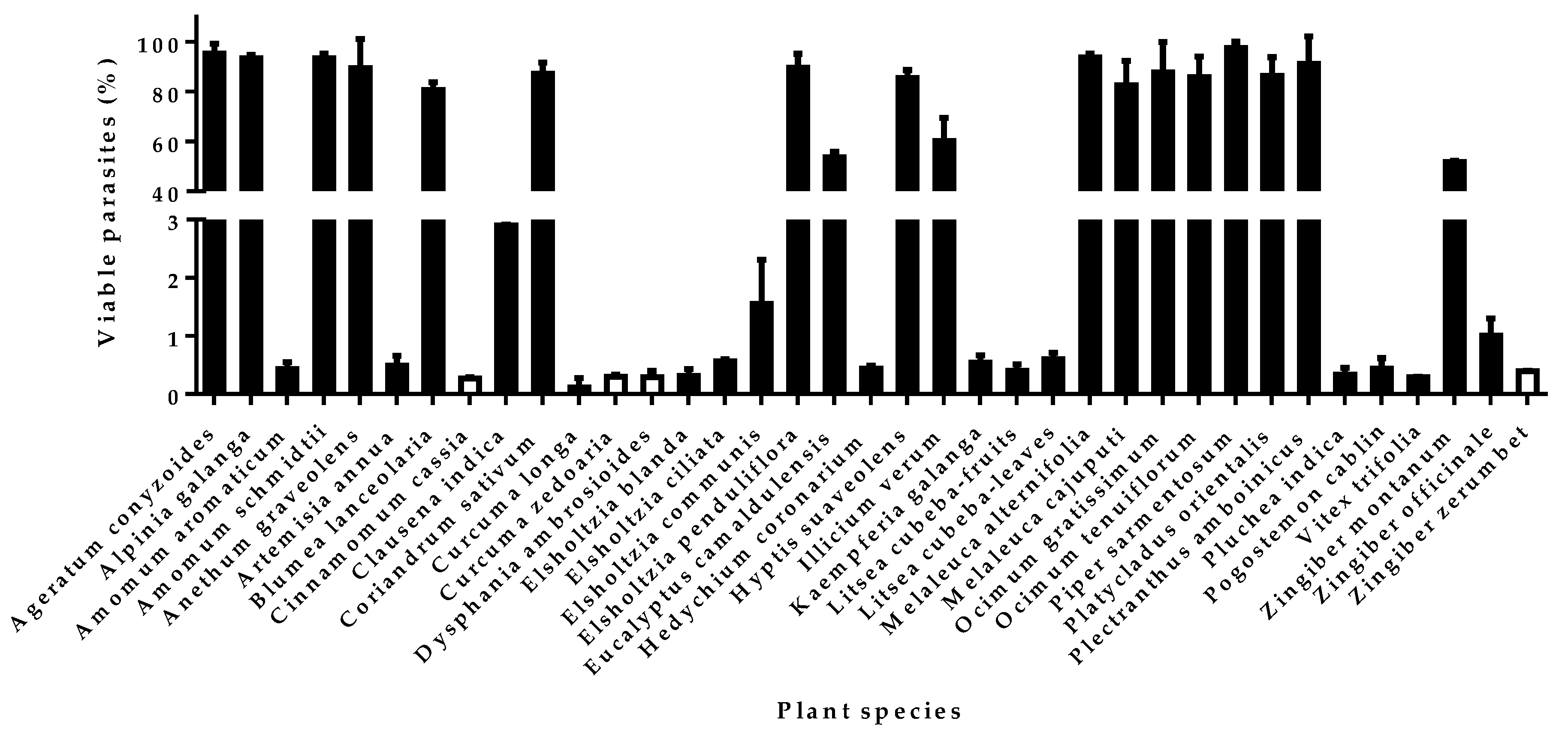
2. Results
3. Discussion
4. Materials and Methods
4.1. Chemicals and Materials
4.2. PLANTS Collection and Essential Oils Extraction
4.3. Parasites, Cells, and Media
4.4. Anti-Trypanosomal Assay
4.5. Cytotoxicity Assay
4.6. Essential Oils Analysis
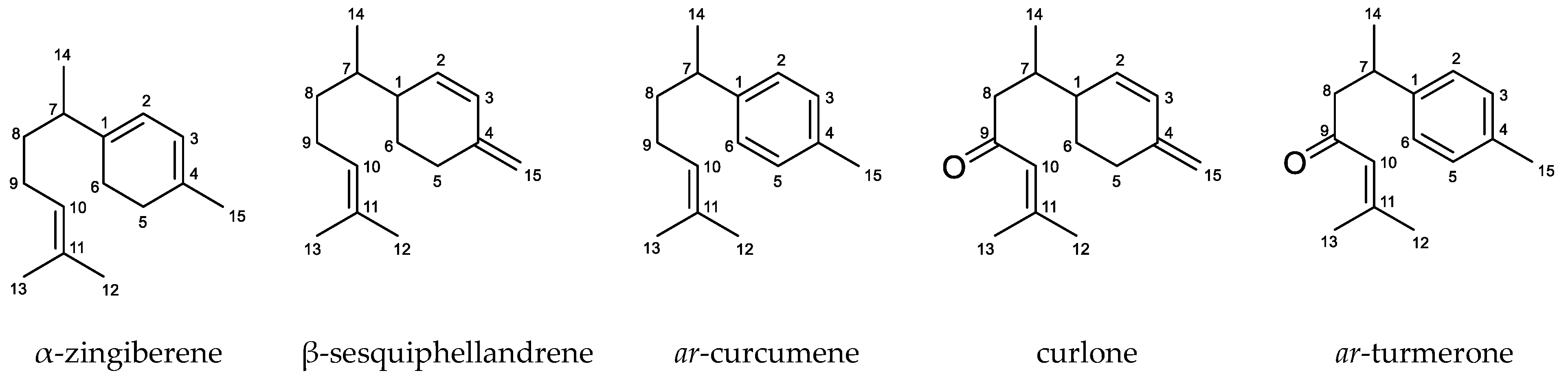
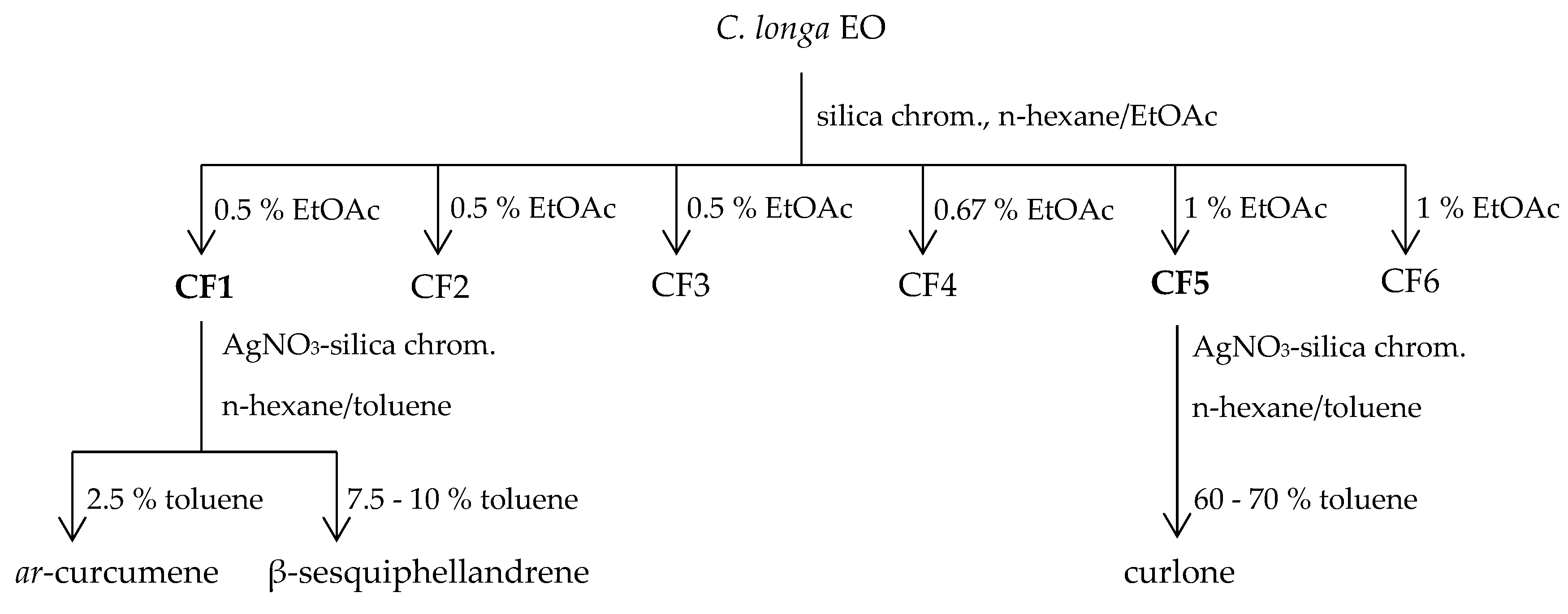
4.7. Components Isolation
5. Conclusions
Author Contributions
Funding
Acknowledgments
Conflicts of Interest
References
- Centers for Disease Control and Prevention (CDC). Parasites—Sleeping Sickness—Epidemiology & Risk Factors; CDC: Atlanta, GA, USA. Available online: https://www.cdc.gov/parasites/sleepingsickness/epi.html (accessed on 23 January 2019).
- Drugs for Neglected Diseases initiative (DNDi). Diseases & Projects—Sleeping Sickness—Fact Sheet. DNDi: Geneva, Switzerland. Available online: https://www.dndi.org/wp-content/uploads/2018/12/Factsheet2018_HAT.pdf (accessed on 23 January 2019).
- Capewell, P.; Cren-Travaillé, C.; Marchesi, F.; Johnston, P.; Clucas, C.; Benson, R.A.; Gorman, T.A.; Calvo-Alvarez, E.; Crouzols, A.; Jouvion, G.; et al. The skin is a significant but overlooked anatomical reservoir for vector-borne African trypanosomes. Elife 2016, 5, e17716. [Google Scholar] [CrossRef]
- Trindade, S.; Rijo-Ferreira, F.; Carvalho, T.; Pinto-Neves, D.; Guegan, F.; Aresta-Branco, F.; Bento, F.; Young, S.A.; Pinto, A.; Van Den Abbeele, J.; et al. Trypanosoma brucei Parasites Occupy and Functionally Adapt to the Adipose Tissue in Mice. Cell Host Microbe 2016, 19, 837–848. [Google Scholar] [CrossRef] [PubMed]
- World Health Organization (WHO). Human African Trypanosomiasis—The Disease—Symptoms, Diagnosis and Treatment; WHO: Geneva, Switzerland; Available online: http://www.who.int/trypanosomiasis_african/disease/diagnosis/en/ (accessed on 23 January 2019).
- Drugs for Neglected Diseases initiative (DNDi). Diseases & Projects—Portfolio—Fexinidazole (HAT). DNDi: Geneva, Switzerland. Available online: https://www.dndi.org/diseases-projects/portfolio/fexinidazole/ (accessed on 23 January 2019).
- Chiara Cristiano, M.; Cosco, D.; Paolino, D. Technological Aspects of Essential Oils. In Aromatherapy: Basic Mechanisms and Evidence-Based Clinical Use; Bagetta, G., Cosentino, M., Sakurada, T., Eds.; CRC Press: Boca Raton, FL, USA, 2016; pp. 152–153. ISBN 978-1-4822-4663-6. [Google Scholar]
- Heuberger, E. Effects of Essential Oils in the Central Nervous System. In Handbook of Essential Oils: Science, Technology and Applications; Can Baser, K.H., Buchbauer, G., Eds.; CRC Press: Boca Raton, FL, USA, 2010; p. 283. ISBN 978-1-4200-6315-8. [Google Scholar]
- World Health Organization (WHO). Human African Trypanosomiasis—African Trypanosomiasis—Drugs; WHO: Geneva, Switzerland; Available online: http://www.who.int/trypanosomiasis_african/drugs/en/ (accessed on 23 January 2019).
- Le, T.B.; Beaufay, C.; Bonneau, N.; Mingeot-Leclercq, M.-P.; Quetin-Leclercq, J. Anti-protozoal activity of essential oils and their constituents against Leishmania, Plasmodium and Trypanosoma. Phytochimie 2018, 1, 1–33. [Google Scholar]
- Montoro, P.; Masullo, M.; Piacente, S.; Pizza, C. Extraction, Sample Preparation, and Analytical Methods for Quality Issues of Essential Oils. In Aromatherapy: Basic Mechanisms and Evidence-Based Clinical Use; Bagetta, G., Cosentino, M., Sakurada, T., Eds.; CRC Press: Boca Raton, FL, USA, 2016; p. 153. ISBN 978-1-4822-4663-6. [Google Scholar]
- Le, T.B.; Beaufay, C.; Nghiem, D.T.; Mingeot-Leclercq, M.-P.; Quetin-Leclercq, J. In vitro anti-leishmanial activity of essential oils extracted from Vietnamese plants. Molecules 2017, 22, 1071. [Google Scholar] [CrossRef] [PubMed]
- Bero, J.; Kpoviessi, S.; Quetin-Leclercq, J. Anti-Parasitic Activity of Essential Oils and their Constituents against Plasmodium, Trypanosoma and Leishmania. In Novel Plant Bioresource: Applications in Food, Medicine and Cosmetic; Gurib-Fakim, A., Ed.; John Wiley & Sons: Oxford, UK, 2014; pp. 455–469. ISBN 978-1-118-46061-0. [Google Scholar]
- Behar, R.Z.; Davis, B.; Wang, Y.; Bahl, V.; Lin, S.; Talbot, P. Identification of toxicants in cinnamon-flavored electronic cigarette refill fluids. Toxicol. In Vitro 2014, 28, 198–208. [Google Scholar] [CrossRef] [Green Version]
- Hérent, M.F.; De Bie, V.; Tilquin, B. Determination of new retention indices for quick identification of essential oils compounds. J. Pharm. Biomed. Anal. 2007, 43, 886–892. [Google Scholar] [CrossRef] [PubMed]
- Babushok, V.I.; Linstrom, P.J.; Zenkevich, I.G. Retention Indices for Frequently Reported Compounds of Plant Essential Oils. J. Phys. Chem. Ref. Data 2011, 40, 043101. [Google Scholar] [CrossRef]
- Monzote, L.; García, M.; Pastor, J.; Gil, L.; Scull, R.; Maes, L.; Cos, P.; Gille, L. Essential oil from Chenopodium ambrosioides and main components: Activity against Leishmania, their mitochondria and other microorganisms. Exp. Parasitol. 2014, 136, 20–26. [Google Scholar] [CrossRef] [PubMed]
- Cheikh-Ali, Z.; Adiko, M.; Bouttier, S.; Bories, C.; Okpekon, T.; Poupon, E.; Champy, P. Composition, and antimicrobial and remarkable antiprotozoal activities of the essential oil of rhizomes of Aframomum sceptrum K. Schum. (Zingiberaceae). Chem. Biodivers. 2011, 8, 658–667. [Google Scholar] [CrossRef] [PubMed]
- Kpadonou Kpoviessi, B.G.H.; Kpoviessi, S.D.S.; Yayi Ladekan, E.; Gbaguidi, F.; Frédérich, M.; Moudachirou, M.; Quetin-Leclercq, J.; Accrombessi, G.C.; Bero, J. In vitro antitrypanosomal and antiplasmodial activities of crude extracts and essential oils of Ocimum gratissimum Linn from Benin and influence of vegetative stage. J. Ethnopharmacol. 2014, 155, 1417–1423. [Google Scholar] [CrossRef] [PubMed]
- Si, L.; Chen, Y.; Han, X.; Zhan, Z.; Tian, S.; Cui, Q.; Wang, Y. Chemical composition of essential oils of Litsea cubeba harvested from its distribution areas in China. Molecules 2012, 17, 7057–7066. [Google Scholar] [CrossRef] [PubMed]
- Wang, H.; Liu, Y. Chemical composition and antibacterial activity of essential oils from different parts of Litsea cubeba. Chem. Biodivers. 2010, 7, 229–235. [Google Scholar] [CrossRef] [PubMed]
- Huang, X.W.; Feng, Y.C.; Huang, Y.; Li, H.L. Potential cosmetic application of essential oil extracted from Litsea cubeba fruits from China. J. Essent. Oil Res. 2013, 25, 112–119. [Google Scholar] [CrossRef]
- Liu, T.T.; Yang, T.S. Antimicrobial impact of the components of essential oil of Litsea cubeba from Taiwan and antimicrobial activity of the oil in food systems. Int. J. Food Microbiol. 2012, 156, 68–75. [Google Scholar] [CrossRef] [PubMed]
- Chen, H.C.; Chang, W.T.; Hseu, Y.C.; Chen, H.Y.; Chuang, C.H.; Lin, C.C.; Lee, M.S.; Lin, M.K. Immunosuppressive effect of Litsea cubeba L. essential oil on dendritic cell and contact hypersensitivity responses. Int. J. Mol. Sci. 2016, 17, 1319. [Google Scholar] [CrossRef] [PubMed]
- Yang, T.S.; Liou, M.L.; Hu, T.F.; Peng, C.W.; Liu, T.T. Antimicrobial activity of the essential oil of Litsea cubeba on cariogenic bacteria. J. Essent. Oil Res. 2013, 25, 120–128. [Google Scholar] [CrossRef]
- Li, Y.; Kong, W.; Li, M.; Liu, H.; Zhao, X.; Yang, S.; Yang, M. Litsea cubeba essential oil as the potential natural fumigant: Inhibition of Aspergillus flavus and AFB1 production in licorice. Ind. Crops Prod. 2016, 80, 186–193. [Google Scholar] [CrossRef]
- Saikia, A.K.; Chetia, D.; Darrigo, M.; Smeriglio, A.; Strano, T.; Ruberto, G. Screening of fruit and leaf essential oils of Litsea cubeba Pers. from north-east India—Chemical composition and antimicrobial activity. J. Essent. Oil Res. 2013, 25, 330–338. [Google Scholar] [CrossRef]
- Kpoviessi, S.; Bero, J.; Agbani, P.; Gbaguidi, F.; Kpadonou-Kpoviessi, B.; Sinsin, B.; Accrombessi, G.; Frédérich, M.; Moudachirou, M.; Quetin-Leclercq, J. Chemical composition, cytotoxicity and in vitro antitrypanosomal and antiplasmodial activity of the essential oils of four Cymbopogon species from Benin. J. Ethnopharmacol. 2014, 151, 652–659. [Google Scholar] [CrossRef]
- Petrelli, R.; Orsomando, G.; Sorci, L.; Maggi, F.; Ranjbarian, F.; Biapa Nya, P.C.; Petrelli, D.; Vitali, L.A.; Lupidi, G.; Quassinti, L.; et al. Biological activities of the essential oil from Erigeron floribundus. Molecules 2016, 21, 1065. [Google Scholar] [CrossRef] [PubMed]
- Hoet, S.; Stévigny, C.; Hérent, M.-F.; Quetin-Leclercq, J. Antitrypanosomal Compounds from the Leaf Essential Oil of Strychnos spinosa. Planta Med. 2006, 72, 480–482. [Google Scholar] [CrossRef] [PubMed]
- Dosoky, N.S.; Setzer, W.N. Chemical composition and biological activities of Essential Oils of Curcuma Species. Nutrients 2018, 10, 1196. [Google Scholar] [CrossRef] [PubMed]
- Petrelli, R.; Ranjbarian, F.; Dall’Acqua, S.; Papa, F.; Iannarelli, R.; Ngahang Kamte, S.L.; Vittori, S.; Benelli, G.; Maggi, F.; Hofer, A.; et al. An overlooked horticultural crop, Smyrnium olusatrum, as a potential source of compounds effective against African trypanosomiasis. Parasitol. Int. 2017, 66, 146–151. [Google Scholar] [CrossRef] [PubMed]
- Yamamoto-Ribeiro, M.M.G.; Grespan, R.; Kohiyama, C.Y.; Ferreira, F.D.; Mossini, S.A.G.; Silva, E.L.; De Abreu Filho, B.A.; Mikcha, J.M.G.; Machinski Junior, M. Effect of Zingiber officinale essential oil on Fusarium verticillioides and fumonisin production. Food Chem. 2013, 141, 3147–3152. [Google Scholar] [CrossRef] [PubMed]
- Bayala, B.; Bassole, I.H.N.; Gnoula, C.; Nebie, R.; Yonli, A.; Morel, L.; Figueredo, G.; Nikiema, J.B.; Lobaccaro, J.M.A.; Simpore, J. Chemical composition, antioxidant, anti-inflammatory and anti-proliferative activities of essential oils of plants from Burkina Faso. PLoS ONE 2014, 9, e92122. [Google Scholar] [CrossRef]
- Noori, S.; Zeynali, F.; Almasi, H. Antimicrobial and antioxidant efficiency of nanoemulsion-based edible coating containing ginger (Zingiber officinale) essential oil and its effect on safety and quality attributes of chicken breast fillets. Food Control 2018, 84, 312–320. [Google Scholar] [CrossRef]
- El-Ghorab, A.H.; Nauman, M.; Anjum, F.M.; Hussain, S.; Nadeem, M. A comparative study on chemical composition and antioxidant activity of ginger (Zingiber officinale) and cumin (Cuminum cyminum). J. Agric. Food Chem. 2010, 58, 8231–8237. [Google Scholar] [CrossRef] [PubMed]
- Martins, A.P.; Salgueiro, L.; Gonçalves, M.J.; da Cunha, A.P.; Vila, R.; Cañigueral, S.; Mazzoni, V.; Tomi, F.; Casanova, J. Essential oil composition and antimicrobial activity of three Zingiberaceae from S Tomé e Príncipe. Planta Med. 2001, 67, 580–584. [Google Scholar] [CrossRef] [PubMed]
- Wohlmuth, H.; Smith, M.K.; Brooks, L.O.; Myers, S.P.; Leach, D.N. Essential oil composition of diploid and tetraploid clones of ginger (Zingiber officinale Roscoe) grown in Australia. J. Agric. Food Chem. 2006, 54, 1414–1419. [Google Scholar] [CrossRef]
- Singh, G.; Kapoor, I.P.S.; Singh, P.; de Heluani, C.S.; de Lampasona, M.P.; Catalan, C.A.N. Chemistry, antioxidant and antimicrobial investigations on essential oil and oleoresins of Zingiber officinale. Food Chem. Toxicol. 2008, 46, 3295–3302. [Google Scholar] [CrossRef] [PubMed]
- Buddhakala, N.; Talubmook, C.; Sriyotha, P.; Wray, S.; Kupittayanant, S. Inhibitory effects of ginger oil on spontaneous and PGF2α- induced contraction of rat myometrium. Planta Med. 2008, 74, 385–391. [Google Scholar] [CrossRef]
- Mulyaningsih, S.; Youns, M.; El-Readi, M.Z.; Ashour, M.L.; Nibret, E.; Sporer, F.; Herrmann, F.; Reichling, J.; Wink, M. Biological activity of the essential oil of Kadsura longipedunculata (Schisandraceae) and its major components. J. Pharm. Pharmacol. 2010, 62, 1037–1044. [Google Scholar] [CrossRef] [PubMed]
- Haddad, M.; Sauvain, M.; Deharo, E. Curcuma as a parasiticidal agent: A review. Planta Med. 2011, 77, 672–678. [Google Scholar] [CrossRef]
- Sun, Y.N.; No, J.H.; Lee, G.Y.; Li, W.; Yang, S.Y.; Yang, G.; Schmidt, T.J.; Kang, J.S.; Kim, Y.H. Phenolic constituents of medicinal plants with activity against Trypanosoma brucei. Molecules 2016, 21, 480. [Google Scholar] [CrossRef]
- Lee, Y. Cytotoxicity Evaluation of Essential Oil and its Component from Zingiber officinale Roscoe. Toxicol. Res. 2016, 32, 225–230. [Google Scholar] [CrossRef] [PubMed]
- Tyagi, A.K.; Prasad, S.; Yuan, W.; Li, S.; Aggarwal, B.B. Identification of a novel compound (β-sesquiphellandrene) from turmeric (Curcuma longa) with anticancer potential: Comparison with curcumin. Investig. New Drugs 2015, 33, 1175–1186. [Google Scholar] [CrossRef] [PubMed]
- Lee, Y. Activation of apoptotic protein in U937 cells by a component of turmeric oil. BMB Rep. 2009, 42, 96–100. [Google Scholar] [CrossRef]
- Russo, R.; Corasaniti, M.T.; Bagetta, G.; Morrone, L.A. Essential Oils Exploited in Cytotoxicity Studies for Translation into Safer and More Effective Cancer Therapeutics. In Aromatherapy: Basic Mechanisms and Evidence-Based Clinical Use; Bagetta, G., Cosentino, M., Sakurada, T., Eds.; CRC Press: Boca Raton, FL, USA, 2016; p. 170. ISBN 978-1-4822-4663-6. [Google Scholar]
- Andrade-Ochoa, S.; Sánchez-Aldana, D.; Chacón-Vargas, K.F.; Rivera-Chavira, B.E.; Sánchez-Torres, L.E.; Camacho, A.D.; Nogueda-Torres, B.; Nevárez-Moorillón, G.V. Oviposition Deterrent and Larvicidal and Pupaecidal Activity of Seven Essential Oils and their Major Components against Culex quinquefasciatus Say (Diptera: Culicidae): Synergism-antagonism Effects. Insects 2018, 9, 25. [Google Scholar] [CrossRef] [PubMed]
- Nibret, E.; Wink, M. Trypanocidal and antileukaemic effects of the essential oils of Hagenia abyssinica, Leonotis ocymifolia, Moringa stenopetala, and their main individual constituents. Phytomedicine 2010, 17, 911–920. [Google Scholar] [CrossRef] [PubMed]
- Ngahang Kamte, S.L.; Ranjbarian, F.; Cianfaglione, K.; Sut, S.; Dall’Acqua, S.; Bruno, M.; Afshar, F.H.; Iannarelli, R.; Benelli, G.; Cappellacci, L.; et al. Identification of highly effective antitrypanosomal compounds in essential oils from the Apiaceae family. Ecotoxicol. Environ. Saf. 2018, 156, 154–165. [Google Scholar] [CrossRef] [PubMed]
- Sliwowski, J.K.; Caspi, E. An improved method of preparation of plates and sheets for thin-layer argentation chromatography. J. Steroid Biochem. 1977, 8, 42–49. [Google Scholar] [CrossRef]
- Sykes, M.L.; Avery, V.M. Development of an Alamar Blue™ Viability Assay in 384-Well Format for High Throughput Whole Cell Screening of Trypanosoma brucei brucei Bloodstream Form Strain 427. Am. J. Trop. Med. Hyg. 2009, 81, 665–674. [Google Scholar] [CrossRef] [PubMed]
- Ioset, L.-R.; Brun, R.; Wenzler, T.; Kaiser, M.; Yardley, V. Drug Screening for Kinetoplastids Diseases, a Training Manual for Screening in Neglected Diseases; The Pan-Asian Screening Network; DNDi: Geneva, Switzerland, 2009; pp. 20–21. [Google Scholar]
- Hirumi, H.; Himuri, K. Axenic culture of African trypanosome bloodstream forms. Parasitol. Today 1994, 10, 80–84. [Google Scholar] [CrossRef]
- Koch, A.; Basar, S.; Richter, R. TLC of Mono- and Sesquiterpenes. In Thin Layer Chromatography in Phytochemistry; Waksmundzka-Hajnos, M., Sherma, J., Kowalska, T., Eds.; CRC Press: Boca Raton, FL, USA, 2008; pp. 459–461. ISBN 978-1-4200-4677-9. [Google Scholar]
- Williams, C.M.; Mander, L.N. Chromatography with silver nitrate. Tetrahedron 2001, 57, 425–447. [Google Scholar] [CrossRef]
- Morris, L.J. Separations of lipids by silver ion chromatography. J. Lipid Res. 1966, 7, 717–732. [Google Scholar] [PubMed]
- Denyer, C.V.; Jackson, P.; Loakes, D.M.; Ellis, M.R.; Young, D.A.B. Isolation of antirhinoviral sesquiterpenes from ginger (Zingiber officinale). J. Nat. Prod. 1994, 57, 658–662. [Google Scholar] [CrossRef] [PubMed]
- Wang, Y.; Harrison, L.J.; Tan, B.C. Terpenoids from the liverwort Chandonanthus hirtellus. Tetrahedron 2009, 65, 4035–4043. [Google Scholar] [CrossRef]
- Fujiwaraj, M.; Yagi, N.; Miyazawa, M. Acetylcholinesterase inhibitory activity of volatile oil from Peltophorum dasyrachis Kurz ex Bakar (Yellow Batai) and bisabolane-type sesquiterpenoids. J. Agric. Food Chem. 2010, 58, 2824–2829. [Google Scholar] [CrossRef] [PubMed]
- Ragasa, C.Y.; Laguardia, M.A.; Rideout, J.A. Antimicrobial sesquiterpenoids and diarylheptanoid from Curcuma domestica. ACGC Chem. Res. Commun. 2005, 18, 21–24. [Google Scholar]
Sample Availability: Samples of essential oils are available from the authors. |


| Plant Species (Studied Parts) | Anti-Trypanosomal Activity (IC50 nL/mL) | Cytotoxicity (IC50 nL/mL) | |||
|---|---|---|---|---|---|
| WI38 | SI | J774 | SI | ||
| Amomum aromaticum (fruits) | 8.75 ± 1.25 | 47.31 ± 0.30 | 5.4 | 22.68 ± 3.22 | 2.6 |
| Artemisia annua (leaves) | 8.99 ± 1.18 | 45.64 ± 1.02 | 5.1 | 38.16 ± 0.21 | 4.2 |
| Cinnamomum cassia (stem barks) | 1.77 ± 0.15 | 11.97 ± 0.93 | 6.8 | 8.97 ± 0.66 | 5.1 |
| Clausena indica (leaves) | 13.22 ± 4.54 | >50.00 | >3.8 | >50.00 | >3.8 |
| Curcuma longa (rhizomes) | 3.17 ± 0.72 | 46.00 ± 0.33 | 14.5 | 44.11 ± 3.13 | 13.9 |
| Curcuma zedoaria (rhizomes) | 2.51 ± 1.08 | 46.64 ± 0.95 | 18.6 | 26.81 ± 1.59 | 10.7 |
| Dysphania ambrosioides (aerial parts) | 2.86 ± 0.32 | >50.00 | >17.5 | 12.29 ± 2.92 | 4.3 |
| Elsholtzia blanda (leaves) | 8.23 ± 1.03 | >50.00 | >6.1 | >50.00 | >6.1 |
| Elsholtzia ciliata (leaves) | 4.26 ± 0.86 | 48.46 ± 0.12 | 11.4 | 13.21 ± 1.48 | 3.1 |
| Elsholtzia communis (leaves) | 18.39 ± 3.32 | >50.00 | >2.7 | 40.68 ± 3.44 | 2.2 |
| Hedychium coronarium (rhizomes) | 9.73 ± 1.43 | >50.00 | >5.1 | 30.00 ± 4.06 | 3.1 |
| Kaempferia galangal (rhizomes) | 15.78 ± 3.29 | >50.00 | >3.2 | >50.00 | >3.2 |
| Litsea cubeba (fruits) | 2.67 ± 1.12 | >50.00 | >18.7 | >50.00 | >18.7 |
| Litsea cubeba (leaves) | 16.47 ± 1.24 | >50.00 | >3.0 | >50.00 | >3.0 |
| Pluchea indica (leaves) | 21.29 ± 1.38 | 27.47 ± 1.49 | 1.3 | 25.05 ± 5.56 | 1.2 |
| Pogostemon cablin (leaves) | 4.07 ± 0.98 | 27.17 ± 3.62 | 6.7 | 28.40 ± 1.81 | 7.0 |
| Vitex trifolia (leaves) | 3.24 ± 0.79 | 31.12 ± 2.83 | 9.6 | 26.64 ± 0.76 | 8.2 |
| Zingiber officinale (rhizomes) | 3.10 ± 0.08 | >50.00 | >16.1 | 37.52 ± 0.05 | 12.1 |
| Zingiber zerumbet (rhizomes) | 6.23 ± 0.73 | 3.65 ± 0.34 | 0.6 | 2.78 ± 0.57 | 0.5 |
| Suramin | 21.53 ± 2.62a | ||||
| Camptothecin | 34.99 ± 9.63 a | 7.32 ± 1.29 a | |||
| No. | Compounds | RI | Relative Percentage (%) | Identification | |||
|---|---|---|---|---|---|---|---|
| L. cubeba | C. zedoaria | Z. officinale | C. longa | ||||
| 1 | α-Pinene m | 536 | 0.74 | 0.11 | 2.29 | - | MS, Co-GC, Ref. |
| 2 | α-Thujene m | 540 | 0.18 | - | - | - | MS, Ref. |
| 3 | Camphene m | 577 | - | 0.26 | 6.94 | - | MS, Ref. |
| 4 | β-Pinene m | 621 | 0.86 | 0.77 | 0.16 | 0.09 | MS, Co-GC, Ref. |
| 5 | Sabinene m | 635 | 0.83 | - | 0.19 | - | MS, Co-GC, Ref. |
| 6 | 3-Carene m | 665 | 0.36 | - | - | - | MS |
| 7 | α-Phellandrene m | 679 | - | - | 0.70 | 0.08 | MS, Co- |
| 8 | Myrcene m | 681 | 1.25 | - | 1.10 | t | MS, Co-GC, Ref. |
| 9 | α-Terpinene m | 697 | 0.51 | - | - | t | MS, Co-GC, Ref. |
| 10 | Limonene m | 714 | 8.72 | 0.18 | 2.06 | 0.19 | MS, Co-GC, Ref. |
| 11 | β-Phellandrene m | 727 | 0.16 | - | 14.78 | - | MS |
| 12 | Eucalyptol m | 727 | 1.37 | 1.61 | 1.79 | 3.15 | MS, Co-GC, Ref. |
| 13 | γ-Terpinene m | 761 | 0.52 | - | t | t | MS, Co-GC, Ref. |
| 14 | p-Cymeme m | 783 | 0.10 | t | t | t | MS, Co-GC, Ref. |
| 15 | Terpinolene m | 798 | 0.31 | - | 0.26 | 1.70 | MS, Co-GC, Ref. |
| 16 | 2-Heptanol | 844 | - | 0.14 | 0.15 | t | MS |
| 17 | 5-Hepten-2-one, 6-methyl- | 854 | 0.35 | - | t | - | MS, Co-GC |
| 18 | 5-Heptenal, 2,6-dimethyl | 867 | 0.68 | - | - | - | MS |
| 19 | 2-Nonanone | 903 | - | 0.43 | 0.15 | t | MS |
| 20 | (E)-2-Octenal | 940 | - | - | t | - | MS |
| 21 | 2-Octanol | 941 | - | t | - | - | MS |
| 22 | p-Cymenene m | 946 | - | - | - | t | MS |
| 23 | 1-Octen-3-ol | 969 | - | t | - | - | MS, Ref. |
| 24 | δ-Elemene s | 980 | - | 0.30 | t | t | MS |
| 25 | Cyclosativene s | 986 | - | - | t | - | MS |
| 26 | Citronellal m | 993 | 43.10 | - | 0.30 | 0.14 | MS, Co-GC, Ref. |
| 27 | α-Copaene s | 999 | - | - | 0.31 | - | MS |
| 28 | Decanone | 1006 | - | t | - | - | MS |
| 29 | Camphor m | 1020 | - | 4.18 | t | - | MS, Co-GC, Ref. |
| 30 | 2-Nonanol | 1038 | - | 2.16 | 0.19 | 0.21 | MS |
| 31 | Linalool m | 1063 | 5.60 | 0.22 | 0.48 | t | MS, Co-GC, Ref. |
| 32 | cis-α-Bergamotene s | 1065 | - | - | t | 0.27 | MS |
| 33 | Pulegol m | 1072 | 6.52 | - | - | - | MS |
| 34 | Isopulegol m | 1082 | 11.10 | - | - | - | MS, Ref. |
| 35 | trans-α-Bergamotene s | 1091 | - | - | - | 0.12 | MS, Ref. |
| 36 | β-Elemene s | 1096 | - | 4.85 | 0.34 | 0.22 | MS |
| 37 | β-Caryophyllene s | 1100 | - | 3.79 | 0.43 | t | MS, Co-GC, Ref. |
| 38 | 2-Undecanone | 1106 | - | - | 0.39 | t | MS |
| 39 | Terpinene-4-ol m | 1109 | 2.58 | 0.31 | 0.22 | 0.13 | MS, Co-GC, Ref. |
| 40 | γ-Elemene s | 1142 | - | 0.32 | - | 0.09 | MS |
| 41 | α-Himachalene s | 1153 | - | - | - | t | MS |
| 42 | γ-Gurjunene s | 1160 | - | - | - | t | MS |
| 43 | α-Humulene s | 1168 | - | 1.28 | - | t | MS, Co-GC, Ref. |
| 44 | (E)-β-Farnesene s | 1174 | - | - | 0.26 | 0.61 | MS |
| 45 | Neral m | 1186 | - | - | 3.16 | - | MS, Co-GC |
| 46 | α-Terpineol m | 1203 | 0.62 | 0.23 | 1.78 | 0.48 | MS, Co-GC, Ref. |
| 47 | Borneol m | 1208 | - | - | 1.35 | - | MS, Co-GC, Ref. |
| 48 | Germacrene D s | 1206 | - | 1.99 | - | t | MS, Ref. |
| 49 | α-Muurolene s | 1217 | - | - | 1.44 | - | MS |
| 50 | β-Selinene s | 1218 | - | 1.76 | - | - | MS |
| 51 | β-Chamigrene s | 1223 | - | 1.47 | - | - | MS |
| 52 | α-Zingiberene s | 1236 | - | - | 27.71 | 25.38 | MS, Co-GC |
| 53 | β-Bisabolene s | 1238 | - | - | 7.27 | 3.38 | MS, Ref. |
| 54 | α-Cubebene s | 1248 | - | - | 0.23 | - | MS |
| 55 | (E,E)-α-Farnesene s | 1259 | - | - | 3.71 | 0.36 | MS |
| 56 | Citronellol m | 1274 | 5.17 | - | - | - | MS, Co-GC, Ref. |
| 57 | β-Sesquiphellandrene s | 1279 | - | - | 8.08 | 18.27 | MS |
| 58 | ar-Curcumene s | 1280 | - | - | 2.71 | 5.22 | MS |
| 59 | ζ-Elemene s | 1323 | - | 2.47 | 0.55 | - | MS |
| 60 | Geraniol m | 1351 | - | - | 0.45 | 0.25 | MS, Co-GC, Ref. |
| 61 | Curzerene s | 1366 | - | 4.87 | - | - | MS |
| 62 | Epiglobulol s | 1468 | - | 0.57 | - | - | MS |
| 63 | (E)-Nerolidol s | 1536 | - | - | 0.32 | - | MS, Co-GC, Ref. |
| 64 | Elemol s | 1569 | - | - | 0.18 | - | MS |
| 65 | Ledene oxide s | 1583 | - | 0.25 | - | - | MS |
| 66 | Bisabolone s | 1595 | - | - | - | 1.08 | MS |
| 67 | Spathulenol s | 1607 | 0.21 | - | - | - | MS |
| 68 | ar-Turmerol s | 1658 | - | - | - | 1.60 | MS |
| 69 | α-Turmerone s | 1667 | - | - | - | 10.28 | MS |
| 70 | Bisabolol s | 1670 | - | - | - | 0.87 | MS |
| 71 | 8,9-Dehydro-9-formyl cycloisolongifolene s | 1703 | - | 29.31 | - | - | MS |
| 72 | Germacrone s | 1710 | - | 8.95 | - | 3.34 | MS |
| 73 | Curlone s | 1723 | - | - | - | 5.15 | MS |
| 74 | ar-Turmerone s | 1739 | - | - | - | 9.93 | MS, Co-GC |
| 75 | Curdione s | 1792 | - | 13.52 | - | - | MS |
| 76 | Farnesol s | 1838 | - | - | 0.20 | - | MS, Ref. |
| Total identified | 91.84 | 86.30 | 92.63 | 92.59 | |||
| Compounds | Anti-Trypanosomal Activity (IC50 µg/mL) | Cytotoxicity (IC50 µg/mL) | |||
|---|---|---|---|---|---|
| WI38 | SI | J774 | SI | ||
| α-Zingiberene | 6.91 ± 2.60 | 28.50 ± 1.43 | 4.1 | 29.64 ± 2.54 | 4.3 |
| β-Sesquiphellandrene | 9.89 ± 1.18 | 19.11 ± 1.58 | 1.9 | 21.02 ± 2.72 | 2.1 |
| ar-Curcumene | 13.38 ± 2.46 | 23.15 ± 1.36 | 1.7 | 24.03 ± 2.64 | 1.8 |
| Curlone | 1.38 ± 0.52 | 43.64 ± 2.45 | 31.7 | 25.06 ± 3.47 | 18.2 |
| ar-Turmerone | 28.83 ± 3.93 | 43.39 ± 3.89 | 1.5 | 44.62 ± 1.41 | 1.6 |
| Suramin | 21.53 ± 2.62 a | ||||
| Camptothecin | 34.99 ± 9.63 a | 7.32 ± 1.29 a | |||
© 2019 by the authors. Licensee MDPI, Basel, Switzerland. This article is an open access article distributed under the terms and conditions of the Creative Commons Attribution (CC BY) license (http://creativecommons.org/licenses/by/4.0/).
Share and Cite
Le, T.B.; Beaufay, C.; Nghiem, D.T.; Pham, T.A.; Mingeot-Leclercq, M.-P.; Quetin-Leclercq, J. Evaluation of the Anti-Trypanosomal Activity of Vietnamese Essential Oils, with Emphasis on Curcuma longa L. and Its Components. Molecules 2019, 24, 1158. https://doi.org/10.3390/molecules24061158
Le TB, Beaufay C, Nghiem DT, Pham TA, Mingeot-Leclercq M-P, Quetin-Leclercq J. Evaluation of the Anti-Trypanosomal Activity of Vietnamese Essential Oils, with Emphasis on Curcuma longa L. and Its Components. Molecules. 2019; 24(6):1158. https://doi.org/10.3390/molecules24061158
Chicago/Turabian StyleLe, Thanh Binh, Claire Beaufay, Duc Trong Nghiem, Tuan Anh Pham, Marie-Paule Mingeot-Leclercq, and Joëlle Quetin-Leclercq. 2019. "Evaluation of the Anti-Trypanosomal Activity of Vietnamese Essential Oils, with Emphasis on Curcuma longa L. and Its Components" Molecules 24, no. 6: 1158. https://doi.org/10.3390/molecules24061158