Non-Targeted Metabolomic Analysis of Methanolic Extracts of Wild-Simulated and Field-Grown American Ginseng


Abstract
:1. Introduction
2. Results
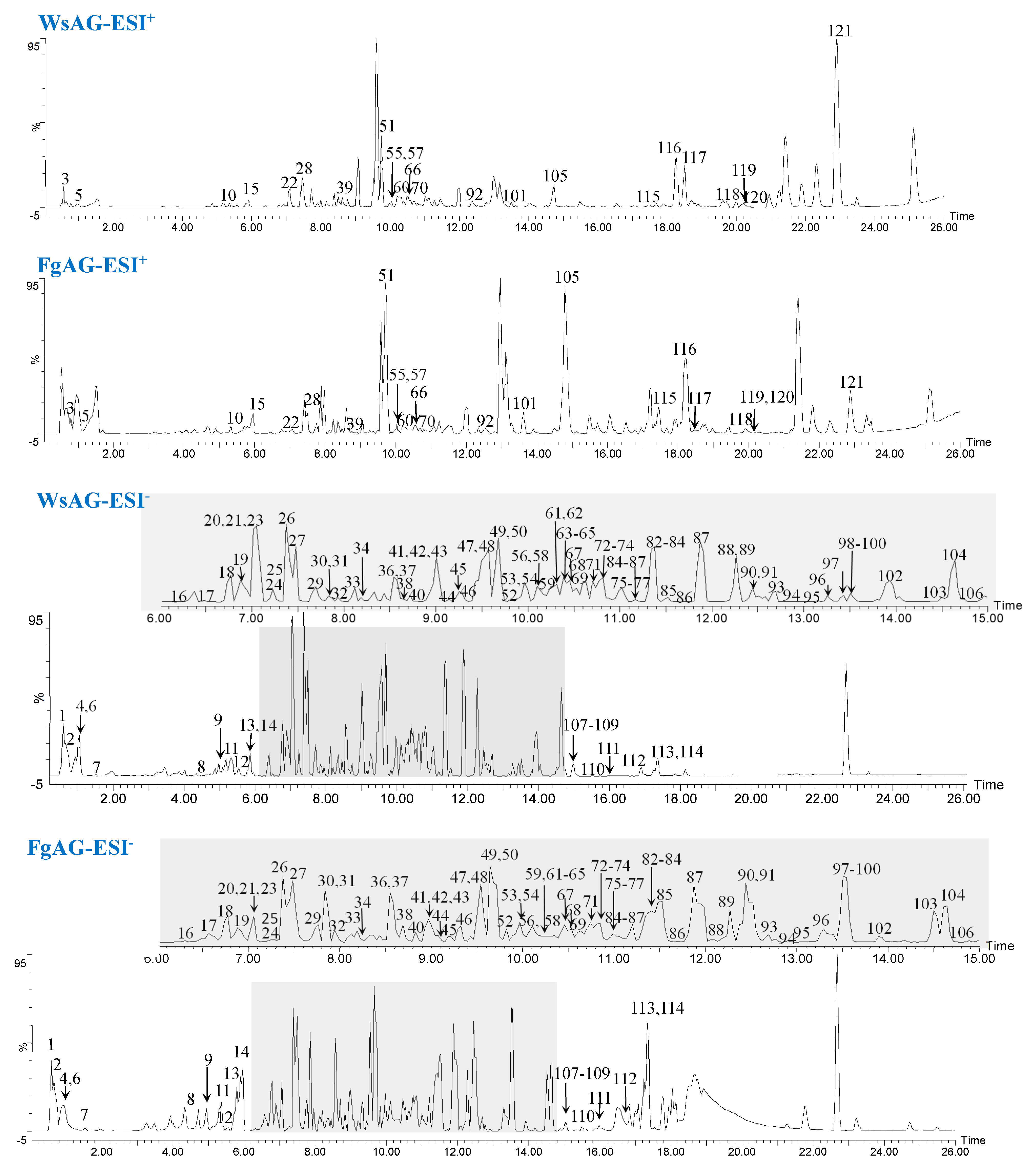
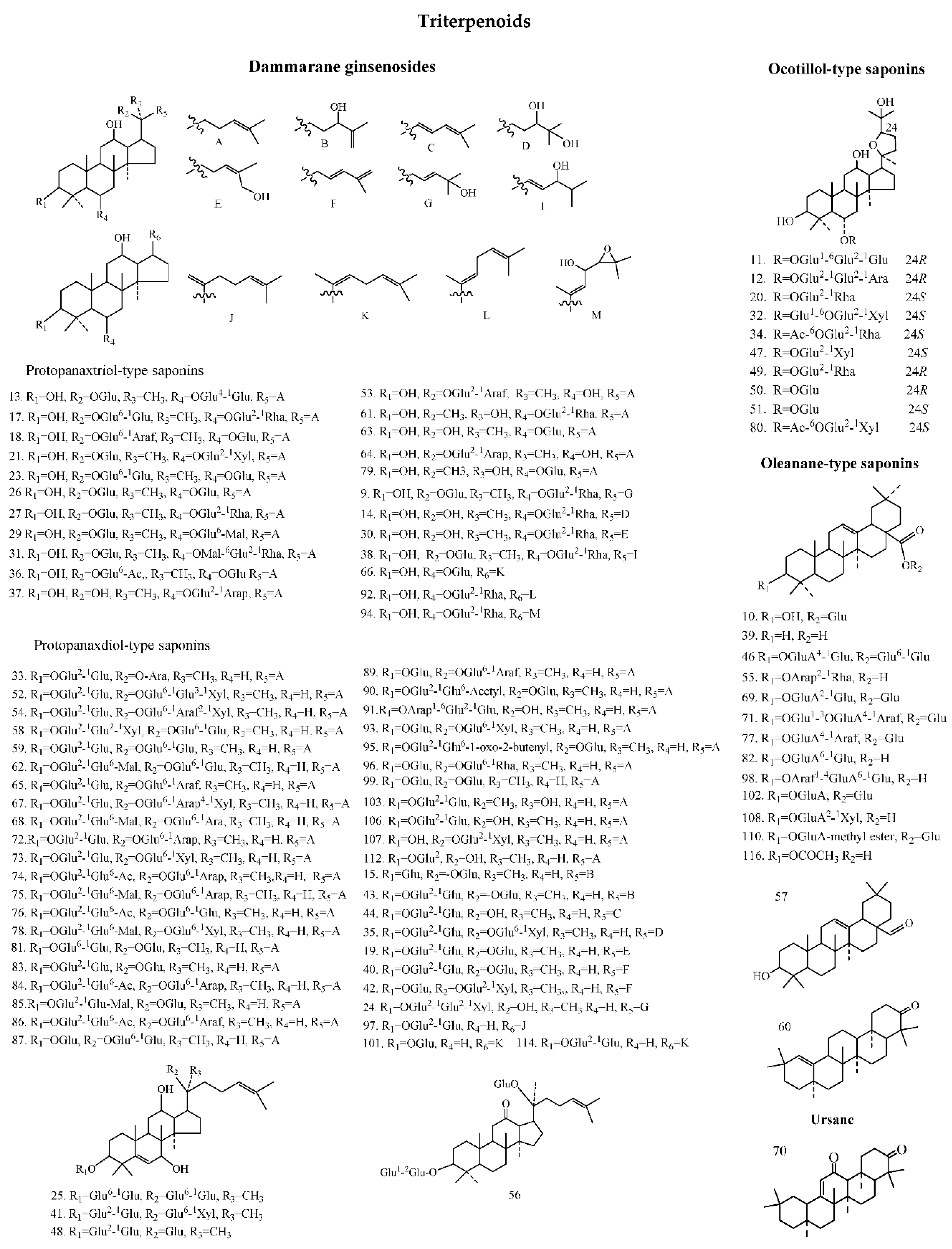
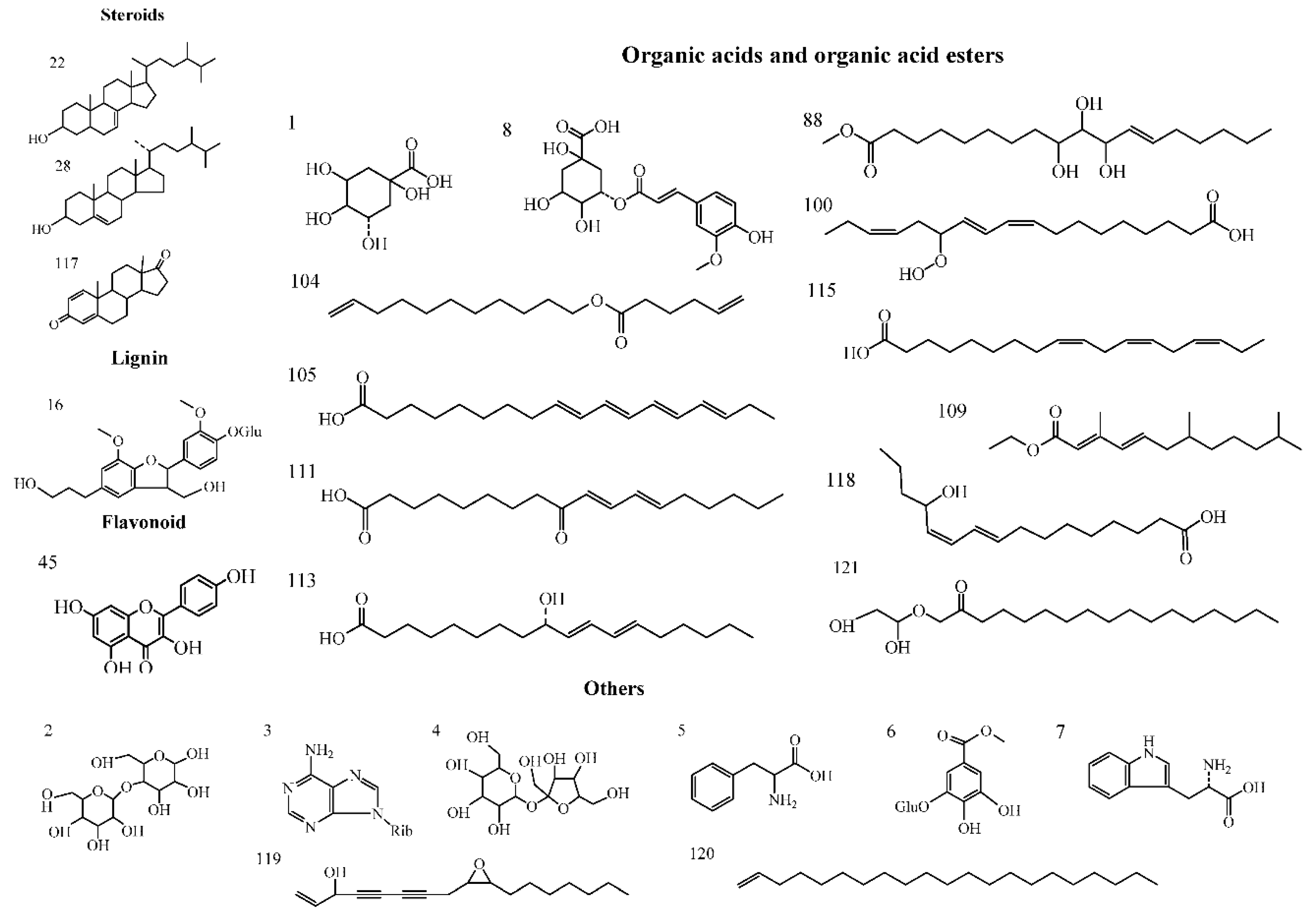
2.1. Identification of Components from FgAG and WsAG Based on the UNIFI Platform
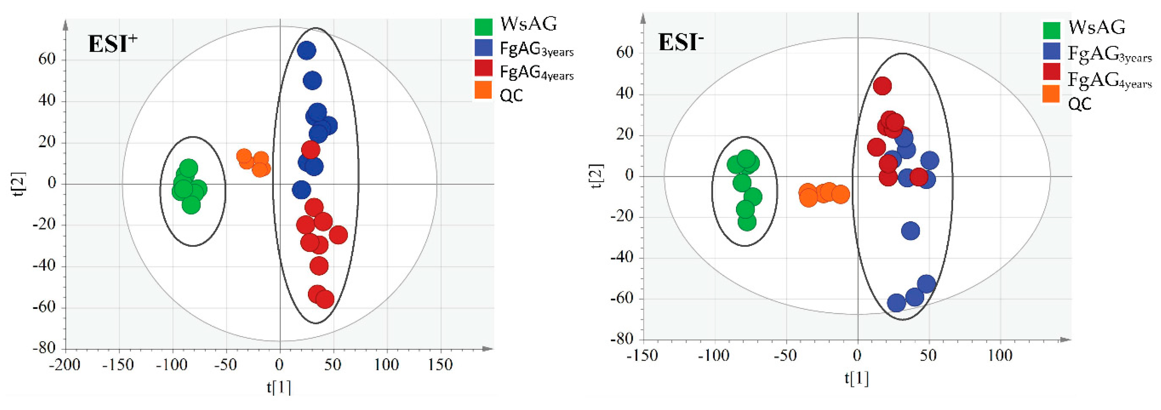
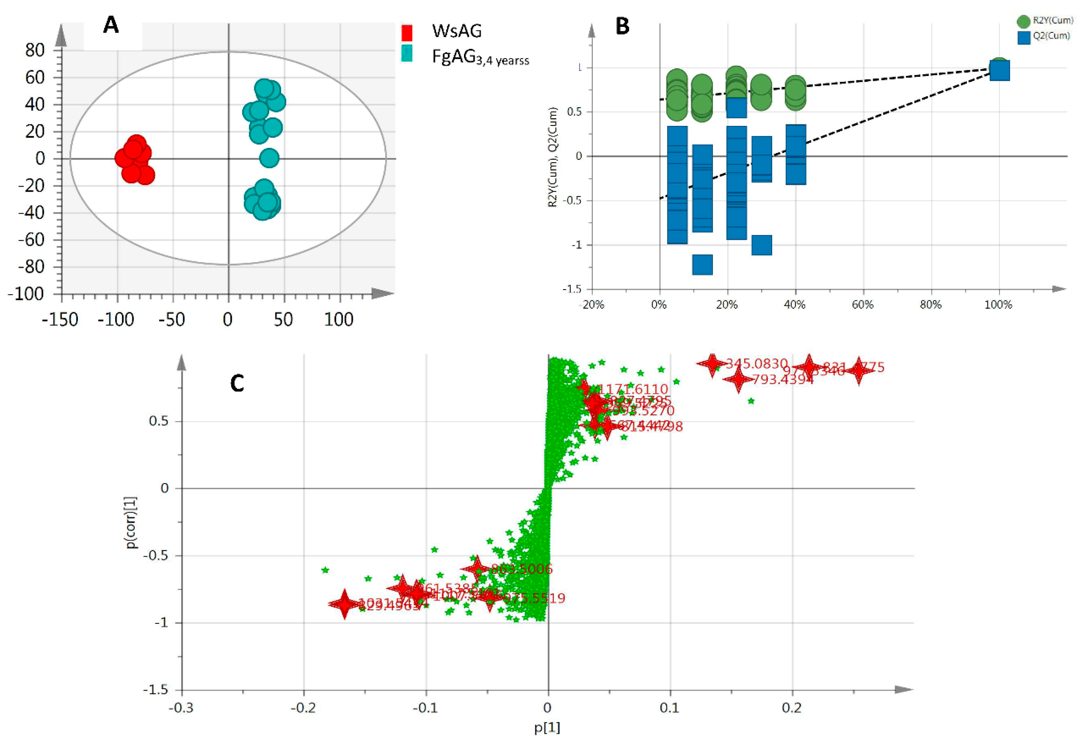
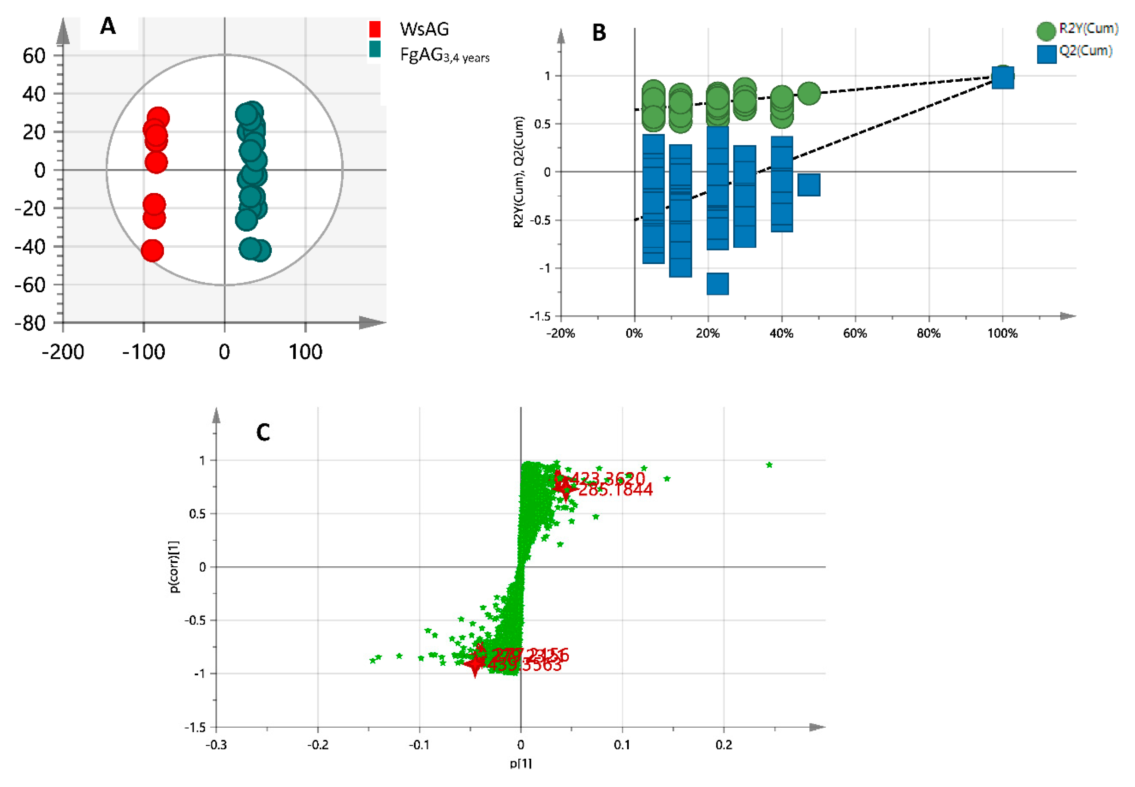
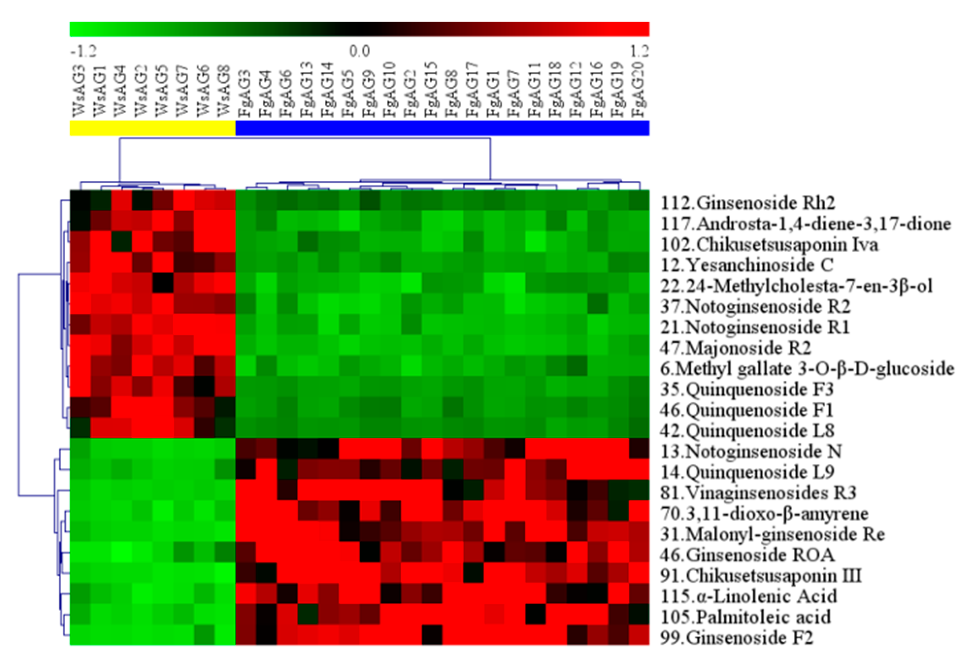
2.2. Biomarker Discovery for FgAG and WsAG
3. Discussion
4. Materials and Methods
4.1. Materials and Reagents
4.2. Sample Preparation and Extraction
4.3. UPLC-QTOF-MS
4.4. Chemical Information Database for the Components of FgAG and WsAG
4.5. The Screening Analysis by the UNIFI Platform
4.6. The Metabolomics Analysis
5. Conclusions
Author Contributions
Funding
Conflicts of Interest
References
- Ko, S.K.; Cho, O.S.; Bae, H.M.; Sohn, U.D.; Im, B.O.; Cho, S.H.; Chung, S.H.; Lee, B.Y. Change of Ginsenoside Composition of Various American Ginseng Roots. J. Korean Soc. Appl. Biol. Chem. 2009, 52, 198–201. [Google Scholar] [CrossRef]
- Lee, D.P. Production Procedures and Economics of the American Ginseng. J. Ginseng Res. 2006, 30, 172–180. [Google Scholar] [Green Version]
- Chen, Y.J.; Zhao, Z.Z.; Chen, H.B.; Brand, E.; Yi, T.; Qin, M.J.; Liang, Z.T. Determination of ginsenosides in Asian and American ginsengs by liquid chromatography-quadrupole/time-of-flight MS: Assessing variations based on morphological characteristics. J. Ginseng Res. 2017, 41, 10–22. [Google Scholar] [CrossRef] [PubMed]
- Nadeau, I.; Simard, R.R.; Olivier, A. The impact of lime and organic fertilization on the growth of wild-simulated American ginseng. Can. J. Plant Sci. 2003, 83, 603–609. [Google Scholar] [CrossRef] [Green Version]
- Lim, W.; Mudge, K.W.; Vermeylen, F. Effects of population, age, and cultivation methods on ginsenoside content of wild American ginseng (Panax quinquefolium). J. Agric. Food Chem. 2005, 53, 8498–8505. [Google Scholar] [CrossRef]
- Charest, P.; Dorais, M.; Gauthier, L.; Khanizadeh, S. The influence of soil preparation, seedling rates and organic mulch on the production of woods-cultivated ginseng. Acta Hortic. 2000, 523, 87–96. [Google Scholar] [CrossRef]
- US Fish and Wildlife Service. American Ginseng. Available online: https://www.fws.gov/international/permits/by-species/american-ginseng.html (accessed on 7 January 2019).
- US Fish and Wildlife Service. American Ginseng Production in Woodlots. Available online: http://digitalcommons.unl.edu/agroforestnotes/ (accessed on 6 January 2019).
- Schlag, E.M.; Mcintosh, M.S. RAPD-based assessment of genetic relationships among and within American ginseng (Panax quinquefolius L.) populations and their implications for a future conservation strategy. Genet. Resour. Crop Evol. 2012, 59, 1553–1568. [Google Scholar] [CrossRef]
- Burkhart, E.P. American ginseng (Panax quinquefolius L.) floristic associations in Pennsylvania: Guidance for identifying calcium-rich forest farming sites. Agrofor. Syst. 2013, 87, 1157–1172. [Google Scholar] [CrossRef]
- Zhu, W.; Han, B.; Sun, Y.; Wang, Z.Y.; Yang, X.H. Immunoregulatory effects of a glucogalactan from the root of Panax quinquefolium L. Carbohydr. Polym. 2012, 87, 2725–2729. [Google Scholar] [CrossRef]
- Duda, R.B.; Zhong, Y.; Navas, V.; Li, M.Z.; Toy, B.R.; Alavarez, J.G. American ginseng and breast cancer therapeutic agents synergistically inhibit MCF-7 breast cancer cell growth. J. Surg. Oncol. 2015, 72, 230–239. [Google Scholar] [CrossRef]
- Barton, D.L.; Soori, G.S.; Bauer, B.A.; Sloan, J.A.; Johnson, P.A.; Figueras, C.; Duane, S.; Mattar, B.; Liu, H.; Atherton, P.J.; et al. Pilot study of Panax quinquefolius (American ginseng) to improve cancer-related fatigue: A randomized, double-blind, dose-finding evaluation: NCCTG trial N03CA. Support. Care Cancer 2010, 18, 179–187. [Google Scholar] [CrossRef]
- Assinewe, V.A.; Baum, B.R.; Gagnon, D.; Arnason, J.T. Phytochemistry of wild populations of Panax quinquefolius L. (North American ginseng). J. Agric. Food Chem. 2003, 51, 4549–4553. [Google Scholar] [CrossRef]
- Vuksan, V.; Sievenpiper, J.L.; Wong, J.; Xu, Z.; Beljan-Zdravkovic, U.; Arnason, J.T.; Assinewe, V.; Stavro, M.P.; Jenkins, A.L.; Leiter, L.A.; et al. American ginseng (Panax quinquefolius L.) attenuates postprandial glycemia in a time-dependent but not dose-dependent manner in healthy individuals. Am. J. Clin. Nutr. 2001, 73, 753–758. [Google Scholar] [CrossRef]
- Oshima, Y.; Sato, K.; Hikino, H. Isolation and hypoglycemic activity of quinquefolans A, B, and C, glycans of Panax quinquefolium roots. J. Nat. Prod. 1987, 50, 188–190. [Google Scholar] [CrossRef]
- Vuksan, V.; Sievenpiper, J.L.; Koo, V.Y.; Francis, T.; Beljan-Zdravkovic, U.; Xu, Z.; Vidgen, E. American ginseng (Panax quinquefolius L) reduces postprandial glycemia in nondiabetic subjects and subjects with type 2 diabetes mellitus. Arch. Intern. Med. 2000, 160, 1009–1013. [Google Scholar] [CrossRef]
- Kitts, D.D.; Wijewickreme, A.N.; Hu, C. Antioxidant properties of a North American ginseng extract. Mol. Cell. Biochem. 2000, 203, 1–10. [Google Scholar] [CrossRef]
- Li, Z.; Guo, Y.Y.; Wu, C.F.; Li, X.; Wang, J.H. Protective Effects of Pseudoginsenoside-F11 on Scopolamine-induced Memory Impairment in Mice and Rats. J. Pharm. Spharmacol. 2010, 51, 435–440. [Google Scholar] [CrossRef]
- Robbins, C.S. Comparative Analysis of Management Regimes and Medicinal Plant Trade Monitoring Mechanisms for American Ginseng and Goldenseal. Conserv. Biol. 2000, 14, 1422–1434. [Google Scholar] [CrossRef]
- Grubbs, H.J.; Case, M.A. Allozyme variation in American ginseng (Panax quinquefolius L.): Variation, breeding system, and implications for current conservation practice. Conserv. Genet. 2004, 5, 13–523. [Google Scholar] [CrossRef]
- Crusesanders, J.M.; Hamrick, J.L. Genetic diversity in harvested and protected populations of wild American ginseng, Panax quinquefolius L. (Araliaceae). Am. J. Bot. 2004, 91, 540–548. [Google Scholar] [CrossRef]
- Voort, M.E.V.D.; Mcgraw, J.B. Effects of harvester behavior on population growth rate affects sustainability of ginseng trade. Biol. Conserv. 2006, 130, 505–516. [Google Scholar] [CrossRef]
- Beyfuss, B. New York State Wild Simulated American Ginseng has been Selling for well over $1000 per Pound. Available online: http://www.ginsenggeek.org/new-york-state-wild-simulated-american-ginseng-Has-been-selling-for-well-over-1000-per-pound/ (accessed on 18 January 2019).
- Anderson, R.C.; Anderson, M.R.; Houseman, G. Wild American Ginseng. Native Plants J. 2002, 3, 93–105. [Google Scholar]
- Predy, G.N.; Goel, V.; Lovlin, R. Efficacy of an extract of North American ginseng containing poly-furanosyl-pyranosyl-saccharides for preventing upper respiratory tract infections: A randomized controlled trial. Cmaj 2005, 173, 1043–1048. [Google Scholar] [CrossRef]
- Zhao, H.; Xu, J.; Ghebrezadik, H.; Hylands, P.J. Metabolomic quality control of commercial Asian ginseng, and cultivated and wild American ginseng using 1H NMR and multi-step PCA. J. Pharm. Biomed. Anal. 2015, 114, 113–120. [Google Scholar] [CrossRef]
- Wang, J.R.; Leung, C.Y.; Ho, H.M.; Chai, S.; Yau, L.F.; Zhao, Z.Z.; Jiang, Z.H. Quantitative Comparison of Ginsenosides and Polyacetylenes in Wild and Cultivated American Ginseng. Chem. Biodivers. 2010, 7, 975–983. [Google Scholar] [CrossRef]
- Wang, C.Z.; Zhang, N.Q.; Wang, Z.Z.; Qi, Z.; Zhu, H.L.; Zheng, B.Z.; Li, P.Y.; Liu, J.P. Nontargeted Metabolomic Analysis of Four Different Parts of Platycodon grandiflorum Grown in Northeast China. Molecules 2017, 22, 1280. [Google Scholar] [CrossRef]
- Wang, C.Z.; Zhang, N.Q.; Wang, Z.Z.; Qi, Z.; Zheng, B.Z.; Li, P.Y.; Liu, J.P. Rapid characterization of chemical constituents of Platycodon grandiflorum and its adulterant Adenophora stricta by UPLC-QTOF-MS/MS. J. Mass Spectrom. 2017, 52, 643–656. [Google Scholar] [CrossRef]
- Zhang, F.X.; Li, M.; Qiao, L.R.; Yao, Z.H.; Li, C.; Shen, X.Y.; Wang, Y.; Yu, K.; Yao, X.S.; Dai, Y. Rapid characterization of Ziziphi Spinosae Semen by UPLC/Q-tof MS with novel informatics platform and its application in evaluation of two seeds from Ziziphus species. J. Pharm. Biomed. Anal. 2016, 122, 59–80. [Google Scholar] [CrossRef]
- Deng, L.; Shi, A.M.; Liu, H.Z.; Meruva, N.; Liu, L.; Hu, H.; Yang, Y.; Huang, C.; Li, P.; Wang, Q. Identification of chemical ingredients of peanut stems and leaves extracts using UPLC-QTOF-MS coupled with novel informatics UNIFI platform. J. Mass Spectrom. 2016, 51, 1157–1167. [Google Scholar] [CrossRef]
- Tang, J.F.; Li, W.X.; Tan, X.J.; Li, P.; Xiao, X.H.; Wang, J.B.; Zhu, M.J.; Li, X.L.; Meng, F. A novel and improved UHPLC-QTOF/MS method for the rapid analysis of the chemical constituents of Danhong Injection. Anal. Methods 2016, 8, 2904–2914. [Google Scholar] [CrossRef]
- Wang, Y.R.; Wang, C.Z.; Lin, H.Q.; Liu, Y.H.; Li, Y.M.; Zhao, Y.; Li, P.Y.; Liu, J.P. Discovery of the Potential Biomarkers for Discrimination between Hedyotis diffusa and Hedyotis corymbosa by UPLC-QTOF/MS Metabolome Analysis. Molecules 2018, 23, 1525. [Google Scholar] [CrossRef]
- Tan, J.; Wang, C.Z.; Zhu, H.L.; Zhou, B.S.; Xiong, L.X.; Wang, F.; Li, P.Y.; Liu, J.P. Comprehensive Metabolomics Analysis of Xueshuan Xinmaining Tablet in Blood Stasis Model Rats Using UPLC-Q/TOF-MS. Molecules 2018, 23, 1650. [Google Scholar] [CrossRef]
- Yang, X.; Yang, L.; Xiong, A.; Li, D.; Wang, Z. Authentication of senecio scandens and s. vulgaris based on the comprehensive secondary metabolic patterns gained by uplc–dad/esi-ms. J. Pharm. Biomed. Anal. 2011, 56, 165–172. [Google Scholar] [CrossRef]
- Wang, D.Q.; Feng, B.S.; Wang, X.B.; Yang, C.R.; Zhou, J. Further study on dammarane saponins of leaves of panax japonicus var.major collected in qinling m ountains china. Acta Pharm. Sin. 1989, 24, 633–637. [Google Scholar]
- Zou, K.; Zhu, S.; Tohda, C.; Cai, S.; Komatsu, K. Dammarane-type triterpene saponins from Panax japonicus. J. Nat. Prod. 2002, 65, 346–351. [Google Scholar] [CrossRef]
- Du, Z.; Li, J.; Zhang, X.; Pei, J.; Huang, L. An integrated LC-MS-based strategy for the quality assessment and discrimination of three panax species. Molecules 2018, 23, 2988. [Google Scholar] [CrossRef]
- Feng, B.S.; Wang, X.B.; Wang, D.Q.; Yang, C.R.; Zhou, J. Dammarane saponins of Panax japonicus var. major collected in Qinling mountain, China. Acta Bot. Yunnanica 1987, 28, 633–636. [Google Scholar]
- Meng, H.Y.; Wang, X.W.; Zhai, C.M.; Jiang, H.; Yang, C.J.; Song, Y.; Wang, Z.B. Isolation and Identification of Lignans from the Fruits of Acanthopanax Sessiliflorus. Inf. Tradit. Chin. Med. 2016, 30, 1–4. [Google Scholar]
- Wang, L.L.; Han, L.F.; Yu, H.S.; Sang, M.M.; Liu, E.W.; Zhang, Y.; Fang, S.M.; Wang, T.; Gao, X.M. Analysis of the Constituents in “Zhu She Yong Xue Shuan Tong” by Ultra High Performance Liquid Chromatography with Quadrupole Time-of-Flight Mass Spectrometry Combined with Preparative High Performance Liquid Chromatography. Molecules 2015, 20, 20518–20537. [Google Scholar] [CrossRef] [Green Version]
- Wang, J.H. Studies on Chemical Constituents and Biological Activities of Stems and Leaves of American Ginseng. Ph.D. Thesis, Shenyang Pharmaceutical University, Shenyang, China, 1999. [Google Scholar]
- Ye, D.Y. Extraction, Separation and Purification of Sterols from Abalone Gland. Master’s Thesis, Fujian Agriculture And Forestry University, Fujian, China, 2015. [Google Scholar]
- Liao, P.Y.; Wang, D.; Zhang, Y.J.; Yang, C.R. Dammarane-Type Glycosides from Steamed Notoginseng. J. Agric. Food Chem. 2008, 56, 1751–1756. [Google Scholar] [CrossRef]
- Zang, Y.W. Studies on the chemical constituents of Schizonepeta mulifida (L.) Briq. China J. Chin. Mater. Med. 1989, 14, 44–45. [Google Scholar]
- Li, G.Y.; Zeng, Y.M.; Meng, H.; Li, X.; Wang, J.H. A new triterpenoid saponin from the leaves and stems of Panax quinquefolium L. Chin. Chem. Lett. 2009, 20, 1207–1210. [Google Scholar] [CrossRef]
- Ha, L.T.; Pawlicki-Jullian, N.; Pillon-Lequart, M.; Boitel-Conti, M.; Duong, H.X.; Gontier, E. Hairy root cultures ofpanax vietnamensis, a promising approach for the production of ocotillol-type ginsenosides. Plant Cell Tissue Org. Cult. 2016, 126, 93–103. [Google Scholar] [CrossRef]
- Wang, J.H.; Lia, W.; Sha, Y.; Tezuka, Y.; Kadota, S.; Li, X. Triterpenoid Saponins from Leaves and Stems of Panax Quinquefolium L. J. Asian Nat. Prod. Res. 2001, 3, 123–130. [Google Scholar] [CrossRef]
- Zou, K.; Zhu, S.; Meselhy, M.R.; Tohda, C.; Cai, S.; Komatsu, K. Dammarane-Type Saponins from Panax japonicus and Their Neurite Outgrowth Activity in SK-N-SH Cells. J. Nat. Prod. 2002, 65, 1288–1292. [Google Scholar] [CrossRef]
- Li, P.Y. Study on Chemical Constituents and Biological Activities of American Ginseng. Ph.D. Thesis, Shenyang Pharmaceutical University, Shenyang, China, 1999. [Google Scholar]
- Liu, J.Y.; Xiao, S.Y.; Shang, W.F.; Xu, L.Z.; Yang, S.L. A new minor triterpene saponin from kaixin-san prescription. J. Asian Nat. Prod. Res. 2005, 7, 643–648. [Google Scholar] [CrossRef]
- Yuan, J.B.; Chen, Y.; Liang, J.; Wang, C.Z.; Liu, X.F.; Yan, Z.H.; Tang, Y.; Li, J.K. Component analysis and target cell-based neuroactivity screening of Panax ginseng, by ultra-performance liquid chromatography coupled with quadrupole-time-of-flight mass spectrometry. J. Chromatogr. B 2016, 1038, 1–11. [Google Scholar] [CrossRef]
- Lu, J.C.; Xu, B.B.; Zhang, X.Y.; Sun, Q.S. Study on chemical constituents of rhizome of Anemone raddeana. Acta Pharm. Sin. 2002, 37, 709–715. [Google Scholar]
- Assimopoulou, A.N.; Papageorgiou, V.P. GC-MS analysis of penta-and tetra-cyclic triterpenes from resins of Pistacia species. Part I. Pistacia lentiscus var. Chia. Biomed. Chromatogr. 2005, 19, 285–311. [Google Scholar] [CrossRef]
- Zhu, H.L.; Lin, H.Q.; Tan, J.; Wang, H.; Wu, F.L.; Dong, Q.H.; Liu, Y.H.; Li, P.Y.; Liu, J.P. UPLC-QTOF/MS-Based Nontargeted Metabolomic Analysis of Mountain- and Garden-Cultivated Ginseng of Different Ages in Northeast China. Molecules 2019, 24, 33. [Google Scholar] [CrossRef]
- Liang, G.Y.; Zhou, Y.; Cao, P.X.; Xu, B.X. Studies on chemical constituents of sabia schumanniana. Chin. Pharm. J. 2005, 39, 900–901. [Google Scholar]
- Zhu, T.T.; Li, F.; Chen, B.; Deng, Y.; Wang, M.K.; Li, L.H. Studies on the saponins from the leaves of Studies on the saponins from the leaves of Panax ginseng. Chin. J. Appl. Environ. Biol. 2016, 22, 70–74. [Google Scholar]
- Duc, N.M.; Nguyen, M.D.; Minh, N.N.T.; Kasai, R.; Ohtani, K.; Kasai, R. Saponins from Vietnamese Ginseng, Panax vietnamensis Haet Grushv. Collected in Central Vietnam. II. Chem. Pharm. Bull. 1994, 42, 115–122. [Google Scholar] [CrossRef]
- Zhao, P.J.; Gan, F.Y.; Zhu, N.; Shen, Y.M. Studies on the Tissue Culture of Cynanchum otophyllum and Calli Chemical Constituents. Chin. Bull. Bot. 2003, 20, 565–571. [Google Scholar]
- Yoshikawa, M.; Murakami, T.; Yashiro, K.; Yamahara, J.; Matsuda, H.; Saijoh, R.; Tanaka, O. Bioactive Saponins and Glycosides. XI. Structures of New Dammarane-Type Triterpene Oligoglycosides, Quinquenosides I, II, III IV, and V, from American Ginseng, the Roots of Panax quinquefolium L. Chem. Pharm. Bull. 1998, 46, 647–654. [Google Scholar] [CrossRef]
- Li, S.L.; Lai, S.F.; Song, J.Z.; Qiao, C.F.; Liu, X.; Zhou, Y.; Cai, H.; Cai, B.C.; Xu, H.X. Decocting-induced chemical transformations and global quality of Du-Shen-Tang, the decoction of ginseng evaluated by UPLC-Q-TOF-MS/MS based chemical profiling approach. J. Pharm. Biomed. Anal. 2010, 53, 946–957. [Google Scholar] [CrossRef]
- Dou, D.; Li, W.; Guo, N.; Fu, R.; Pei, Y.; Koike, K.; Nikaido, T. Ginsenoside Rg8, a New Dammarane-Type Triterpenoid Saponin from Roots of Panax quinquefolium. Chem. Pharm. Bull. 2006, 54, 751–753. [Google Scholar] [CrossRef]
- Ritter, A.; Goulitquer, S.; Salaün, J.P.; Tonon, T.; Correa, J.A.; Potin, P. Stress Induces Biosynthesis of Octadecanoid and Eicosanoid Oxygenated Derivatives in the Brown Algal Kelp Laminaria digitata. New Phytol. 2008, 180, 809–821. [Google Scholar] [CrossRef] [PubMed]
- Wang, J.Y.; Li, X.G.; Zheng, Y.N.; Yang, X.W. Isoginsenoside-rh3, a new triterpenoid saponin from the fruits of panax ginseng CA Mey. J. Asian Nat. Prod. Res. 2004, 6, 289–293. [Google Scholar] [CrossRef]
- He, K.; Liu, Y.; Yang, Y.; Peng, L.; Ling, Y. A Dammarane Glycoside Derived from Ginsenoside Rb3. Chem. Pharm. Bull. 2005, 53, 177–179. [Google Scholar] [CrossRef]
- Sun, M.; Salomon, R.G. Oxidative Fragmentation of Hydroxy Octadecadienoates Generates Biologically Active γ-Hydroxyalkenals. J. Am. Chem. Soc. 2004, 126, 5699–5708. [Google Scholar] [CrossRef] [PubMed]
- Xu, G.H.; Choo, S.J.; Ryoo, I.J.; Kim, Y.H.; Paek, K.Y.; Yoo, I.D. Polyacetylenes from the Tissue Cultured Adventitious Roots of Panax ginseng C.A. Meyer. Nat. Prod. Sci. 2008, 14, 177–181. [Google Scholar]
- Qiu, N.N.; Li, P.Y. Studies on the Chemical Constituents, Fingerprint and Bioactivities of Purple Red Ginseng. Master’s Thesis, Jilin University, Jilin, China, 2013. [Google Scholar]
- Liu, J.P.; Li, P.Y. Studies on Isolation, Structure Modification and Pharmacological Activities of Saponins from the Leaves and Stems of Panax quiquefolium L. Cultivated in China. Ph.D. Thesis, Shenyang Pharmaceutical University, Shenyang, China, 2005. [Google Scholar]
- Li, P.; Liu, J.P.; Lu, D. Standard NMR Spectrum of Ginsenosides; Chemical Industry Press: Beijing, China, 2012. [Google Scholar]
- Lee, J.W.; Ji, S.H.; Choi, B.R.; Choi, D.J.; Lee, Y.G.; Kim, H.G.; Kim, G.S.; Kim, K.; Lee, Y.H.; Baek, N.I.; et al. UPLC-QTOF/MS-Based Metabolomics Applied for the Quality Evaluation of Four Processed Panax ginseng Products. Molecules 2018, 23, 2062. [Google Scholar] [CrossRef]
- Zhao, Y.Y.; Cheng, X.L.; Wei, F.; Xiao, X.Y.; Sun, W.J.; Zhang, Y.M.; Lin, R.C. Serum metabonomics study of adenine-induced chronic renal failure in rats by ultra performance liquid chromatography coupled with quadrupole time-of-flight mass spectrometry. Biomarkers 2012, 17, 48–55. [Google Scholar] [CrossRef] [PubMed]
- Pang, Z.Q.; Wang, G.Q.; Ran, N.; Lin, H.Q.; Wang, Z.Y.; Guan, X.W.; Yuan, Y.Z.; Fang, K.Y.; Liu, J.P.; Wang, F. Inhibitory Effect of Methotrexate on Rheumatoid Arthritis Inflammation and Comprehensive Metabolomics Analysis Using Ultra-Performance Liquid Chromatography-Quadrupole Time of Flight-Mass Spectrometry (UPLC-Q/TOF-MS). Int. J. Mol. Sci. 2018, 19, 2894. [Google Scholar] [CrossRef] [PubMed]
Sample Availability: Samples of the compounds are not available from the authors. |


| No. | tR (min) | Formula | Calculated Mass (Da) | TheoreticalMass (Da) | Mass Error (ppm) | MSE Fragmentation | Identification | Sources | Ref. |
|---|---|---|---|---|---|---|---|---|---|
| 1 | 0.57 | C7H12O6 | 192.0636 | 192.0634 | 1.0 | 191.0563[M − H]−, 173.0454[M − H-H2O]−, 127.0407[M − H-H2O-HCOOH]−, 109.0452[M − H-2H2O-HCOOH]−, 91.0352[M − H-3H2O-HCOOH]− | Quinic acid | WsAG, FgAG | s |
| 2 | 0.64 | C12H22O11 | 342.1162 | 342.1165 | 0.8 | 341.1092[M − H]−, 179.0562[M − H-Glu]− | α-Maltose | WsAG, FgAG | s |
| 3 | 0.77 | C10H13N5O4 | 267.0959 | 267.0968 | −3.3 | 268.1031[M+H]+, 237.0874[M + H-CH2OH]+, 226.0898[M + H-CN2H2]+, 136.0612[M + H-Rib]+, 130.0495[M + H-CH2OH-C4N4H3]+ | Adenosine | WsAG, FgAG | s |
| 4 | 0.93 | C12H22O11 | 342.1168 | 342.1162 | 1.7 | 341.1095[M − H]−, 287.1097[M − H-3H2O]−, 179.0563[M − H-Glu]− | Sucrose | WsAG, FgAG | s |
| 5 | 0.95 | C9H11NO2 | 165.0777 | 165.0782 | −3.0 | 166.0850[M + H]+, 150.0589[M + H-NH2]+, 132.0486[M + H-H2O-NH2]+, 120.0807[M + H-HCOOH]+, 91.0559[M + H-CH-NH2-HCOOH]+ | L-Phenylalanine | WsAG, FgAG | s |
| 6 * | 1.02 | C14H18O10 | 346.0903 | 346.0900 | 1.0 | 345.0830[M − H]−, 327.0598[M − H-H2O]−, 309.0728[M − H-2H2O]−, 165.0195[M − H-Glu]−, 150.0115[M − H-Glu-CH3]− | Methyl gallate 3-O-β-d-glucoside | WsAG > FgAG VIP: 14.18 p < 0.001 | s |
| 7 | 1.51 | C11H12N2O2 | 204.0899 | 204.0899 | −0.1 | 203.0826[M − H]−, 141.0660[M − H-HCOOH-NH2]−, 129.0506[M − H-C3H6O2]− | L-Tryptophane | WsAG, FgAG | s |
| 8 | 4.71 | C17H20O9 | 368.1106 | 368.1107 | −0.4 | 367.1033[M − H]−, 191.0754[M − H-C10H9O3]−, 193.0466[M − H-GluA]−, 177.0758[M − H-GluA-CH3]−, 127.0350[M − H-GluA-H2O-OCH3]− | 3-O-trans-Feruloylquinic Acid | WsAG, FgAG | [36] |
| 9 | 4.92 | C48H82O19 | 962.5440 | 962.5450 | −1.0 | 1007.5432[M + HCOO]−, 763.2898[M − H-2H2O-Glu]−, 815.4784[M − H-Rha]−, 781.3155[M − H-Glu]−, 635.2307[M − H-Glu-Rha-H2O]−, 437.1863[M − H-2Glu-Rha-3H2O]− | Majoroside F6 | WsAG, FgAG | [37] |
| 10 | 5.18 | C36H58O8 | 618.4130 | 618.4132 | −0.3 | 619.4203[M + H]+, 439.3712[M + H-Glu]+, 422.3451[M + H-Glu-OH]+, 383.2823[M + H-Glu-C4H8]+, 297.2336[M + H-Glu-C9H16O]+ | Oleanolic acid -28-O-β-d-glucopyranoside | WsAG, FgAG | s |
| 11 | 5.32 | C48H82O20 | 978.5399 | 978.5397 | −0.3 | 1023.5379[M + HCOO]−, 997.5331[M − H]−,815.4972[M − H-Glu]−, 797.4718[M − H-Glu-H2O]−, 653.3389[M − H-2Glu]−, 491.2724[M − H-3Glu]− | Yesanchinoside B | WsAG, FgAG | [38] |
| 12 * | 5.54 | C47H80O19 | 948.5305 | 948.5294 | 1.2 | 993.5270[M + HCOO]−, 815.4921[M − H-Ara]−, 653.3392[M − H-2Glu-Ara]−, 473.3030[M − H-2Glu-Ara]−, 455.3922[M − H-2Glu-Ara-H2O]−, 391.2582[M − H-2Glu-Ara-C6H12O]− | Yesanchinoside C | WsAG > FgAG VIP: 6.18 p < 0.001 | [38] |
| 13 # | 5.78 | C48H82O19 | 962.5443 | 962.5450 | −1.0 | 1007.5436[M + HCOO]−, 961.5388[M − H]−, 799.4784[M − H-Glu]−, 637.2307[M − H-2Glu]−, 475.5863[M − H-3Glu]− | Notoginsenoside N | WsAG < FgAG VIP: 11.83 p < 0.001 | [39] |
| 14 # | 5.94 | C42H74O15 | 818.5031 | 818.5028 | 0.4 | 863.5006[M + HCOO]−, 667.4323[M − H-Rha]−, 533.2329[M − H-C6H13O2-C11H19O]−, 506.3845[M − H-Glu-Rha]−, 477.2169[M − H-C20H36O4]− | Quinquenoside L9 | WsAG < FgAG VIP: 7.20 p = 0.0002 | s |
| 15 | 5.94 | C42H72O14 | 800.4915 | 800.4922 | −0.9 | 801.4988[M + H]+, 621.4983[M + H-Glu-H2O]+, 459.3659[M + H-2Glu]+, 423.3450[M + H-2Glu-3H2O]+ | Majoroside F2 | WsAG, FgAG | [40] |
| 16 | 6.25 | C26H34O11 | 522.2097 | 522.2101 | −0.7 | 567.2079[M + HCOO]−, 521.2204[M − H]−, 458.2935[M − H-H2O-C2H5O]−, 341.1396[M − H-Glu-H2O]−, 178.0559[M − H-C20H23O5]− | Urolignoside | WsAG, FgAG | [41] |
| 17 | 6.48 | C54H92O23 | 1108.6034 | 1108.6029 | 0.4 | 1153.6016[M + HCOO]−, 961.5452[M − H-Rha]−, 799.4902[M − H-Glu-Rha]−, 637.2950[M − H-2Glu-Rha]−, 475.2681[M − H-3Glu-Rha]− | Yesanchinoside E | WsAG, FgAG | [42] |
| 18 | 6.77 | C47H80O18 | 932.5348 | 932.5345 | 0.3 | 977.5318[M + HCOO]−, 931.5271[M − H]−, 799.4697[M − H-Ara]−, 769.4734[M − H-Glu]−, 637.3146[M − H-Glu-Ara]− | Quinquenoside F6 | WsAG, FgAG | [37] |
| 19 | 6.88 | C48H82O19 | 962.5450 | 962.5450 | −0.1 | 1007.5432[M + HCOO]−, 859.4881[M − H-C5H10O2]−, 799.4204[M − H-Glu]−, 696.4328[M − H-Glu-C5H9O]−, 637.3158[M − H-2Glu]−, 601.2316[M − H-2Glu-2H2O]− | Quinquenoside L2 | WsAG, FgAG | [43] |
| 20 | 7.01 | C42H72O14 | 800.4914 | 800.4922 | −1.0 | 845.4896[M + HCOO]−, 799.4836[M − H]−, 653.4319[M − H-Rha]−, 491.2475[M − H-Glu-Rha]− | (24S)-Pseudoginsenoside F11 | WsAG, FgAG | s |
| 21 * | 7.05 | C47H80O18 | 932.5349 | 932.5345 | 0.4 | 977.5346[M + HCOO]−, 840.4930[M − H-2H2O-C4H7]−, 799.4859[M − H-Xyl]−, 769.4735[M − H-Glu]−, 637.4321[M − H-Glu-Xyl]− | Notoginsenoside R1 | WsAG > FgAG VIP: 24.59 p < 0.001 | s |
| 22 * | 7.10 | C28H48O | 400.3723 | 400.3705 | 4.3 | 423.3620[M+Na]+, 382.2862[M + H-CH3]+, 339.2934[M + H-H2O-C3H7]+, 255. 2948[M + H-H2O-C9H19]+ | Methylcholesta-7-en-3β-ol | WsAG > FgAG VIP: 4.69 p < 0.001 | [44] |
| 23 | 7.12 | C48H82O19 | 962.5435 | 962.5450 | −1.5 | 1007.54171[M + HCOO]−, 961.5329[M − H]−, 799.3722[M − H-Glu]−, 637.4321[M − H-2Glu]−, 475.3722[M − H-3Glu]−, 391.4833[M − H-3Glu-C6H12]− | Notoginsenoside R6 | WsAG, FgAG | [42] |
| 24 | 7.25 | C47H80O18 | 932.5335 | 932.5345 | −1.0 | 977.5317[M + HCOO]−, 931.5282[M − H]−, 799.4701[M − H-Xyl]−, 673.3294[M − H-Xyl-Glu]−, 475.2148[M − H-Xyl-2Glu]− | Notoginsenoside ST5 | WsAG, FgAG | [45] |
| 25 | 7.29 | C54H90O24 | 1122.5818 | 1122.5822 | 0.4 | 1167.5812[M + HCOO]−, 1121.5747[M − H]−, 959.5120[M − H-Glu]−, 797.4669[M − H-2Glu]−, 473.4334[M − H-4Glu]− | Quinquenoside IV | WsAG, FgAG | [42] |
| 26 | 7.40 | C42H72O14 | 800.4918 | 800.4922 | −0.5 | 845.4900[M + HCOO]−, 784.4683[M − H-CH3]−, 637.4340[M − H-Glu]−, 471.3787[M − H-2Glu]− | Ginsenoside Rg1 | WsAG, FgAG | s |
| 27 | 7.47 | C48H82O18 | 946.5491 | 946.5501 | −1.0 | 991.5473[M + HCOO]−, 945.5413[M − H]−, 783.5142[M − H-Glu]−, 637.4125[M − H-Glu-Rha]−, 475.5147[M − H-2Glu-Rha]− | Ginsenoside Re | WsAG, FgAG | s |
| 28 | 7.47 | C28H48O | 400.3715 | 400.3705 | 2.4 | 423.3617[M+Na]+, 401.3540[M + H]+, 383.2861[M + H-H2O]+, 325.2982[M + H-H2O-CH3-C3H7]+, 284.1420[M + H-H2O-C7H15]+, 175.1221[M + H-H2O-C15H28]+ | Campesterol | WsAG, FgAG | a |
| 29 | 7.69 | C45H74O17 | 886.4926 | 886.4925 | −0.1 | 885.4853[M − H]−, 799.4748[M − H-Mal]−, 637.4303[M − H-Mal-Glu]−, 475.3751[M − H-Mal-2Glu]− | Malonyl-ginsenoside Rg1 | WsAG, FgAG | [46] |
| 30 | 7.85 | C42H72O14 | 800.4929 | 800.4922 | 0.8 | 845.4911[M + HCOO]−, 799.4837[M − H]−, 653.3687[M − H-Rha]−, 491.2354[M − H-Rha-Glu]− | Quinquenoside L11 | WsAG, FgAG | s |
| 31 # | 7.84 | C51H84O21 | 1032.5505 | 1032.5532 | 2.6 | 1031.5414[M − H]−, 945.5212[M − H-Mal]−, 783.4173[M − H-Mal-Glu]−, 637.4385[M − H-Mal-Rha-Glu-Ac]−, 475.3932[M − H-Mal-2Glu-Rha]− | Malonyl-ginsenoside Re | WsAG < FgAG VIP: 5.65 p = 0.0060 | [39] |
| 32 | 7.94 | C47H80O19 | 948.5282 | 948.5294 | −1.2 | 947.5209[M − H]−, 815.4786[M − H-Xyl]−, 653.2758[M − H-Glu-Xyl]−, 491.1787[M − H-2Glu-Xyl]− | Vinaginsenoside R6 | WsAG, FgAG | [39] |
| 33 | 8.15 | C47H80O17 | 916.5409 | 916.5396 | 1.4 | 961.5377[M + HCOO]−, 915.5306[M − H]−, 783.4819[M − H-Ara]−, 753.4732[M − H-Glu]−, 621.4290[M − H-Ara-Glu]−, 459.4687[M − H-2Glu-Ara]− | Quinquenoside L14 | WsAG, FgAG | [47] |
| 34 | 8.23 | C44H74O15 | 842.4996 | 842.5028 | −3.6 | 887.4978[M + HCOO]−, 841.4939[M − H]−, 799.4833[M − H-Ac]−, 695.4459[M − H-Glu]−, 653.4321[M − H-Ac-Rha]−, 684.3932[M − H-CH3-C8H14O2]−, 491.4219[M − H-Rha-Glu-Ac]− | Vinaginsenoside R1 | WsAG, FgAG | [48] |
| 35 * | 8.26 | C54H94O24 | 1126.6162 | 1126.6135 | 2.4 | 1171.6110[M + HCOO]−, 1125.6094[M − H]−, 975.5349 [M − H-Xyl-H2O]−, 963.5502[M − H-Glu]−, 801.3547[M − H-2Glu]−, 831.3214[M − H-Glu-Xyl]−, 507.3214[M − H-3Glu-Xyl]− | Quinquenoside F3 | WsAG > FgAG VIP: 3.35 p < 0.001 | s |
| 36 | 8.57 | C44H74O15 | 842.5028 | 842.5012 | −1.7 | 887.4995[M + HCOO]−, 841.4939[M − H]−, 637.4321[M − H-Glu-Ac]−, 475.3030[M − H-2Glu-Ac]−, 391.4158[M − H-2Glu-Ac-C6H11]− | Acetyl-Ginsenoside Rg1 | WsAG, FgAG | [39] |
| 37 * | 8.59 | C41H70O13 | 770.4808 | 770.4816 | −1.0 | 815.4798[M + HCOO]−, 769.4722[M − H]−, 637.4321[M − H-Ara]−, 475.2678[M − H-Ara-Glu]−, 391.1748[M − H-Ara-Glu-C6H11]− | Notoginsenoside R2 | WsAG > FgAG VIP: 4.83 p < 0.001 | [42] |
| 38 | 8.63 | C48H82O19 | 962.5423 | 962.5450 | −2.7 | 1007.5405[M + HCOO]−, 961.5371[M − H]−, 815.4317[M − H-Rha]−, 799.4622[M − H-Glu]−, 653.4385[M − H-Glu-Rha]−, 617.4316[M − H-Glu-Rha-H2O]−, 491.2912[M − H-2Glu-Rha]− | Majoroside F5 | WsAG, FgAG | [37] |
| 39 | 8.64 | C30H48O2 | 440.3646 | 440.3654 | −1.9 | 441.3719[M + H]+, 423.3606[M + H-H2O]+, 339.2908[M + H-HCOOH-C4H8]+, 248.2948[M + H-C14H24]+, 203.1849[M + H-HCOOH-C14H24]+ | Deoxyoleanolic acid | WsAG, FgAG | [46] |
| 40 | 8.78 | C48H80O18 | 944.5338 | 944.5345 | −0.6 | 989.5320[M + HCOO]−, 943.5250[M − H]−, 781.4541[M − H-Glu]−, 619.4143[M − H-2Glu]−, 457.5876[M − H-3Glu]− | Quinquenoside L1 | WsAG, FgAG | [49] |
| 41 | 8.83 | C53H88O23 | 1092.5718 | 1092.5716 | 0.2 | 1137.5696[M + HCOO]−, 1091.5641[M − H]−, 959.5571[M − H-Xyl]−, 929.4601[M − H-Glu]−, 797.4852[M − H-Glu-Xyl]− | Yesanchinoside G | WsAG, FgAG | [50] |
| 42 * | 8.89 | C47H78O17 | 914.5225 | 914.5239 | −1.5 | 959.5225[M + HCOO]−, 913.5147[M − H]−, 733.2547[M − H-H2O-Glu]−, 619.4527[M − H-Glu-Xyl]−, 457.3254[M − H-2Glu-Xyl]− | Quinquenoside L8 | WsAG > FgAG VIP: 3.97 p = 0.0004 | s |
| 43 | 8.97 | C48H82O19 | 962.5438 | 962.5450 | −1.2 | 1007.5420[M + HCOO]−, 946.5212[M − H-CH3]−, 781.4533[M − H-Glu-H2O]−, 637.4321[M − H-2Glu]−, 475.3932[M − H-3Glu]− | Majoroside F1 | WsAG, FgAG | [40] |
| 44 * | 9.14 | C42H70O13 | 782.4325 | 782.4816 | 1.2 | 781.4747[M − H]−, 619.4181[M − H-Glu]−, 457.4798[M − H-2Glu]−, 376.4797[M − H-2Glu-C6H9]− | Quinquenoside F1 | WsAG > FgAG VIP: 4.10 p = 0.0004 | [51] |
| 45 | 9.27 | C15H10O6 | 286.0480 | 286.0477 | 0.8 | 285.0407[M − H]−, 227.0521[M − H-C2H2O2]−, 151.0037[M − H-C8H6O2]−, 106.0148[M − H-C9H7O4]−, 112.0351[M − H-C9H5O5]− | Kaempferol | WsAG, FgAG | s |
| 46 # | 9.32 | C53H86O24 | 1118.5514 | 1118.5509 | 0.5 | 1117.5436[M − H]−, 1040.5481[M − H-CH3OH]−, 955.3219[M − H-Glu]−, 793.2905[M − H-2Glu]−, 453.1095[M − H-3Glu-GluA]− | Ginsenoside ROA | WsAG < FgAG VIP: 12.60 p < 0.001 | [52] |
| 47 * | 9.54 | C41H70O14 | 786.4775 | 786.4766 | 1.1 | 831.4775[M + HCOO]−, 767.4297[M − H-H2O]−, 653.4318[M − H-Xyl]−, 491.2015[M − H-Glu-Xyl]− | Majonoside R2 | WsAG > FgAG VIP: 25.80 p < 0.001 | [39] |
| 48 | 9.61 | C48H80O19 | 960.5285 | 960.5294 | −0.9 | 1005.5267[M + HCOO]−, 941.5316[M − H-H2O]−, 797.4287[M − H-Glu]−, 635.3221[M − H-2Glu]−, 473.2684[M − H-3Glu]− | Notoginsenoside G | WsAG, FgAG | [42] |
| 49 | 9.68 | C42H72O14 | 800.4922 | 800.4922 | 0.0 | 845.4904[M + HCOO]−, 799.4844[M − H]−, 783.2451[M − H-Rha]−, 621.3547[M − H-Glu-Rha]− | Pseudo-ginsenoside F11 | WsAG, FgAG | s |
| 50 | 9.73 | C36H62O10 | 654.4340 | 654.4343 | −0.4 | 699.4322[M + HCOO]−, 653.4262[M − H]−, 635.4312[M − H-H2O]−, 491.3254[M − H-Glu]− | Pseudo-ginsenoside RT5 | WsAG, FgAG | s |
| 51 | 9.78 | C36H62O10 | 654.4337 | 654.4343 | −0.9 | 655.4410[M + H]+, 599.4418[M − H-3H2O]+, 493.3437[M − H-Glu]+, 457.2651[M − H-Glu-2H2O]+ | Pseudo-ginsenoside RT4 | WsAG, FgAG | [39] |
| 52 | 9.82 | C59H100O27 | 1240.6458 | 1240.6452 | 0.5 | 1239.6380[M − H]−, 1107.6376[M − H-Xyl]−, 954.6930[M − H-Xyl-Glu]−, 783.4833[M − H-Xyl-2Glu]−, 621.4431[M − H-Xyl-3Glu]−, 459.4943[M − H-Xyl-4Glu]− | Ginsenoside Ra3 | WsAG, FgAG | [53] |
| 53 | 9.96 | C41H70O13 | 770.4810 | 770.4816 | −0.8 | 815.4804[M + HCOO]−, 751.4804[M − H-H2O]−, 637.4904[M − H-Ara]−, 475.3804[M − H-Glu-Ara]− | Ginsenoside F5 | WsAG, FgAG | [39] |
| 54 | 9.98 | C58H98O26 | 1210.6350 | 1210.6346 | 0.3 | 1209.6272[M − H]−, 1077.5814[M − H-Xyl]−, 945.4706[M − H-Xyl-Ara]−, 783.4803[M − H-Xyl-Ara-Glu]−, 459.4799[M − H-Xyl-Ara-3Glu]− | Ginsenoside Ra2 | WsAG, FgAG | [53] |
| 55 | 10.02 | C41H66O11 | 734.4590 | 734.4605 | −2.1 | 735.4663[M + H]+, 589.3646[M + H-Rha]+, 457.3705[M + H-Rha-Ara]+, 441.5712[M + H-Rha-Ara-HCOOH]+ | Eleutheroside K | WsAG, FgAG | [54] |
| 56 | 10.04 | C48H80O18 | 944.5320 | 944.5345 | −2.6 | 989.5302[M + HCOO]−, 943.5263[M − H]−, 781.4839[M − H-Glu]−, 619.4206[M − H-2Glu]−, 457.5701[M − H-3Glu]− | Quinquenoside L6 | WsAG, FgAG | - |
| 57 | 10.07 | C30H48O2 | 440.3638 | 440.3654 | −3.6 | 441.3717[M + H]+, 394.3508[M + H-H2O-CHO]+, 328.3504[M + H-CHO-C6H12]+, 219.1792[M + H-C15H26O]+, 205.1619[M + H-H2O-C15H22O]+ | 3β-Hydroxyolean-12-en-28-al | WsAG, FgAG | a |
| 58 | 10.14 | C59H100O27 | 1240.6452 | 1240.6462 | 0.8 | 1285.6444[M + HCOO]−, 1107.5976[M − H-Xyl]−, 945.4900[M − H-Xyl-Glu]−, 783.4835[M − H-Xyl-2Glu]−, 459.4929[M − H-Xyl-4Glu]− | Notoginsenoside Fa | WsAG, FgAG | [53] |
| 59 | 10.24 | C54H92O23 | 1108.6039 | 1108.6029 | 0.8 | 1153.6021[M + HCOO]−, 1107.5961[M − H]−, 943.5414[M − H-Glu]−, 763.4784[M − H-2Glu]−, 615.4417[M − H-3Glu]− | Ginsenoside Rb1 | WsAG, FgAG | s |
| 60 | 10.24 | C30H48O | 424.3692 | 424.3705 | −3.1 | 425.3765[M + H]+, 409.3102[M + H-H2O]+, 371.3759[M + H-CH3-C3H5]+, 189.1614[M + H-C16H26O]+, 205.1775[M + H-C15H26O]+ | Olean-18-en-3-one | WsAG, FgAG | [55] |
| 61 | 10.31 | C57H94O26 | 1194.6054 | 1194.6033 | 1.7 | 1193.5981[M − H]−, 1077.5402[M − H-mal]−, 945.5097[M − H-mal-Glu]−, 783.4906[M − H-mal-2Glu]−, 621.4906[M − H-mal-3Glu]− | Malonyl-ginsenoside Rb1 | WsAG, FgAG | [53] |
| 62 | 10.33 | C42H72O13 | 784.4975 | 784.4973 | 0.3 | 829.4957[M + HCOO]−, 768.4744[M − H-CH3]−, 635.4330[M − H-Rha]−, 471.3782[M − H-Glu-Rha]− | 20(R)-Ginsenoside Rg2 | WsAG, FgAG | s |
| 63 | 10.35 | C36H62O9 | 638.4391 | 638.4394 | −0.4 | 683.4373[M + HCOO]−, 637.4313[M − H]−, 475.2658[M − H-Glu]−, 457.2235[M − H-Glu-H2O]− | 20(S)-Ginsenoside Rh1 | WsAG, FgAG | s |
| 64 | 10.36 | C41H70O13 | 770.4817 | 770.4816 | 0.0 | 815.4799[M + HCOO]−, 678.4450[M − H-2H2O-C4H7]−, 637.4321[M − H-Ara]−, 590.2706[M − H-C4H7-C9H16]−, 475.2622[M − H-Glu-Ara]− | Ginsenoside F3 | WsAG, FgAG | [39] |
| 65 | 10.39 | C53H90O22 | 1078.5931 | 1078.5924 | 0.6 | 1123.5913[M + HCOO]−, 943.5423[M − H-Araf]−, 854.4890[M − H-H2O-Araf-C4H7]−, 763.4850[M − H-Glu-Araf]− | Ginsenoside Rc | WsAG, FgAG | s |
| 66 | 10.42 | C36H60O8 | 620.4276 | 620.4288 | −1.9 | 621.4349[M + H]+, 603.4238[M + H-H2O]+, 441.3714[M + H-Glu]+, 423.3612[M + H-Glu-H2O]+, 350.2971[M + H-Glu-2H2O-C4H7]+, 341.1160[M + H-Glu-C6H12O]+ | Ginsenoside Rh4 | WsAG, FgAG | [56] |
| 67 | 10.46 | C58H98O26 | 1210.6353 | 1210.6346 | 0.6 | 1209.6275[M − H]−, 1077.5914[M − H-Xyl]−, 945.4807[M − H-Xyl-Ara]−, 783.4687[M − H-Xyl-Ara-Glu]−, 459.4329[M − H-Xyl-Ara-3Glu]− | Ginsenoside Ra1 | WsAG, FgAG | [53] |
| 68 | 10.58 | C56H92O25 | 1164.5932 | 1164.5928 | 0.3 | 1163.5859[M − H]−, 1119.5976[M − H-CO2]−, 1077.6021[M − H-Mal]−, 1031.5694[M − H-Araf]−, 945.4900[M − H-Araf-Mal]−, 783.4835[M − H-Glu-Araf-Mal]− | Malonyl-ginsenoside Rc | WsAG, FgAG | [53] |
| 69 | 10.62 | C48H76O19 | 956.4976 | 956.4981 | −0.5 | 955.4903[M − H]−, 783.4214[M − H-GluA]−, 631.4157[M − H-2Glu]−, 459.4174[M − H-2Glu-GluA]− | Ginsenoside Ro | WsAG, FgAG | s |
| 70 # | 10.69 | C30H46O2 | 438.3486 | 438.3498 | −2.7 | 439.3563[M + H]+, 424.3600[M + H-CH3]+, 411.1114[M + H-CO]+, 233.1676[M + H-C15H24]+, 205.1928[M + H-C15H22O2]+, 190.1778[M + H-C16H23O2]+ | 3,11-dioxo-β-amyrene | WsAG < FgAG VIP: 6.49 p < 0.001 | [57] |
| 71 | 10.70 | C53H84O23 | 1088.5402 | 1088.5403 | −0.1 | 1087.5326[M − H]−, 955.5235[M − H-Ara]−, 925.4610[M − H-Glu]−, 793.2350[M − H-Ara-Glu]−, 455.4611[M − H-Ara-2Glu-GluA]− | Stipuleanoside R2 | WsAG, FgAG | [39] |
| 72 | 10.78 | C53H90O22 | 1078.5924 | 1078.5924 | 0.0 | 1123.5906[M + HCOO]−, 913.5403[M − H-Glu]−, 779.4886[M − H-Glu-Ara]−, 615.4431[M − H-2Glu-Ara]− | Ginsenoside Rb2 | WsAG, FgAG | s |
| 73 | 10.79 | C53H90O22 | 1078.5924 | 1078.5924 | 0.0 | 1123.5320[M + HCOO]−, 913.4581[M − H-Glu]−, 779.3696[M − H-Glu-Xyl]−, 615.4912[M − H-2Glu-Xyl]−, 451.3672[M − H-3Glu-Xyl]− | Ginsenoside Rb3 | WsAG, FgAG | s |
| 74 | 10.89 | C55H92O23 | 1120.6009 | 1120.6029 | −1.8 | 1165.5991[M + HCOO]−, 1077.3151[M − H-Xyl]−, 945.5076[M − H-Ara-Xyl]−, 783.3942[M − H-Ara-Xyl-Glu]−, 621.4742[M − H-Ara-Xyl-2Glu]− | Notoginsenoside Fc | WsAG, FgAG | [53] |
| 75 | 10.94 | C56H92O25 | 1164.5937 | 1164.5928 | 0.8 | 1163.5864[M − H]−, 1077.5570[M − H-Mal]−, 945.5302[M − H-Ara-Mal]−, 783.4540[M − H-Glu-Ara-Mal]−, 621.4570[M − H-2Glu-Ara-Mal]− | Malonyl-ginsenoside Rb2 | WsAG, FgAG | [53] |
| 76 | 11.01 | C56H94O24 | 1150.6138 | 1150.6135 | 0.3 | 1195.6120[M + HCOO]−, 1149.6060[M − H]−, 1107.4997[M − H-Ac]−, 987.4976[M − H-Glu]−, 945.6047[M − H-Glu-Ac]−, 783.4864[M − H-2Glu-Ac]− | Quinquenoside R1 | WsAG, FgAG | [53] |
| 77 | 11.02 | C47H74O18 | 926.4864 | 926.4875 | −1.2 | 925.4791[M − H]−, 793.4272[M − H-Ara]−, 612.3784[M − H-GluA-Ara]−, 540.3784[M − H-Glu-C14H21O]−, 455.2841[M − H-Glu-Ara-GluA]− | Chikusetsu saponin IV | WsAG, FgAG | [39] |
| 78 | 11.14 | C56H92O25 | 1164.4967 | 1164.4958 | 0.8 | 1163.4925[M − H]−, 1077.5760[M − H-Mal]−, 945.5503[M − H-Mal-Xyl]−, 783.4735[M − H-Mal-Xyl-Glu]−, 621.4269[M − H-Mal-Xyl-2Glu]− | Malonyl-ginsenoside Rb3 | WsAG, FgAG | [53] |
| 79 | 11.16 | C36H62O9 | 638.4395 | 638.4394 | 0.2 | 683.4366[M + HCOO]−, 637.4317[M − H]−, 475.2574[M − H-Glu]−, 457.2147[M − H-Glu-H2O]− | 20(R)-ginsenoside Rh1 | WsAG, FgAG | s |
| 80 | 11.18 | C43H72O15 | 828.4864 | 828.4871 | −0.8 | 873.4846[M + HCOO]−, 784.4798[M − H-COCH3]−, 695.2912[M − H-Xyl]−, 491.4938[M − H-Xyl-Glu-Ac]−, 455.2535[M − H-Xyl-Glu-Ac-2H2O]− | Vinaginsenoside R2 | WsAG, FgAG | [39] |
| 81 # | 11.20 | C48H82O17 | 930.5546 | 930.5552 | −0.7 | 929.5474[M − H]−, 767.4642[M − H-Glu]−, 605.4365[M − H-2Glu]−, 443.1196[M − H-3Glu]− | Vinaginsenosides R3 | WsAG < FgAG VIP: 7.60 p < 0.001 | [58,59] |
| 82 | 11.34 | C42H66O14 | 794.4447 | 794.4453 | −0.8 | 793.4368[M − H]−, 631.3279[M − H-Glu]−, 613.4222[M − H-Glu-H2O]−, 569.2927[M − H-Glu-HCOOH]−, 455.1562[M − H-Glu-GluA]− | Chikusetsu saponin II | WsAG, FgAG | [39] |
| 83 | 11.36 | C48H82O18 | 946.5508 | 946.5501 | 0.7 | 991.5490[M + HCOO]−, 945.5430[M − H]−, 783.5147[M − H-Glu]−, 459.3241[M − H-3Glu]− | Ginsenoside Rd | WsAG, FgAG | s |
| 84 | 11.39 | C55H92O23 | 1120.6029 | 1120.6049 | 1.7 | 1119.5941[M − H]−, 1077.5699[M − H-Ac]−, 943.4874[M − H-Ac-Ara]−, 779.1224[M − H-Ac-Ara-Glu]−, 451.2649[M − H-Ac-Ara-3Glu]− | Ginsenoside Rs1 | WsAG, FgAG | s |
| 85 | 11.52 | C51H84O21 | 1032.5503 | 1032.5505 | −0.2 | 1031.5425[M − H]−, 987.5520[M − H-CO2]−, 945.5192[M − H-mal]−, 783.3540[M − H-mal-Glu]−, 621.2570[M − H-mal-2Glu]−, 459.3458[M − H-mal-3Glu]− | Malonyl-ginsenoside Rd | WsAG, FgAG | [53] |
| 86 | 11.70 | C55H92O23 | 1120.6039 | 1120.6029 | 0.8 | 1165.6021[M + HCOO]−, 1119.5961[M − H]−, 987.3684[M − H-Ara]−, 914.4587[M − H-Glu-Ac]−, 458.5471[M − H-3Glu-Ara-Ac]− | Ginsenoside Rs2 | WsAG, FgAG | s |
| 87 | 11.88 | C48H82O18 | 946.5501 | 946.5500 | −0.1 | 991.5482[M + HCOO]−, 783.4871[M − H-Glu]−, 603.4416[M − H-2Glu]− | Gypenoside XVII | WsAG, FgAG | s |
| 88 | 12.20 | C19H36O5 | 344.2565 | 344.2563 | 0.6 | 343.2486[M − H]−, 329.0232[M − H-CH3]−, 311.2112[M − H-H2O-CH3]−, 294.1609[M − H-H2O-OCH3]−, 255.1494[M − H-OCH3-C4H9]−, 242.1610[M − H-OCH3-C5H11]−, 228.2523[M − H-OCH3-C6H12]− | Methyl-9,10,11-trihydroxy-12- octadecenoate | WsAG, FgAG | [60] |
| 89 | 12.27 | C47H80O17 | 916.5398 | 916.5396 | 0.2 | 961.5380[M + HCOO]−, 866.4901[M − H-H2O-CH2OH]−, 783.4749[M − H-Ara]−, 753.4664[M − H-Glu]−, 621.4616[M − H-Glu-Ara]−, 459.4008[M − H-2Glu-Ara]− | Notoginsenoside Fe | WsAG, FgAG | [39] |
| 90 | 12.38 | C50H84O19 | 988.5594 | 988.5607 | −1.3 | 1033.5576[M + HCOO]−, 987.5539[M − H]−, 945.5428[M − H-COCH3]−, 809.4326[M − H-2H2O-C8H14O2]−, 797.4813[M − H-Glu-C2H4]− | Quinquenoside III | WsAG, FgAG | [61] |
| 91 # | 12.48 | C47H80O17 | 916.5385 | 916.5396 | −1.1 | 961.5385[M + HCOO]−, 900.5146[M − H-CH3]−, 783.49.6[M − H-Ara]−, 630.4290[M − H-Glu-2H2O-C5H9]−, 621.3500[M − H-Glu-Ara]−, 459.2328[M − H-2Glu-Ara]− | Chikusetsu saponin III | WsAG < FgAG VIP: 12.78 p < 0.001 | [39] |
| 92 | 12.57 | C30H46O2 | 766.4855 | 766.4867 | −1.6 | 767.4928[M + H]+, 749.3674[M + H-H2O]+, 621.2398[M + H-Rha]+, 459.2280[M + H-Glu-Rha]+, 207.1780[M + H-Glu-Rha-C16H26O]+ | (20E)-Ginsenoside F4 | WsAG, FgAG | [62] |
| 93 | 12.68 | C47H80O17 | 916.5399 | 916.5396 | 0.3 | 961.5381[M + HCOO]−, 814.4616[M − H-H2O-C6H11]−, 783.4923[M − H-Xyl]−, 621.4390[M − H-Glu-Xyl]− | Gypenoside IX | WsAG, FgAG | [53] |
| 94 | 13.14 | C42H70O14 | 798.4748 | 798.4765 | −2.1 | 797.4676[M − H]−, 651.4246[M − H-Rha]−, 489.4158[M − H-Glu-Rha]− | Ginsenoside Rg8 | WsAG, FgAG | [63] |
| 95 | 13.18 | C52H86O19 | 1014.5756 | 1014.5763 | −0.6 | 1059.5738[M + HCOO]−, 1013.5685[M − H]−, 945.5933[M − H-C4H5O]−, 851.4510[M − H-Glu]−, 833.4903[M − H-Glu-H2O]−, 620.2875[M − H-2Glu-C4H5O]−, 458.2663[M − H-3Glu-C4H5O]− | Quinquenoside I | WsAG, FgAG | [61] |
| 96 | 13.31 | C48H82O17 | 930.5548 | 930.5552 | −0.4 | 929.5470[M − H]−, 783.4642[M − H-Rha]−, 767.4695[M − H-Glu]−, 621.4365[M − H-Glu-Rha]−, 459.3956[M − H-2Glu-Rha]− | Gypenoside X | WsAG, FgAG | - |
| 97 | 13.43 | C42H70O12 | 766.4861 | 766.4867 | −0.8 | 765.4783[M + HCOO]−, 610.2361[M − H-CH2OH-C8H13-CH3]−, 603.2375[M − H-Glu]−, 441.1811[M − H-2Glu]−, 340.2323[M − H-C30H49O]− | Ginsenoside Rk1 | WsAG, FgAG | [39] |
| 98 | 13.50 | C47H74O18 | 926.4862 | 926.4875 | −1.4 | 925.4789[M − H]−, 793.4879[M − H-Ara]−, 731.4457 [M − H-Ara-HCOOH]−, 727.4338[M − H-Glu-H2O]−, 659.4254[M − H-Glu-C3H4-HCOOH]−, 569.4945 [M − H-Ara-Glu-HCOOH]−, 455. 4979 [M − H-Ara- Glu-GluA]− | Chikusetsu saponin Ib | WsAG, FgAG | [39] |
| 99 # | 13.52 | C42H72O13 | 784.4974 | 784.4973 | −0.1 | 783.4896[M − H]−, 737.4755[M − H-CH2OH-CH3]−, 660.4330[M − H-3H2O-C5H9]−, 621.4361[M − H-Glu]−, 459.3782[M − H-2Glu]− | Ginsenoside F2 | WsAG < FgAG VIP: 17.01 p < 0.001 | [39] |
| 100 | 13.57 | C18H30O4 | 310.2141 | 310.2144 | −1.0 | 309.2068[M − H]−, 291.1960[M − H-H2O]−, 185.1181[M − H-COOH-C6H9]−, 171.1024[M − H-CH2COOH-C6H9]− | 13S-hydroperoxy-9Z,11E,15Z- octadecatrienoic acid | WsAG, FgAG | [64] |
| 101 | 13.62 | C36H60O7 | 604.4334 | 604.4339 | −0.8 | 605.4407[M + H]+, 586.4285[M + H-H2O]+, 443.3860[M + H-Glu]+, 405.3657[M + H-Glu-H2O]+, 333.0939[M + H-2H2O-C16H26O]+, 296.1006[M + H-Glu-H2O-C8H13]+ | Isoginsenoside Rh3 | WsAG, FgAG | [65] |
| 102 * | 13.93 | C42H66O14 | 794.4449 | 794.4453 | −0.5 | 793.4387[M − H]−, 613.3751[M − H-Glu]−, 569.3830[M − H-Glu-H2O-CO2]− | Chikusetsusaponin Iva | WsAG > FgAG VIP:16.17 p < 0.001 | [53] |
| 103 | 14.51 | C42H72O13 | 784.4967 | 784.4973 | −0.7 | 829.4951[M + HCOO]−, 783.4892[M − H]−, 621.4442[M − H-Glu]−, 459.3684[M − H-2Glu]− | 20(R)-Ginsenoside Rg3 | WsAG, FgAG | s |
| 104 | 14.64 | C17H30O2 | 266.2246 | 266.2246 | −0.1 | 311.2228[M + HCOO]−, 168.1023[M − H-C7H13]−, 154.1074[M − H-C8H15]−, 137.2250[M − H-C7H13-OCH3]−, 115.0352[M − H-C4H7-C6H9O]−, 96.0352[M − H-C11H21O]− | 5-Hexenoic acid, 10-undecenyl ester | WsAG, FgAG | a |
| 105 # | 14.74 | C18H28O2 | 276.2081 | 276.2089 | −2.9 | 277.2156[M + H]+, 150.1312[M + H-C7H11O2]+, 137.0951[M + H-H2O-C9H14]+, 110.1017[M + H-C10H15O2]+ | Palmitoleic acid | WsAG < FgAG VIP: 8.57 p < 0.001 | s |
| 106 | 14.75 | C42H72O13 | 784.4940 | 784.4973 | −4.1 | 829.4951[M + HCOO]−, 783.4865[M − H]−, 621.4942[M − H-Glu]−, 459.4578[M − H-2Glu]−, 441.5214[M − H-2Glu-H2O]− | 20(S)-Ginsenoside Rg3 | WsAG, FgAG | s |
| 107 | 14.90 | C41H70O12 | 754.4874 | 754.4867 | 0.8 | 799.4856[M + HCOO]−, 621.3141[M − H-Xyl]−, 459.3887[M − H-Glu-Xyl]−, 351.2556[M − H-Xyl-H2O-C16H28O2]−, 275.1442[M − H-Glu-Xyl-2C6H11]− | Gypenoside XIII | WsAG, FgAG | [66] |
| 108 | 14.98 | C41H64O13 | 764.4345 | 764.4347 | −0.3 | 763.4272[M − H]−, 613.3766[M − H-Xyl]−, 569.3856[M − H-Xyl-HCOOH]− | Pseudo-ginsenoside Rp1 | WsAG, FgAG | [39] |
| 109 | 15.05 | C17H30O2 | 266.2244 | 266.2246 | −0.7 | 311.2226[M + HCOO]−, 222.1128[M − H-C3H7]−, 139.0826[M − H-C9H19]−, 127.1127[M − H-C8H11O2]− | (2E,4E)-Hydroprene | WsAG, FgAG | a |
| 110 | 15.75 | C43H68O14 | 808.4610 | 808.4609 | 0.1 | 807.4532[M − H]−, 609.3820[M − H-Glu-H2O]−, 455.3519[M − H-Glu-Glu acid methyl ester]−, 319.1792[M − H-Glu acid methyl ester-C21H32]− | Chikusetsusaponin IVa methyl ester | WsAG, FgAG | [39] |
| 111 | 15.98 | C18H30O3 | 294.2194 | 294.2195 | −0.3 | 293.2122[M − H]−, 275.2013[M − H-H2O]−, 171.1024[M − H-C9H15]−, 121.1020[M − H-C9H15O3]− | (E,E)-9-Oxooctadeca-10,12-dienoic acid | WsAG, FgAG | [34] |
| 112 * | 16.90 | C36H62O8 | 622.4431 | 622.4445 | −2.1 | 667.4442[M + HCOO]−, 621.4360[M − H]−, 459.2656[M − H-Glu]−, 441.4772[M − H-Glu-H2O]− | Ginsenoside Rh2 | WsAG > FgAG VIP: 4.68 p = 0.0032 | s |
| 113 | 17.34 | C18H32O3 | 296.2347 | 296.2351 | 1.4 | 295.2274[M − H]−, 278.2172[M − H-H2O]−, 233.2273[M − H-HCOOH-O]−, 184.1182[M − H-C8H15]−, 171.1023[M − H-C9H16]−, 148.1125[M − H-C8H15O2]−, 125.1174[M − H-H2O-C10H17O]− | 9-Hydroxyoctadeca-10,12-dienoic acid | WsAG, FgAG | [67] |
| 114 | 17.37 | C42H70O12 | 766.4860 | 766.4867 | −0.9 | 811.4933[M + HCOO]−, 747.4834[M − H-H2O]−, 603.4833[M − H-Glu]−, 585.4309[M − H-Glu]−, 459.0768[M − H-Glu-Rha]−, 421.4457[M − H-Glu-Rha]− | Ginsenoside Rg5 | WsAG, FgAG | [53] |
| 115 # | 17.45 | C18H30O2 | 278.2245 | 278.2246 | −0.1 | 279.2321[M + H]+, 218.1936[M + H-HCOOH-CH3]+, 184.1479[M + H-C7H11]+ | α-Linolenic Acid | WsAG < FgAG VIP: 5.24 p < 0.001 | s |
| 116 | 18.24 | C32H50O4 | 498.3722 | 498.3709 | 2.4 | 521.3614[M+Na]+, 484.3365[M + H-CH3]+, 439.3322[M + H-C2H3O2]+, 303.3080[M-C12H20O2]+, 263.2783[M-C15H24O2]+, 248.2610[M + H-C16H26O2]+, 203.0991[M + H-C2H3O2-C15H22O2]+ | 3-O-Acetyloleanolic acid | WsAG, FgAG | a |
| 117 * | 18.50 | C19H24O2 | 284.1773 | 284.1776 | −1.1 | 285.1843[M + H]+, 259.2243[M + H-C2H2]+, 243.1701[M + H-C2H2O]+, 159.1308[M + H-C8H13O]+, 122.1168[M + H-C11H11O]+ | Androsta-1,4-diene-3,17-dione | WsAG > FgAG VIP: 6.16 p < 0.001 | a |
| 118 | 19.97 | C16H28O3 | 268.2039 | 268.2038 | 0.4 | 291.1950[M+Na]+, 223.1536[M + H-HCOOH]+, 123.0441[M + H-H2O-C7H13O2]+, 95.0141[M + H-C4H9O-C5H9O2] | 13-Hydroxy-9,11-hexadecanedioic acid | WsAG, FgAG | [34] |
| 119 | 20.15 | C17H24O2 | 260.1766 | 260.1776 | −3.8 | 261.1839[M + H]+, 243.1708[M + H-H2O]+, 221.1479[M + H-CH3-C2H3]+, 159.0791[M + H-H2O-C6H13]+ | Panaxydol | WsAG, FgAG | [68] |
| 120 | 20.49 | C17H26O3 | 280.3138 | 280.3130 | 3.3 | 303.3030[M+Na]+, 252.2401[M + H-C2H5]+, 149.1310[M + H-C10H21]+,140.1322[M + H-C10H21]+, 97.1025[M + H-C13H27]+ | 1-Eicosene | WsAG, FgAG | a |
| 121 | 22.88 | C19H38O4 | 330.2766 | 330.2770 | −1.2 | 353.2658[M+Na]+, 313.2725[M + H-H2O]+, 280.2603[M + H-2H2O-CH3]+, 239.2352[M + H-C3H7O3]+, 99.0871[M + H-C4H7O4-C8H17]+ | Monopalmitin | WsAG, FgAG | [30] |
| Species and the Morphological Features | Source | Growth Year | Collection Time | Batch No. |
|---|---|---|---|---|
| FgAGs Main roots 9~15 cm (length) × 1.5~3.0 cm (diameter); 2~3 branch roots with diameters of 2~3.5 cm; fibrous roots with diameters of 0.1~0.2 cm; 3~4 stem scars in rhizomes; no adventitious roots. | Ji’an City, Jilin Province, China | 3, 4 | 2017.09–2017.10 | FgAG1, 11 |
| Fusong County, Jilin Province, China | 3, 4 | 2017.09–2017.10 | FgAG2, 12 | |
| Tonghua City, Jilin Province, China | 3, 4 | 2017.09–2017.10 | FgAG3,13 | |
| Jingyu Country, Jilin Province, China | 3, 4 | 2017.09–2017.10 | FgAG4, 14 | |
| Antu Country, Jilin Province, China | 3, 4 | 2017.09–2017.10 | FgAG5, 15 | |
| Hunchun City, Jilin Province, China | 3, 4 | 2017.09–2017.10 | FgAG6, 16 | |
| Helong City, Jilin Province, China | 3, 4 | 2017.09–2017.10 | FgAG7, 17 | |
| Huadian City, Jilin Province, China | 3, 4 | 2017.09–2017.10 | FgAG8, 18 | |
| Huairou District, Beijing Province, China | 3, 4 | 2017.10–2017.11 | FgAG9, 19 | |
| Wendeng Area, Shandong Province, China | 3, 4 | 2017.10–2017.11 | FgAG10, 20 | |
| WsAGs Main roots 5.0~6.0 cm (length) × 1.5~2.0 cm (diameter); 2~3 branch roots with diameters of 0.5~0.9 cm; fibrous roots with diameters of 0.1~0.2 cm; 15~25 stem scars in rhizomes; adventitious roots with diameters of 0.5~0.8 cm | Lawton Coumtry, Michigan State, American | >15 | 2017.09–2017.11 | WsAG1, 3, 6 |
| Schoharie County, Catskill region, American | >15 | 2017.09–2017.10 | WsAG2, 4, 8 | |
| Monongalia County, West Virginia, American | >15 | 2017.10–2017.11 | WsAG5, 7 |
© 2019 by the authors. Licensee MDPI, Basel, Switzerland. This article is an open access article distributed under the terms and conditions of the Creative Commons Attribution (CC BY) license (http://creativecommons.org/licenses/by/4.0/).
Share and Cite
Lin, H.; Zhu, H.; Tan, J.; Wang, H.; Dong, Q.; Wu, F.; Liu, Y.; Li, P.; Liu, J. Non-Targeted Metabolomic Analysis of Methanolic Extracts of Wild-Simulated and Field-Grown American Ginseng. Molecules 2019, 24, 1053. https://doi.org/10.3390/molecules24061053
Lin H, Zhu H, Tan J, Wang H, Dong Q, Wu F, Liu Y, Li P, Liu J. Non-Targeted Metabolomic Analysis of Methanolic Extracts of Wild-Simulated and Field-Grown American Ginseng. Molecules. 2019; 24(6):1053. https://doi.org/10.3390/molecules24061053
Chicago/Turabian StyleLin, Hongqiang, Hailin Zhu, Jing Tan, Han Wang, Qinghai Dong, Fulin Wu, Yunhe Liu, Pingya Li, and Jinping Liu. 2019. "Non-Targeted Metabolomic Analysis of Methanolic Extracts of Wild-Simulated and Field-Grown American Ginseng" Molecules 24, no. 6: 1053. https://doi.org/10.3390/molecules24061053