Article

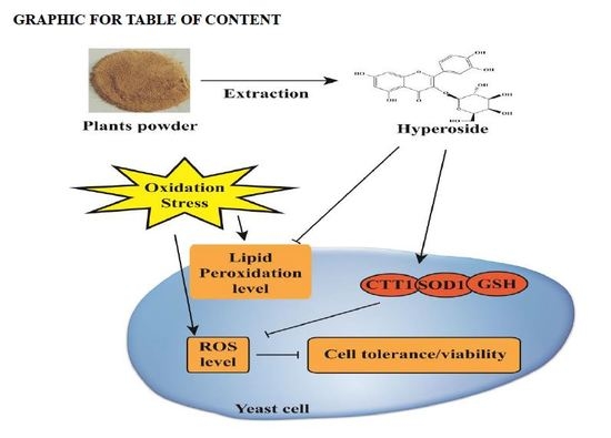
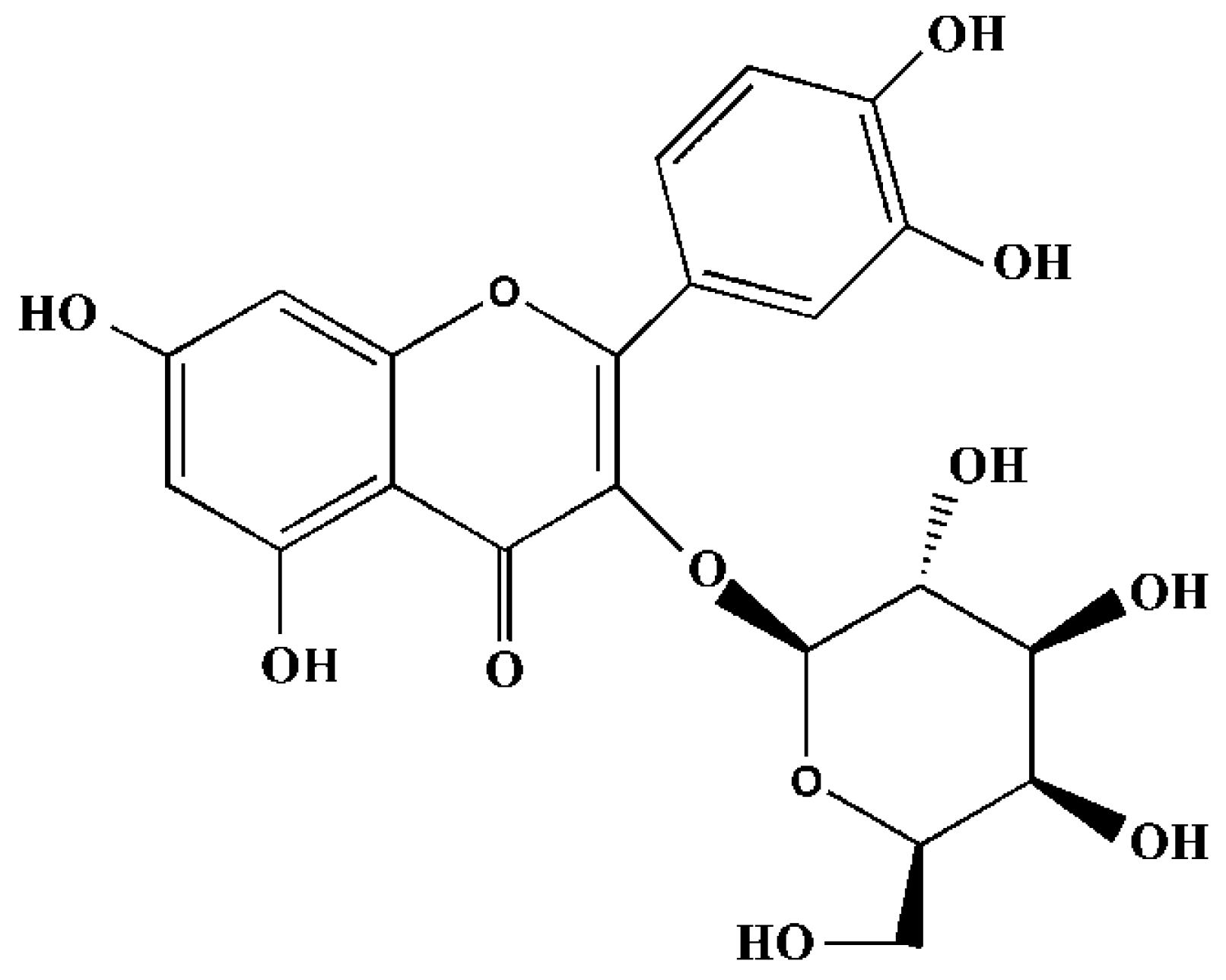
Antioxidant Activity Evaluation of Dietary Flavonoid Hyperoside Using Saccharomyces Cerevisiae as a Model

1
Tianjin Key Laboratory of Food and Biotechnology, School of Biotechnology and Food Science, Tianjin University of Commerce, Tianjin 300134, China
2
Centre for Food Research and Innovation, Department of Wine, Food and Molecular Bioscience, Lincoln University, Lincoln 7647, New Zealand
3
The Third Central Clinical College, Tianjin Medical University, Jintang Road, Hedong, Tianjin 300170, China
*
Authors to whom correspondence should be addressed.
These authors contributed equally to this work.
Molecules 2019, 24(4), 788; https://doi.org/10.3390/molecules24040788
Submission received: 24 January 2019 / Revised: 19 February 2019 / Accepted: 21 February 2019 / Published: 22 February 2019

Abstract

:
Oxidative stress leads to various diseases, including diabetes, cardiovascular diseases, neurodegenerative diseases, and even cancer. The dietary flavonol glycoside, hyperoside (quercetin-3-O-galactoside), exerts health benefits by preventing oxidative damage. To further understand its antioxidative defence mechanisms, we systemically investigated the regulation of hyperoside on oxidative damage induced by hydrogen peroxide, carbon tetrachloride, and cadmium in Saccharomyces cerevisiae. Hyperoside significantly increased cell viability, decreased lipid peroxidation, and lowered intracellular reactive oxygen species (ROS) levels in the wild-type strain (WT) and mutants gtt1∆ and gtt2∆. However, the strain with ctt1∆ showed variable cell viability and intracellular ROS-scavenging ability in response to the hyperoside treatment upon the stimulation of H2O2 and CCl4. In addition, hyperoside did not confer viability tolerance or intercellular ROS in CdSO4-induced stress to strains of sod1∆ and gsh1∆. The results suggest that the antioxidative reactions of hyperoside in S. cerevisiae depend on the intercellular ROS detoxification system.

Graphical Abstract

1. Introduction

Oxidative stress reflects an imbalance between an organism’s excessive production of oxygen radicals and its reduced capacity to detoxify. Oxidative intermediates, such as OH, O2, and NO, attack cell components and cause inflammation, cancer, ageing, and metabolic diseases [1,2]. It has been commonly accepted that the prevention of oxidative stress benefits human health [3,4]. For example, the ingestion of natural plants such as vegetables, fruits, and tea [5,6,7,8,9,10] has been well identified to counteract the amount of oxidative stress that cells encounter and to reduce the incidence of diseases related to oxidative damage [11,12,13,14]. The positive effect of a phytogenic diet on human health can be mainly attributed to the abundance of bioactive flavonols with pharmaceutical properties.
Within the flavonoid group, hyperoside (quercetin-3-O-β-d-galactopyranoside, Figure 1) is a member of the family of Hypericaceae, Rosaceae, and Labiata and the genus of Crataegus [15,16,17]. Hyperoside has been suggested to attenuate oxidative stress-related diseases [18,19]. For example, Piao elucidated the cytoprotective effects of hyperoside against H2O2-induced cell damage in Chinese hamster lung fibroblast cells, in which the scavenging mechanisms include elevation of the expression of HO-1 and activation of nuclear factor erythroid 2-related factor 2 (Nrf2). Furthermore, Xing [13] suggested that hyperoside prevents oxidative damage through GSK-3β inactivation. Of note, the major site of generation of reactive oxygen species (ROS) is located in the mitochondrial respiratory chain (RC), where the surrounding molecules (i.e., superoxide dismutase (SOD) and cytosolic catalase 1 (CTT1)) react with the electrons of free radicals in regulating the cellular bioenergetic status under physiological or pathological conditions. Mitochondrion usually acts as the target responding to oxidative stress, resulting in the functional decline of cells. It has been suggested that hyperoside regulates the mitochondrial apoptotic pathway to prevent oxidative damage [20,21]. However, few studies have investigated the role of hyperoside in mitochondrial detoxification for the prevention of oxidative stress.
In the present study, we used Saccharomyces cerevisiae as a model organism for studying antioxidant protection because yeast has developed antioxidant defence systems, including an enzymatic ROS detoxification system that involves SOD, CTT1, GPX, and a non-enzymatic system mainly including glutathione (GSH), to maintain intracellular redox status and ROS levels [2]. Therefore, we attempted to validate the response mechanism by which hyperoside decreases oxidative damage in S. cerevisiae (BY4741WT and its isogenic mutants gsh1∆, ctt1∆, sod1∆, gtt1∆, gtt2∆) caused by oxidants (CdSO4, H2O2 and CCl4), and to provide further evidence for hyperoside to be used as a health food product.

2. Results and Discussion

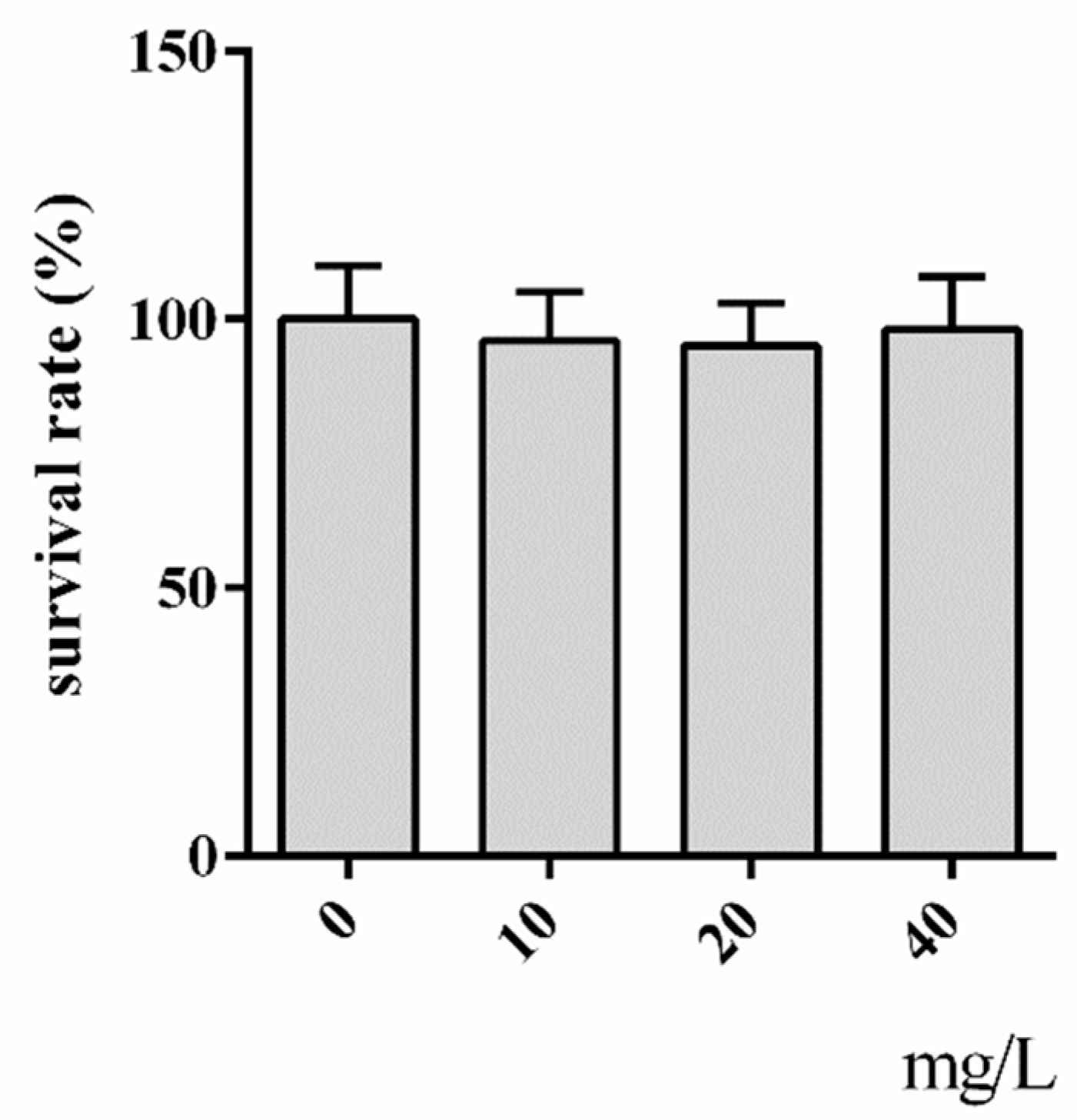
2.1. Cytotoxicity Assay of Hyperoside

To evaluate the toxic impact of hyperoside on the yeast cells, we first determined the cell viability of WT cells exposed to hyperoside for 2 h. As expected, we did not find hyperoside to be toxic to the yeast cells within the observed range of 10–40 mg/L, and the cells continued to reach 100% tolerance (Figure 2).

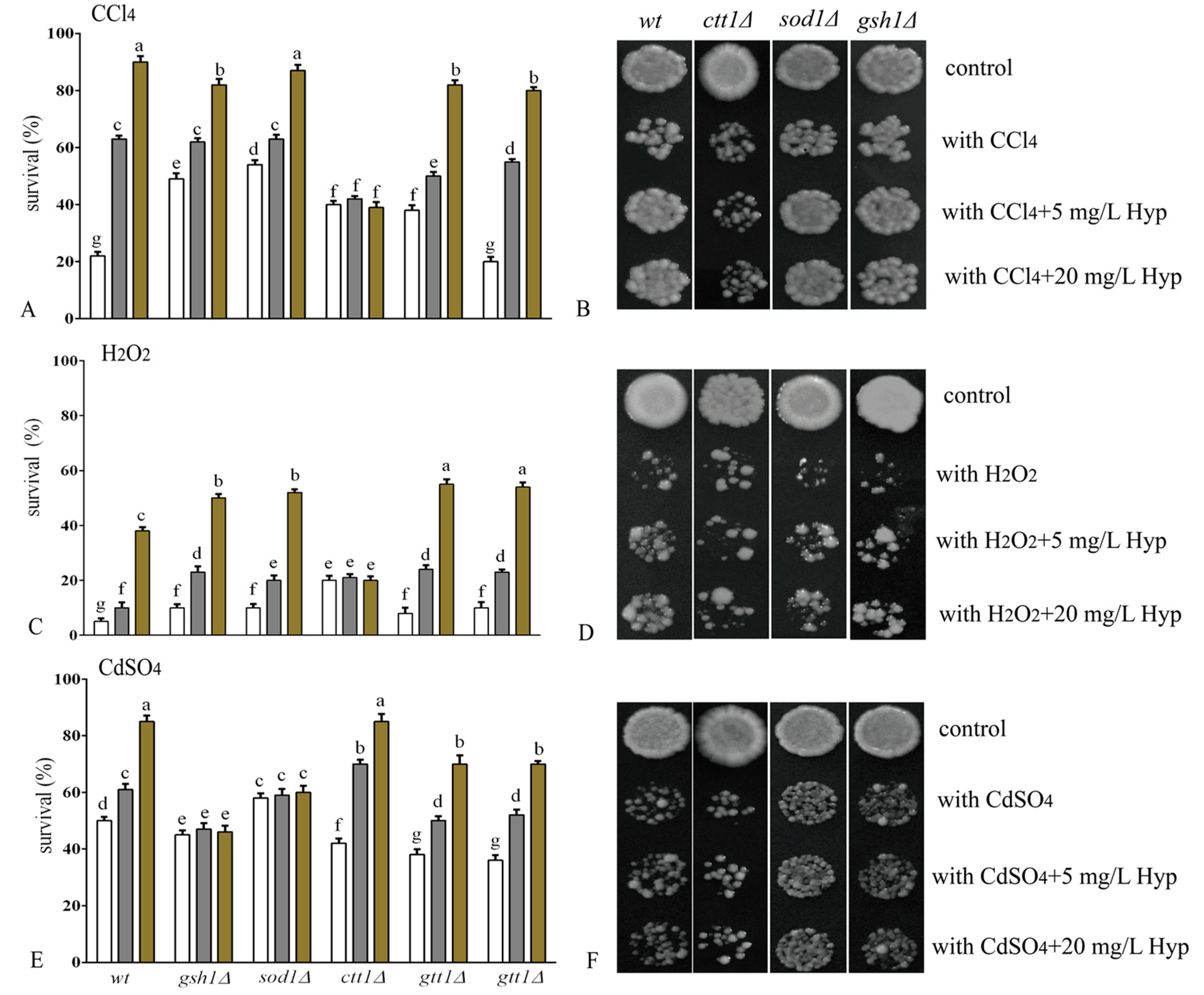
2.2. Hyperoside Increases Oxidative Stress Tolerance in S. cerevisiae

The stimuli used in our study are well-recognized oxidants of cells [22,23,24]. The survival rate of the WT strain consistently dropped significantly when exposed to the oxidants CCl4, H2O2, or CdSO4 (Figure 3). However, when the cells of the WT strain were preadapted with hyperoside, there was a concentration-dependent tolerance to all stresses observed in our experiments (Figure 3). The data suggested a protective effect of hyperoside in yeast against oxidative stimulation.
In theory, yeast cells control the intracellular redox balance through enzymatic and non-enzymatic defence systems [2]. To determine the defensive targets of hyperoside in yeast, a range of isogenic mutant strains (deficient in the genes CTT1, SOD1, GSH, GTT1, or GTT2), which present the phenotypes of enzymes involved in ROS detoxification, were used to assess the responses to oxidative stimuli.
Initially, the effect of hyperoside on the survival inhibition of CCl4 in the yeast strains was determined. The toxicity of CCl4 is usually caused by its reductive dehalogenation by cytochrome P450 into trichloromethyl free radicals (·CCl3) and peroxy trichloromethyl radicals (OOCCl3). Trichloromethyl free radicals react with sulfhydryl groups (glutathione and proteinthiols) and antioxidant enzymes (CAT and SOD), resulting in the excessive generation of ROS and membrane lipid peroxidation [22]. When hyperoside was used in combination with cells exposed to CCl4, an increased tolerance was observed in the strains of gsh1∆, sod1∆, gtt1∆ and gtt2∆ with improved survival rates of 30~60%. The mutant strain ctt1∆ showed a much weaker response to the addition of hyperoside compared with the other strains mentioned above (Figure 3A); no difference in growth was observed for ctt1∆, as shown in Figure 3B. Thus, the data indicated that hyperoside-induced prevention of CCl4 may involve intercellular catalase.
Intercellular H2O2 or its derivatives OH• and O•2− are tightly linked and attack cell membrane components, proteins, and DNA [25]. Given the importance of H2O2 in cell damage and death, we determined whether hyperoside was capable of increasing the tolerance of yeast trains to H2O2. Hyperoside improved the viability of the strains gsh1∆, gtt2∆, sod1∆, and gtt1∆, whereas this action was not observed in the mutant strain ctt1∆ (Figure 3C); the same condition can be observed in Figure 3D. The data suggested that the protective effect of hyperoside against H2O2-induced stress may be related to catalase, which catalyses H2O2 to generate H2O and O2 to alleviate tissue injury [26].
The heavy metal cadmium (Cd2+) is extensively found in food materials due to its industrial use. Environmental and occupational exposure to Cd2+ may be an important risk factor for a broad spectrum of illnesses affecting body organs such as the liver, kidneys, and cardiovascular systems [27]. Growing evidence suggests that oxidative stress induced by Cd2+ alters the free radical-scavenging effects of N-acetyl cysteine, resulting in cellular damage via increasing genome mutations and recombination, and accelerating lipid peroxidation [23]. Consistent with the findings of CCl4 and H2O2, cadmium (Cd2+) also significantly inhibited the growth of yeast cells, as shown in Figure 3E. Treatment with hyperoside significantly improved the viability of both the wild type and mutant strains, including ctt1∆, gtt1∆, and gtt2∆. However, the gsh1∆ and sod1∆ strains did not improve sufficiently in response to hyperoside, indicating that the protective effect of hyperoside against CdSO4-induced stress was related to glutathione (GSH) and superoxide dismutase (SOD1) (Figure 3E,F). Interestingly, the results suggest potential solutions to cadmium toxicity, which occurs in the body mainly through decreasing cellular SOD activity and GSH expression [28].

2.3. Hyperoside Attenuates the Level of Intracellular Oxidation

Intercellular ROS are usually generated from the mitochondria, and excessive accumulation of ROS is characterized by the presence of highly reactive molecules that regulate cell death and ageing [29]. Previous studies have demonstrated that the ability to scavenge intercellular ROS from natural compounds plays an important role in mediating the viability of organisms in response to oxidative stress [30,31,32]. Hence, the mechanism whereby hyperoside protects against oxidative stress-induced loss of tolerance in yeast cells may be mediated by the clearance of intercellular ROS.
Our results confirm that hyperoside increased oxidative stress tolerance in S. cerevisiae. In particular, the intercellular antioxidant enzyme CTT1 was important for modulating oxidative stress tolerance in response to CCl4 and H2O2, whereas the other two enzymes, SOD1 and GSH, were responsible for cell tolerance in response to CdSO4. Therefore, we checked whether the effects of intercellular ROS and antioxidant enzymes were consistent with the above performances of the yeast phenotypes. The fluorescence intensity of the H2DCF-DA probe was used to indicate the intracellular ROS level and significantly increased when the cells were treated with the indicated oxidants (Table 1). The addition of hyperoside reversed the increase in intracellular ROS intensity in all the strains we tested. Furthermore, CTT1 deficiency neutralized the influence of hyperoside on intercellular ROS in response to CCl4 or H2O2 stimulation. The expression of cytosolic catalase CTT1 is thus usually required for detoxification in S. cerevisiae upon exposure to a variety of stress conditions, and reduces intracellular free radicals [33,34].
In fact, the toxicity of CCl4 and H2O2 is largely attributed to their ability to morph deftly into ROS. Via the reductive dehalogenation reaction, CCl4 can be transformed into trichloromethyl free radicals by cytochrome P450, which readily interacts with molecular oxygen and creates trichloromethyl peroxyl radicals [35]. With regard to H2O2, it can produce hydroxyl radicals, the most reactive and toxic ROS. CTT generally stays at a very low level in cells, but can be dramatically activated by peroxides over the threshold concentration, thereby maintaining a controlled intracellular peroxide concentration and protecting membranes from oxidative damage [36]. Thus, intercellular CTT1 deficiency might remain at a high ROS level in yeasts, even with hyperoside pre-adaption.
In contrast to CCl4 or H2O2 stimulation, SOD1 or GSH deficiency significantly counteracted the reduction of intercellular ROS by hyperoside, resulting in a response to CdSO4 stimulation, which is in agreement with the results shown in Figure 3E,F. Although Cd2+ is not a redox-active metal, it mediates in ROS production indirectly by weakening the enzymatic (e.g., SOD, glutathione peroxidase (GPx), and glutathione reductase (GR)) and non-enzymatic (e.g., small molecule antioxidants: reduced glutathione (GSH), vitamins C and E) antioxidative barrier of cells [37,38,39,40]. Indeed, the antioxidant enzyme SOD1 is located in the cytoplasm and mitochondrial membrane space sod1∆ during abnormal oxidative damage because amino acids containing met and lys are hyperoside-sensitive to oxidative stress [41]. Furthermore, as the most abundant sulfhydryl donor, γ-glutamylcysteinylglycine (GSH) is an essential metabolite in both rodent and yeast cells that acts as a cofactor for the antioxidative enzymes glutathione peroxidase and glutathione transferase [42]. In the absence of exogenous GSH, cells with mutants lacking GSH are deficient in resisting toxic attacks caused by heavy metals such as cadmium [43].
Except hyperoside, a wide spectrum of polyphenols demonstrated antioxidant activity to fight against toxicities from heavy metals including cadmium [37,44,45,46,47]. The antioxidative and chelating properties were presented to explain the protective action of polyphenolic compounds against Cd2+ toxicity [48]. On the one hand, polyphenols directly increased the activities of antioxidative and pro-oxidative enzymes. On the other hand, polyphenols had the capacity to chelate Cd2+ and lower the cell burden of Cd2+. Our current study suggests that hyperoside exerts anti-oxidative protection on yeasts. However, whether or not the detailed action mechanisms of hyperoside against Cd2+ are mediated via the above pathways still needs further investigation.

2.4. Hyperoside Attenuates S. cerevisiae Cell Membrane Lipid Peroxidation

In addition to the above intercellular oxidative enzymes, cellular membrane lipids, mainly unsaturated fatty acids, represent common targets of oxidative attack. Free radicals resulting from oxidative stress, such as CCl4, H2O2, and CdSO4 [49,50,51], initiate a cascading lipid peroxidation process and eventually lead to membrane damage, including leakage and even cell death [52,53]. Because hyperoside possesses the flavonoid chemical structure of a potential hydrogen donor, we inferred that hyperoside may serve as a protector of membrane fatty acids against lipid peroxidation [54].
To further check the speculation regarding the protective role of hyperoside, membrane lipid peroxidation in yeast cells subjected to CCl4, H2O2, or CdSO4 was examined. As expected (Table 2) for the WT strains, the membrane lipid peroxidation level of the yeast cells was significantly increased under the stimulation of the three oxidants. The WT yeast cells were also re-evaluated with hyperoside prior to culturing with the oxidant, and alleviation of oxidative damage to membrane peroxidation was observed, which was in accordance with the tolerance assay (Figure 3) and indicated the capacities of hyperoside for increasing the integrity of the cell membrane.
Having identified the attenuation of lipid peroxidation in the WT yeasts, we investigated possible cellular enzymes involved in the redox regulation of membrane lipids. Hyperoside was observed to protect the lipid peroxidation in yeast cells from oxidative stress regardless of the antioxidant enzyme (Table 2). Indeed, it is suggested that the antioxidant enzymes located in the cytoplasm or mitochondria lack definitive preventive effects on membrane lipids [2], and the potential mechanism is worth exploring further.
Interestingly, lipid peroxidation caused by Cd2+ was more alleviated in the mutants, including sod1∆, ctt1∆, and gsh1∆ than in the WT strain. This could be attributed, at least partly, to the super expression of other antioxidant systems as a form of compensation. Several other studies indicated that a deficiency in one antioxidant system may be overcome by an increase in the remaining defence system [55,56].

3. Materials and Methods

3.1. Chemicals and Reagents

H2O2, CdSO4, and CCl4 were purchased from YuanLi Chemical Co., Ltd. (Tianjin, China). Dimethyl sulfoxide (DMSO), yeast extract, and peptone were purchased from Oxoid Company (Basingstoke, UK). Glucose was purchased from Amresco (Solon, OH, USA). Hyperoside was purchased from Biopurify Technology Development Co., Ltd. (Chendu, China), the structure and the purification (99.13%) of hyperoside was confirmed by 1H-NMR and 13C-NMR and MS (Figures S1–S4. Tables S1 and S2).

3.2. S. cerevisiae Strains, Media, and Growth Conditions

The wild-type (WT) strain of S. cerevisiae BY4741 (Matα his3∆1 leu2∆0 met15∆0 ura3∆0) [57] was purchased from EUROSCARF (European S. cerevisiae Archive for Functional Analysis, Institute of Molecular Biosciences, Johann Wolfgang Goethe-University Frankfurt, Frankfurt, Germany), and its isogenic mutants ctt1∆, sod1∆, gsh1∆, gtt1∆, and gtt2∆ harbouring the genes CTT1, SOD1, GSH1, GTT1, and GTT2, respectively, were obtained from Dr. Pereira (Departamento de Bioquímica, Universidade Federal do Rio de Janeiro, Rio de Janeiro, Brazil). Stocks of the strains were maintained on solid Yeast Extract-Peptone-Dextrose (YPD) (1% yeast extract, 2% glucose, 2% peptone, and 2% agar), and the medium also contained 0.02% geneticin for the mutant strains. For all experiments, the cells were grown in liquid YPD medium using an orbital shaker (Aosheng, Hangzhou, Zhejiang, China) at 28 °C/160 rpm.

3.3. Cytotoxicity Assay of Hyperoside

Yeast cells in the mid-log phase (106 cells/mL) were reinoculated in fresh medium (the initial cell concentration was 105 cells/mL) with or without hyperoside (10 mg/L, 20 mg/L, or 40 mg/L) and incubated at 28 °C/160 rpm for 2 h. Then, diluted (1:1000) yeast cells were spotted adjacently on YPD agar plates and incubated at 28 °C for approximately 72 h. The number of colonies was counted and calculated according to methods published in the literature [58].

3.4. Tolerance Assay

Yeast cells in the mid-log phase (106 cells/mL) were reinoculated in fresh medium to make a suspension of yeast cells with a density of 105 cells/mL, either with or without hyperoside (5 mg/L and 20 mg/L), and were incubated for 2 h at 28 °C/160 rpm. Then, the cells were subjected to oxidative stress (2 mmol/L H2O2, 10 mmol/L CCl4, or 3 mmol/L CdSO4) for 1 h. Cell viability was analysed by plating the appropriately diluted (1000×) cells in triplicate on solidified 2% YPD medium. The plates were incubated at 28 °C for 72 h, and then the colonies were counted. Survival expressed as a percentage was determined before and after oxidative stress exposure using cells treated with hyperoside and untreated control cells [59].

3.5. Cell Growth Assay

Yeast cells in mid-log phase (106 cells/mL) were reinoculated in fresh medium and then treated with hyperoside for 2 h at 28 °C/160 rpm after treatment with oxidative stress for 1 h. The cells were plated in quadruplicate in 96-well plates and spotted on solid YPD media. The plates were incubated at 28 °C for 3 days before photography, and each assay was repeated at least three times [60].

3.6. Determination of Cell Membrane Lipid Peroxidation

Yeast cells (106 cells/mL) exposed directly to oxidative stress for 1 h either pre-treated for 2 h with hyperoside or not treated were incubated at 28 °C /160 rpm. The cells (50 mg) were harvested by cooling on ice, centrifuged (5000 rpm/5 min/4 °C), and washed twice with distilled water. Cell pellets were resuspended in 0.5 mL of a 10% trichloroacetic acid solution (TCA) solution (w/v), and 1.5 g glass beads were added. Then, the cell walls were broken with cell breakers for 3 min at 5 degrees, and the supernatant was mixed with 0.1 mL of a 0.1 mol/L ethylenediaminetetraacetic acid (EDTA) solution and 0.6 mL of a 1% (w/v) thiobarbituric acid (TBA) solution in 0.05 mol/L NaOH. The reaction mixture was incubated in a boiling water bath for 15 min, and the mixture was cooled afterwards. The extracts were used to detect malondialdehyde (MDA) by measuring the absorbance at 532 nm [61,62].

3.7. Determination of Intracellular Oxidation

The intracellular oxidation level was assayed using the oxidant-sensitive probe 2′7′-dichlorofluorescein diacetate (H2DCF-DA) according to our previously established method [63]. Briefly, yeast cells with or without hyperoside were incubated for 2 h at 28 °C/160 rpm and then stressed with an oxidative agent for 1 h. The cells were incubated with the probe, harvested, washed, and resuspended in 0.5 mL water containing 1.5 g glass beads. Next, the cell walls were broken, and the supernatant solutions were centrifuged at 25,000× g for 5 min and diluted 6-fold with water before fluorescence was measured (an excitation wavelength of 504 nm and an emission wavelength of 524 nm).

3.8. Statistical Analyses

The statistical analyses were conducted using analysis of variance and Tukey’s test with the SPSS 12.0 package (SPSS, Inc., Chicago, IL, USA). The latter denoted homogeneity between the experimental groups at p < 0.05. To evaluate the levels of lipid peroxidation and intracellular oxidation, we compared homogeneity between stressed and non-stressed cells of each strain at p < 0.05. In all figures and tables, different letters indicate significantly different results.

4. Conclusions

Taken together, this study indicated that the dietary flavonoid hyperoside serves as a protector for S. cerevisiae in response to the stimulation of oxidative stress. More importantly, we identified that three antioxidant genes—CTT1, SOD1, and GSH—constructed a tight relationship between intercellular ROS and cellular viability regarding the antioxidant mechanisms of hyperoside through the yeast model. However, the three genes did not achieve a decrease in lipid peroxidation according to our results. It would be worthwhile to further investigate the mechanism to eventually better understand and develop dietary flavonoid products, including hyperoside.

Supplementary Materials

The following are available online at https://www.mdpi.com/1420-3049/24/4/788/s1, Figure S1: The 1H-NMR spectra (DMSO, 400 MHz) of hyperoside, FigureS2: The 13C-NMR spectra (DMSO, 100 MHz) of hyperoside, Figure S3: The HPLC spectra of hyperoside, Table S1: The analytical result of hyperoside.

Author Contributions

Conceptualization, H.Z. and G.D.; methodology, L.F.; investigation, L.F., Y.G., X.W. and R.L.; data curation, L.F. and Y.G.; writing—original draft preparation, Y.G., H.Z., W.G., G.D., M.B., C.B. and H.G.; writing—review and editing, H.Z., M.B. and C.B.; supervision, H.Z., C.B. and G.D.; funding acquisition, H.Z., W.G., G.D., J.L., M.W.

Funding

This study was supported by the National Natural Science Foundation of China (grant nos. 31571832, 31772019, and 81803548), Tianjin Innovative Research Team Grant (TD13-5087), Tianjin Natural Science Grant (16JCQNJC14600 and 16JCZDJC34000), Key Program of Tianjin Municipal Health Bureau (no. 2013-GG-05), Open Project of Tianjin Key Laboratory of Food Biotechnology and Talent Grant of TJCU (R170106), and College Student Innovation and Entrepreneurship Training (201710069071). We also appreciate Pereira (Departamento de Bioquímica, Instituto de Química, Universidade Federal do Rio de Janeiro, Rio de Janeiro, RJ, Brazil 21941-909) for his generous gifts of the mutant strains, which are key to our research.

Conflicts of Interest

The authors declare no conflict of interest.

Abbreviations

SODssuperoxide dismutases
GPXsglutathione peroxidases
CTT1cytosolic catalaseT1
Prxsperoxiredoxins
GSHglutathione
ROSreactive oxygen species
S.cerevisiaesaccharomyces cerevisiae
GTT1glutathione transferase1
GTT2glutathione transferase2
YPDyeast extract peptone dextrose
TCAtrichloroacetic acid
TBAthiobarbituric acid
MDAmalondialdehyde
H2DCF-DA2′7′-dichlorofluorescein diacetate
WTwild type

References

  1. Meng, D.; Zhang, P.; Zhang, L.L.; Wang, H.; Ho, C.T.; Li, S.M.; Shahidi, F.; Zhao, H. Detection of cellular redox reactions and antioxidant activity assays. J. Funct. Foods 2017, 37, 467–479. [Google Scholar] [CrossRef]
  2. Meng, D.; Zhang, P.; Li, S.M.; Ho, C.T.; Zhao, H. Antioxidant activity evaluation of dietary phytochemicals using Saccharomyces cerevisiae as a model. J. Funct. Foods 2017, 38, 36–44. [Google Scholar] [CrossRef]
  3. Wang, L.W.; Wang, J.H.; Fang, L.Y.; Zheng, Z.L.; Zhi, D.X.; Wang, S.Y.; Li, S.M.; Ho, C.T.; Zhao, H. Anticancer activities of citrus peel polymethoxyflavones related to angiogenesis and others. Biomed. Res. Int. 2014, 2014, 453972. [Google Scholar] [CrossRef] [PubMed]
  4. Li, Y.R.; Li, S.M.; Lin, C.C. Effect of resveratrol and pterostilbene on aging and longevity. Biofactors 2018, 44, 69–82. [Google Scholar] [CrossRef] [PubMed]
  5. Wang, L.W.; Pan, M.H.; Lo, C.Y.; Zhao, H.; Li, S.M.; Ho, C.T.; Yang, G.L. Anti-fibrotic activity of polyphenol-enriched sugarcane extract in rats via inhibition of p38 and JNK phosphorylation. Food Funct. 2018, 9, 951–958. [Google Scholar]
  6. Su, D.X.; Li, N.; Chen, M.; Yang, Y.; He, S.; Wang, Y.; Wu, Q.H.; Li, L.; Yang, H.L.; Zeng, Q.Z. Effects of in vitro digestion on the composition of flavonoids and antioxidant activities of the lotus leaf at different growth stages. Int. J. Food Sci. Technol. 2018, 53, 1631–1639. [Google Scholar] [CrossRef]
  7. Shukor, N.A.A.; Ablat, A.; Muhamad, N.A.; Mohamad, J. In vitro antioxidant and in vivo xanthine oxidase inhibitory activities of Pandanus amaryllifolius in potassium oxonate-induced hyperuricemic rats. Int. J. Food Sci. Technol. 2018, 53, 1476–1485. [Google Scholar] [CrossRef] [Green Version]
  8. Quan, W.; Tao, Y.D.; Lu, M.; Yuan, B.; Chen, J.; Zeng, M.M.; Qin, F.; Guo, F.X.; He, Z.Y. Stability of the phenolic compounds and antioxidant capacity of five fruit (apple, orange, grape, pomelo and kiwi) juices during in vitro simulated gastrointestinal digestion. Int. J. Food Sci. Technol. 2018, 53, 1131–1139. [Google Scholar] [CrossRef]
  9. Elijah, A.A.; Daniel, M.C.; Porfirio, M.; Mercedes, S.; Helios, E.; Marıa, E.M.; Azalia, A.N.; Ivan, C.; Myrna, B. Comparative study of antioxidant and antibacterial properties of the edible mushrooms Pleurotus levis, P. ostreatus, P. pulmonarius and P. tuber-regium. Int. J. Food Sci. Technol. 2018, 53, 1316–1330. [Google Scholar]
  10. Ahmad, H.S.M.; Ismail, A.; Kassim, N.K.; Hamid, M.; Mat, A.M.S. Underutilised fruits: A review of phytochemistry and biological properties. J. Food Bioact. 2018, 1, 28. [Google Scholar]
  11. Mukherjee, S.; Das, S.K.; Vasudevan, D.M. Protective role of extracts of grape skin and grape flesh on ethanol-induced oxidative stress, inflammation and histological alterations in rat brain. Arch. Physiol. Biochem. 2015, 121, 144–151. [Google Scholar] [CrossRef] [PubMed]
  12. Li, W.; Liu, M.; Xu, Y.F.; Fang, Y.; Che, J.P.; Wang, G.C.; Zheng, J.H. Combination of quercetin and hyperoside has anticancer effects on renal cancer cells through inhibition of oncogenic microRNA-27a. Oncol. Rep. 2014, 31, 117–124. [Google Scholar] [CrossRef] [PubMed]
  13. Xing, H.Y.; Cai, Y.Q.; Wang, X.F.; Wang, L.L.; Li, P.; Wang, G.Y.; Chen, J.H. The Cytoprotective Effect of Hyperoside against Oxidative Stress Is Mediated by the Nrf2-ARE Signaling Pathway through GSK-3beta Inactivation. PLoS ONE 2015, 10, e0145183. [Google Scholar] [CrossRef] [PubMed]
  14. Yang, F.Q.; Liu, M.; Li, W.; Che, J.P.; Wang, G.C.; Zheng, J.H. Combination of quercetin and hyperoside inhibits prostate cancer cell growth and metastasis via regulation of microRNA21. Mol. Med. Rep. 2015, 11, 1085–1092. [Google Scholar] [CrossRef] [PubMed]
  15. Saddiqe, Z.; Maimoona, A.; Abbas, G.; Naeem, I.; Shahzad, M. Pharmacological screening of Hypericum androsaemum extracts for antioxidant, anti-lipid peroxidation, antiglycation and cytotoxicity activity. Pak. J. Pharm. Sci. 2016, 29, 397–405. [Google Scholar] [PubMed]
  16. Wang, L.W.; Yue, Z.W.; Guo, M.Z.; Fang, L.Y.; Bai, L.; Li, X.Y.; Tao, Y.Q.; Wang, S.Y.; Liu, Q.; Zhi, D.X.; et al. Dietary Flavonoid Hyperoside Induces Apoptosis of Activated Human LX-2 Hepatic Stellate Cell by Suppressing Canonical NF-kappaB Signaling. BioMed. Res. Int. 2016, 2016, 1068528. [Google Scholar] [PubMed]
  17. Wen, L.; Lin, Y.L.; Lv, R.M.; Yan, H.J.; Yu, J.Q.; Zhao, H.Q.; Wang, X.; Wang, D.J. An Efficient Method for the Preparative Isolation and Purification of Flavonoids from Leaves of Crataegus pinnatifida by HSCCC and Pre-HPLC. Molecules 2017, 22, 767. [Google Scholar] [CrossRef] [PubMed]
  18. Li, Y.L.; Wang, Y.W.; Li, L.; Kong, R.; Pan, S.H.; Ji, L.; Liu, H.; Chen, H.; Sun, B. Hyperoside induces apoptosis and inhibits growth in pancreatic cancer via Bcl-2 family and NF-kappaB signaling pathway both in vitro and in vivo. Tumor. Biol. 2016, 37, 7345–7355. [Google Scholar] [CrossRef] [PubMed]
  19. Piao, M.J.; Kang, K.A.; Zhang, R.; Ko, D.O.; Wang, Z.H.; You, H.J.; Kim, H.S.; Kim, J.S.; Kang, S.S.; Hyun, J.W. Hyperoside prevents oxidative damage induced by hydrogen peroxide in lung fibroblast cells via an antioxidant effect. Bba Biomembranes 2008, 1780, 1448–1457. [Google Scholar] [CrossRef] [PubMed]
  20. Bernatoniene, J.; Kopustinskiene, D.M.; Jakstas, V.; Majiene, D.; Baniene, R.; Kuršvietiene, L.; Masteikova, R.; Savickas, A.; Toleikis, A.; Trumbeckaite, S. The effect of Leonurus cardiaca herb extract and some of its flavonoids on mitochondrial oxidative phosphorylation in the heart. Planta Med. 2014, 80, 525–532. [Google Scholar] [CrossRef] [PubMed]
  21. Guon, T.E.; Chung, H.S. Hyperoside and rutin of Nelumbo nucifera induce mitochondrial apoptosis through a caspase-dependent mechanism in HT-29 human colon cancer cells. Oncol. Lett. 2016, 11, 2463–2470. [Google Scholar] [CrossRef] [PubMed] [Green Version]
  22. Hafez, M.M.; Shabanah, O.A.A.; Harbi, N.A.O.; Harbi, M.M.A.; Rejaie, S.S.A.; Alsurayea, S.M.; Ahmed, M.M.S. Association between paraoxonases gene expression and oxidative stress in hepatotoxicity induced by CCl4. Oxid. Med. Cell Longev. 2014, 2014, 893212. [Google Scholar] [CrossRef] [PubMed]
  23. Guo, L.; Ghassemian, M.; Komives, E.A.; Russell, P. Cadmium-induced proteome remodeling regulated by Spc1/Sty1 and Zip1 in fission yeast. Toxicol. Sci. 2012, 129, 200–212. [Google Scholar] [CrossRef] [PubMed]
  24. Zou, Y.; Lu, Y.; Wei, D. Antioxidant Activity of a Flavonoid-Rich Extract of Hypericum perforatum L. in Vitro. J. Agri. Food Chem. 2004, 52, 5032–5039. [Google Scholar] [CrossRef] [PubMed]
  25. Cobb, C.A.; Cole, M.P. Oxidative and nitrative stress in neurodegeneration. Neurobiol dis. 2015, 84, 4–21. [Google Scholar] [CrossRef] [PubMed] [Green Version]
  26. Debattisti, V.; Gerencser, A.A.; Saotome, M.; Das, S.; Hajnoczky, G. ROS Control Mitochondrial Motility through p38 and the Motor Adaptor Miro/Trak. Cell Rep. 2017, 21, 1667–1680. [Google Scholar] [CrossRef] [PubMed]
  27. Mohajeri, M.; Rezaee, M.; Sahebkar, A. Cadmium-induced toxicity is rescued by curcumin: A review. Biofactors 2017, 43, 645–661. [Google Scholar] [CrossRef] [PubMed]
  28. Srivastava, S.; Pant, A.; Trivedi, S.; Pandey, R. Curcumin and beta-caryophellene attenuate cadmium quantum dots induced oxidative stress and lethality in Caenorhabditis elegans model system. Environ. Toxicol. Phar. 2016, 42, 55–62. [Google Scholar] [CrossRef] [PubMed]
  29. Marchi, S.; Giorgi, C.; Suski, J.M.; Agnoletto, C.; Bononi, A.; Bonora, M.; Marchi, E.D.; Missiroli, S.; Patergnani, S.; Poletti, F.; et al. Mitochondria-ros crosstalk in the control of cell death and aging. J. Recept. Signal Transduct. Res. 2012, 2012, 329635. [Google Scholar] [CrossRef]
  30. Czerniewicz, P.; Sytykiewicz, H.; Durak, R.; Sobkowiak, B.B. Role of phenolic compounds during antioxidative responses of winter triticale to aphid and beetle attack. Plant Physiol. Bioch. 2017, 118, 529–540. [Google Scholar] [CrossRef]
  31. Sandoval-Acuna, C.; Ferreira, J.; Speisky, H. Polyphenols and mitochondria: An update on their increasingly emerging ROS-scavenging independent actions. Arch. Biochem. Biophys. 2014, 559, 75–90. [Google Scholar] [CrossRef] [PubMed]
  32. Zhang, N.; Ying, M.D.; Wu, Y.P.; Zhou, Z.H.; Ye, Z.M.; Li, H.; Lin, D.S. Hyperoside, a flavonoid compound, inhibits proliferation and stimulates osteogenic differentiation of human osteosarcoma cells. PLoS ONE 2014, 9, e98973. [Google Scholar] [CrossRef] [PubMed]
  33. Asimi, O.A.; Sahu, N.P.; Pal, A.K. Antioxidant capacity of crude water and ethylacetate extracts of some Indian spices and their antimicrobial activity against Vibrio vulnificus and Micrococcus luteus. J. Med. Plants Res. 2013, 7, 1907–1915. [Google Scholar]
  34. Jennifer Spencer, T.G.P.; Katherine, A.S.; Greetham, D. Tolerance of pentose utilising yeast to hydrogen peroxide-induced oxidative stress. BMC Res. Notes 2014, 7, 1–12. [Google Scholar]
  35. Alan, T.; Williams, M.D.; Raymond, F.; Burk, M.D. Carbon Tetrachloride Hepatotoxicity: An Example of Free Radical-Mediated Injury. Seminars Liver Dis. 1990, 10, 279–284. [Google Scholar]
  36. Halliwell, B.; Gutteridge, J.M.C. Free Radicals in Biology and Medicine, 3rd ed.; Clarendon Press: Oxford, UK, 1999. [Google Scholar]
  37. Amamou, F.; Nemmiche, S.; Nemmiche, R.K.; Didi, A.; Yazit, S.M.; Sari, D.C. Protective effect of olive oil and colocynth oil against cadmium-induced oxidative stress in the liver of Wistar rats. Food Chem. Toxicol. 2015, 78, 177–184. [Google Scholar] [CrossRef] [PubMed]
  38. Pari, L.; Shagirtha, K. Hesperetin protects against oxidative stress related hepatic dysfunction by cadmium in rats. Exp. Toxicol. Pathol. 2012, 64, 513–520. [Google Scholar] [CrossRef] [PubMed]
  39. Renugadevi, J.; Prabu, S.M. Cadmium-induced hepatotoxicity in rats and the protective effect of naringenin. Exp. Toxicol. Pathol. 2010, 62, 171–181. [Google Scholar] [CrossRef] [PubMed]
  40. Sakr, S.A.; Bayomy, M.F.; El-Morsy, A.M. Rosemary extract ameliorates cadmium-induced histological changes and oxidative damage in the liver of albino rat. J. Basic Appl. Zool. 2015, 71, 1–9. [Google Scholar] [CrossRef] [Green Version]
  41. Culotta, V.C.; Yang, M.; O’Halloran, T.V. Activation of superoxide dismutases: Putting the metal to the pedal. BBA Biomembranes 2006, 1763, 747–758. [Google Scholar] [CrossRef] [Green Version]
  42. Morano, K.A.; Grant, C.M.; Moye-Rowley, W.S. The response to heat shock and oxidative stress in Saccharomyces cerevisiae. Gene 2012, 190, 1157–1195. [Google Scholar] [CrossRef] [PubMed]
  43. Thorsen, M.; Perrone, G.G.; Kristiansson, E.; Traini, M.; Ye, T.; Dawes, I.W.; Nerman, O.; Tamás, M.J. Genetic basis of arsenite and cadmium tolerance in Saccharomyces cerevisiae. BMC Genomics 2009, 10, 105. [Google Scholar] [CrossRef] [PubMed]
  44. Rasines-Perea, Z.; Teissedre, P.L. Grape Polyphenols’ Effects in Human Cardiovascular Diseases and Diabetes. Molecules 2017, 22, 68. [Google Scholar] [CrossRef] [PubMed]
  45. Venancio, V.P.; Cipriano, P.A.; Kim, H.; Antunes, L.M.G.; Talcott, S.T.; Mertens-Talcott, S.U. Cocoplum (Chrysobalanus icaco L.) anthocyanins exert anti-inflammatory activity in human colon cancer and non-malignant colon cells. Food Funct. 2017, 8, 307–314. [Google Scholar] [CrossRef] [PubMed]
  46. Bozzetto, L.; Annuzzi, G.; Pacini, G.; Costabile, G.; Vetrani, C.; Vitale, M.; Griffo, E.; Giacco, A.; Natale, C.D.; Cocozza, S.; et al. Polyphenol-rich diets improve glucose metabolism in people at high cardiometabolic risk: a controlled randomised intervention trial. Diabetologia 2015, 58, 1551–1560. [Google Scholar] [CrossRef] [PubMed]
  47. Henning, S.M.; Wang, P.; Said, J.W.; Huang, M.; Grogan, T.; Elashoff, D.; Carpenter, C.L.; Heber, D.; Aronson, W.J. Randomized clinical trial of brewed green and black tea in men with prostate cancer prior to prostatectomy. Prostate 2015, 75, 550–559. [Google Scholar] [CrossRef] [PubMed]
  48. Mezynska, M.; Brzoska, M.M. Review of polyphenol-rich products as potential protective and therapeutic factors against cadmium hepatotoxicity. J. App. Toxicol. 2019, 39, 117–145. [Google Scholar] [CrossRef]
  49. Jancic, S.A.; Stosic, B.Z. Cadmium effects on the thyroid gland. Vitam. Horm. 2014, 94, 391–425. [Google Scholar]
  50. Repetto, M.; Semprine, J.; Boveris, A. Lipid Peroxidation: Chemical Mechanism, Biological Implications and Analytical Determination. Lipid Peroxidation 2012. [Google Scholar] [Green Version]
  51. Knockaert, L.; Berson, A.; Ribault, C.; Prost, P.E.; Fautrel, A.; Pajaud, J.; Lepage, S.; Lucas-Clerc, C.; Begue´, J.M.; Fromenty, B.; et al. Carbon tetrachloride-mediated lipid peroxidation induces early mitochondrial alterations in mouse liver. Lab. Invest. 2012, 92, 396–410. [Google Scholar] [CrossRef]
  52. Sultana, R.; Perluigi, M.; Butterfield, D.A. Lipid peroxidation triggers neurodegeneration: A redox proteomics view into the Alzheimer disease brain. Free Radical. Bio. Med. 2013, 62, 157–169. [Google Scholar] [CrossRef] [PubMed] [Green Version]
  53. Atiba, A.S.; Abbiyesuku, F.M.; Niran-atiba, T.A.; Oparinde, D.P.; Ajose, O.A.; Akindele, R.A. Free Radical Attack on Membrane Lipid and Antioxidant Vitamins in the Course of Pre-Eclamptic Pregnancy. Ethiop. J. Health Dev. 2014, 24, 35. [Google Scholar] [CrossRef] [Green Version]
  54. Hritcu, L.; Ionita, R.; Postu, P.A.; Gupta, G.K.; Turkez, H.; Lima, T.C.; Carvalho, C.U.S.; Sousa, D.P. Antidepressant Flavonoids and Their Relationship with Oxidative Stress. Oxid. Med. Cell Longev. 2017, 2017, 5762172. [Google Scholar] [CrossRef] [PubMed]
  55. Fernandes, P.N.; Mannarino, S.C.; Silva, C.G.; Pereira, M.D.; Panek, A.D.; Eleutherio, E.C.A. Oxidative stress response in eukaryotes: effect of glutathione, superoxide dismutase and catalase on adaptation to peroxide and menadione stresses in Saccharomyces cerevisiae. Redox Rep. 2007, 12, 236–244. [Google Scholar] [CrossRef] [PubMed]
  56. Mauro Braga Franca, A.D.P.; Eleutherio, E.C.A. The role of cytoplasmic catalase in dehydration tolerance of Saccharomyces cerevisiae. Cell Stress Chaper. 2015, 10, 3. [Google Scholar]
  57. Kaosaar, S.; Kahru, A.; Mantecca, P.; Kasemets, K. Profiling of the toxicity mechanisms of coated and uncoated silver nanoparticles to yeast Saccharomyces cerevisiae BY4741 using a set of its 9 single-gene deletion mutants defective in oxidative stress response, cell wall or membrane integrity and endocytosis. TIV 2016, 35, 149–162. [Google Scholar]
  58. Pereira, M.D.; Herdeiro, R.S.; Fernandes, P.N.; Eleutherio, E.C.A.; Panek, A.D. Targets of oxidative stress in yeast sod mutants. BBA Gen. Subjects 2003, 1620, 245–251. [Google Scholar] [CrossRef]
  59. Silva, C.G.; Herdeiro, R.S.; Mathias, J.C.; Panek, A.D.; Silveira, C.S.; Rodrigues, V.P.; Renno, M.N.; Falc˜ao, D.Q.; Cerqueira, D.M.; Minto, A.B.M.; et al. Evaluation of antioxidant activity of Brazilian plants. Pharmacol. Res. 2005, 52, 229–233. [Google Scholar] [CrossRef]
  60. Bompiani, K.M.; Tsai, C.Y.; Achatz, F.P.; Liebig, J.K.; Howell, S.B. Copper transporters and chaperones CTR1, CTR2, ATOX1, and CCS as determinants of cisplatin sensitivity. Metallomics 2016, 8, 951–962. [Google Scholar] [CrossRef] [Green Version]
  61. Baron, J.A.; Chen, J.S.; Culotta, V.C. Cu/Zn superoxide dismutase and the proton ATPase Pma1p of Saccharomyces cerevisiae. Biochem. Hem. Bio. Res. Co. 2015, 462, 251–260. [Google Scholar] [CrossRef]
  62. Dani, C.; Bonatto, D.; Salvador, M.; Pereira, M.D.; Henriques, J.A.P.; Eleutherio, E. Antioxidant Protection of Resveratrol and Catechin in Saccharomyces cerevisiae. J. Agri. Food Chem. 2008, 56, 4268–4272. [Google Scholar] [CrossRef] [PubMed]
  63. Wang, M.Y.; Meng, D.; Zhang, P.; Wang, X.X.; Du, G.; Brennan, C.; Li, S.M.; Ho, C.H.; Zhao, H. Antioxidant Protection of Nobiletin, 5-Demethylnobiletin, Tangeretin, and 5-Demethyltangeretin from Citrus Peel in Saccharomyces cerevisiae. J. Agri. Food Chem. 2018, 66, 3155–3160. [Google Scholar] [CrossRef] [PubMed]
Figure 1. The structure of hyperoside.
Molecules 24 00788 g001
Figure 2. Survival of exponential WT cells exposed to increased concentrations of hyperoside.
Molecules 24 00788 g002
Figure 3. Effect of hyperoside on yeast cells under indicated stress. A, C, E: Effect of 10 mM CCl4, 2 mM H2O2, and 3 mM CdSO4 on the viability in Saccharomyces cerevisiae cells (wild type and mutant strains ctt1Δ, sod1Δ, gtt1Δ, gtt2Δ, and gsh1Δ) and the anti-oxidative effect of hyperoside pre-treatment. The concentration of hyperoside used was 0 mg/L (white bars), 5 mg/L (grey bars), and 20 mg/L (brown bars). B, D, F: Effect of 10 mM CCl4, 2 mM H2O2, and 3 mM CdSO4 on the growth of S. cerevisiae cells (wild type and mutant strains ctt1Δ, sod1Δ, and gsh1Δ) and the anti-oxidative effect of hyperoside pre-treatment. The results represent the means ± SD from three independent experiments. The statistically different results were represented by different letters in each oxidative stress group, p < 0.05.
Molecules 24 00788 g003
Table 1. Effect of 20 mg/L hyperoside on the level of ROS in yeast cells.
StressTreatmentWTsod1Δctt1Δgsh1Δ
CCl4without hyperoside4.77 ± 0.53 p a q3.35 ± 0.43b2.49 ± 0.47c2.41 ± 0.31c
with hyperoside1.62 ± 0.40d1.69 ± 0.23d2.36 ± 0.22c1.45 ± 0.16d
H2O2without hyperoside14.23 ± 0.31a6.99 ± 0.20b5.84 ± 0.39c8.87 ± 0.52b
with hyperoside4.39 ± 0.33c1.87 ± 0.45d5.86 ± 0.14c1.24 ± 0.17d
CdSO4without hyperoside1.91 ± 0.23a1.84 ± 0.56a2.00 ± 0.15a1.60 ± 0.30a
with hyperoside0.70 ± 0.57d1.72 ± 0.24a1.13 ± 0.17c1.44 ± 0.29a
p The results were expressed as a ratio between fluorescence of stressed, adapted or not with polyphenol, and non-stressed cells. The data represent the means ± SDs of at least three independent experiments. q Each stress was analyzed separately to determine statistical differences (different letters mean statistically different results at p < 0.05).
Table 2. Effect of 20 mg/L hyperoside on membrane lipid peroxidation in yeast cells.
StressTreatmentWTsod1Δctt1Δgsh1Δ
CCl4without hyperoside1.9 ± 0.3paq1.5 ± 0.2a1.7 ± 0.2a1.6 ± 0.3a
with hyperoside0.7 ± 0.2c0.8 ± 0.3b0.6 ± 0.3c0.5 ± 0.2c
H2O2without hyperoside2.3 ± 0.3a2.1 ± 0.2a2.5 ± 0.2a2.4 ± 0.3a
with hyperoside1.0 ± 0.2c1.2 ± 0.3b1.3 ± 0.1b1.2 ± 0.2b
CdSO4without hyperoside1.5 ± 0.2a1.2 ± 0.1a1.3 ± 0.2a1.4 ± 0.2a
with hyperoside1.1 ± 0.3b0.6 ± 0.2c0.8 ± 0.2c0.7 ± 0.1c
p The results were expressed as a ratio between lipid peroxidation levels of stressed, adapted or not with hyperoside, and non-stressed cells. The data represent the means ± SDs of at least three independent experiments. q Each stress was analyzed separately to determine statistical differences (different letters mean statistically different results at p < 0.05).

