Article

Synthesis and Telomeric G-Quadruplex-Stabilizing Ability of Macrocyclic Hexaoxazoles Bearing Three Side Chains

1
Department of Biotechnology and Life Science, Tokyo University of Agriculture and Technology, 2-24-16 Naka-cho, Koganei, Tokyo 184-8588, Japan
2
Department of Chemistry, Chiba University, 1-33 Yayoi, Inage, Chiba 263-8522, Japan
3
Transborder Medical Reserch Center, University of Tsukuba, 1-1-1 Tennodai, Tsukuba 305-8575, Japan
4
Division of Biomedical Science, University of Tsukuba, 1-1-1 Tennodai, Tsukuba 305-8575, Japan
5
Molecular Profiling Research Center for Drug Discovery, National Institute of Advanced Industrial Science and Technology, 2-4-7 Aomi, Koto-ward, Tokyo 135-0064, Japan
6
Laboratoire de Biologie et Pharmacologie appliquée, CNRS UMR 8113, ENS paris-saclay 60 avenue du president Wilson, 94230 Cachan, France
7
Division of Physics and Applied Physics, Nanyang Technological University, Singapore 637371, Singapore
*
Author to whom correspondence should be addressed.
Molecules 2019, 24(2), 263; https://doi.org/10.3390/molecules24020263
Submission received: 13 December 2018 / Revised: 4 January 2019 / Accepted: 10 January 2019 / Published: 11 January 2019
(This article belongs to the Special Issue G-Quadruplex Ligands and Cancer)

Abstract

:
G-quadruplexes (G4s), which are structures formed in guanine-rich regions of DNA, are involved in a variety of significant biological functions, and therefore “sequence-dependent” selective G4-stabilizing agents are required as tools to investigate and modulate these functions. Here, we describe the synthesis of a new series of macrocyclic hexaoxazole-type G4 ligand (6OTD) bearing three side chains. One of these ligands, 5b, stabilizes telomeric G4 preferentially over the G4-forming DNA sequences of c-kit and K-ras, due to the interaction of its piperazinylalkyl side chain with the groove of telomeric G4.

Graphical Abstract

1. Introduction

G-quadruplex (G4) is one of the characteristic higher-order structures of non-B DNAs [1]. G4-forming DNA sequences have been found in telomeres, CpG islands, and gene-promoter regions [2,3,4,5,6,7], and play critical roles in many biological processes, including replication [8,9], gene transcription [10], and translation [11,12,13]. Thus, compounds that selectively stabilize G4s are promising tools for investigating those functions, as well as candidate therapeutic agents for diseases related to the G4s. Since the G4 structure consists of two or three G-quartet planes with grooves, most G4 ligands so far reported have targeted the G-quartet or the grooves [14,15]. For example, the G4 ligands TMPyP4 [16,17], PhenDC3 [18], and pyridostatin [14,19] interact with the G-quartet in G4, whereas TOxaPy [20] and distamycin A [21] recognize the groove in telomeric G4.
We have developed a series of macrocyclic hexaoxazoles, called 6OTD [22], as G4 ligands based upon the structure of the natural G4 ligand telomestatin [23,24]. Among these derivatives, we have recently shown that 4,2-L2H2-6OTD [25] and L2G2-2M2EG-6OTD [26], obtained by altering the connectivity of the oxazoles or by introducing side chains into the 6OTD core structure, interact selectively with anti-parallel and parallel-type topologies, respectively. Here, we describe the synthesis of a new series of 6OTD derivatives bearing side chains that recognize both the G-quartet and the grooves of G4. One of these ligands, 5b, stabilizes telomeric G4 preferentially over the G4-forming DNA sequences of c-kit and K-ras, due to the interaction of a piperazinylalkyl side chain with the groove of telomeric G4 [27,28,29,30,31]. Selective stabilization of telomeric G4 would lead to the senescence and apoptosis of cancer cells through dissociation of telomeric protective proteins such as TRF2 and POT1 from telomeric DNA [32,33,34,35,36].

2. Results and Discussion

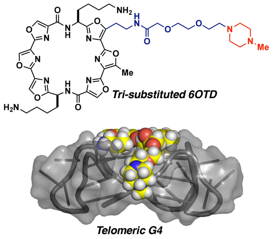
2.1. Design of 6OTD Derivatives for Targeting Telomeric G4

G-quadruplex structures form a variety of topologies depending upon their sequences, and a promising strategy to design “sequence-dependent” selective ligands [37,38,39] is to target both the G-quartet and the G4 grooves. We previously analyzed the interaction mode of L2H2-6M(2)OTD 1 with telomeric DNA by NMR, which is shown in Figure 1a [40], and found that the core structure of the macrocyclic moiety in 6OTD 1 interacts with the G-quartet of telomeric G4 through a π-π interaction. In addition, the two aminoalkyl side chains in 1 interact with the phosphate backbone of DNA. Notably, the architecture, as shown in Figure 1a, includes an additional potential ligand-binding site, i.e., the groove circled in blue. Thus, based on the structure depicted in Figure 1b, we designed tri-substituted 6OTD 2, which has an additional side chain at C5 of L2H2-6M(2)OTD 1. We expected that this modification would increase the affinity for telomeric G4 and the specificity of the interaction. Specifically, we designed three types of novel tri-substituted L2H2-6OTD derivatives 35, bearing an aminoethyl group, an aminoethoxyethyl group, and a piperazinyethoxyethyl group at C5 in 2, respectively, and examined their specific interactions with telomeric DNA, as shown in Figure 2.

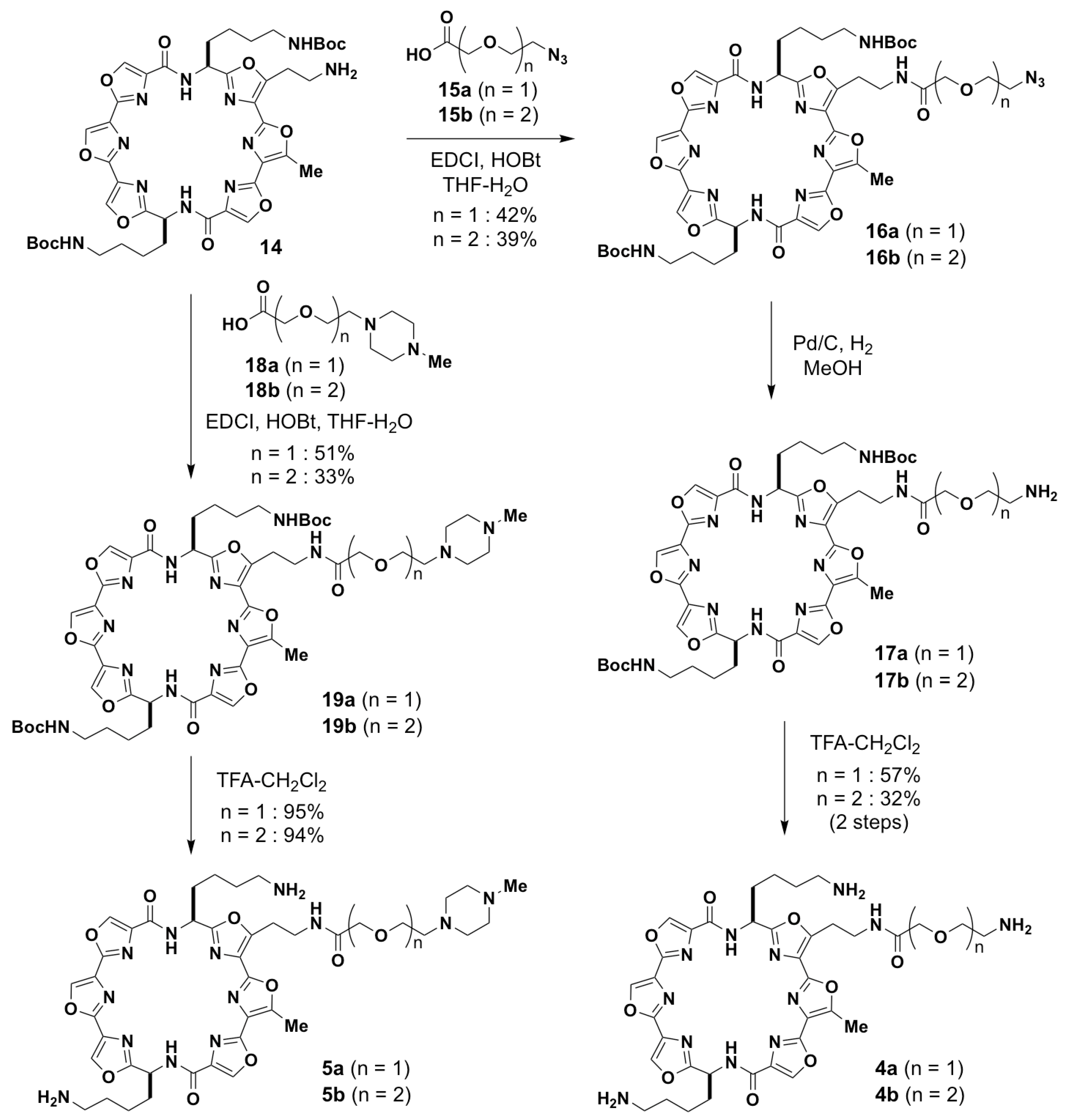
2.2. Synthesis of Tri-Substituted 6OTD Derivatives 35

We synthesized tri-substituted 6OTD derivatives 35 as summarized in Scheme 1 and Scheme 2. Firstly, the synthesis of tri-amine 3 was carried out. Briefly, the bis-Boc-protected tri-substituted 6OTD 14 was synthesized from trioxazoles 6 and 8 based upon the procedure developed by our group [41,42]. Then, the two Boc groups in 14 were deprotected with TFA to give tri-amine 3 in 81% yield from 13. Compounds 4 and 5 were synthesized as follows: The amine 14 was reacted with carboxylic acids 15a (n = 1) and 15b (n = 2) bearing an azide functional group in the presence of EDCI and HOBt, and the corresponding amides 16a and 16b were obtained in 42% and 39% yields, respectively. After reduction of the azide group in 16 under hydrogen in the presence of Pd/C as a catalyst, the resulting amine 17 was deprotected with TFA to give tri-amines 4a and 4b in 94% and 90% yields from 16, respectively. The tri-substituted L2H2-6OTD derivatives of 5a and 5b, bearing an N-methylpiperazine-substituted side chain, were synthesized similarly to 4 from amine 14 and carboxylic acids 18a (n = 1) and 18b (n = 2).

2.3. Stabilization Abilities of 35 for G4-Forming DNA Sequences by Fluorescence Resonance Energy Transfer (FRET) Melting Analysis

Next, the abilities of 35 to stabilize telomere, c-kit, and K-ras G4-forming DNA sequences were compared with that of L2H2-6M(2)OTD 1 by means of FRET melting assay, with results shown in Table 1 [43]. In the case of telomeric G4, telo21, the ΔTm value was increased by ligands 35 bearing additional side chains compared with L2H2-6M(2)OTD 1, except for with 5a. In regards to other G4-forming DNA sequences, slight increases of ΔTm for c-kit were observed with ligands 3, 4a, and 4b, while significant decreases were observed with 5a and 5b. In the case of K-ras, the ΔTm values were slightly decreased with ligands 5a and 5b, respectively. These results suggest that 5b interacts selectively with telomeric G4 over the G4-forming DNA sequences of c-kit and K-ras, among the ligands and DNA sequences we examined.

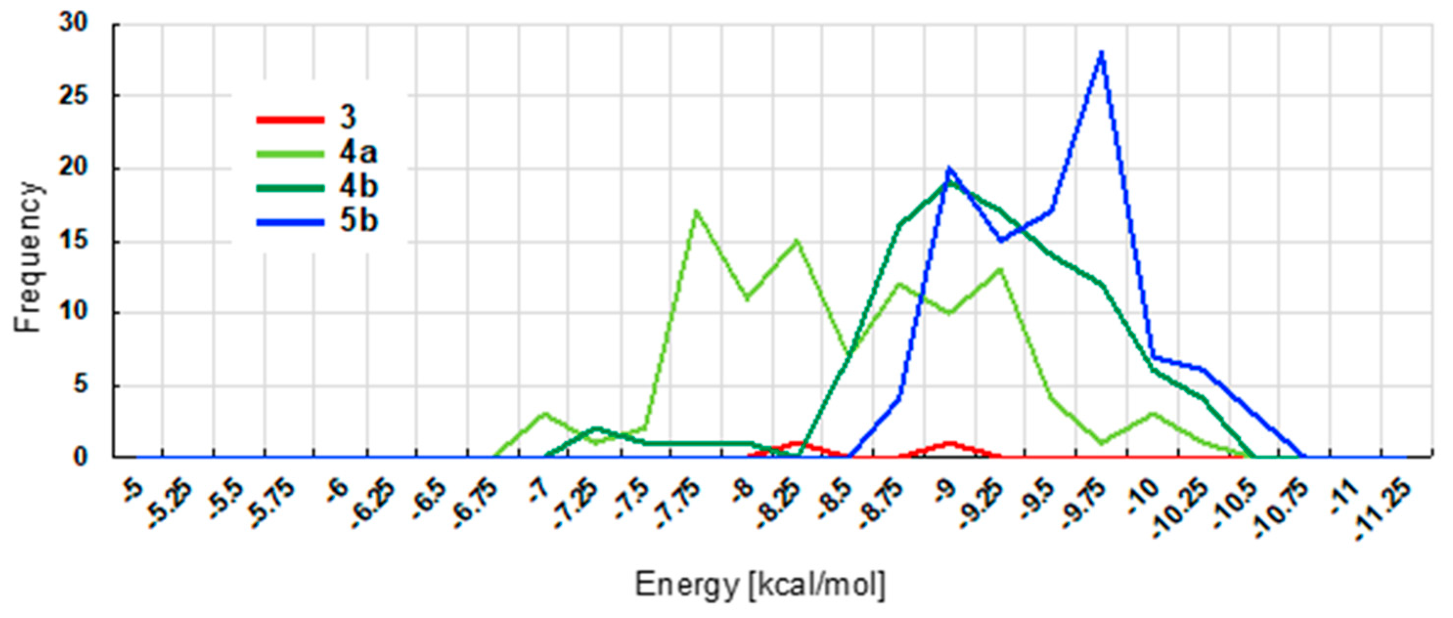
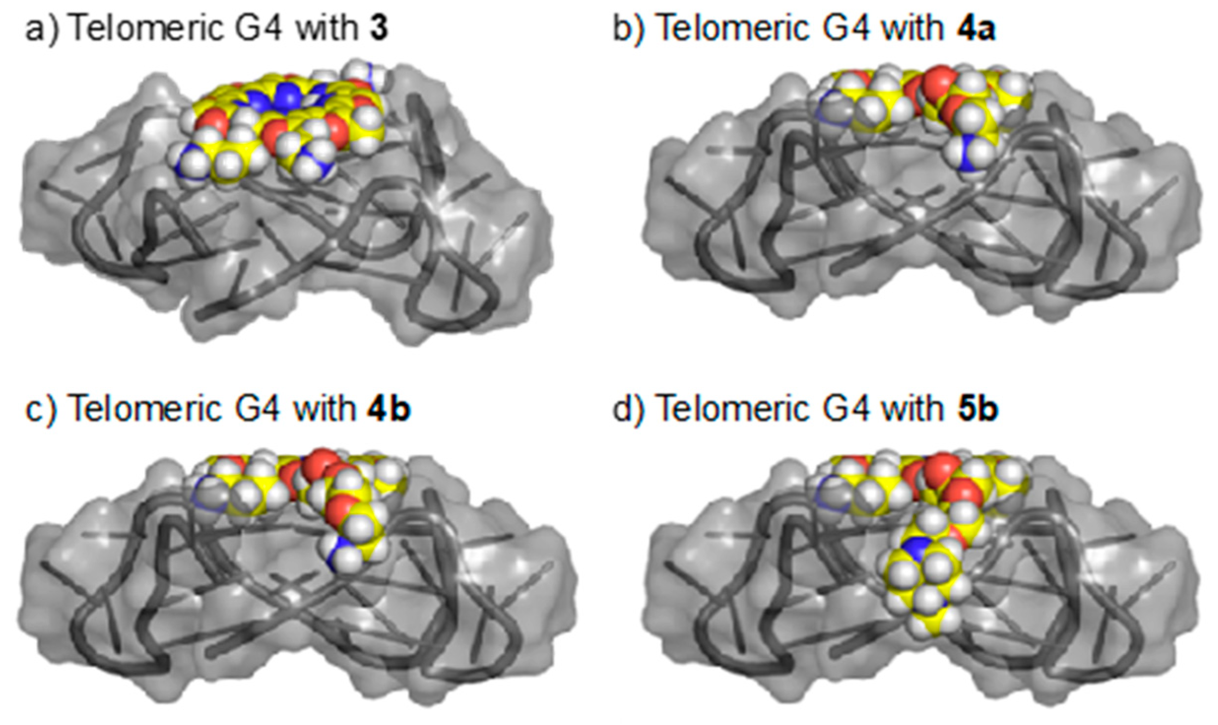
2.4. Docking Studies of Ligands 3, 4, and 5b with Telomeric G4

To gain insight into the differences in the stabilizing abilities of 3, 4, and 5b, docking studies with telomeric G4 were carried out. Since the three topologies of telomeric DNA [44] (i.e., anti-parallel, hybrid, and parallel) are affected by ionic conditions, solvents, and ligands, we first analyzed the telomeric G4 topologies induced by ligands 35 by means of Circular Dichroism (CD) spectroscopy [45]. As shown in Figure 3, telomeric G4 was induced predominantly into the parallel form in the presence of ligands 3, 4, and 5b under cation-free conditions. On the other hand, in case of ligand 5a, the telomeric G4 was induced mainly into parallel-type topology, but did not show a single, distinctive spectrum, in contrast to the other ligands [46].
With the CD analysis results in hand, docking studies were carried out between parallel-type telomeric G4 and ligands 3, 4, and 5b. The docking energies between the ligands and parallel-type telomeric G4 were calculated, and the distribution of the energies for the top 100 poses is shown in Figure 4 [47]. In these calculations, 5b mainly showed the lowest-energy docking (ca. −9 to −10.5 kcal/mol) among the ligands tested. The next lowest energy was found for 4b, followed by 4a, and 3, which is consistent with the results of the FRET melting analysis, which were shown in Table 1.
Docking models with the lowest energy scores for ligands 3, 4, and 5b are depicted in Figure 5. In every case, the hexaoxazole moiety in the ligands stacks with the G-quartet, and the newly introduced side chain is suggested to interact with the groove in the parallel-type telomeric G4. The two nitrogens of piperazine in the side chain in 5b were suggested to interact efficiently with the phosphate backbone of DNA, which may contribute to 5b’s potent stabilizing ability for telomeric DNA, compared to 3 and 4. In the case of 5a, the newly introduced side chain may have an inappropriate chain length for binding with the groove, resulting in a weak stabilizing ability for parallel-type telomeric G4.

3. Materials and Methods

3.1. General

Flash chromatography was performed on Silica gel 60 (spherical, particle size 0.040–0.100 mm; Kanto Co., Inc., Tokyo, Japan). Preparative-TLC (PLC) was performed using PLC Silica gel 60 F254 (0.5 mm, Merck Ltd., Darmstadt, Germany). Optical rotations were measured on a JASCO P-2200 polarimeter. 1H- and 13C-NMR spectra were recorded on JEOL JNM-AL300 (300 MHz), JEOL JNM-ECX 400 (400 MHz), and JEOL JNM-ECA 500 (500 MHz). The spectra were referenced internally according to the residual solvent signals of CDCl3 (1H-NMR, δ = 7.26 ppm; 13C-NMR, δ = 77.0 ppm) and DMSO-d6 (1H-NMR, δ = 2.50 ppm; 13C-NMR, δ = 39.5 ppm). Dates for 1H-NMR were recorded as follows: Chemical shift (δ, ppm), multiplicity (s, singlet; t, triplet; m, multiplet; br, broad), integration, and coupling constant (Hz). Dates for 13C-NMR were reported in terms of chemical shift (δ, ppm). Mass spectra were recorded on a JEOL JMS-T100LC spectrometer with ESI-MS mode, using MeOH as solvent.

3.2. Synthesis Methods

Compound 10: To a solution of trioxazole 6 [41] (1.00 g, 1.36 mmol) in THF-H2O (3:1, 80 mL) was added LiOH·H2O (85.5 mg, 2.04 mmol) at room temperature, and stirred for 30 min. The reaction was acidified with 3 N HCl to give carboxylic acid 7 as a THF-H2O solution, which was used without further purification. To a solution of trioxazole 8 [42] (1.20 g, 1.40 mmol) in MeOH-THF (1:3, 80 mL) was added 10% Pd/C (120 mg), and the mixture was stirred at room temperature under a hydrogen atmosphere for 10 min. The reaction mixture was filtered through a pad of Celite® (FUJIFILM Wako Pure Chemical Co., Osaka, Japan) and eluted with CHCl3-MeOH (9:1). The filtrates were concentrated in vacuo to give an amine 9, which was used without further purification. To a solution of carboxylic acid 7 (THF-H2O) was added NMM (451 µL, 4.08 mmol), DMT-MM (1.12 g, 4.08 mmol), and amine 9, and the mixture was stirred at room temperature. After being stirred overnight, to the reaction mixture was added H2O, and the organic layer was extracted with CHCl3, washed with 1.2 N HCl, dried over MgSO4, and filtered. The filtrates were concentrated in vacuo, and the residue was purified by column chromatography on silica gel (CHCl3:MeOH = 200:1) to give bistrioxazole 10 (1.20 g, 75%, two steps). Spectral data for 10: [ α ] D 25 = −17.8 (c 1.1, CHCl3); 1H-NMR (400 MHz, CDCl3) δ 8.31–8.27 (m, 4H), 8.06 (d, J = 8.7 Hz, 1H), 7.26–7.21 (m, 5H), 5.90 (m, 1H), 5.47 (m, 1H), 5.33–5.20 (m, 2H), 5.03–4.94 (m, 3H), 4.66–4.57 (m, 3H), 3.95 (s, 3H), 3.73–3.62 (m, 2H), 3.45 (t, J = 6.4 Hz, 2H), 3.10 (br, 4H), 2.71 (s, 3H), 2.26–1.78 (m, 4H), 1.52–1.40 (m, 26H); 13C-NMR (100 MHz, CDCl3) δ 165.0, 161.1, 160.2, 156.3, 155.9, 155.8, 155.2, 151.2, 150.9, 143.8, 141.1, 139.5, 139.2, 139.1, 136.4, 136.1, 134.1, 132.4, 130.5, 129.5, 128.2, 127.8, 127.6, 126.5, 124.3, 117.7, 78.9, 78.8, 77.2, 66.5, 65.8, 54.5, 52.2, 49.0, 46.7, 40.0, 39.8, 38.7, 33.4, 32.5, 29.3, 28.2, 26.7, 22.7, 22.3, 11.6; HRMS (ESI, M + Na) calcd. for C56H67N11O17Na 1188.4614, found 1188.4628.
Compound 11: To a solution of bistrioxazole 10 (400 mg, 340 µmol) in THF (100 mL) was added morpholine (298 µL, 3.43 mmol) and Pd(PPh3)4 (118 mg, 100 µmol) at room temperature under argon atmosphere and stirred for 10 min. The reaction mixture was concentrated in vacuo, and the residue was purified by column chromatography on silica gel (CHCl3:MeOH = 100:3) to give amine 11 (350 mg, 84%). Spectral data for 11: [ α ] D 25 = +5.33 (c 1.4, CHCl3); 1H-NMR (400 MHz, CDCl3) δ 8.31 (s, 1H), 8.30 (s, 1H), 8.27 (s, 1H), 8.07 (d, J = 8.7 Hz, 1H), 7.26–7.22 (m, 7H), 5.50–5.44 (m, 1H), 5.24 (br, 1H), 5.04–4.96 (m, 2H), 4.66 (br, 1H), 4.55 (br, 1H), 4.04 (t, J = 6.9 Hz, 2H), 3.95 (s, 3H), 3.75–3.44 (m, 4H), 3.12–3.10 (br, 4H), 2.72 (s, 3H), 2.25–0.86 (m, 30H); 13C-NMR (100 MHz, CDCl3) δ 166.3, 165.1, 161.2, 160.3, 156.3, 155.9, 155.3, 151.1, 143.8, 141.1, 139.6, 139.2, 136.5, 136.1, 134.3, 130.6, 129.6, 128.3, 128.0, 127.7, 79.0, 78.9, 77.2, 70.5, 66.7, 52.3, 49.9, 46.8, 40.2, 40.0, 38.9, 35.4, 32.6, 29.7, 29.3, 28.3, 26.8, 22.9, 22.7, 11.8; HRMS (ESI, M + Na) calcd. for C52H63N11O15Na 1104.4403, found 1104.4370.
Compound 3: To a solution of 13 (70.0 mg, 66.7 µmol) in MeOH (6 mL) was added 10% Pd/C (50.0 mg), and the mixture was stirred at room temperature under a hydrogen atmosphere for 10 min. The reaction mixture was filtered through a pad of Celite® and eluted with CHCl3-MeOH (9:1). The filtrates were concentrated in vacuo to give amine 14, which was used without further purification. The amine 14 was dissolved in CH2Cl2-TFA (10:1, 11 mL), and the resulting solution was stirred at room temperature for 10 min. The reaction mixture was concentrated in vacuo to give 3 (38.7 mg, 81%, two steps). Spectral data for 3: [ α ] D 25 = +46.1 (c 1.1, MeOH); 1H-NMR (400 MHz, DMSO-d6) δ 9.14–9.12 (m, 2H), 8.92–8.90 (m, 2H), 8.33–8.27 (m, 2H), 8.15 (br, 2H), 7.75 (br, 4H), 5.45 (dt, J = 12.8, 5.5 Hz, 1H), 5.37 (dt, J = 12.8, 5.5 Hz, 1H), 3.75–3.29 (m, 4H), 2.81–2.73 (m, 7H), 2.11–1.88 (m, 4H), 1.54–1.19 (m, 8H); 13C-NMR (100 MHz, DMSO-d6) δ 164.4, 162.0, 158.9, 158.8, 158.7, 158.3, 155.7, 155.3, 155.1, 154.6, 151.9, 149.8, 142.5, 142.4, 141.8, 141.1, 136.0, 136.0, 129.7, 128.5, 125.9, 123.8, 118.4, 47.2, 47.1, 36.9, 33.3, 26.7, 23.9, 20.9, 11.5; HRMS (ESI, M + H) calcd. for C33H38N11O8 716.2904, found 716.2918.
Compound 16a: To a solution of 15a (10.0 mg, 66.6 µmol) in THF-H2O (2:1, 3 mL) was added EDCI (38.0 mg, 200 µmol) and HOBt (27.0 mg, 200 µmol) at room temperature. After stirring for 1 h, to the reaction mixture was added amine 14 (61.0 mg, 66.6 µmol). The mixture was stirred for 24 h and quenched with 1.2 N HCl, and the organic layer was extracted with CHCl3, dried over MgSO4, and filtered. The filtrates were concentrated in vacuo, and the residue was purified by column chromatography on silica gel (CHCl3:MeOH = 80:1) to give 16a (29.0 mg, 42%). Spectral data for 16a: [ α ] D 25 = −14.5 (c 0.58, CHCl3); 1H-NMR (300 MHz, DMSO-d6) δ 9.12 (s, 1H), 9.09 (s, 1H), 8.91 (s, 1H), 8.89 (s, 1H), 8.34-8.27 (m, 2H), 7.98 (t, J = 6.2 Hz, 1H), 6.77 (t, J = 5.2 Hz, 2H), 5.42 (dt, J = 12.4, 7.2 Hz, 1H), 5.32 (dt, J = 12.4, 7.2 Hz, 1H), 3.84 (d, J = 15.5 Hz, 1H), 3.78 (d, J = 15.1 Hz, 1H), 3.61–3.27 (m, 6H), 2.84 (br, 4H), 2.75 (s, 3H), 2.04 (br, 2H), 1.90 (br, 2H), 1.33–1.03 (m, 28H); 13C-NMR (125 MHz, DMSO-d6) δ 169.0, 164.5, 162.5, 158.9, 158.7, 155.7, 155.5, 154.5, 154.3, 152.5, 151.0, 142.5, 142.3, 141.7, 141.0, 136.0, 135.9, 129.8, 128.4, 124.8, 124.6, 79.2, 77.2, 69.7, 69.5, 49.8, 47.4, 47.2, 36.6, 33.5, 33.4, 29.2, 28.2, 26.1, 21.1, 11.5; HRMS (ESI, M + Na) calcd. for C47H58N14O14Na 1065.4155, found 1065.4143.
Compound 4a: To a solution of 16a (20.0 mg, 19.2 µmol) in MeOH (4 mL) was added 10% Pd/C (20.0 mg), and the mixture was stirred at room temperature under a hydrogen atmosphere for 30 min. The reaction mixture was filtered through a pad of Celite® and eluted with CHCl3-MeOH (9:1). The filtrates were concentrated in vacuo to give amine 17a, which was used without further purification. The amine 17a was dissolved in CH2Cl2-TFA (2:1, 3 mL), and the resulting solution was stirred at room temperature for 10 min. The reaction mixture was concentrated in vacuo to give 4a (8.90 mg, 57%, two steps). Spectral data for 4a: [ α ] D 25 = +87.8 (c 2.3, MeOH); 1H-NMR (500 MHz, DMSO-d6) δ 9.15 (s, 1H), 9.14 (s, 1H), 8.94 (s, 1H), 8.92 (s, 1H), 8.31 (d, J = 7.5 Hz, 1H), 8.28 (d, J = 7.5 Hz, 1H), 8.23 (t, J = 5.7 Hz, 1H), 7.79 (br, 6H), 5.44 (dt, J = 12.6, 7.5 Hz, 1H), 5.34 (dt, J = 12.6, 7.5 Hz, 1H), 3.84 (d, J = 14.9 Hz, 1H), 3.80 (d, J = 15.5 Hz, 1H), 3.62-3.21 (m, 4H), 3.00–2.92 (m, 2H), 2.77–2.73 (m, 7H), 2.09–1.91 (m, 4H), 1.53–1.21 (m, 10H); 13C-NMR (125 MHz, DMSO-d6) δ 169.0, 164.5, 158.9, 158.8, 155.7, 155.6, 155.7, 155.6, 154.6, 154.3, 152.5, 151.2, 142.6, 142.4, 141.9, 141.2, 136.0, 135.9, 129.7, 128.5, 124.7, 124.6, 69.7, 66.9, 47.2, 47.1, 38.6, 36.6, 33.5, 33.4, 26.7, 26.2, 20.9, 11.5; HRMS (ESI, M + Na) calcd. for C37H44N12O10Na 839.3201, found 839.3196.
Compound 16b: To a solution of 15b (15.0 mg, 77.3 µmol) in THF-H2O (4:1, 2.5 mL) was added EDCI (44.6 mg, 230 µmol) and HOBt (31.3 mg, 230 µmol) at room temperature. After stirring for 1 h, to the reaction mixture was added amine 14 (70.0 mg, 77.3 µmol). The mixture was stirred for 24 h and quenched with 1.2 N HCl, and the organic layer was extracted with CHCl3, dried over MgSO4, and filtered. The filtrates were concentrated in vacuo, and the residue was purified by column chromatography on silica gel (CHCl3:MeOH = 80:1) to give 16b (32.7 mg, 39%). Spectral data for 16b: [ α ] D 25 = −7.6 (c 1.7, CHCl3); 1H-NMR (500 MHz, DMSO-d6) δ 9.12 (s, 1H), 9.10 (s, 1H), 8.91 (s, 1H), 8.89 (s, 1H), 8.34–8.28 (m, 2H), 7.91 (t, J = 6.3 Hz, 1H), 6.78 (t, J = 5.7 Hz, 2H), 5.42 (dt, J = 12.6, 7.5 Hz, 1H), 5.32 (dt, J = 12.6, 7.5 Hz, 1H), 3.80 (d, J = 15.5 Hz, 1H), 3.75 (d, J = 14.9 Hz, 1H), 3.61–3.22 (m, 10H), 2.90–2.80 (m, 4H), 2.74 (s, 3H), 2.03 (br, 2H), 1.89 (br, 2H), 1.41–0.85 (m, 28H); 13C-NMR (125 MHz, DMSO-d6) δ 169.3, 164.6, 162.5, 158.9, 158.7, 155.7, 155.5, 154.5, 154.3, 152.5, 151.0, 142.5, 142.3, 141.7, 141.0, 136.0, 135.9, 129.8, 128.4, 124.8, 124.6, 79.2, 77.2, 70.1, 70.0, 69.3, 69.2, 50.0, 47.4, 47.2, 36.6, 33.5, 33.4, 29.2, 28.2, 29.1, 21.1, 21.0, 11.5; HRMS (ESI, M + Na) calcd. for C49H62N14O15Na 1109.4417, found 1109.4387.
Compound 4b: To a solution of 16b (8.00 mg, 7.35 µmol) in MeOH (1 mL) was added 10% Pd/C (10.0 mg), and the mixture was stirred at room temperature under a hydrogen atmosphere for 10 min. The reaction mixture was filtered through a pad of Celite® and eluted with CHCl3-MeOH (9:1). The filtrates were concentrated in vacuo to give amine 17b, which was used without further purification. The amine 17b was dissolved in CH2Cl2-TFA (2:1, 3 mL), and the resulting solution was stirred at room temperature for 10 min. The reaction mixture was concentrated in vacuo to give 4b (2.03 mg, 32%, 2 steps). Spectral data for 4b: [ α ] D 25 = +58.2 (c 2.1, MeOH); 1H-NMR (500 MHz, DMSO-d6) δ 9.15 (s, 1H), 9.14 (s, 1H), 8.94 (s, 1H), 8.92 (s, 1H), 8.30 (d, J = 7.5 Hz, 1H), 8.28 (d, J = 8.0 Hz, 1H), 8.07 (br, 1H), 7.76 (br, 6H), 5.44 (dt, J = 12.6, 6.9 Hz, 1H), 5.35 (dt, J = 12.6, 7.5 Hz, 1H), 3.82–2.63 (m, 19H), 2.09–1.87 (m, 4H), 1.53–1.22 (m, 10H); 13C-NMR (125 MHz, DMSO-d6) δ 169.5, 164.5, 162.4, 159.0, 158.8, 158.4, 158.1, 155.7, 155.6, 154.6, 154.4, 152.6, 151.2, 142.6, 142.4, 141.9, 141.2, 137.4, 136.0, 135.9, 129.7, 128.9, 128.5, 128.2, 125.4, 124.7, 124.6, 69.9, 69.3, 66.7, 47.2, 47.0, 38.6, 36.6, 33.5, 33.4, 26.8, 26.1, 21.1, 21.0, 20.9, 11.6; HRMS (ESI, M + Na) calcd. for C39H48N12O11Na 883.3463, found 883.3479.
Compound 19a: To a solution of 18a (9.60 mg, 47.6 µmol) in THF-H2O (2:1, 3 mL) was added EDCI (27.4 mg, 143 µmol) and HOBt (19.3 mg, 143 µmol) at room temperature. After stirring for 1 h, to the reaction mixture was added amine 14 (43.6 mg, 47.6 µmol). The mixture was stirred for 24 h and quenched with 1.2 N HCl, and the organic layer was extracted with CHCl3, dried over MgSO4, and filtered. The filtrates were concentrated in vacuo, and the residue was purified by column chromatography on silica gel (CHCl3:MeOH = 10:1) to give 19a (40.0 mg, 76%). Spectral data for 19a: [ α ] D 25 = −0.25 (c 1.0, CHCl3); 1H-NMR (500 MHz, DMSO-d6) δ 9.12 (s, 1H), 9.09 (s, 1H), 8.91 (s, 1H), 8.90 (s, 1H), 8.32 (d, J = 8.0 Hz, 1H), 8.29 (d, J = 7.5 Hz, 1H), 8.12 (t, J = 6.3 Hz, 1H), 6.78 (t, J = 5.7 Hz, 2H), 5.42 (dt, J = 12.6, 7.5 Hz, 1H), 5.31 (dt, J = 12.6, 7.5 Hz, 1H), 3.77 (d, J = 15.5 Hz, 1H), 3.72 (d, J = 14.9 Hz, 1H), 3.62-3.24 (m, 4H), 2.84 (br, 4H), 2.74 (s, 3H), 2.36–1.88 (m, 17H), 1.33–1.03 (m, 28H); 13C-NMR (125 MHz, DMSO-d6) δ 169.7, 164.5, 162.5, 158.9, 158.7, 155.7, 155.5, 154.5, 154.3, 152.5, 151.0, 142.5, 142.3, 141.7, 141.0, 136.1, 136.0, 136.1, 135.9, 129.8, 128.5, 124.8, 124.7, 79.2, 77.3, 70.0, 68.5, 56.9, 54.7, 52.9, 47.4, 47.3, 45.7, 36.6, 33.4, 29.2, 28.2, 26.1, 21.1, 11.6; HRMS (ESI, M + Na) calcd. for C52H69N13O14Na 1122.4985, found 1122.4991.
Compound 5a: To a solution of 19a (6.70 mg, 6.01 μmol) in CH2Cl2 (2 mL) was added TFA (1 mL) at room temperature, and the mixture was stirred for 10 min. The reaction mixture was concentrated in vacuo to give 5a (5.14 mg, 95%). Spectral data for 5a: [ α ] D 25 = +85.6 (c 3.4, MeOH); 1H-NMR (500 MHz, DMSO-d6) δ 9.16 (s, 1H), 9.14 (s, 1H), 8.94 (s, 1H), 8.92 (s, 1H), 8.32 (d, J = 7.5 Hz, 1H), 8.30 (d, J = 8.0 Hz, 1H), 8.04 (t, J = 6.3 Hz, 1H), 7.71 (br, 4H), 5.44 (dt, J = 12.6, 6.9 Hz, 1H), 5.34 (dt, J = 12.6, 7.5 Hz, 1H), 3.81 (d, J = 15.5 Hz, 1H), 3.76 (d, J = 14.9 Hz, 1H), 3.62–3.19 (m, 15H), 2.98 (br, 2H), 2.75 (br, 7H), 2.07–1.87 (m, 4H), 1.53–1.23 (m, 10H); 13C-NMR (125 MHz, DMSO-d6) δ 169.1, 164.5, 162.5, 159.0, 158.8, 158.5, 155.7, 155.6, 154.6, 154.4, 152.6, 151.2, 142.6, 142.4, 141.9, 141.2, 137.4, 136.0, 135.9, 129.8, 129.0, 128.5, 128.3, 125.4, 124.8, 124.7, 117.7, 115.3, 69.5, 47.3, 47.1, 38.6, 33.5, 33.4, 26.8, 26.7, 26.0, 21.1, 21.0, 11.5; HRMS (ESI, M + H) calcd. for C42H54N13O10 900.4117, found 900.4098.
Compound 19b: To a solution of 18b (9.40 mg, 38.1 µmol) in THF-H2O (2:1, 3 mL) was added EDCI (22.0 mg, 114 µmol) and HOBt (15.0 mg, 114 µmol) at room temperature. After stirring for 1 h, to the reaction mixture was added amine 14 (34.9 mg, 38.1 µmol). The mixture was stirred for 24 h and quenched with 1.2 N HCl, and the organic layer was extracted with CHCl3, dried over MgSO4, and filtered. The filtrates were concentrated in vacuo, and the residue was purified by column chromatography on silica gel (CHCl3:MeOH = 10:1) to give 19b (14.4 mg, 33%). Spectral data for 19b: [ α ] D 25 = −6.7 (c 0.65, CHCl3); 1H-NMR (500 MHz, DMSO-d6) δ 9.12 (s, 1H), 9.09 (s, 1H), 8.91 (s, 1H), 8.90 (s, 1H), 8.34-8.28 (m, 2H), 7.95 (t, J = 5.7 Hz, 1H), 6.78 (t, J = 5.7 Hz, 2H), 5.42 (dt, J = 12.6, 7.5 Hz, 1H), 5.32 (dt, J = 12.6, 7.5 Hz, 1H), 3.79 (d, J = 15.5 Hz, 1H), 3.73 (d, J = 15.5 Hz, 1H), 3.60–3.16 (m, 8H), 2.86–2.84 (m, 4H), 2.74 (s, 3H), 2.37–1.88 (m, 17H), 1.33–1.04 (m, 28H); 13C-NMR (125 MHz, DMSO-d6) δ 1 169.9, 165.1, 163.0, 162.8, 159.4, 159.2, 156.2, 156.0, 155.0, 154.8, 153.0, 151.5, 143.0, 142.8, 142.2, 141.5, 136.6, 136.4, 130.3, 129.0, 125.3, 125.1, 79.7, 77.7, 70.5, 70.4, 69.9, 68.7, 57.7, 55.2, 53.5, 47.9, 47.7, 46.3, 37.1, 36.3, 34.0, 33.9, 29.7, 28.7, 26.6, 21.6, 21.5, 12.0; HRMS (ESI, M + Na) calcd. for C54H73N13O15Na 1166.5247, found 1166.5238.
Compound 5b: To a solution of 19b (14.4 mg, 12.6 μmol) in CH2Cl2 (2 mL) was added TFA (1 mL) at room temperature, and the mixture was stirred for 10 min. The reaction mixture was concentrated in vacuo to give 5b (11.2 mg, 94%). Spectral data for 5b: [ α ] D 25 = +29.7 (c 4.4, MeOH); 1H-NMR (500 MHz, DMSO-d6) δ 9.15 (s, 1H), 9.14 (s, 1H), 8.94 (s, 1H), 8.92 (s, 1H), 8.31 (d, J = 7.5 Hz, 1H), 8.28 (d, J = 7.5 Hz, 1H), 8.02 (br, 1H), 7.73 (br, 4H), 5.45 (dt, J = 12.6, 7.5 Hz, 1H), 5.34 (dt, J = 12.6, 7.5 Hz, 1H), 3.80 (d, J = 15.5 Hz, 1H), 3.76 (d, J = 15.5 Hz, 1H), 3.60–3.19 (m, 19H), 2.75 (br, 9H), 2.08–1.87 (m, 4H), 1.53–1.17 (m, 10H); 13C-NMR (125 MHz, DMSO-d6) δ 169.4, 164.5, 162.4, 158.8, 155.7, 152.7, 151.2, 141.9, 141.2, 136.0, 128.9, 128.3, 124.8, 117.7, 115.3, 79.2, 69.9, 69.8, 69.3, 47.2, 47.1, 38.6, 33.5, 33.4, 26.8, 26.7, 26.1, 21.1, 21.0, 20.9, 11.5; HRMS (ESI, M + H) calcd. for C44H58N13O11 944.4379, found 944.4344.

3.3. FRET Melting Analysis

Fluorescence resonance energy transfer (FRET) melting assay was performed with an excitation wavelength of 470–505 nm and a detection wavelength of 523–543 nm using the DNA Engine Option 2 Real-Time Cycler PCR detection system (Biorad, Hercules, CA, USA). The dual fluorescently labeled oligonucleotides were used in this protocol (Table S1). The donor fluorophore was 6-carboxyfluorescein (FAM) and the acceptor fluorophore was 6-carboxytetramethylrhodamine (TAMRA). All purified nucleotides (Sigma Genosys, Tokyo, Japan) were dissolved as stock solutions (100 μM) in MilliQ water to be used without further purification. Further dilutions of the oligonucleotides were performed with a 60 mM potassium cacodylate buffer (pH 7.4), and FRET experiments were carried out with a 0.4 μM oligonucleotide solution. Dual-labeled DNA was annealed by heating at 99 °C for 5 min, and then slowly cooled to room temperature. Ligands were prepared as DMSO stock solutions (10 mM) and diluted to 1 mM using DMSO, and then diluted to 100 μM using a 60 mM potassium cacodylate buffer (pH 7.4). Next, the annealed DNA (20 μL, 0.4 μM) and the compound solution (20 μL, 2 μM) were distributed across 96-well plates (Takara), with a total volume of 40 μL, with the labeled oligonucleotide (0.2 μM) and the compound (1.0 μM). The plates were incubated at 25 °C for 12 h. Subsequent experiments used the following temperature procedure in RT-PCR, finishing as follows: 25 °C for 20 min, and then a stepwise increase of 1 °C every minute from 25 °C until 99 °C. During the procedures, we measured the FAM fluorescence after each step. The change in the melting temperature at 1.0 μM compound concentration (ΔTm (1.0 μM)) was calculated from at least three experiments by subtraction of the blank from the averaged melting temperature of each compound.

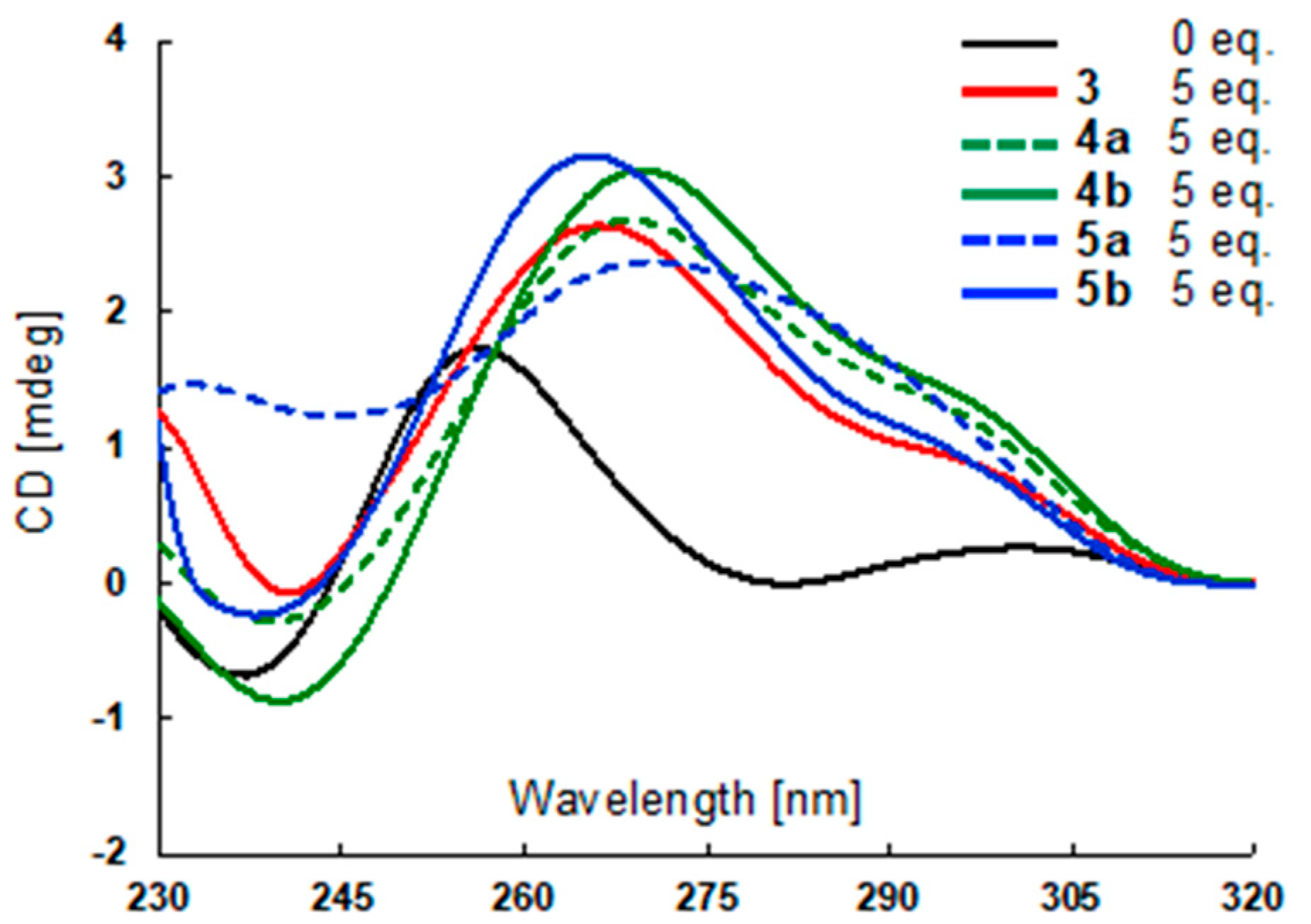
3.4. CD Spectrometry

Circular Dichroism (CD) spectra were recorded on a J-720 spectropolarimeter (JASCO, Tokyo, Japan), using a quartz cell of 1 mm optical path length and an instrument scanning speed of 500 nm/min with a response time of 1 s, and over a wavelength range of 220–320 nm. The oligonucleotide telo24 used in this protocol (Table S1) was dissolved as a 1.0 mM stock solution in MilliQ water, to be used without further purification. Further dilution of the nucleotide was done with a 50 mM Tris-HCl buffer (without ions or with 50 mM KCl) from 1 mM stock solutions, to give a concentration of 10 μM. The solution was annealed by heating at 99 °C for 5 min, and then slowly cooled to room temperature, and then titrated into the oligonucleotide samples up to 5 mol equivalents using ligands, and incubated overnight. Finally, the CD spectra are representative of five averaged scans taken at 25 °C.

3.5. Computational Analysis

For the initial coordinates of compounds 3, 4, and 5, ionization and energy minimization were performed by the OPLS3 force field in the LigPrep Script in the Maestro (Schrödinger, LLC, New York, NY, USA). These minimized structures were employed as input structures for docking simulations. The human telomeric G4 structures with parallel-type topologies (PDB ID: 1KF1 and 3UYH) were refined for docking simulations using constrained energy refinements in the OPLS-AA force field (Schrödinger LLC). Docking simulations were performed using the Glide [48,49] SP docking program (Schrödinger, LLC, New York, NY, USA).

4. Conclusions

In conclusion, we have newly synthesized tri-substituted 6OTD derivatives 35 by introducing aminoethyl, aminoethoxyethyl, and piperazinyethoxyethyl groups at C5 of L2H2-6M(2)OTD 1, respectively. Among these ligands, 5b stabilizes telomeric G4 preferentially over c-kit and K-ras G4s. Docking studies with parallel-type telomeric G4 revealed that the hexaoxazole moiety of 5b stacks with the G-quartet of telomeric G4, and the newly introduced side chain efficiently interacts with the groove.

Supplementary Materials

The following are available online.

Author Contributions

Conceptualization, A.T.P. and K.N.; investigation, Y.M., K.I., and S.S.; molecular calculation, T.H. and B.H.; writing—original draft preparation, Y.M. and K.N.; writing—review and editing, K.N.

Funding

This research was funded by Grants-in-Aid for Scientific Research, Japan Society for the Promotion of Science (JSPS) (Scientific Research (B) 26282214 to K.N.; Scientific Research (C) 18K05349 to K.I.), a Grant-in-Aid for Scientific Research on Innovative Areas “Middle Molecular Strategy” (18H04387 to K.N.), a Grant-in-Aid for JSPS Research Fellow (16J08216 to Y.M.), funds from the Platform Project for Supporting Drug Discovery and Life Science Research (Basis for Supporting Innovative Drug Discovery and Life Science Research (BINDS)) from the Japan Agency for Medical Research and Development (AMED, JP18am0101114 to TH), and grants from Nanyang Technological University (to A.T.P.).

Conflicts of Interest

The authors declare no conflict of interest.

References and Notes

  1. Davis, J.T. G-quartets 40 years later: From 5’-GMP to molecular biology and supramolecular chemistry. Angew. Chem. Int. Ed. 2004, 43, 668–698. [Google Scholar] [CrossRef] [PubMed]
  2. Todd, A.K.; Johnston, M.; Neidle, S. Highly prevalent putative quadruplex sequence motifs in human DNA. Nucleic Acids Res. 2005, 33, 2901–2907. [Google Scholar] [CrossRef] [PubMed] [Green Version]
  3. Huppert, J.L.; Balasubramanian, S. Prevalence of quadruplexes in the human genome. Nucleic Acids Res. 2005, 33, 2908–2916. [Google Scholar] [CrossRef] [PubMed] [Green Version]
  4. Huppert, J.L.; Balasubramanian, S. G-quadruplexes in promoters throughout the human genome. Nucleic Acids Res. 2007, 35, 406–413. [Google Scholar] [CrossRef] [PubMed]
  5. Huppert, J.L.; Bugaut, A.; Kumari, S.; Balasubramanian, S. G-quadruplexes: The beginning and end of UTRs. Nucleic Acids Res. 2008, 36, 6260–6268. [Google Scholar] [CrossRef] [PubMed]
  6. Verma, A.; Yadav, V.K.; Basundra, R.; Kumar, A.; Chowdhury, S. Evidence of genome-wide G4 DNA-mediated gene expression in human cancer cells. Nucleic Acids Res. 2009, 37, 4194–4204. [Google Scholar] [CrossRef] [PubMed] [Green Version]
  7. Chambers, V.S.; Marsico, G.; Boutell, J.M.; Di Antonio, M.; Smith, G.P.; Balasubramanian, S. High-throughput sequencing of DNA G-quadruplex structures in the human genome. Nat. Biotechnol. 2015, 33, 877–881. [Google Scholar] [CrossRef] [Green Version]
  8. Paeschke, K.; Capra, J.A.; Zakian, V.A. DNA replication through G-quadruplex motifs is promoted by the Saccharomyces cerevisiae Pif1 DNA helicase. Cell 2011, 145, 678–691. [Google Scholar] [CrossRef]
  9. Kanoh, Y.; Matsumoto, S.; Fukatsu, R.; Kakusho, N.; Kono, N.; Renard-Guillet, C.; Masuda, K.; Iida, K.; Nagasawa, K.; Shirahige, K.; et al. Rif1 binds to G quadruplexes and suppresses replication over long distances. Nat. Struct. Mol. Biol. 2015, 22, 889–897. [Google Scholar] [CrossRef]
  10. Siddiqui-Jain, A.; Grand, C.L.; Bearss, D.J.; Hurley, L.H. Direct evidence for a G-quadruplex in a promoter region and its targeting with a small molecule to repress c-MYC transcription. Proc. Natl. Acad. Sci. USA 2002, 99, 11593–11598. [Google Scholar] [CrossRef] [Green Version]
  11. Patel, D.J.; Phan, A.T.; Kuryavyi, V. Human telomere, oncogenic promoter and 5’-UTR G-quadruplexes: Diverse higher order DNA and RNA targets for cancer therapeutics. Nucleic Acids Res. 2007, 35, 7429–7455. [Google Scholar] [CrossRef] [PubMed]
  12. Lipps, H.J.; Rhodes, D. G-quadruplex structures: In vivo evidence and function. Trends Cell Biol. 2009, 19, 414–422. [Google Scholar] [CrossRef] [PubMed]
  13. Bugaut, A.; Balasubramanian, S. 5′-UTR RNA G-quadruplexes: Translation regulation and targeting. Nucleic Acids Res. 2012, 40, 4727–4741. [Google Scholar] [CrossRef] [PubMed]
  14. Le, D.D.; Di Antonio, M.; Chan, L.K.; Balasubramanian, S. G-quadruplex ligands exhibit differential G-tetrad selectivity. Chem. Commun. 2015, 51, 8048–8050. [Google Scholar] [CrossRef] [PubMed] [Green Version]
  15. Ranjan, N.; Davis, E.; Xue, L.; Arya, D.P. Dual recognition of the human telomeric G-quadruplex by a neomycin-anthraquinone conjugate. Chem. Commun. 2013, 49, 5796–5798. [Google Scholar] [CrossRef] [PubMed]
  16. Wei, C.Y.; Jia, G.Q.; Zhou, J.; Han, G.Y.; Li, C. Evidence for the binding mode of porphyrins to G-quadruplex DNA. Phys. Chem. Chem. Phys. 2009, 11, 4025–4032. [Google Scholar] [CrossRef] [PubMed]
  17. Phan, A.T.; Kuryavyi, V.; Gaw, H.Y.; Patel, D.J. Small-molecule interaction with a five-guanine-tract G-quadruplex structure from the human MYC promoter. Nat. Chem. Biol. 2005, 1, 167–173. [Google Scholar] [CrossRef] [Green Version]
  18. Chung, W.J.; Heddi, B.; Hamon, F.; Teulade-Fichou, M.P.; Phan, A.T. Solution structure of a G-quadruplex bound to the bisquinolinium compound Phen-DC3. Angew. Chem. Int. Ed. 2014, 53, 999–1002. [Google Scholar] [CrossRef]
  19. Marchand, A.; Granzhan, A.; Iida, K.; Tsushima, Y.; Ma, Y.; Nagasawa, K.; Teulade-Fichou, M.P.; Gabelica, V. Ligand-Induced Conformational Changes with Cation Ejection upon Binding to Human Telomeric DNA G-Quadruplexes. J. Am. Chem. Soc. 2015, 137, 750–756. [Google Scholar] [CrossRef]
  20. Hamon, F.; Largy, E.; Guedin-Beaurepaire, A.; Rouchon-Dagois, M.; Sidibe, A.; Monchaud, D.; Mergny, J.L.; Riou, J.F.; Nguyen, C.H.; Teulade-Fichou, M.P. An Acyclic Oligoheteroaryle That Discriminates Strongly between Diverse G-Quadruplex Topologies. Angew. Chem. Int. Ed. 2011, 50, 8745–8749. [Google Scholar] [CrossRef]
  21. Martino, L.; Virno, A.; Pagano, B.; Virgilio, A.; Di Micco, S.; Galeone, A.; Giancola, C.; Bifulco, G.; Mayol, L.; Randazzo, A. Structural and thermodynamic studies of the interaction of distamycin A with the parallel quadruplex structure [d(TGGGGT)]4. J. Am. Chem. Soc. 2007, 129, 16048–16056. [Google Scholar] [CrossRef]
  22. Iida, K.; Nagasawa, K. Macrocyclic Polyoxazoles as G-Quadruplex Ligands. Chem. Rec. 2013, 13, 539–548. [Google Scholar] [CrossRef]
  23. Kim, M.Y.; Vankayalapati, H.; Shin-Ya, K.; Wierzba, K.; Hurley, L.H. Telomestatin, a potent telomerase inhibitor that interacts quite specifically with the human telomeric intramolecular g-quadruplex. J. Am. Chem. Soc. 2002, 124, 2098–2099. [Google Scholar] [CrossRef] [PubMed]
  24. Shin-ya, K.; Wierzba, K.; Matsuo, K.; Ohtani, T.; Yamada, Y.; Furihata, K.; Hayakawa, Y.; Seto, H. Telomestatin, a novel telomerase inhibitor from Streptomyces anulatus. J. Am. Chem. Soc. 2001, 123, 1262–1263. [Google Scholar] [CrossRef]
  25. Sakuma, M.; Ma, Y.; Tsushima, Y.; Iida, K.; Hirokawa, T.; Nagasawa, K. Design and synthesis of unsymmetric macrocyclic hexaoxazole compounds with an ability to induce distinct G-quadruplex topologies in telomeric DNA. Org. Biomol. Chem. 2016, 14, 5109–5116. [Google Scholar] [CrossRef] [PubMed]
  26. Ma, Y.; Tsushima, Y.; Sakuma, M.; Sasaki, S.; Iida, K.; Okabe, S.; Seimiya, H.; Hirokawa, T.; Nagasawa, K. Development of G-quadruplex ligands for selective induction of a parallel-type topology. Org. Biomol. Chem. 2018, 16, 7375–7382. [Google Scholar] [CrossRef]
  27. Teulade-Fichou, M.P.; Carrasco, C.; Guittat, L.; Bailly, C.; Alberti, P.; Mergny, J.L.; David, A.; Lehn, J.M.; Wilson, W.D. Selective recognition of G-qQuadruplex telomeric DNA by a bis(quinacridine) macrocycle. J. Am. Chem. Soc. 2003, 125, 4732–4740. [Google Scholar] [CrossRef] [PubMed]
  28. Di Leva, F.S.; Zizza, P.; Cingolani, C.; D’Angelo, C.; Pagano, B.; Amato, J.; Salvati, E.; Sissi, C.; Pinato, O.; Marinelli, L.; et al. Exploring the chemical space of G-quadruplex binders: Discovery of a novel chemotype targeting the human telomeric sequence. J. Med. Chem. 2013, 56, 9646–9654. [Google Scholar] [CrossRef]
  29. Maji, B.; Kumar, K.; Kaulage, M.; Muniyappa, K.; Bhattacharya, S. Design and synthesis of new benzimidazole-carbazole conjugates for the stabilization of human telomeric DNA, telomerase inhibition, and their selective action on cancer cells. J. Med. Chem. 2014, 57, 6973–6988. [Google Scholar] [CrossRef]
  30. Lavrado, J.; Ohnmacht, S.A.; Correia, I.; Leitao, C.; Pisco, S.; Gunaratnam, M.; Moreira, R.; Neidle, S.; Santos, D.J.; Paulo, A. Indolo [3,2-c]quinoline G-quadruplex stabilizers: A structural analysis of binding to the human telomeric G-quadruplex. Chem. Med. Chem. 2015, 10, 836–849. [Google Scholar] [CrossRef]
  31. Abraham Punnoose, J.; Ma, Y.; Li, Y.; Sakuma, M.; Mandal, S.; Nagasawa, K.; Mao, H. Adaptive and Specific Recognition of Telomeric G-Quadruplexes via Polyvalency Induced Unstacking of Binding Units. J. Am. Chem. Soc. 2017, 139, 7476–7484. [Google Scholar] [CrossRef] [PubMed]
  32. Pennarun, G.; Granotier, C.; Gauthier, L.R.; Gomez, D.; Boussin, F.D. Apoptosis related to telomere instability and cell cycle alterations in human glioma cells treated by new highly selective G-quadruplex ligands. Oncogene 2005, 24, 2917–2928. [Google Scholar] [CrossRef] [PubMed] [Green Version]
  33. Ruden, M.; Puri, N. Novel anticancer therapeutics targeting telomerase. Cancer Treat. Rev. 2013, 39, 444–456. [Google Scholar] [CrossRef] [PubMed]
  34. Xu, Y. Chemistry in human telomere biology: Structure, function and targeting of telomere DNA/RNA. Chem. Soc. Rev. 2011, 40, 2719–2740. [Google Scholar] [CrossRef] [PubMed]
  35. Balasubramanian, S.; Neidle, S. G-quadruplex nucleic acids as therapeutic targets. Curr. Opin. Chem. Biol. 2009, 13, 345–353. [Google Scholar] [CrossRef] [PubMed] [Green Version]
  36. Hansel-Hertsch, R.; Di Antonio, M.; Balasubramanian, S. DNA G-quadruplexes in the human genome: Detection, functions and therapeutic potential. Nat. Rev. Mol. Cell Biol. 2017, 18, 279–284. [Google Scholar] [CrossRef] [PubMed]
  37. Chen, S.B.; Hu, M.H.; Liu, G.C.; Wang, J.; Ou, T.M.; Gu, L.Q.; Huang, Z.S.; Tan, J.H. Visualization of NRAS RNA G-Quadruplex Structures in Cells with an Engineered Fluorogenic Hybridization Probe. J. Am. Chem. Soc. 2016, 138, 10382–10385. [Google Scholar] [CrossRef]
  38. Hu, M.H.; Chen, S.B.; Wang, B.; Ou, T.M.; Gu, L.Q.; Tan, J.H.; Huang, Z.S. Specific targeting of telomeric multimeric G-quadruplexes by a new triaryl-substituted imidazole. Nucleic Acids Res. 2017, 45, 1606–1618. [Google Scholar] [CrossRef]
  39. Hu, M.H.; Wang, Y.Q.; Yu, Z.Y.; Hu, L.N.; Ou, T.M.; Chen, S.B.; Huang, Z.S.; Tan, J.H. Discovery of a New Four-Leaf Clover-Like Ligand as a Potent c-MYC Transcription Inhibitor Specifically Targeting the Promoter G-Quadruplex. J. Med. Chem. 2018, 61, 2447–2459. [Google Scholar] [CrossRef]
  40. Chung, W.J.; Heddi, B.; Tera, M.; Iida, K.; Nagasawa, K.; Phan, A.T. Solution structure of an intramolecular (3 + 1) human telomeric G-quadruplex bound to a telomestatin derivative. J. Am. Chem. Soc. 2013, 135, 13495–13501. [Google Scholar] [CrossRef]
  41. Iida, K.; Majima, S.; Ohtake, T.; Tera, M.; Shin-ya, K.; Nagasawa, K. Design and synthesis of g-quadruplex ligands bearing macrocyclic hexaoxazoles with four-way side chains. Heterocycles 2012, 84, 401–411. [Google Scholar]
  42. Tera, M.; Ishizuka, H.; Takagi, M.; Suganuma, M.; Shin-ya, K.; Nagasawa, K. Macrocyclic hexaoxazoles as sequence- and mode-selective G-quadruplex binders. Angew. Chem. Int. Ed. 2008, 47, 5557–5560. [Google Scholar] [CrossRef] [PubMed]
  43. De Cian, A.; Guittat, L.; Kaiser, M.; Sacca, B.; Amrane, S.; Bourdoncle, A.; Alberti, P.; Teulade-Fichou, M.P.; Lacroix, L.; Mergny, J.L. Fluorescence-based melting assays for studying quadruplex ligands. Methods 2007, 42, 183–195. [Google Scholar] [CrossRef] [PubMed]
  44. Ou, T.M.; Lu, Y.J.; Tan, J.H.; Huang, Z.S.; Wong, K.Y.; Gu, L.Q. G-quadruplexes: Targets in anticancer drug design. ChemMedChem 2008, 3, 690–713. [Google Scholar] [CrossRef] [PubMed]
  45. Vorlickova, M.; Kejnovska, I.; Sagi, J.; Renciuk, D.; Bednarova, K.; Motlova, J.; Kypr, J. Circular dichroism and guanine quadruplexes. Methods 2012, 57, 64–75. [Google Scholar] [CrossRef] [PubMed]
  46. The sterically hindered side chains introduced in the compounds 4 and 5 might cause the topology change from hybrid into parallel structure by interfering with the loops.
  47. To evaluate the docking accuracy of our protocol, we carried out the re-docking study using the known x-ray structure of G-quadruplex-ligand complex (PDB ID: 3UYH). In the re-docking experiments, we obtained near native poses (RMSD < 3 Å) in top 100 candidates, however, correlation between the docking scores and the RMSD plots were low. To avoid the choices of the false positive poses from the best-rank only, we decided to consider 100 poses together with the docking scores. See Figure S2.
  48. Friesner, R.A.; Banks, J.L.; Murphy, R.B.; Halgren, T.A.; Klicic, J.J.; Mainz, D.T.; Repasky, M.P.; Knoll, E.H.; Shelley, M.; Perry, J.K.; et al. Glide: A new approach for rapid, accurate docking and scoring. 1. Method and assessment of docking accuracy. J. Med. Chem. 2004, 47, 1739–1749. [Google Scholar] [CrossRef] [PubMed]
  49. Halgren, T.A. Identifying and characterizing binding sites and assessing druggability. J. Chem. Inf. Model. 2009, 49, 377–389. [Google Scholar] [CrossRef]
Sample Availability: Samples of all compounds are available from the authors.
Figure 1. (a) Structure of L2H2-6M(2)OTD 1 and its interaction with telomeric G-quadruplex (G4), as elucidated by NMR analysis; (b) Structure of tri-substituted macrocyclic hexaoxazole (6OTD) 2, and its possible interaction with telomeric G4.
Molecules 24 00263 g001
Figure 2. Structures of novel 6OTD analogs 35 targeting the G-quartet and the groove of telomeric G4.
Molecules 24 00263 g002
Scheme 1. Synthesis of tri-aminoalkyl-substituted 6OTD 3.
Molecules 24 00263 sch001
Scheme 2. Synthesis of tri-substituted 6OTDs 4 and 5.
Molecules 24 00263 sch002
Figure 3. Circular Dichroism (CD) spectra of 3, 4 and 5 in the presence of telomeric G4.
Molecules 24 00263 g003
Figure 4. Distribution of docking energies of the top 100 poses of 3, 4a, 4b, and 5b with parallel-type telomeric G4.
Molecules 24 00263 g004
Figure 5. Docking models of parallel-type telomeric G4 with compounds (a) 3, (b,c) 4, and (d) 5b.
Molecules 24 00263 g005
Table 1. Fluorescence resonance energy transfer (FRET) melting analysis of 6OTDs 35 (1.0 µM) with G4-forming DNA sequences (0.2 µM) *.
DNAΔTm Values (°C)
L2H2-6M(2)OTD (1)34a4b5a5b
telo2118.1 ± 0.023.4 ± 0.123.5 ± 0.224.4 ± 0.518.0 ± 0.926.2 ± 0.3
c-kit18.3 ± 0.420.8 ± 0.219.5 ± 0.320.7 ± 0.112.9 ± 0.612.1 ± 0.3
K-ras10.3 ± 0.413.0 ± 1.011.9 ± 0.213.5 ± 0.29.0 ± 0.28.2 ± 0.4
dsDNA0.1 ± 0.00.0 ± 0.10.1 ± 0.00.0 ± 0.20.0 ± 0.10.0 ± 0.1
* Melting temperatures were calculated from the average and SEM of at least three experiments.

