Article

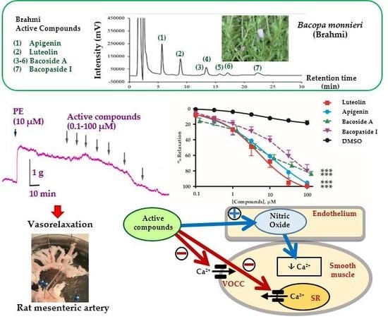
Vasodilatory Effects and Mechanisms of Action of Bacopa monnieri Active Compounds on Rat Mesenteric Arteries

by
Natakorn Kamkaew
1,2,
Tamkeen Urooj Paracha
3,
Kornkanok Ingkaninan
4,
Neti Waranuch
5 and
Krongkarn Chootip
1,*
1
Department of Physiology, Faculty of Medical Science, Naresuan University, Phitsanulok 65000, Thailand
2
Division of Physiology, School of Medical Sciences, University of Phayao, Phayao 56000, Thailand
3
Department of Pharmacy Practice, Faculty of Pharmaceutical Sciences, Naresuan University, Phitsanulok 65000, Thailand
4
Department of Pharmaceutical Chemistry and Pharmacognosy, Faculty of Pharmaceutical Sciences and Center of Excellence for Innovation in Chemistry, Naresuan University, Phitsanulok 65000, Thailand
5
Cosmetics and Natural Products Research Center, Department of Pharmaceutical Technology and Center of Excellence for Innovation in Chemistry, Faculty of Pharmaceutical Sciences, Naresuan University, Phitsanulok 65000, Thailand
*
Author to whom correspondence should be addressed.
Molecules 2019, 24(12), 2243; https://doi.org/10.3390/molecules24122243
Submission received: 17 May 2019 / Revised: 7 June 2019 / Accepted: 11 June 2019 / Published: 15 June 2019
(This article belongs to the Special Issue Flavonoids and Their Disease Prevention and Treatment Potential)

Abstract

:
B. monnieri extract (BME) is an abundant source of bioactive compounds, including saponins and flavonoids known to produce vasodilation. However, it is unclear which components are the more effective vasodilators. The aim of this research was to investigate the vasorelaxant effects and mechanisms of action of saponins and flavonoids on rat isolated mesenteric arteries using the organ bath technique. The vasorelaxant mechanisms, including endothelial nitric oxide synthase (eNOS) pathway and calcium flux were examined. Saponins (bacoside A and bacopaside I), and flavonoids (luteolin and apigenin) at 0.1–100 µM caused vasorelaxation in a concentration-dependent manner. Luteolin and apigenin produced vasorelaxation in endothelial intact vessels with more efficacy (Emax 99.4 ± 0.7 and 95.3 ± 2.6%) and potency (EC50 4.35 ± 1.31 and 8.93 ± 3.33 µM) than bacoside A and bacopaside I (Emax 83.6 ± 2.9 and 79.9 ± 8.2%; EC50 10.8 ± 5.9 and 14.6 ± 5.4 µM). Pretreatment of endothelial intact rings, with L-NAME (100 µM); an eNOS inhibitor, or removal of the endothelium reduced the relaxant effects of all compounds. In K+-depolarised vessels suspended in Ca2+-free solution, these active compounds inhibited CaCl2-induced contraction in endothelial denuded arterial rings. Moreover, the active compounds attenuated transient contractions induced by 10 µM phenylephrine in Ca2+-free medium containing EGTA (1 mM). Thus, relaxant effects occurred in both endothelial intact and denuded vessels which signify actions through both endothelium and vascular smooth muscle cells. In conclusion, the flavonoids have about twice the potency of saponins as vasodilators. However, in the BME, there is ~20 × the amount of vaso-reactive saponins and thus are more effective.

Graphical Abstract

1. Introduction

Bacopa monnieri (L.) Wettst. or Brahmi, is an Ayurvedic medicine traditionally used as a memory enhancer. Along with memory improvement, it is known to promote mental health, as a neurotonic and cardiotonic agent. B. monnieri extract (BME) clearly has a cognitive enhancing potential and neuroprotective effects [1,2,3,4,5,6,7,8,9,10,11,12,13,14,15,16]. It has been shown to be antioxidant in rat brain [17,18] and to possess several pharmacological actions such as anti-depressant [19,20,21], anti-dementia [9], anti-cholinesterase [8,9], anti-hyperglycaemic [22] and anti-hyperlipidaemia [23]. B. monnieri appears to be non-toxic using haematological and blood biochemical diagnostics [24,25,26]. BME demonstrated cardioprotection, improved coronary blood flow, and protection against myocardial ischemia reperfusion injury [27,28]. Our recent work showed that BME acted as a vasodilator by releasing nitric oxide (NO) from endothelium and inhibiting Ca2+ influx and Ca2+ release from the sarcoplasmic reticulum (SR). These mechanisms mediated an acute decrease in blood pressure [29]. Also, daily oral BME (40 mg/kg) in rats for 8 weeks showed a significant increase in cerebral blood flow [30], which implies cerebrovascular dilation.
BME contains an abundance of bioactive compounds. They include dammarane-type triterpenoid saponins, jujubogenin and pseudojujubogenin glycosides. These saponins are predominantly bacopaside I and bacoside A, a mixture of bacoside A3, bacopaside II, jujubogenin isomer of bacopasaponin C, and bacopasaponin C [31,32,33]. Other than saponins, flavonoids, essentially luteolin and apigenin are also present in B. monnieri [10,34,35,36]. Bacoside A3 and bacopaside II relax rat mesenteric arteries [29] but the mechanism(s) of their relaxation are presently unknown. The flavonoids found in B. monnieri also relax rat aortae [37,38,39,40,41] but these experiments used a variety of protocols and vascular preparations. Therefore, it is important to make a side-by-side comparison of these flavonoids with the B. monnieri saponins using a resistance vessel type. For this we choose the mesenteric artery which better exemplifies actions on regional blood flow and systemic blood pressure than the aorta. This work provides evidence to clarify the effective B. monnieri components for vasorelaxation which could be related to the improvement of blood flow or memory enhancement.

2. Results

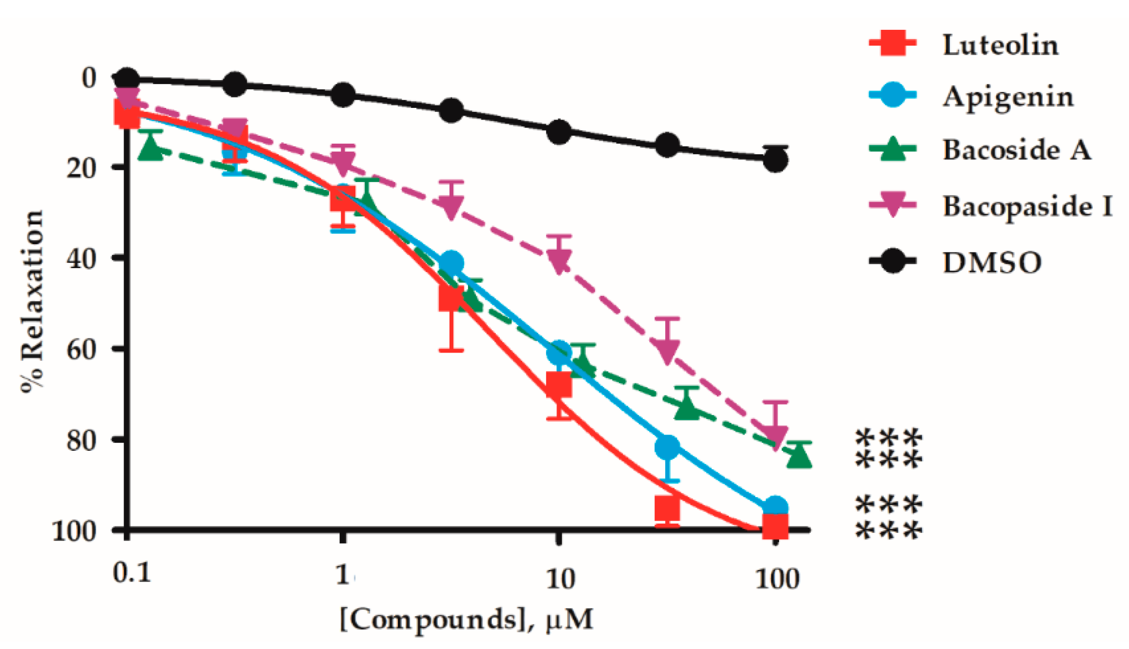
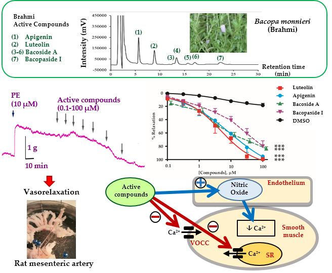
2.1. Vasorelaxant Effects of the B. monnieri Active Compounds

Mesenteric arteries of rats were isolated and mounted in an organ bath via intraluminal wire hooks connected to a force transducer. The vessels were pre-contracted with 10 µM phenylephrine (PE), before adding B. monnieri compounds including flavonoids (luteolin and apigenin), bacopaside I, and the saponin mixture (bacoside A) at 0.1–100 µM. B. monnieri compounds caused vasorelaxation of endothelial intact arteries (+EC) in a concentration-dependent manner (Figure 1) with EC50 and Emax values shown in Table 1.

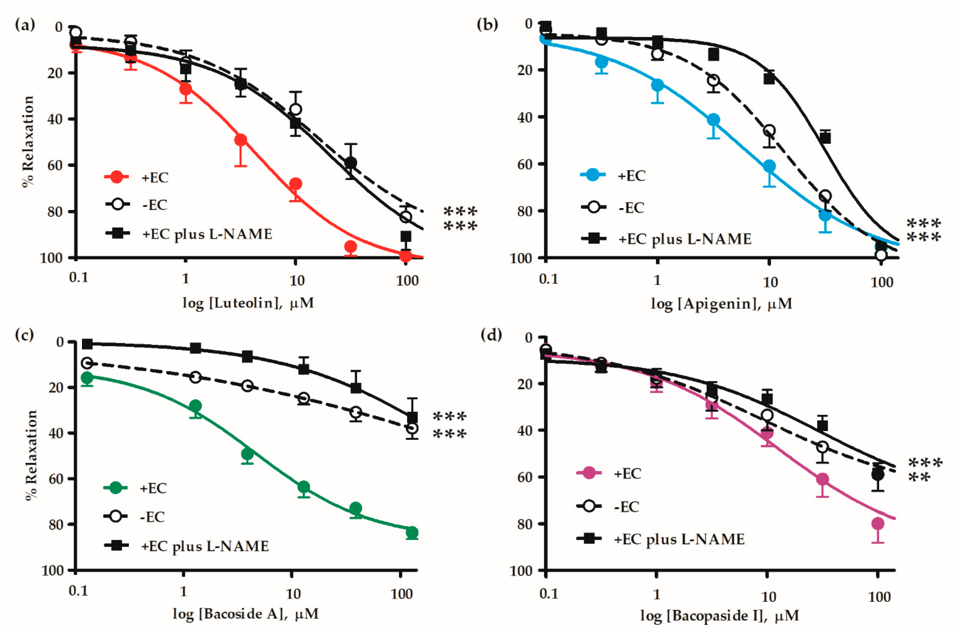
2.2. Mechanisms of Vasorelaxation by B. monnieri Compounds

All the B. monnieri compounds caused vasorelaxation in both endothelial intact (+EC) and endothelial denuded (-EC) mesenteric arterial rings. The relaxations were reduced by the removal of endothelium, implying that these compounds acted via an effect on endothelial vasodilators. However, the compounds still produced some vasorelaxations of the endothelial denuded arterial rings due to a direct action on vascular smooth muscle cells. For intact vessels, L-NAME (inhibitor of endothelial NO synthase; eNOS inhibitor), also reduced the vasorelaxations (Figure 2, Table 2). These reductions suggest that some or all the vasorelaxations were mediated through production and release of NO by endothelial cells.

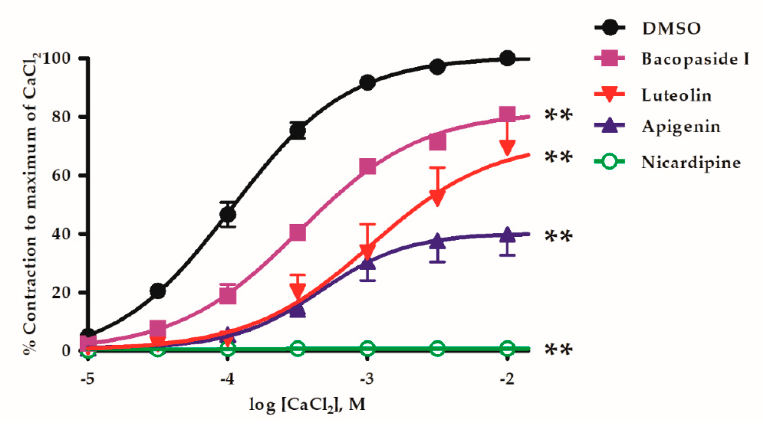
2.3. B. monnieri Compounds and Ca2+ Influx

Voltage-operated Ca2+ channels (VOCCs) were activated by depolarising denuded vessels with 80 mM K+ in Ca2+-free Krebs’ solution. Then vascular contraction elicited by CaCl2 accumulatively added at increasing concentrations (0.01–10 mM). In the same vessel, the protocol was repeated by pre-incubation with 10 µM B. monnieri compounds for 15 min and these CaCl2-induced contractions were inhibited and seen as a rightward shift of the plots and reduced Emax from control (Figure 3).
The maximum contraction (Emax) of control, bacopaside I, luteolin and apigenin were 100 ± 1.3, 81.9 ± 1.7, 72.0 ± 6.7 and 40.2 ± 3.5%, respectively. Positive control, L-type Ca2+-channel blocker, nicardipine (1 µM) completely abolished this CaCl2-induced vasoconstriction (Figure 3).

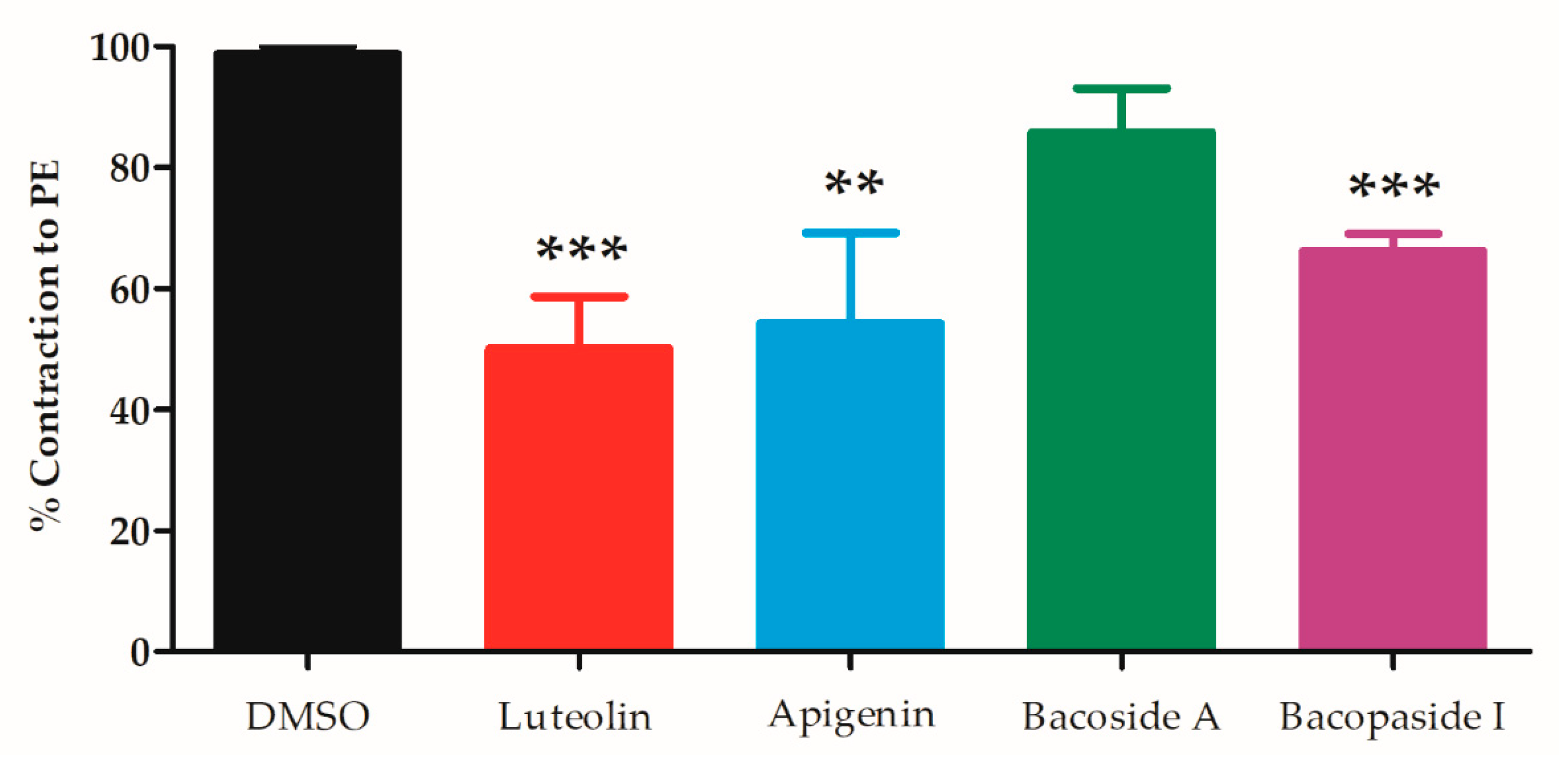
2.4. B. monnieri Compounds and Intracellular Ca2+ Release

The release of intracellular Ca2+ from the sarcoplasmic reticulum is another important trigger of vascular contraction. Denuded arterial rings were pre-incubated in Ca2+-free Krebs’ solution for 10 min and then 10 µM PE added thereby eliciting a transient contraction. Then the protocol was repeated with the same arterial ring in the presence of the test compounds (control, apigenin, luteolin, bacoside A and bacopaside I) producing reduced contractions (98.8 ± 1.2, 50.1 ± 8.5, 54.3 ± 14.9, 85.8 ± 7.2 and 66.2 ± 2.9%, respectively) (Figure 4). Luteolin, apigenin and bacopaside I caused significant decrease in PE-induced contraction compared to the vehicle control (p < 0.001, <0.01 and <0.001, respectively).

3. Discussion

This is the first study comparing the vasodilatory mechanisms elicited by saponins (particularly bacoside A and bacopaside I) and the principal flavonoids (luteolin and apigenin) were the most potent (EC50 4.4 and 8.9 µM) (Figure 1). However, these are present in BME at only about 1/20th the contents of the bacoside A saponins and bacopaside I (Figure S1 and Table S1) [42]. Thus in terms of the overall actions of the complete BME, the saponins would be expected to make a larger contribution to the vasorelaxation than the flavonoids.
However, higher potency of aglycone flavonoids compared to saponin glycosides may be due to sugar moieties interfering with the molecule interacting with the binding sites responsible for the vasorelaxation as suggested by previously, i.e., lipophilic groups in the ring skeleton of flavonoids increased their vasorelaxant activity [43]. This provides a basis for study of the molecular mechanisms of vasorelaxation of flavonoids.
We investigated the mechanisms of flavonoid- and saponin-induced relaxation by endothelial denudation in mesenteric arterial rings which impaired vasorelaxation (Figure 2). Role of NO was investigated using the eNOS inhibitor (L-NAME) with the test compounds. L-NAME increased EC50 and reduced Emax which imitated the effect of endothelial denudation, suggesting the relaxation was mainly medicated by NO. This accords with observations made by Jin et al. that a cyclooxygenase (COX) inhibitor did not affect the relaxation induced by apigenin [44], and consistent with our previous study of B. monnieri extract, where indomethacin had no effect on vasorelaxation [29]. There were some important concentration dependent differences between flavonoids and saponins. Firstly, denudation or blockade of eNOS reduced the effect of bacoside A more than bacopaside I, luteolin and apigenin. Perhaps this was a reflection of bacoside A being a mixture of saponins. However, curiously the responses of luteolin and apigenin to denudation and L-NAME where the latter had a greater effect.
Vascular smooth muscle express plasma membrane L-type Ca2+ channels that allow depolarisation dependent Ca2+ entry to trigger contraction. All three compounds (luteolin, apigenin and bacopaside I) tested in denuded vessels depressed this mechanism of contraction that can also explain in part, the vasorelaxant effect. But here, apigenin appeared to be more effective than luteolin while it was less effective in relaxation studies suggesting some heterogeneity in the mechanism of flavonoid action.
Ca2+ release from intracellular stores also regulates contraction via inositol trisphosphate (IP3) or ryanodine receptors (RyR) associated channels in the SR membranes. IP3 associated channels are commonly activated by plasma membrane G-protein coupled receptors including α1-receptors which are activated by PE. RyR channels are activated by Ca2+ itself. The three pure compounds also inhibited Ca2+ released from stores which can account for at least some vasorelaxation of vessels precontracted by PE. However, the bacoside A was without clear effect again suggesting some heterogeneity between the four test substances. Other Ca2+-channels may also be involved, for example T-channels and TRP channels, especially TRPC4 which is activated by α1-receptor activation.
K+ channels also play a role in regulation of vascular tone, i.e., voltage-dependent K+ (Kv) channels open upon depolarization of the plasma membrane in vascular smooth muscle cells, and thus inhibits Ca2+ influx through VOCCs, resulting in vasodilation [45]. Jiang et al. also reported that luteolin inhibited Ca2+ channels, inhibited release of stored Ca2+ while K+ channels were activated, specifically via KATP, KCa, KV and KIR [40] therefore the effects of apigenin, bacoside A and bacopaside I involving K+ channels deserve further investigation. Our findings support those of Si et al. that luteolin can directly act on vascular endothelial cells, by inducing eNOS phosphorylation at Ser1177, leading to NO production [41]. The flavonoids evoke relaxations and also protect endothelial dependent vasorelaxation against oxidative stress [44,46,47] and diabetes [48], however vasoprotective effects of saponins needs further comprehensive investigation.

4. Materials and Methods

4.1. General Information

Tissues were from male Wistar rats (200–300 g) which were obtained from Nomura Siam International Co. Ltd. (Bangkok, Thailand). Experiments were approved by the Naresuan University Animal Care and Use Committee (NUACUC), protocol number NU-AE 600710. The rats were housed under the environmental conditions at 22 ± 1 °C, 12-h light and dark cycle, fed with standard rodent diet and tap water in Naresuan University Center for Animal Research (NUCAR) according to the guidelines for care and use of laboratory animals (Institute of Laboratory Animal Research, eighth edition 2011. Rats were anesthetized by intraperitoneal injection of thiopental sodium (100 mg/kg BW) and killed. The mesenteric arteries were excised, cleaned of surrounding loose connective tissue and cut into rings of 3–5 mm width. In some experiments, endothelial cells were mechanically removed by gently rubbing the lumen with a stainless steel wire. The mesenteric rings were mounted on a pair of intraluminal wires in organ chambers containing physiological Krebs’ solution (mM): NaCl, 122; KCl, 5; [N-(2-hydroxyethyl) piperazine N’-(2-ethanesulfonic acid)] HEPES, 10; KH2PO4, 0.5; NaH2PO4, 0.5; MgCl2, 1; glucose, 11; and CaCl2, 1.8 (pH 7.3), at 37 °C and aerated [29,49,50,51]. The vessel segments were allowed to equilibrate for 1-h at a resting tension of 1–1.3 g during which the solution was replaced every 15 min. Changes in isometric tension were measured using force transducer lever (CB Sciences Inc., Milford, MA, USA) connected to a MacLab A/D converter (Chart V7; A.D. Instruments, Castle Hill, NSW, Australia), stored and displayed on a personal computer. Following stabilization, the arterial rings were tested for viability by the application of 10 µM PE. Upon development of a steady contraction, the endothelium status was tested with 10 µM ACh. The vessel was considered endothelial intact when the ACh induced >70% relaxation. After establishing the status of the endothelium, the rings were then rinsed with Krebs’ solution for 30 min and one of the following protocols was initiated. Luteolin (lot 126M4061V) and apigenin (lot WE445301/1) were purchased from Sigma Aldrich (St. Louis, MO, USA). Bacoside A (lot 00002005-003) and bacopaside I (lot 00002002-T17H) were purchased from ChromaDex, Inc. (Irvine, CA, USA).

4.2. Vasorelaxant Effects of B. monnieri Active Compounds on Endothelial Intact Arteries

Following stabilization, endothelial intact rings of mesenteric arteries were pre-contracted with 10 µM PE. After the contraction had become constant, the B. monnieri active compounds (0.1–100 µM), including luteolin, apigenin, bacoside A or bacopaside I were added cumulatively.

4.3. Vasorelaxant Effects of B. monnieri Active Compounds on Endothelial Denuded Arteries

Successful endothelial denudation was confirmed by the absence of relaxation upon addition of 10 µM ACh. For investigation of the role of endothelium in 0.1–100 µM B. monnieri active compounds (luteolin, apigenin, bacoside A or bacopaside I) induced vasorelaxation, endothelial denuded arteries were used. The data of effect of active compounds were presented as %relaxation.

4.4. Study of Vasorelaxant Mechanisms of B. monnieri Active Compounds via eNOS Pathway

The role of the endothelial relaxing factor, NO, in B. monnieri active compounds (luteolin, apigenin, bacoside A or bacopaside I) induced vasorelaxation were evaluated in endothelial intact ring pre-treated with NG-nitro-L-arginine methyl ester (L-NAME, 100 µM), an inhibitor of eNOS, for 30 min prior to 10 µM PE exposure.

4.5. Study of Vasorelaxant Mechanisms of B. monnieri Active Compounds on Extracellular Ca2+ Influx

Endothelial denuded mesenteric arteries were equilibrated in Ca2+-free Krebs’ solution (containing (mM): ethylene glycol-bis (ß-aminoethyl ether)-N,N,N,N tetra acetic acid (EGTA), 0.01; NaCl, 122; KCl, 5; HEPES, 10; KH2PO4, 0.5; NaH2PO4, 0.5; MgCl2, 1 and glucose, 11 (pH 7.3)) for 30 min followed by replacing with Ca2+-free Krebs’ solution containing 80 mM K+ for 10 min which depolarizes the vascular smooth muscle cells, thus opening VOCCs. Various concentrations of CaCl2 were then added (0.01–10 mM) in a logarithmic progression. After obtaining the maximum response, the baths were washed out and replenished with Ca2+-free Krebs’ solution for 30 min. The Ca2+-free 80 mM K+ solution was then re-applied following pre-incubation for 10 min with either: 10 µM active compounds or 1 µM nicardipine (antagonist of VOCCs). Concentration-response curves to cumulative addition of CaCl2 were then repeated and compared with maximum contraction evoked by previous control CaCl2 challenges.

4.6. Study of Vasorelaxant Mechanisms of B. monnieri Active Compounds on Intracellular Ca2+ Release

To stimulate initial Ca2+ loading of the SR Ca2+ stores, endothelial denuded mesenteric arteries were exposed to 80 mM K+ solution for 5 min, and then washed out with Ca2+-free Krebs’ solution containing 1 mM EGTA for 10 min. The arterial rings were then challenged with 10 µM PE (acting through phospholipase C/IP3 signaling) which release Ca2+ from the SR thereby eliciting a transient contraction [29]. The same protocol was then repeated to ensure that similar transient contractions to PE could be obtained. Then, the arterial rings were challenged again with 80 mM K+ solution for 5 min, and washed out with Ca2+-free Krebs’ solution containing 1 mM EGTA and 10 µM active compounds for 10 min. The arterial rings were again challenged with 10 µM PE. The PE-induced contractions were compared in the presence or absence of active compounds.

4.7. Statistical Analyses

Statistical analyses used GraphPad Prism version 5.00 for Windows, (GraphPad Software Inc., La Jolla, CA, USA). Data from each concentration-effect curve was analysed using non-repeated two-way ANOVA. Curve fitting in the figures was generated by the same software using non-linear regression. EC50 and Emax were compared using unpaired Student’s t test. Values are expressed as mean ± SEM. A p-value < 0.05 was considered significant. ‘n’ is the number of vascular rings used, each ring originating from a different animal.

5. Conclusions

This study demonstrated that B. monnieri active components, including both saponins and flavonoids, produced vasodilatory effects on rat isolated mesenteric arteries partially via endothelial dependent release of vasodilators and also by direct effects on vascular smooth muscle cells via blockade of Ca2+ influx and its release from SR. This study for the first time reports the comparative vasodilatory effects of saponins and flavonoids found in B. monnieri extract. However, B. monnieri extract, flavonoids i.e., luteolin and apigenin would be more potent vasodilators but saponins have a greater effect because of their greater contents. Accordingly, the clinical benefits on enhanced blood flow and cognitive function may arise from a combination of flavonoids and particularly the saponins.

Supplementary Materials

Supplementary materials are available online. Figure S1: Representative HPLC-UV chromatogram of mixed seven standards at 20 µg/mL for 1 and 2 and 100 µg/mL for 3–7 (A) and Brahmi extract (2 mg/mL) (B); luteolin (1), apigenin (2), bacoside A3 (3), bacopaside II (4), bacopaside X (5), bacopasaponin C (6) and bacopaside I (7), Table S1: Amount of each compound in 95% ethanolic extract of Brahmi analyzed by HPLC.

Author Contributions

Conceptualization, K.I., N.W. and K.C.; methodology and experimental design, N.K., T.U.P. and K.C.; software, N.K.; validation, N.K., T.U.P. and K.C.; formal analysis, N.K. and K.C.; investigation, N.K.; resources, K.I., N.W. and K.C.; data curation and interpretation, N.K. and K.C.; writing—original draft preparation, N.K.; manuscript writing—review and editing, T.U.P. and K.C.; visualization, K.I., N.W. and K.C.; supervision, K.I., N.W. and K.C.; project administration, K.C.; funding acquisition, K.C.

Funding

This research was funded by National Research Council of Thailand (NRCT), grant No. R2561B032.

Acknowledgments

We would like to acknowledge the Center of Excellence for Innovation in Chemistry (PERCH-CIC) and the International Research Network (IRN61W0005) on research facility support. We would like to thank C. Norman Scholfield for his constructive criticism of the manuscript.

Conflicts of Interest

The authors declared no conflict of interest.

References

  1. Bacopa monniera. Monograph. Altern. Med. Rev. J. Clin. Ther. 2004, 9, 79–85.
  2. Russo, A.; Borrelli, F. Bacopa monniera, a reputed nootropic plant: An overview. Phytomedicine 2005, 12, 305–317. [Google Scholar] [CrossRef] [PubMed]
  3. Kumar, V. Potential medicinal plants for CNS disorders: An overview. Phytother. Res. PTR 2006, 20, 1023–1035. [Google Scholar] [CrossRef]
  4. Rajan, K.E.; Preethi, J.; Singh, H.K. Molecular and functional characterization of Bacopa monniera: A retrospective review. Evid.-Based Complement. Altern. Med. eCAM 2015, 2015, 945217. [Google Scholar] [CrossRef] [PubMed]
  5. Aguiar, S.; Borowski, T. Neuropharmacological review of the nootropic herb Bacopa monnieri. Rejuvenation Res. 2013, 16, 313–326. [Google Scholar] [CrossRef] [PubMed]
  6. Singh, H.K. Brain enhancing ingredients from Ayurvedic medicine: Quintessential example of Bacopa monniera, a narrative review. Nutrients 2013, 5, 478–497. [Google Scholar] [CrossRef] [PubMed]
  7. Mathur, D.; Goyal, K.; Koul, V.; Anand, A. The molecular links of re-emerging therapy: A review of evidence of Brahmi (Bacopa monniera). Front. Pharmacol. 2016, 7, 44. [Google Scholar] [CrossRef]
  8. Das, A.; Shanker, G.; Nath, C.; Pal, R.; Singh, S.; Singh, H. A comparative study in rodents of standardized extracts of Bacopa monniera and Ginkgo biloba: Anticholinesterase and cognitive enhancing activities. Pharmacol. Biochem. Behav. 2002, 73, 893–900. [Google Scholar] [CrossRef]
  9. Dhanasekaran, M.; Tharakan, B.; Holcomb, L.A.; Hitt, A.R.; Young, K.A.; Manyam, B.V. Neuroprotective mechanisms of ayurvedic antidementia botanical Bacopa monniera. Phytother. Res. PTR 2007, 21, 965–969. [Google Scholar] [CrossRef]
  10. Limpeanchob, N.; Jaipan, S.; Rattanakaruna, S.; Phrompittayarat, W.; Ingkaninan, K. Neuroprotective effect of Bacopa monnieri on beta-amyloid-induced cell death in primary cortical culture. J. Ethnopharmacol. 2008, 120, 112–117. [Google Scholar] [CrossRef]
  11. Uabundit, N.; Wattanathorn, J.; Mucimapura, S.; Ingkaninan, K. Cognitive enhancement and neuroprotective effects of Bacopa monnieri in Alzheimer’s disease model. J. Ethnopharmacol. 2010, 127, 26–31. [Google Scholar] [CrossRef] [PubMed]
  12. Vollala, V.R.; Upadhya, S.; Nayak, S. Enhancement of basolateral amygdaloid neuronal dendritic arborization following Bacopa monniera extract treatment in adult rats. Clinics (Sao Paulo Brazil) 2011, 66, 663–671. [Google Scholar] [CrossRef] [PubMed]
  13. Vollala, V.R.; Upadhya, S.; Nayak, S. Learning and memory-enhancing effect of Bacopa monniera in neonatal rats. Bratisl. Lek. Listy 2011, 112, 663–669. [Google Scholar] [PubMed]
  14. Vollala, V.R.; Upadhya, S.; Nayak, S. Enhanced dendritic arborization of hippocampal CA3 neurons by Bacopa monniera extract treatment in adult rats. Rom. J. Morphol. Embryol. 2011, 52, 879–886. [Google Scholar] [PubMed]
  15. Vollala, V.R.; Upadhya, S.; Nayak, S. Enhanced dendritic arborization of amygdala neurons during growth spurt periods in rats orally intubated with Bacopa monniera extract. Anat. Sci. Int. 2011, 86, 179–188. [Google Scholar] [CrossRef] [PubMed]
  16. Kongkeaw, C.; Dilokthornsakul, P.; Thanarangsarit, P.; Limpeanchob, N.; Norman Scholfield, C. Meta-analysis of randomized controlled trials on cognitive effects of Bacopa monnieri extract. J. Ethnopharmacol. 2014, 151, 528–535. [Google Scholar] [CrossRef] [PubMed]
  17. Anbarasi, K.; Vani, G.; Balakrishna, K.; Devi, C.S. Effect of bacoside A on brain antioxidant status in cigarette smoke exposed rats. Life Sci. 2006, 78, 1378–1384. [Google Scholar] [CrossRef] [PubMed]
  18. Jyoti, A.; Sharma, D. Neuroprotective role of Bacopa monniera extract against aluminium-induced oxidative stress in the hippocampus of rat brain. Neurotoxicology 2006, 27, 451–457. [Google Scholar] [CrossRef]
  19. Mannan, A.; Abir, A.B.; Rahman, R. Antidepressant-like effects of methanolic extract of Bacopa monniera in mice. BMC Complement. Altern. Med. 2015, 15, 337. [Google Scholar] [CrossRef]
  20. Sairam, K.; Dorababu, M.; Goel, R.K.; Bhattacharya, S.K. Antidepressant activity of standardized extract of Bacopa monniera in experimental models of depression in rats. Phytomedicine 2002, 9, 207–211. [Google Scholar] [CrossRef]
  21. Kadali, S.R.M.; Das, M.C.; Rao, A.S.; Sri, G.K. Antidepressant activity of brahmi in albino mice. J. Clin. Diagn. Res. JCDR 2014, 8, 35–37. [Google Scholar] [CrossRef] [PubMed]
  22. Udhaya Lavinya, B.; Sabina, E.P. Anti-hyperglycaemic effect of Brahmi (Bacopa monnieri L.) in streptozotocininduced diabetic rats: A study involving antioxidant, biochemical and haematological parameters. J. Chem. Pharm. Res. 2015, 7, 531–534. [Google Scholar]
  23. Kamesh, V.; Sumathi, T. Antihypercholesterolemic effect of Bacopa monniera Linn. on high cholesterol diet induced hypercholesterolemia in rats. Asian Pac. J. Trop. Med. 2012, 5, 949–955. [Google Scholar] [CrossRef]
  24. Sireeratawong, S.; Jaijoy, K.; Khonsung, P.; Lertprasertsuk, N.; Ingkaninan, K. Acute and chronic toxicities of Bacopa monnieri extract in Sprague-Dawley rats. BMC Complement. Altern. Med. 2016, 16, 249. [Google Scholar] [CrossRef] [PubMed]
  25. Joshua Allan, J.; Damodaran, A.; Deshmukh, N.S.; Goudar, K.S.; Amit, A. Safety evaluation of a standardized phytochemical composition extracted from Bacopa monnieri in Sprague–Dawley rats. Food Chem. Toxicol. 2007, 45, 1928–1937. [Google Scholar] [CrossRef] [PubMed]
  26. Pravina, K.; Ravindra, K.R.; Goudar, K.S.; Vinod, D.R.; Joshua, A.J.; Wasim, P.; Venkateshwarlu, K.; Saxena, V.S.; Amit, A. Safety evaluation of BacoMind in healthy volunteers: A phase I study. Phytomedicine 2007, 14, 301–308. [Google Scholar] [CrossRef] [PubMed]
  27. Srimachai, S.; Devaux, S.; Demougeot, C.; Kumphune, S.; Ullrich, N.D.; Niggli, E.; Ingkaninan, K.; Kamkaew, N.; Scholfield, C.N.; Tapechum, S.; et al. Bacopa monnieri extract increases rat coronary flow and protects against myocardial ischemia/reperfusion injury. BMC Complement. Altern. Med. 2017, 17, 117. [Google Scholar] [CrossRef]
  28. Nandave, M.; Ojha, S.K.; Sujata, J.; Kumari, S.; Arya, D.S. Cardioprotective effect of Bacopa monneira against isoproterenol-induced myocardial necrosis in rats. Int. J. Pharmacol. 2007, 3, 385–392. [Google Scholar]
  29. Kamkaew, N.; Scholfield, C.N.; Ingkaninan, K.; Maneesai, P.; Parkington, H.C.; Tare, M.; Chootip, K. Bacopa monnieri and its constituents is hypotensive in anaesthetized rats and vasodilator in various artery types. J. Ethnopharmacol. 2011, 137, 790–795. [Google Scholar] [CrossRef]
  30. Kamkaew, N.; Norman Scholfield, C.; Ingkaninan, K.; Taepavarapruk, N.; Chootip, K. Bacopa monnieri increases cerebral blood flow in rat independent of blood pressure. Phytother. Res. PTR 2013, 27, 135–138. [Google Scholar] [CrossRef]
  31. Phrompittayarat, W.; Putalun, W.; Tanaka, H.; Jetiyanon, K.; Wittaya-Areekul, S.; Ingkaninan, K. Determination of pseudojujubogenin glycosides from Brahmi based on immunoassay using a monoclonal antibody against bacopaside I. Phytochem. Anal. PCA 2007, 18, 411–418. [Google Scholar] [CrossRef] [PubMed]
  32. Phrompittayarat, W.; Putalun, W.; Tanaka, H.; Wittaya-Areekul, S.; Jetiyanon, K.; Ingkaninan, K. An enzyme-linked immunosorbant assay using polyclonal antibodies against bacopaside I. Anal. Chim. Acta 2007, 584, 1–6. [Google Scholar] [CrossRef] [PubMed]
  33. Nuengchamnong, N.; Sookying, S.; Ingkaninan, K. LC-ESI-QTOF-MS based screening and identification of isomeric jujubogenin and pseudojujubogenin aglycones in Bacopa monnieri extract. J. Pharm. Biomed. Anal. 2016, 129, 121–134. [Google Scholar] [CrossRef] [PubMed]
  34. Honnegowda, S.; Bagul, M.S.; Padh, H.; Rajani, M. A rapid densitometric method for the quantification of luteolin in medicinal plants using HPTLC. Chromatographia 2004, 60, 131–134. [Google Scholar]
  35. Deepak, M.; Sangli, G.K.; Arun, P.C.; Amit, A. Quantitative determination of the major saponin mixture bacoside A in Bacopa monnieri by HPLC. Phytochem. Anal. PCA 2005, 16, 24–29. [Google Scholar] [CrossRef] [PubMed]
  36. Rajasekaran, A. Simultaneous estimation of luteolin and apigenin in methanol leaf extract of Bacopa monnieri Linn by HPLC. Br. J. Pharm. Res. 2014, 4, 1629–1637. [Google Scholar] [CrossRef]
  37. Chan, E.C.; Pannangpetch, P.; Woodman, O.L. Relaxation to flavones and flavonols in rat isolated thoracic aorta: Mechanism of action and structure-activity relationships. J. Cardiovasc. Pharmacol. 2000, 35, 326–333. [Google Scholar] [CrossRef]
  38. Calderone, V.; Chericoni, S.; Martinelli, C.; Testai, L.; Nardi, A.; Morelli, I.; Breschi, M.C.; Martinotti, E. Vasorelaxing effects of flavonoids: Investigation on the possible involvement of potassium channels. Naunyn-Schmiedeberg’s Arch. Pharmacol. 2004, 370, 290–298. [Google Scholar] [CrossRef]
  39. Je, H.D.; Kim, H.-D.; La, H.-O. The inhibitory effect of apigenin on the agonist-induced regulation of vascular contractility via calcium desensitization-related pathways. Biomol. Ther. 2014, 22, 100–105. [Google Scholar] [CrossRef]
  40. Jiang, H.; Xia, Q.; Wang, X.; Song, J.; Bruce, I.C. Luteolin induces vasorelaxion in rat thoracic aorta via calcium and potassium channels. Die Pharm. 2005, 60, 444–447. [Google Scholar]
  41. Si, H.; Wyeth, R.P.; Liu, D. The flavonoid luteolin induces nitric oxide production and arterial relaxation. Eur. J. Nutr. 2014, 53, 269–275. [Google Scholar] [CrossRef] [PubMed]
  42. Saesong, T.; Temkitthawon, P.; Nangngam, P.; Ingkaninan, K. Pharmacognostic and physico-chemical investigations of the aerial part of Bacopa monnieri (L.) Wettst. SJST 2019, 41, 397–404. [Google Scholar]
  43. Wu, H.; Jiang, H.; Wang, L.; Hu, Y. Relationship between vasorelaxation of flavonoids and their retention index in RP-HPLC. Die Pharm. 2006, 61, 667–669. [Google Scholar]
  44. Jin, B.H.; Qian, L.B.; Chen, S.; Li, J.; Wang, H.P.; Bruce, I.C.; Lin, J.; Xia, Q. Apigenin protects endothelium-dependent relaxation of rat aorta against oxidative stress. Eur. J. Pharmacol. 2009, 616, 200–205. [Google Scholar] [CrossRef] [PubMed]
  45. Ko, E.A.; Han, J.; Jung, I.D.; Park, W.S. Physiological roles of K+ channels in vascular smooth muscle cells. J. Smooth Muscle Res. 2008, 44, 65–81. [Google Scholar] [CrossRef] [PubMed]
  46. Ma, X.; Li, Y.F.; Gao, Q.; Ye, Z.G.; Lu, X.J.; Wang, H.P.; Jiang, H.D.; Bruce, I.C.; Xia, Q. Inhibition of superoxide anion-mediated impairment of endothelium by treatment with luteolin and apigenin in rat mesenteric artery. Life Sci. 2008, 83, 110–117. [Google Scholar] [CrossRef]
  47. Qian, L.B.; Wang, H.P.; Chen, Y.; Chen, F.X.; Ma, Y.Y.; Bruce, I.C.; Xia, Q. Luteolin reduces high glucose-mediated impairment of endothelium-dependent relaxation in rat aorta by reducing oxidative stress. Pharmacol. Res. 2010, 61, 281–287. [Google Scholar] [CrossRef]
  48. El-Bassossy, H.M.; Abo-Warda, S.M.; Fahmy, A. Chrysin and luteolin attenuate diabetes-induced impairment in endothelial-dependent relaxation: Effect on lipid profile, AGEs and NO generation. Phytother. Res. PTR 2013, 27, 1678–1684. [Google Scholar] [CrossRef]
  49. Wisutthathum, S.; Kamkaew, N.; Inchan, A.; Chatturong, U.; Paracha, T.U.; Ingkaninan, K.; Wongwad, E.; Chootip, K. Extract of Aquilaria crassna leaves and mangiferin are vasodilators while showing no cytotoxicity. J. Tradit. Complement. Med. 2018, in press. [Google Scholar] [CrossRef]
  50. Wisutthathum, S.; Demougeot, C.; Totoson, P.; Adthapanyawanich, K.; Ingkaninan, K.; Temkitthawon, P.; Chootip, K. Eulophia macrobulbon extract relaxes rat isolated pulmonary artery and protects against monocrotaline-induced pulmonary arterial hypertension. Phytomedicine 2018, 50, 157–165. [Google Scholar] [CrossRef]
  51. Wisutthathum, S.; Chootip, K.; Martin, H.; Ingkaninan, K.; Temkitthawon, P.; Totoson, P.; Demougeot, C. Vasorelaxant and hypotensive effects of an ethanolic extract of Eulophia macrobulbon and Its main compound 1-(4′-hydroxybenzyl)-4,8-dimethoxyphenanthrene-2,7-diol. Front. Pharmacol. 2018, 9, 484. [Google Scholar] [CrossRef] [PubMed]
Sample Availability: Samples of the compounds, luteolin, apigenin, bacoside A and bacopaside I are not available from the authors.
Figure 1. Relaxations induced by luteolin, apigenin, bacoside A, and bacopaside I (0.1–100 µM) and vehicle (DMSO) in endothelial intact mesenteric arteries precontracted with phenylephrine (10 µM). Values are mean ± SEM of 6–9 individual arterial rings. *** indicates p < 0.001 comparing relaxation for each compound with the control (DMSO) using two-way ANOVA (n = 6–9). Lines were fitted by non-linear regression.
Molecules 24 02243 g001
Figure 2. Cumulative concentration-response curves of (a) luteolin, (b) apigenin, (c) bacoside A and (d) bacopaside I in concentrations (0.1–100 µM) in endothelial intact (+EC), denuded (-EC) mesenteric arterial rings and endothelial intact vessels pre-incubated in L-NAME (100 µM). The graphs are expressed as %relaxation of vessel pre-contracted with 10 µM PE. Values are mean ± SEM of 6–9 individual arteries. ** p < 0.01, *** p < 0.001 each compound compared with intact vessels (+EC) using two-way ANOVA (n = 6–9).
Molecules 24 02243 g002
Figure 3. CaCl2-induced contractions of denuded mesenteric arteries pre-incubated in high K+, Ca2+-free media in the conditions of pre-incubation with DMSO (negative control), 10 µM bacopaside I, 10 µM luteolin, 10 µM apigenin, and 1 µM nicardipine (positive control). Y-axis, % contraction compared to the contraction achieved with the highest Ca2+ concentration during the initial run without a B. monnieri compound in the same vessel. Values are mean ± SEM of 4–6 individual arteries. ** p < 0.01 each of the active compounds compared to DMSO using two-way ANOVA (n = 4–6).
Molecules 24 02243 g003
Figure 4. PE-induced contraction induced by Ca2+ release from sarcoplasmic reticulum of endothelial denuded mesenteric arteries in the presence of DMSO (control), 10 µM of luteolin, apigenin, bacoside A and bacopaside I. The data is % contraction to 10 µM PE induced contraction compared to contractions produced by the initial protocol without test compound. Values are mean ± SEM of 5–6 individual arteries. ** p < 0.01, *** p < 0.001 each of the active compounds compared with control using unpaired Student’s t-test (n = 5–6).
Molecules 24 02243 g004
Table 1. The EC50 and Emax of B. monnieri active compounds on relaxation of endothelial intact rat mesenteric arteries.
Active CompoundsEC50 (µM)Emax (%)np-Value Whole Graph Curves
FlavonoidsLuteolin4.35 ± 1.3199.4 ± 0.76-
Apigenin8.93 ± 3.3395.3 ± 2.69NS
SaponinsBacoside A10.8 ± 5.983.6 ± 2.9 ††7< 0.05 †
Bacopaside I14.6 ± 5.479.9 ± 8.2 †7< 0.01 ††
VehicleDMSO-17.4 ± 3.1 ††7< 0.01 ††
Significantly different compared with luteolin † p < 0.05, †† p < 0.01 using unpaired Student’s t-test (n = 6–9).
Table 2. The EC50 and Emax of B. monnieri compounds on relaxations of endothelial intact (+EC), denuded (-EC) mesenteric arterial rings or endothelial intact arteries with L-NAME.
Active CompoundsEC50 (µM)Emax (%)n
Luteolin
+EC4.35 ± 1.3199.35 ± 0.666
-EC21.90 ± 5.86 †82.42 ± 4.65 ††6
+EC plus L-NAME14.99 ± 3.56 †90.85 ± 5.856
Apigenin
+EC8.93 ± 3.3395.27 ± 2.619
-EC12.80 ± 2.5498.81 ± 1.198
+EC plus L-NAME25.62 ± 3.38 ††94.40 ± 2.107
Bacoside A
+EC10.81 ± 5.9583.60 ± 2.867
-EC14.50 ± 6.3037.90 ± 4.72 ††6
+EC plus L-NAME33.81 ± 6.25 †33.16 ± 8.41 ††5
Bacopaside I
+EC14.63 ± 5.3679.94 ± 8.177
-EC17.29 ± 4.7558.97 ± 7.05 †7
+EC plus L-NAME25.38 ± 4.3358.45 ± 4.21 †7
Comparison of EC50 or Emax of each component +EC vs. -EC or +EC plus L-NAME. † p < 0.05, †† p < 0.01 using unpaired Student’s t-test.

