Article

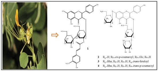
Four New Flavonoids Isolated from the Aerial Parts of Cadaba rotundifolia Forssk. (Qadab)

by
Gadah Abdulaziz Al-Hamoud
1,2,
Raha Saud Orfali
2,*,
Sachiko Sugimoto
1,
Yoshi Yamano
1,
Nafee Alothyqi
3,
Ali Mohammed Alzahrani
4 and
Katsuyoshi Matsunami
1,*
1
Department of Pharmacognosy, Graduate School of Biomedical and Health Sciences, Hiroshima University, 1-2-3 Kasumi, Minami-ku, Hiroshima 734-8553, Japan
2
Department of Pharmacognosy, College of Pharmacy, King Saud University, 11495 Riyadh, Saudi Arabia
3
Department of Biology, Umm Al-Qura University, 1109 Makkah Al-Mukarramah, Saudi Arabia
4
Department of Biology, Arts and Sciences in Qilwah, Al-Baha University, 1988 Al-Baha, Saudi Arabia
*
Authors to whom correspondence should be addressed.
Molecules 2019, 24(11), 2167; https://doi.org/10.3390/molecules24112167
Submission received: 19 May 2019 / Revised: 5 June 2019 / Accepted: 6 June 2019 / Published: 9 June 2019
(This article belongs to the Special Issue Natural Product Isolation, Identification and Biological Activity)

Abstract

:
Cadaba rotundifolia (Forssk.) (family: Capparaceae; common name: Qadab) is one of four species that grow in the Red Sea costal region in the Kingdom of Saudi Arabia. The roots and leaves of C. rotundifolia is traditionally used to treat tumors and abscesses in Sudan. A previous phytochemical study of the roots yielded a quaternary alkaloid, but no report on chemical constituents of the aerial parts of the C. rotundifolia growing in Saudi Arabia has been issued so far. Oxidative stress and advanced glycation end products (AGEs) are thought as causal factors in many degenerative diseases, such as Alzheimer’s disease, diabetes, atherosclerosis and aging. In this study, a total of twenty compounds, including four previously undescribed acylated kaempferol glucosides, were isolated from the aerial parts of C. rotundifolia collected in Saudi Arabia. These new compounds were identified as kaempferol 3-O-[2-O-(trans-feruloyl)-3-O-β-d-glucopyranosyl]-β-d-glucopyranoside (1), kaempferol 3-O-β-neohesperidoside-7-O-[2-O-(cis-p-coumaroyl)-3-O-β-d-glucopyranosyl]-β-d-glucopyranoside (2), kaempferol 3-O-[2,6-di-O-α-l-rhamnopyranosyl]-β-d-glucopyranoside-7-O-[6-O-(trans-feruloyl)]-β-d-glucopyranoside (3) and kaempferol 3-O-[2,6-di-O-α-l-rhamnopyranosyl]-β-d-glucopyranoside-7-O-[6-O-(trans-p-coumaroyl)]-β-d-glucopyranoside (4). Their structures were established based on UV-visible, 1D, 2D NMR, and HR-ESI-MS analyses. Of the assayed compounds, 17 and 18 showed potent radical scavenging activity with IC50 values of 14.5 and 11.7 µM, respectively, and inhibitory activity toward AGEs together with compound 7 with IC50 values 96.5, 34.9 and 85.5 µM, respectively.

Graphical Abstract

1. Introduction

Oxidative stress and advanced glycation end products (AGEs) are thought as causal factors of many diseases, such as diabetes, atherosclerosis, neurodegenerative disorders and aging. Therefore, the discovery of antioxidant and anti-AGEs molecules are important for preventing these age-related pathogenesis. Cadaba (Forssk.) is one of genera in the Capparaceae family, with about 30 species distributed in southern Africa, India, Malaysia, and Australia [1,2]. Among them four species grow in the Red Sea costal region in the Kingdom of Saudi Arabia [3]. Phytochemical studies of several Cadaba species led to isolation of various secondary metabolites, such as alkaloids, terpenoids and flavonoids [4,5,6,7]. Cadaba rotundifolia Forssk., known by the common name Qadab, is an erect shrub with numerous drooping branches with young twigs covered with short glandular hair [8]. In Sudan, C. rotundifolia roots and leaves are traditionally used to treat tumors and abscesses. A previous chemical study of the ethanolic extract of C. rotundifolia root yielded a quaternary alkaloid (3-hydroxystachydrine) [9]. However, no report on chemical constituents of the aerial parts of C. rotundifolia has been issued so far. In this work, we have investigated the chemical profile of the aerial parts of C. rotundifolia collected in Saudi Arabia. As a result, twenty flavonoids were obtained, including four undescribed acylated kaempferol glucosides 14 and 16 known compounds. This paper describes on the isolation, structural elucidation, and evaluation of antioxidant (DPPH) and inhibitory activity toward AGEs formation.

2. Results and Discussion

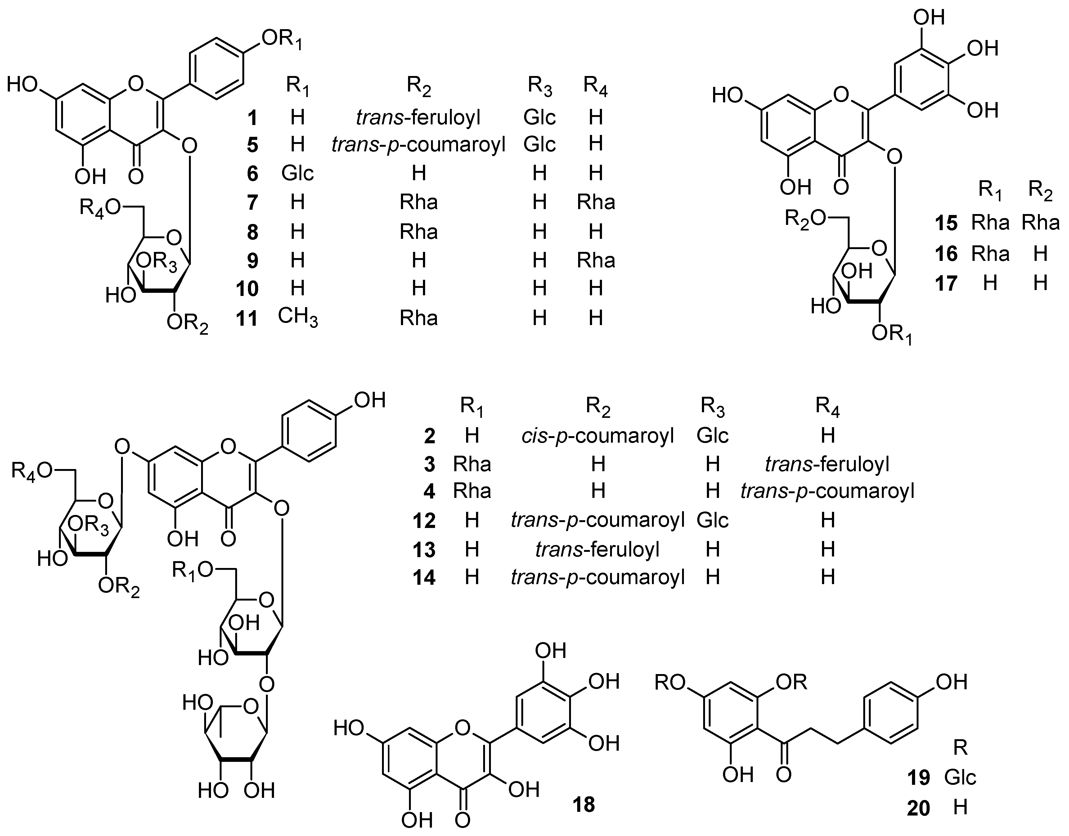
2.1. Isolation and Spectroscopic Analyses of the Compounds

The aerial parts of C. rotundifolia were extracted with MeOH at room temperature till exhausted. The MeOH extract (800 g) was further separated by solvent fractionation, various column chromatographies (CC), and HPLC to afford compounds 120 (Figure 1). The chemical structures of the isolated compounds were investigated and identified through intensive spectroscopic analyses based on UV-visible, 1D and 2D NMR, and HR-ESI-MS data as follows (Figures S1–S35).

2.1.1. Chemical Structure of Compound 1

Compound 1 was isolated as a yellow amorphous powder, soluble in MeOH with a negative optical rotation ([α]22D −79.0). Its molecular formula, C37H38O19, was determined by positive HR-ESI-MS analysis (m/z 809.1888 [M + Na]+,calcd for C37H38O19Na 809.1899). It had a UV spectrum typical of an acylated kaempferol glycoside (λmax 327, 267 nm) [10,11]. The IR spectrum suggested the presence of hydroxy (3388 cm−1), α,β-unsaturated carbonyl ester (1710 cm−1), α,β-unsaturated ketone (1658 cm−1), aromatic ring (1513, 1446 cm−1) and ether (1176, 1070 cm−1) functions. The proton and carbon resonances in its 1H and 13C-NMR spectra clarified the aromatic and glycosidic nature of 1. It was found that the 1H-NMR spectrum of 1 displayed feature resembling to those of 5 [12], as both the aglycone and glycoside units were almost identical. The major differences were in the signals related to the acyl group of 5. The 1H-NMR spectrum of 1 (Table 1) exhibited meta-coupled resonances at δH 6.17 and 6.37 (each 1H, d, J = 2.0 Hz), characteristic of H-6 and H-8 of the A-ring, respectively. The 1H–1H correlation spectrum (COSY, Figure 2) and 1H-NMR spectrum of 1 displayed two ortho-coupled doublet resonances at δH 8.01 and 6.91 (each 2H, d, J = 8.8 Hz) assignable to H-2′/6′ and H-3′/5′ of the B-ring, respectively, which indicated the presence of a kaempferol aglycone (Table 1).
The 1H-NMR spectrum of 1 (Table 1) also gave signals at δH 6.83 (1H, d, J = 8.2 Hz), 7.09 (1H, d, J = 8.2, 1.8 Hz) and 7.20 (1H, d, J = 1.8 Hz) attributable to an ABX coupling system for a 1,3,4-trisubstituted aromatic moiety, and two trans-coupled olefinic protons at δH 6.40 (1H, br d, J = 15.9 Hz) and 7.68 (1H, br d, J = 15.9 Hz), together with a singlet signal for a methoxy group at δH 3.92 which suggested the presence of a feruloyl or isoferuloyl function. The two anomeric proton resonances, which occur at δH 5.73 (1H, d, J = 8.0 Hz) and 4.43 (1H, d, J = 7.8 Hz) and 12 carbon signals (δc 104.9, 100.5, 84.9, 78.4, 78.1 , 77.7, 74.8, 74.6, 71.4, 70.0, 62.5 and 62.4) correspond to two glucose moieties. Acid hydrolysis of 1 released d- glucose and the β-anomeric configuration for the sugars was determined from the magnitude of the J1,2 coupling constants. In addition, alkaline hydrolysis afforded ferulic acid, which was identified by comparing with an authentic sample based on their HPLC retention times. The 13C-NMR spectrum (Table 2) of 1 showed 37 carbon resonances, 12 of which assigned to two β-glucose units, 15 signals were consistent with kaempferol aglycone and the remaining 10 signals were ascribed to acyl groups. The correlations observed between the proton and carbon resonances of the acyl moiety from H-7′′′′ (δH 7.68) to C-2′′′′ (δc 111.7), C-6′′′′ (δc 124.3) and an ester carbonyl C-9′′′′ (δc 168.5), and from the protons of methoxy group (δH 3.92) to C-3′′′′ (δc 149.3) in HMBC spectrum (Figure 2) indicated the presence of a feruloyl unit.
According to the coupling constant of the olefinic protons at H-7′′′′ and H-8′′′′ (J = 15.9 Hz), the configuration of the feruloyl moiety was determined as trans. Full assignments of the 1H and 13C resonances of the structural fragments of 1 were achieved by analysis of COSY, HSQC and HMBC spectra. A correlation between the anomeric H-1′′ proton (δH 5.73) and δc 134.8 in the HMBC spectrum (Figure 2) defined C-3 of kaempferol as a site of O-glucosylation. Moreover, the interglucosidic linkage of the disaccharide moiety was also determined by the HMBC cross-peak between H-3′′ (δH 3.91) and C-1′′′ (δc 104.9). The E-feruloyl unit was linked to C-2′′ based on the correlations of the H-2′′ (δH 5.23) with C-1′′ (δc 100.5), C-3′′ (δc 84.9) and ester carbonyl carbon C-9′′′′ (δc 168.5). Therefore, the structure of compound 1 was determined as kaempferol 3-O-[2-O-(trans-feruloyl)-3-O-β-d-glucopyranosyl]-β-d-glucopyranoside.

2.1.2. Chemical Structure of Compound 2

Compound 2 was isolated as a yellow amorphous powder, with a positive optical rotation ([α] 22D +14.5). Its molecular formula was determined to be C48H56O27 by positive HR-ESI-MS (m/z 1087.2875 ([M + Na]+, calcd for C48H56O27Na 1087.2901). The UV profile of 2 displayed λmax 316, 267 nm, which was similar to those of the kaempferol glycoside acylated with a hydroxycinnamic acid [10,11,13]. The IR spectrum indicated absorptions of hydroxy (3397 cm−1), α,β-unsaturated carbonyl ester (1721 cm−1), α,β-unsaturated ketone (1657 cm−1), aromatic ring (1512, 1442 cm−1) and ether (1157, 1072 cm−1) functions. The above features were confirmed by the 1H-NMR spectrum which revealed a set of kaempferol signals, a p-coumaroyl group and glycoside moieties. The kaempferol aglycone of 2 was represented in the 1H-NMR spectrum (Table 1) by two ortho-coupled resonances of the B-ring at δH 8.06 and 6.89 (each 1H, d, J = 8.9 Hz), typical of H-2′/6′ and H-3′/5′, respectively, in addition to two meta-coupled proton resonances at δH 6.37 and 6.65 (each 1H, br s), attributed to H-6 and H-8 of the A-ring, respectively. In comparison with literature, the downfield shifts of H-6 and H-8 in the 1H-NMR spectrum suggested the presence of substitution at C-7 of kaempferol, while the substitution at C-3 was evident from the downfield shift of C-2 to δC 159.2 in the 13C-NMR spectrum. Moreover, the 1H-NMR spectrum (Table 1) exhibited two olefinic protons at δH 5.83 and 6.86 with a coupling constant of 12.9 Hz. Similarly, the AA′BB′ aromatic coupling system at δH 6.67 and 7.55 (each 1H, d, J = 8.6 Hz) were attributed to a p-coumaroyl unit, which was confirmed by the correlation of the olefinic protons (δH 5.83 and 6.86) with the ester carbonyl carbon at δc 167.2 and aromatic carbons (δC 127.6; C-1′′′′′′ and 133.4; C-2/6′′′′′′) in the HMBC spectrum (Figure 2). The sugar units in 2 were represented in the 1H-NMR spectra by four anomeric proton signals at δH 4.40 (1H, d, J = 7.8 Hz), 5.22 (1H, d, J = 1.3 Hz), 5.29 (1H, d, J = 7.8 Hz), 5.72 (1H, d, J = 7.6 Hz) and one methyl group at 0.96 (3H, d, J = 6.18 Hz), which indicated the tetraglycosidic nature of 2. Identification of the sugar residues was carried out by careful analysis of the 1D and 2D NMR data as well as comparison with the reported chemical shift values. The absolute stereochemistry of the sugars was determined by acid hydrolysis of 2 and HPLC analysis with an optical rotation detector, and the magnitudes of J1,2 coupling constants that revealed β-d-glucopyranose and α-l-rhamnopyranose moieties. The spectroscopic data of 2 (Table 1 and Table 2) indicated that it is an isomer of 12 [13]. The relatively smaller coupling constant (J = 12.9 Hz) and the chemical shift values of H-7′′′′′′ and H-8′′′′′′ (δH 6.86 and 5.83, respectively) indicated 2 as a cis isomer. In addition, alkaline hydrolysis of 2 afforded cis-p-coumaric acid. The positions of glycosylation and the cis-p-coumaroyl moiety on the kaempferol aglycone were decided using the HMBC spectrum (Figure 2). The anomeric protons of two glucose moieties at δH 5.72 and 5.29 have long-range correlations with C-3 (δc 134.7) and C-7 (δc 163.8) of the kaempferol aglycone, respectively, indicating the connectivity of the sugars. The COSY correlation (Figure 2) of H-2′′ at (δH 3.60) with H-1′′ at (δH 5.72), together with correlation observed in the HMBC spectrum between H-2′′and C-1′′′′ of rhamnose at (δc 102.6) confirmed the presence of a β-neohesperidoside structure at C-3 of kaempferol. Moreover, the correlations of H-2′′′ (δH 5.25) with H-1′′′ (δH 5.29) and H-3′′′ (δH 3.92) in COSY spectrum, and the correlations from H-2′′′ to C-9′′′′′′ (δc 167.2), and from H-3′′′ to C-1′′′′′ (δc 105.0) in HMBC spectrum confirmed the presence of 7-O-[2-O-(cis-p-coumaroyl)-3-O-β-d-gluco-pyranosyl]-β-d-glucopyranose as the kaempferol aglycone. The structure of 2 was therefore identified as kaempferol 3-O-β-neohesperidoside-7-O-[2-O-(cis-p-coumaroyl)-3-O-β-d-glucopyranosyl]-β-d-glucopyranoside.

2.1.3. Chemical Structure of Compound 3

Compound 3 was obtained as a yellow amorphous powder with a negative optical rotation ([α]22D −82.5). The molecular formula, C49H58O27, was deduced from the sodiated molecular ion peak at m/z 1101.3021 (calcd for C49H58O27Na 1101.3058) by HR-ESI-MS measurement. It displayed characteristic UV spectrum of an acylated kaempferol glycoside with λmax at 329, 268 nm, similar to those of 1. The IR spectrum exhibited absorptions of hydroxy (3389 cm−1), α,β-unsaturated ketone (1651 cm−1), aromatic ring (1513, 1455 cm−1) and ether (1180, 1071 cm−1) functions. Analysis of the 1H- and 13C-NMR spectra of 3 (Table 1 and Table 2) indicated the presence of a kaempferol residue, four hexose sugar moieties, and an E-feruloyl unit. The 1H spectrum (Table 1) displayed four downfield doublet signals at δH 6.46 (1H, d, J = 2.1 Hz), 6.61 (1H, d, J = 2.1 Hz), 6.76 (2 H, d, J = 8.9 Hz) and 7.93 (2 H, d, J = 8.9 Hz) assignable to H-6, 8, 3′/5′ and 2′/6′, respectively, belonging to kaempferol aglycone. Moreover, the characteristic signals of the E-feruloyl moiety were observed in 1H-NMR spectrum as five downfield doublets at δH 6.28 (1H, br d, J = 15.8 Hz), 6.67 (1H, d, J = 8.2 Hz), 6.85 (1H, d, J = 8.2, 1.7 Hz), 6.98 (1H, d, J = 1.7 Hz) and 7.50 (1H, br d, J = 15.8 Hz), corresponding to H-8′′′′′′, 5′′′′′′, 6′′′′′′, 2′′′′′′ and 7′′′′′′, respectively, in addition to a singlet signal of a methoxy group at δH 3.75. Full assignment of the 1D NMR spectra of 3 was achieved by analysis of COSY, HSQC and HMBC data, which confirmed presence of a 3,7-O-glycosidic kaempferol residue together with the E-feruloyl unit. Acid hydrolysis of 3 with 1 M HCl released d-glucose and l-rhamnose moieties, while alkaline hydrolysis afforded a ferulic acid moiety, identified by HPLC comparison with authentic samples. The 1H-NMR spectrum of 3 exhibited resonances for anomeric protons of O-linked sugars at δH 4.98 (1H, d, J = 7.4 Hz) and 5.51 (1H, d, J = 7.6 Hz), corresponding to two d-glucoses, and 4.38 (1H, d, J = 1.3 Hz) and 5.14 (1H, d, J = 1.1 Hz), together with typical doublets of two methyl groups at δH 0.88 (3H, d, J = 6.2 Hz) and 0.96 (3H, d, J = 6.2 Hz), corresponding to two l-rhamnose moieties. Full assignment of the 1H and 13C-NMR data of each sugar was achieved using standard 2D NMR experiments (Table 1 and Table 2). The configurations of these sugars were identified from the magnitude of their J1,2 coupling constants and the chemical shifts as β-d-glucopyranose and α-l-rhamnopyranose. The glycosidation sites on the kaempferol aglycone of 3 were determined from the long-range correlations exhibited in the HMBC spectrum from H-1′′ δH 5.51 to C-3 (δc 134.6), and from H-1′′′ 4.98 to C-7 (164.4). Both C-2′′ and C-6′′ of the β-glucose attached at C-3 were shifted in the 13C-NMR spectrum (Table 2) to δc 79.7 and 68.3, respectively, and glycosylated by two α-l-rhamnopyranosyl moieties (Rha I and Rha II, respectively), according to the HMBC correlations (Figure 2), their connectivities were detected by the cross peaks between H-2′′ (δH 3.52) and C-1′′′′ of Rha I (δc 102.5), and between H2-6′′ (δH 3.27 and 3.71) and C-1′′′′′ of Rha II (δc 102.2). Furthermore, the HMBC correlations from significant downfield shifted protons of H2-6′′′ of the β-glucose attached at C-7 at δH 4.17 and 4.57 to C-9′′′′′′ at (δc 169.1) indicated the position of acylation, thus defining the E-feruloyl linkage site. Therefore, the structure of compound 3 was elucidated as kaempferol 3-O-[2,6-di-O-α-l-rhamnopyranosyl]-β-d-glucopyranoside-7-O-[6-O-(trans-feruloyl)]-β-d-glucopyranoside.

2.1.4. Chemical Structure of Compound 4

Compound 4 was isolated as a yellow amorphous powder, with a negative optical rotation ([α]22D −81.6). Its molecular formula was established as C48H56O26 by its positive HR-ESI-MS spectrum (m/z 1071.2925 [M + Na]+, calcd for C48H56O26Na 1071.2952), suggesting the lack of one methoxy group compared to 3. Analysis of 2D experiments of 4 including COSY, HSQC and HMBC, and comparison of its 1H- and 13C-NMR values with those of 3 (Table 1 and Table 2) showed that 4 was a kaempferol tetraglycoside acylated with a cinnamic acid derivative. The 1H- and 13C-NMR spectra of 4 revealed an identical glycosylation profile to that of 3. Comparison of the HMBC correlations of 4 with those of 3 (Figure 2) detected the same pattern of connectivity defining the linkages between the glycosyl moieties and the kaempferol residue, and between the glycosyl moieties themselves. Thus, the difference between 3 and 4 must be located at the acyl substituent. In particular, the 1H-NMR spectrum (Table 1) showed the typical coupling pattern (AA′BB′ system) of a 1,4-disubstituted aromatic moiety at δH 6.66 and 7.25 (each 2H, d, J = 8.6 Hz) and two trans-coupled double bond protons at δH 6.24 and 7.50 (each 1H, br d, J = 15.9 Hz), as well as to an ester carbonyl (δc 169.1) in the 13C-NMR spectrum assignable to a p-coumaroyl unit. In the HMBC spectrum (Figure 2), the cross peaks between H2-6′′′ (δH 4.18 and 4.54) and C-9′′′′′′ (δc 169.1) placed the p-coumaroyl unit at C-6′′′.The sugar units were identified as D-glucose and l-rhamnose by acid hydrolysis of 4, followed by HPLC analysis with chiral detector in comparison with authentic samples. Anomeric configurations were determined from the magnitude of the J1,2 coupling constants in the 1H-NMR spectrum. Therefore, the structure of 4 was a trans-p-coumaroylated kaempferol tetraglycoside, i.e., kaempferol 3-O-[2,6-di-O-α-L-rhamnopyranosyl]-β-d-glucopyranoside-7-O-[6-O-(trans-p-coumaroyl)]-β-d-gluco-pyranoside.

2.2. Biological Activities of the Isolated Compounds

Isolated compounds 120 from the n-BuOH fraction of C. rotundifolia were evaluated for DPPH radical scavenging assay activity (Table 3) using Trolox as a positive control (IC50: 29.2 ± 0.39 µM). The results showed that compounds 17 and 18 exhibited potent radical scavenging activity with IC50 values of 14.5 and 11.7 µM, respectively. On the other hand, compounds 15 and 16 showed moderate activity with IC50 values of 43.0 and 31.8 µM, respectively. These results are consistent with previous studies on the effectiveness of myricetin as a potent antioxidant agent which could prevent and slow the progress of ageing or various diseases associated with oxidative stress.
Inhibitory effects of isolated compounds (120) on AGEs formation were also evaluated using a fluorescence method [14]. As shown in Table 3, compound 7 (IC50: 85.5 ± 3.5 µM), 17 (IC50: 96.5 ± 1.8 µM) and 18 (IC50: 34.9 ± 1.2 µM) showed strong inhibitory activity than that of a positive control, aminoguanidine hydrochloride (IC50: 7818 ± 34.4 µM). The results revealed that the most inhibitory effect was exhibited by myricetin (18). Comparison of IC50 values of myricetin (18) and its tri-, di-, and mono-glycosides (15, 16, and 17, respectively) indicated that the number of sugar moieties at C-3 of myricetin aglycone decreases the inhibition activity of AGEs formation. The other compounds did not show any inhibitory activity.

3. Materials and Methods

3.1. General Methods

Optical rotations were measured on a JASCO P-1030 polarimeter (Jasco, Tokyo, Japan). UV and IR spectra were obtained on a Jasco V-520 UV/Vis spectrophotometer and a Horiba FT-710 Fourier transform infrared spectrophotometer (Horiba, Kyoto, Japan), respectively. NMR experiments were measured on a Bruker Avance 500, 600 and 700 MHz spectrometers (Bruker, Billerica, MA, USA), with tetramethylsilane (TMS) as an internal standard. Positive ion HR-ESI-MS spectra were recorded using a LTQ Orbitrap XL mass spectrometer (Thermo Fisher Scientific, Waltham, MA, USA). Column chromatography (CC) was performed on Diaion HP-20 (Mitsubishi Chemical Corp., Tokyo, Japan), silica gel 60 (230–400 mesh, Merck, Darmstadt, Germany), and octadecyl silica (ODS) gel (Cosmosil 75C18-OPN (Nacalai Tesque, Kyoto, Japan; Φ = 35 mm, L = 350 mm), and TLC was performed on precoated silica gel plates 60 GF254 (0.25 mm in thickness, Merck). HPLC was performed on ODS gel (Inertsil ODS-3, GL-science, 10 mm × 250 mm, flow rate 2.5 mL/min) with a mixture of H2O, acetone and MeOH, and the eluate was monitored by refractive index and/or a UV detector. An amino column (Shodex Asahipak NH2P-50 4E (4.6 mm × 250 mm), CH3CN-H2O (3:1) 1 mL/min) together with a chiral detector (Jasco OR-2090plus) was used for HPLC analysis of sugars obtained after hydrolysis [15].

3.2. Plant Material

Aerial parts of Cadaba rotundifolia were collected from eastern region, near Makkah, Saudi Arabia, in April 2017. The collected plant was identified and classified by a plant taxonomist at Biology Department–Umm Al-Qura University, Makkah, Saudi Arabia. A voucher specimen (No. 15501) was deposited in the Herbarium of the Department of Pharmacognosy, Pharmacy College, King Saud University.

3.3. Extraction and Isolation

The air-dried powdered aerial parts (6.0 Kg) of C. rotundifolia Forssk. were extracted with MeOH (10 L × 7) and then concentrated under reduced pressure to give a viscous gummy material (800 g). This residue was suspended in 900 mL of distilled water and fractionated by shaking with petroleum ether (bp. 60–80 °C) (3L × 5, 132.9 g), CH2Cl2 (3 L × 5, 6 g), EtOAc (3 L × 5, 10.9 g) and n-BuOH (3 L × 5, 118 g), respectively. Part of n-BuOH fraction (112 g) was fractionated by Diaion HP-20 column chromatography (Φ = 6 cm, L = 35 cm, 900 g). The column was eluted initially with H2O, then with a MeOH/H2O stepwise gradient with increasing MeOH content (20, 40, 60, 80, 100% MeOH, respectively, 3 L each), yielding five fractions (Frs. Cr1–Cr5). The fraction Cr2 (10.7 g) was subjected to silica gel column chromatography (Φ = 3 cm, L = 15 cm, 100 g) eluting with increasing amount of MeOH in CHCl3 (20:1, 10:1, 7:1, 5:1, 3:1, 1:1, finally with 100% MeOH, 500 mL each) yielding seven fractions (Frs. Cr2.1–Cr2.7). Fraction Cr2.4 (3.5 g) was subjected to open reversed phase (ODS) column chromatography with a linear gradient of 10% aq. MeOH (400 mL)–100% methanol (400 mL), affording eight fractions (Frs. Cr2.4.1–Cr2.4.8). The residue of fraction Cr2.4.4 (187 mg) was purified by preparative HPLC using 46% aq. acetone to give 18 (5.0 mg) and 20 (25.1 mg). Fraction Cr2.5 (2.4 g) was subjected to ODS column chromatography eluting with a linear gradient of 10% aq. MeOH (400 mL)–100% MeOH (400 mL), to give seven fractions (Frs. Cr2.5.1–Cr2.5.7). The residue of fraction Cr2.5.4 (554 mg) was purified by preparative HPLC (25% aq. acetone) to give 6 (4.2 mg). Fraction Cr2.7 (2.6 g) was subjected to ODS column chromatography (linear gradient of 10% aq. MeOH (400 mL)–100% MeOH (400 mL), giving six fractions (Frs. Cr2.7.1–Cr2.7.6). The residue of fraction Cr2.7.2 (632 mg) was purified by preparative HPLC (25% aq. acetone) to give 15 (5.6 mg) and 16 (11.0 mg). Then residue of fraction Cr2.7.3 (1.2 g) was also purified by preparative HPLC (28% aq. acetone) to give 7 (17.3 mg) and 19 (9.2 mg). Fraction Cr3 (15.5 g) was subjected to silica gel column chromatography (Φ = 3 cm, L = 20 cm, 150 g) with increasing amounts of MeOH in CHCl3 (20:1, 10:1, 7:1, 5:1, 3:1, 1:1, then 100% MeOH, 400 mL each), yielding seven fractions (Frs. Cr3.1–Cr3.7). Fraction Cr3.3 (2.9 g) was subjected to ODS column chromatography (linear gradient of 10% aq. MeOH (400 mL)–100% MeOH (400 mL), affording nine fractions (Frs. Cr3.3.1–Cr3.3.9). The residue of fraction Cr3.3.4 (645 mg) was purified by preparative HPLC (28% aq. acetone) to give 10 (30.1 mg). Fraction Cr3.4 (2.8 g) was subjected to ODS column chromatography (linear gradient of 10% aq. MeOH (400 mL)–100% MeOH (400 mL)) leading to seven fractions (Frs. Cr3.4.1–Cr3.4.7). The residue of fraction Cr3.4.2 (1.2 g) was purified by preparative HPLC (28% aq. acetone) to give 17 (15.2 mg). The residue of fraction Cr3.4.4 (187 mg) was also purified by preparative HPLC (40% aq. acetone) to give 1 (7.4 mg) and 5 (2.0 mg). Fraction Cr3.5 (4.4 g) was subjected to ODS column chromatography (linear gradient of 10% aq. MeOH (400 mL)–100% MeOH (400 mL)) providing seven fractions (Frs. Cr3.5.1–Cr3.5.7). The residue of fraction Cr3.5.4 (692 mg) was purified by preparative HPLC (28% aq. acetone) to give 8 (39.0 mg) and 9 (20.0 mg). The other residue of fraction Cr3.5.5 (65.7 mg) was purified by preparative HPLC (32% aq. acetone) to give 11 (3.0 mg). Fraction Cr3.6 (760 mg) was subjected to ODS column chromatography (linear gradient of 10% aq. MeOH (400 mL)–100% MeOH (400 mL)), affording seven fractions (Frs. Cr3.6.1–Cr3.6.7). The residue of fraction Cr3.6.4 (923 mg) was purified by preparative HPLC (25% aq. acetone). The following compounds were separated: 2 (5.0 mg), 12 (12.0 mg), 13 (2.0 mg) and 14 (10.0 mg). The residue of fraction Cr3.6.5 (963 mg) was purified by preparative HPLC (35% aq. acetone) to give 3 (7.8 mg) and 4 (2.0 mg).
The known compounds 520 were identified by comparison of their spectroscopic data with those reported in the literature as kaempferol 3-O-[2-O-(trans-p-coumaroyl)-3-O-β-d-gluco-pyranosyl]-β-d-glucopyranoside (5) [12], kaempferol 3,4′-di-O-β-d-glucoside (6) [16], kaempferol 3-O-[2,6-di-O-α-l-rhamnopyranosyl]-β-d-glucoside (7) [17], kaempferol 3-O-β-neohesperidoside (8) [17], kaempferol 3-O-β-rutinoside (9) [17], kaempferol 3-O-β-d-glucoside (10) [17], rhamnocitrin 3-O-β-neohesperidoside (11) [18], kaempferol 3-O-β-neohesperidoside-7-O-[2-O-(trans-p-coumaroyl)-3-O-β-d-glucopyranosyl]-β-d-glucopyranoside (12) [13], kaempferol 3-O-β-neohesperidoside-7-O-[2-O-(trans-feruloyl)]-β-d-glucopyranoside (13) [13], kaempferol 3-O-β-neohesperidoside-7-O-[2-O-(trans-p-coumaroyl)]-β-d-glucopyranoside (14) [13], myricetin 3-O-[2,6-di-O-α-l-rhamnopyranosyl]-β-d-glucoside (15) [17], myricetin 3-O-β-neohesperidoside (16) [17], myricetin 3-O-β-D-glucoside (17) [17], myricetin (18) [19], beitingxinhuagtong C (19) [20], phloretin (20) [21].

3.4. Spectroscopic Data of Compounds 14

Kaempferol 3-O-[2-O-(trans-feruloyl)-3-O-β-d-glucopyranosyl]-β-d-glucopyranoside (1)
Yellow amorphous powder [α]22D −79.0 (c 0.42, MeOH); HR-ESI-MS: m/z: 809.1888 [M + Na]+ (calcd for C37H38O19Na 809.1899); UV λmax (MeOH) nm (log ɛ): 327 (4.21), 299 (4.12), 267 (4.13), 231 (4.23); IR (film) νmax: 3388, 2936, 1710, 1658, 1604, 1513, 1446, 1363, 1176, 1070, 1028 cm−1; 1H and 13C data see Table 1 and Table 2.
Kaempferol 3-O-β-neohesperidoside-7-O-[2-O-(cis-p-coumaroyl)-3-O-β-d-glucopyranosyl]-β-d-gluco-pyranoside (2)
Yellow amorphous powder [α]22D +14.5 (c 0.60, MeOH); HR-ESI-MS: m/z: 1087.2875 [M + Na]+ (calcd for C48H56O27Na 1087.2901); UV λmax (MeOH) nm (log ɛ): 351 (3.97), 316 (4.14), 267 (4.04), 229 (3.99); IR (film) νmax: 3397, 2924, 1721, 1657, 1603, 1512, 1442, 1367, 1157, 1072, 1024 cm−1; 1H and 13C data see Table 1 and Table 2.
Kaempferol 3-O-[2,6-di-O-α-l-rhamnopyranosyl]-β-d-glucopyranoside-7-O-[6-O-(trans-feruloyl)]-β-d-gluco pyranoside (3)
Yellow amorphous powder [α]22D −82.5 (c 1.02, MeOH); HR-ESI-MS: m/z: 1101.3026 [M + Na]+ (calcd for C49H58O27Na 1101.3058); UV λmax (MeOH) nm (log ɛ): 329 (4.17), 295 (4.00), 268 (4.05), 233 (4.11); IR (film) νmax: 3389, 2932, 1712, 1651, 1601, 1513, 1455, 1371, 1180, 1071, 1024 cm−1; 1H and 13C data see Table 1 and Table 2.
Kaempferol 3-O-[2,6-di-O-α-l-rhamnopyranosyl]-β-d-glucopyranoside-7-O-[6-O-(trans-p-coumaroyl)]-β-d-glucopyranoside (4)
Yellow amorphous powder [α]22D −81.6 (c 0.36, MeOH); HR-ESI-MS: m/z: 1071.2925 [M + Na]+ (calcd for C48H56O26Na 1071.2952); UV λmax (MeOH) nm (log ɛ): 348 (4.01), 317 (4.15), 267 (4.05), 228 (4.09); IR (film) νmax: 3379, 2912, 1660, 1601, 1499, 1445, 1372, 1172, 1074, 1034 cm−1; 1H and 13C data see Table 1 and Table 2.

3.5. Acid Hydrolysis

The isolated compounds 14 (1.0 mg each) were hydrolyzed with 1 M HCl (1.0 mL) at 80 °C for 3 h, then cooled. The reaction mixtures were neutralized with Amberlite IRA96SB (OH form), then filtered to remove the resin. The filtrates were extracted with EtOAc. The aqueous layers were analyzed by HPLC with an amino column [Asahipak NH2P-50 4E, CH3CN-H2O (3:1), 1 mL/min] and a chiral detector (JASCO OR-2090plus) in comparison with authentic standards of D-glucose and L-rhamnose. The aqueous layers of the hydrolyzed compounds showed a peak at tR 8.15 min which coincided with that of d-glucose (positive optical rotation). The tR of the l-rhamnose (negative optical rotation) was 6.5 min [15].

3.6. Alkaline Hydrolysis

Solutions of 13 (3.0 mg each) in 50% aqueous 1,4-dioxane (0.5 mL) were treated with 10% aqueous KOH (0.5 mL) and stirred at 37 °C for 1 h. The reaction mixtures were neutralized with an ion-exchange resin Amberlite IR-120B (H+-form), then filtered and evaporated. The residues were dissolved in EtOAc and subjected to HPLC analysis [Cosmosil C18-PAQ, MeOH-H2O (45:50), 1 mL/min)] with UV (245 nm) detection to identify ferulic acid (tR 16.7 min) from 1 and 3, and p-coumaric acid (tR 15.1 min) from 2 [22]. The authentic cis-p-coumaric acid was prepared according to the literature [23].

3.7. DPPH Radical Scavenging Activity

Free radical scavenging activity were evaluated by using a quantitative DPPH assay. The absorbance of various concentrations of test compounds dissolved in 100 µL of MeOH in 96-well microtiter plate were measured at 515 nm as (Ablank). Then, a 100 µL of DPPH solution (200 µM) was added to each well. The plate was incubated in dark chamber at room temperature for 30 min before measuring the absorbance (Asample) again. The following equation was used to calculate % inhibition of free radicals:
% Inhibition = [1 − (Asample − Ablank)/(Acontrol − Ablank)] × 100
where Acontrol is the absorbance of control (DMSO and all reagents, except the test compound). Their IC50 values were determined based on three independent experiments. Trolox was used as reference compound [24].

3.8. Determination of In Vitro AGEs Formation

The reaction mixture including 0.5 M ribose and 10 mg/mL bovine serum albumin in 50 mM phosphate buffer (pH 7.4)- 0.02% sodium azide was mixed with the test compounds, and incubated for 24 h at 37 °C. The formation of fluorescent AGEs were measured with a fluorometric microplate reader (Ex: 370 nm, Em: 440 nm, EnSpire, PerkinElmer, Inc., Waltham, MA, USA). Experiments were performed in triplicate and IC50 values were calculated by linear regression [14].

4. Conclusions

In conclusion, four new flavonoid glycosides 14 together with 16 known compounds 520 were isolated from the aerial parts of Cadaba rotundifolia (Forssk.) (Qadab) collected in Saudi Arabia. The chemical structures of the new compounds were elucidated by intensive spectroscopic analyses and chemical reactions. Among the isolated compounds, 1518 exhibited significant radical scavenging activity and compounds 7, 17 and 18 showed strong inhibitory activity toward AGEs formation. These results suggest that C. rotundifolia and their chemical constituents may ameliorate oxidative stress in various degenerative disorders including Alzheimer’s disease, diabetes, atherosclerosis and aging. Finally, it is noteworthy that most of the known compounds, 58, 1116, 19, and 20, were isolated from the family Capparaceae for the first time. The remaining three known compounds, 9, 10, 17, also have not been isolated from the title plant so far. These results indicated that the characteristic circumstances of the oases surrounded by desert in Saudi Arabia provide promising and interesting natural resources for future drug development.

Supplementary Materials

The following are available online, Figures S1–S9: HR-ESI-MS, 1H, 13C NMR, DEPT, COSY, HSQC, HMBC, UV and IR spectra of 1, Figures S10–S18: Spectra of 2, Figures S19–S27: Spectra of 3, Figures S28–S35: Spectra of 4.

Author Contributions

K.M. and G.A.A.-H.; conceived and designed the experiment, G.A.A.-H.; implement the experiments, G.A.A.-H.; analyzed the data and wrote the original draft; K.M., S.S., and Y.Y.: helped in experiments and contributed reagents/material and analysis tools; K.M. and R.S.O., supervision, K.M.: write, review and editing; N.A. and A.M.A.: collection of plant.

Funding

This work was supported in part by King Saud University External Joint Supervision Program (EJSP), Kingdom of Saudi Arabia, and by Grants-in-Aid from the Ministry of Education, Culture, Sports, Science and Technology of Japan, and the Japan Society for the Promotion of Science (Nos. 17K15465 and 17K08336).

Acknowledgments

The measurements of NMR and HR-ESI-MS were performed with Bruker avance series and LTQ Orbitrap XL spectrometer, respectively, at the Natural Science Center for Basic Research and Development (N-BARD), Hiroshima University. The other experimental facilities and useful suggestions for publication of this work was kindly supported by the research center in King Saud University.

Conflicts of Interest

The authors report no conflicts of interest.

References

  1. Hall, J.C.; Sytsma, K.J.; Iltis, H.H. Phylogeny of Capparaceae and Brassicaceae based on chloroplast sequence data. Am. J. Bot. 2002, 89, 1826–1842. Available online: https://doi.org/10.3732/ajb.89.11.1826 (accessed on 5 June 2019). [CrossRef] [PubMed]
  2. Kers, L.E. Capparceae. In The Families and Genera of Vascular Plants. Flowering Plants, Dicotyledons: Malvales, Capparales, and Non-betalain Caryophyllales.; Kubitzki, K., Bayer, C., Eds.; Springer: Berlin/Heidelberg, Germany, 2003; Volume 5, pp. 36–56. [Google Scholar]
  3. Migahid, A.; Hammouda, M. Flora of Saudi Arabia, 1st ed.; Riyadh University Publication: Riyadh, Saudi Arabia, 1974; p. 42. [Google Scholar]
  4. Ahmad, V.; Amber, A.; Arif, S.; Chen, M.H.; Clardy, J. Cadabicine, an alkaloid from Cadaba farinose. Phytochemistry 1985, 24, 2709–2711. Available online: https://doi.org/10.1016/S0031-9422(00)80700-1 (accessed on 5 June 2019). [CrossRef]
  5. Al-Musayeib, N.M.; Mohamed, G.A.; Ibrahim, S.R.; Ross, S.A. Lupeol-3-O-decanoate, a new triterpene ester from Cadaba farinose Frossk. Growing in Saudi Arabia. Med. Chem. Res. 2013, 22, 5297–5302. [Google Scholar] [CrossRef]
  6. Mohamed, G.A.; Ibrahim, S.R.; Al-Musayeib, N.M.; Ross, S.A. New anti-inflammatory flavonoids from Cadaba glandulosa Frossk. Arch. Pharm. Res. 2014, 37, 459–466. [Google Scholar] [CrossRef] [PubMed]
  7. Velmurugan, P.; Kamaraj, M.; Prema, D. Phytochemical constituents of Cadaba trifoliata Roxb. root extract. Int. J. Phytomed. 2010, 2, 379–384. [Google Scholar]
  8. Kamel, W.M.; El-Ghani, M.M.; El-Bous, M.M. Taxonomic study of Capparaceae from Egypt: Revisited. AJPSB 2009, 3, 27–35. [Google Scholar]
  9. Yousif, G.; Iskander, G.M.; Eisa, E. Alkaloid components in the Sudan flora. Part II. Alkaloid of Cadaba farinose and C. rotundifolia. Fitoterapia 1986, 55, 117–118. [Google Scholar]
  10. Harbaum, B.; Hubbermann, E.M.; Wolf, C.; Herges, R.; Zhu, Z.; Schwarz, K. Identification of flavonoids and hydroxycinnamic acid in pak choi varieties (Brassica campestris L. ssp. chinesis var. communis) by HPLC-ESI-MSn and NMR and their quantification by HPLC-DAD. J. Agric. Food Chem. 2007, 3, 8251–8260. [Google Scholar] [CrossRef] [PubMed]
  11. Gossan, D.P.; Alabdul Majed, A.; Yao-Kouassi, P.A.; Coffy, A.A.; Harakat, D.; Voutaquenne-Nazabadioko, L. New acylated flavonol glycosides from the aerial parts of Gouania longipetala. Phytochem. Lett. 2015, 11, 306–310. [Google Scholar] [CrossRef]
  12. Corea, G.; Fattorusso, E.; Lanzotti, V. Saponins and flavonoids of Allium triquetrum. J. Nat. Prod. 2003, 66, 1405–1411. [Google Scholar] [CrossRef] [PubMed]
  13. Carotenuto, A.; Feo, V.D.; Fattorusso, E.; Lanzotti, V.; Magnot, S.; Cicala, C. The flavonoids of Allium ursinum. Phytochemistry 1996, 41, 531–536. [Google Scholar] [CrossRef]
  14. Séro, L.; Sanguinet, L.; Blanchard, P.; Dang, B.T.; Morel, S.; Richomme, P.; Séraphin, D.; Séverine, D. Tuning a 96-well microtiter plate fluorescence-based assay to identify AGE inhibitors in crude plant extracts. Molecules 2013, 18, 14320–14339. [Google Scholar] [CrossRef] [PubMed]
  15. Sugimoto, S.; Wanas, A.S.; Mizuta, T.; Matsunami, K.; Kamel, M.S.; Otsuka, H. Structure elucidation of secondary metabolites isolated from the leaves of lxora undulate and their inhibitory activity toward advanced glycation end-products formation. Phytochemistry 2014, 108, 189–195. [Google Scholar] [CrossRef] [PubMed]
  16. Lee, K.T.; Choi, J.H.; Kim, D.H.; Son, K.H.; Kim, W.B.; Kown, S.H.; Park, H.J. Constituents and antitumor principle of Allium victorialis var. platyphyllum. Arch. Pharm. Res. 2011, 24, 44–50. [Google Scholar] [CrossRef]
  17. Kazuma, K.; Noda, N.; Suzuki, M. Malonylated flavonol glycosides from the petals of Clitoria ternatea. Phytochemistry 2003, 62, 229–237. [Google Scholar] [CrossRef]
  18. Walter, A.; Sequin, U. Flavonoids from the leaves of Boscia salicifolia. Phytochemistry 1990, 29, 2561–2563. [Google Scholar] [CrossRef]
  19. Mabry, T.J.; Markham, K.R.; Thomas, M.B. The Systematic Identification of Flavonoids; Springer: New York, NY, USA, 1970; pp. 253–273. [Google Scholar]
  20. Zheng, X.; Li, M.; Zeng, M.; Zhang, J.; Zhao, X.; Lv, J.; Zhang, Z.; Feng, W. Extraction Method of Beitingxinhuangtong C from Lipidium apetalum and Its Application in Preparing Estrogenic Drug. Faming Zhuanli Shenging Gongkai Shoumingshu CN 1076024640 A 201180119, 19 January 2018. [Google Scholar]
  21. Qin, X.; Xing, Y.F.; Zhou, Z.; Yao, Y. Dihydrochalcone compounds isolated from Crabapple leaves showed anticancer effects on human cancer cell line. Molecules 2015, 20, 21193–21230. [Google Scholar] [CrossRef] [PubMed]
  22. Yoshikawa, M.; Sugimoto, S.; Nakamura, S.; Matsuda, H. Medicinal flowers. XXII structures of chakasaponins V and VI, chakanoside I, and chakaflavonoside A from flower buds of Chinese tea plant (Camellia sinensis). Chem. Pharm. Bull. 2008, 56, 1297–1303. [Google Scholar] [CrossRef] [PubMed]
  23. Mitani, T.; Mimura, H.; Ikeda, K.; Nishide, M.; Yamaguchi, M.; Koyama, H.; Hayashi, Y.; Sakamoto, H. Process for the purification of cis-p-coumaric acid by cellulose column chromatography after the treatment of the trans isomer with ultraviolet irradiation. Anal. Sci. 2018, 34, 1195–1199. Available online: https://doi.org/10.2116/analsci.18P102 (accessed on 5 June 2019). [CrossRef] [PubMed]
  24. Matsunami, K.; Takamori, I.; Shinzato, T.; Aramoto, M.; Kondo, K.; Otsuka, K.; Takeda, Y. Radical-scavenging activities of new megastimane glucosides from Macaranga tanarius (L.) MULL.-ARG. Chem. Pharm. Bull. 2006, 54, 1403–1407. [Google Scholar] [CrossRef] [PubMed]
Sample Availability: Samples of the compounds 14 are available from the authors.
Figure 1. Compounds 120 isolated from Cadaba rotundifolia.
Molecules 24 02167 g001
Figure 2. COSY and HMBC correlations of 14.
Molecules 24 02167 g002
Table 1. 1H-NMR data of compounds 14 (600 or 700 * MHz, CD3OD, δ in ppm, J in Hz).
Position1 * 2 3 4 Position1 * 2 3 4
66.17 d (2.0)6.37 br s6.46 d (2.1)6.45 d (2.1)1′′′′-5.22 d (1.3)5.14 d (1.1)5.14 br s
86.37 d (2.0)6.65 br s6.61 d (2.1)6.62 d (2.1)2′′′′7.20 d (1.8)3.99 m3.90 m3.90 m
2′8.01 d (8.8)8.06 d (8.9)7.93 d (8.9)7.95 d (8.8)3′′′′-3.76 m3.69 m3.69 m
3′6.91 d (8.8)6.89 d (8.9)6.76 d (8.9)6.77 d (8.8)4′′′′-3.33 t (9.6)3.25 m3.42 m
5′6.91 d (8.8)6.89 d (8.9)6.76 d (8.9)6.77 d (8.8)5′′′′6.83 d (8.2)4.02 dq (9.6, 6.18)3.93 dq (9.5, 6.2)3.68 dq (9.6, 6.2)
6′8.01 d (8.8)8.06 d (8.9)7.93 d (8.9)7.95 d (8.8)6′′′′7.09 dd (8.2, 1.8)0.96 d (6.18)0.88 d (6.2)0.96 d (6.2)
7′′′′7.68 br d (15.9)
1′′5.73 d (8.0)5.72 d (7.6)5.51 d (7.6)5.51 d (7.6)8′′′′6.40 br d (15.9)
2′′5.23 dd (9.5, 8.0)3.60 m3.52 dd (9.6, 7.6)3.52 dd (9.6, 7.6)OCH33.92 s
3′′3.91 t (9.5)3.55 t (9.0)3.45 m3.44 m
4′′3.55 t (9.5)3.26 t (9.0)3.13 t (9.12)3.19 m1′′′′′ 4.40 d (7.8)4.38 d (1.3)4.36 br s
5′′3.39 ddd (9.5, 5.5, 2.0)3.31 m3.24 m3.24 m2′′′′′ 3.20 dd (8.5, 7.8)3.38 m3.37 m
6′′3.62 dd (12.0, 5.5)3.47 dd (12.0, 5.9)3.27 m3.27 m3′′′′′ 3.31 m3.34 dd (9.4, 3.4)3.33 dd (9.4, 3.4)
3.81 dd (12.0, 2.0)3.71 dd (12.0, 2.1)3.71 m3.71 m4′′′′′ 3.29 m3.12 t (9.4)3.25 m
5′′′′′ 3.22 m3.30 m3.25 m
1′′′4.43 d (7.8)5.29 d (7.8)4.98 d (7.4)4.98 d (7.3)6′′′′′ 3.87 dd (11.5, 5.4) 0.96 d (6.2)0.88 d (6.2)
2′′′3.21 dd (8.8, 7.8)5.25 dd (9.0, 7.8)3.42 m3.41 t (9.4) 3.64 dd (11.5, 1.3)
3′′′3.31 t (8.8)3.92 t (9.0)3.43 m3.42 m
4′′′3.28 t (8.8)3.76 m3.34 m3.34 t (9.4)2′′′′′′ 7.55 d (8.6)6.98 d (1.7)7.25 d (8.6)
5′′′3.34 m3.78 m3.72 m3.72 m3′′′′′′ 6.67 d (8.6)-6.66 d (8.6)
6′′′3.62 dd (11.9, 2.2)3.75 dd (12.0, 5.1) 4.17 dd (12.0, 2.0)4.18 dd (12.0, 2.1)5′′′′′′ 6.67 d (8.6)6.67 d (8.2)6.66 d (8.6)
3.88 dd (11.9, 6.0)3.94 dd (12.0, 1.6)4.57 dd (12.0, 6.9)4.54 dd (12.0, 7.0)6′′′′′′ 7.55 d (8.6)6.85 d (8.2, 1.7)7.25 d (8.6)
7′′′′′′ 6.86 br d (12.9)7.50 br d (15.8)7.50 br d (15.9)
8′′′′′′ 5.83 br d (12.9)6.28 br d (15.8)6.24 br d (15.9)
OCH3 3.75 s
m: multiplet or overlapped signals.
Table 2. 13C-NMR data of compounds 14 (150 or 175 * MHz, CD3OD, δ in ppm).
Position1 * 2 3 4 Position1 * 2 3 4
2158.4 159.2 159.4 159.4 1′′′′127.8 102.6 102.5 102.5
3134.8 134.7 134.6 134.6 2′′′′111.7 72.4 72.3 72.3
4179.2 179.5 179.5 179.5 3′′′′149.3 72.3 72.3 72.3
5163.2 163.0 162.9 162.9 4′′′′150.6 74.0 74.0 74.7
699.8 100.4 100.7 100.7 5′′′′116.4 69.9 69.9 69.9
7165.8 163.8 164.4 164.4 6′′′′124.3 17.6 17.6 17.6
894.6 95.7 96.3 96.2 7′′′′147.5
9158.5 157.9 157.8 157.8 8′′′′115.5
10105.8 108.0 107.7 107.7 9′′′′168.5
1′122.7 122.8 122.8 122.8 OCH356.4
2′132.2 132.3 132.2 132.2
3′116.3 116.2 116.2 116.9 1′′′′′ 105.0 102.2 102.2
4′161.6 161.6 161.4 161.5 2′′′′′ 74.7 72.0 72.0
5′116.3 116.2 116.2 116.9 3′′′′′ 77.8 72.0 72.0
6′132.2 132.3 132.2 132.2 4′′′′′ 71.4 73.8 74.0
5′′′′′ 78.9 69.7 69.7
1′′100.5 100.2 100.3 100.3 6′′′′′ 62.5 17.8 17.8
2′′74.6 80.0 79.7 79.8
3′′84.9 78.9 79.0 79.0 1′′′′′′ 127.6 127.6 127.1
4′′70.0 71.8 72.0 71.7 2′′′′′′ 133.4 111.6 131.2
5′′78.4 78.1 77.2 77.2 3′′′′′′ 115.9 149.2 116.2
6′′62.4 62.6 68.3 68.3 4′′′′′′ 160.0 150.5 161.2
5′′′′′′ 115.9 116.5 116.2
1′′′104.9 99.6 101.6 101.6 6′′′′′′ 133.4 124.2 131.2
2′′′74.8 73.4 74.7 73.8 7′′′′′′ 145.0 147.3 147.1
3′′′77.7 84.3 77.8 77.8 8′′′′′′ 116.6 115.1 114.7
4′′′71.4 69.8 71.7 71.8 9′′′′′′ 167.2 169.1 169.1
5′′′78.1 78.2 75.8 75.7 OCH3 56.5
6′′′62.5 62.2 64.6 64.6
Table 3. Bioactivities of 7 and 1518 from C. rotundifolia.
Isolated CompoundsDPPH (IC50, µM)AGEs (IC50, µM)
Kaempferol 3-O-[2,6-di-O-α-l-rhamnopyranosyl]-β-d-glucoside (7)>10085.5 ± 3.5
Myricetin 3-O-[2,6-di-O-α-l-rhamnopyranosyl]-β-d-glucoside (15)43.0 ± 1.15>100
Myricetin 3-O-β-neohesperidoside (16)31.8 ± 0.63>100
Myricetin 3-O-β-d-glucoside (17)14.5 ± 1.1596.5 ± 1.8
Myricetin (18)11.7 ± 1.834.9 ± 1.2
Trolox29.2 ± 0.39n.d.
Aminoguanidine hydrochloriden.d.7818 ± 34.4
n.d.: not determined.

