New Diterpenes from Arenga pinnata (Wurmb.) Merr. Fruits

1
Institute of Chinese Materia Medica, Heilongjiang Academy of Chinese Medicine Sciences, Harbin 150036, China
2
National Resource Center for Chinese Materia Medica, China Academy of Chinese Medical Sciences, Beijing 100700, China
*
Authors to whom correspondence should be addressed.
Molecules 2019, 24(1), 87; https://doi.org/10.3390/molecules24010087
Submission received: 29 November 2018
/
Revised: 24 December 2018
/
Accepted: 26 December 2018
/
Published: 27 December 2018
(This article belongs to the Section Natural Products Chemistry)
Abstract
:Three new ent-kauran-type diterpenes (1–3), named arenterpenoids A–C, and five known ones (4–8) were isolated and identified from Arenga pinnata (Wurmb.) Merr. Fruits. The structures of these compounds were established by 1D and 2D NMR spectra and HR-ESI-MS. To the best of our knowledge, this is the first scientific report of diterpenes from Arenga genus.
1. Introduction
Arenga pinnata (Wurmb.) Merr. are tall evergreen trees belonging to the genus Arenga of the family Palmae. They are widely distributed in Southeast Asian countries, including China. A. pinnata fruits are the fruits of the A. pinnata. [1]. As a kind of folk medicine, it was first recorded in the Song Dynasty’s “Kai Bao Ben Cao” and the Ming Dynasty’s “Ben Cao Hui Yan” [2,3]. In the folk literature, A. pinnata fruits are made into medicinal liquor, which is rapid and significant in relieving pain [4]. A. pinnata fruits have significant effects on local neuropathic pain, rheumatism, bone pain and traumatic pain [5]. It is abundant and there is a huge development space [6]. At present, the secondary metabolites from A. pinnata fruits have not been reported, and its main effective medicinal ingredients are still not clear. Palm plants contain terpenes, alcohols, alkanes, esters, phenols, quinones, aldehydes and alkaloids, etc [7,8,9]. In recent years, studies have shown that diterpenes have excellent anti-tumor effects in vivo and in vitro [10,11]. The total diterpene of Rabdosia excisa has significant inhibitory effects on P-388, H-22, and Lewis B-16 tumor cells [12]; the diterpenes of Pteris semipinnata L. have significant inhibitory effects on A 549 and CNE-2 tumor cells [13]. This study is the first separation of the Arenga genus by silica gel and ODS column chromatography. We analyzed the chemical constituents of A. pinnata fruits and identified three new diterpenes named arenterpenoids A (1), B (2), and C (3) together with known pseudaminic acids (4) [14], 12α-(β-d-glucopyranosyl)-7β-hydroxy-kaurenolide (5) [15], paniculoside (6) [16], agittarioside b (7) [17], and orychoside B (8) [18]. This report covers the separation and structural analysis of these compounds 1–8 Figure 1.
2. Results
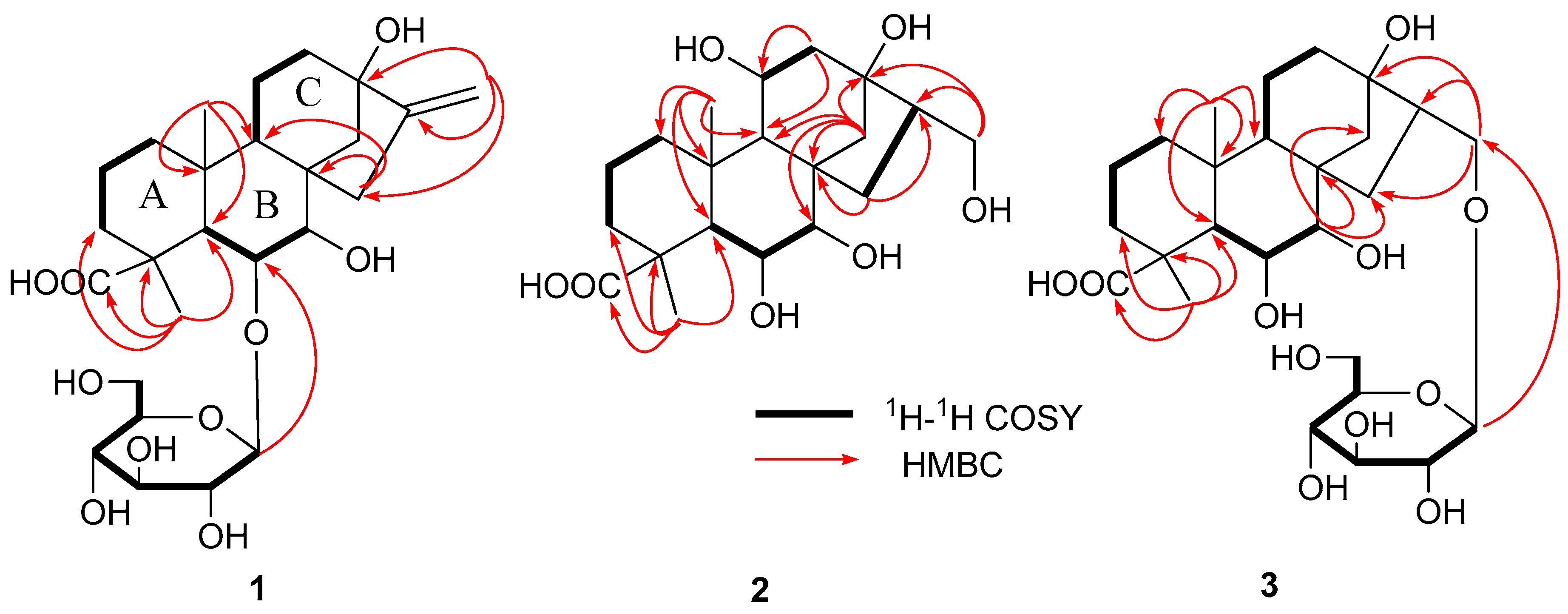
Compound 1 possessed the molecular formula of C26H40O10 according to the HR-ESI-MS at m/z 513.2651 [M + H]+. The acid hydrolysis of 1 liberated d-glucose, which was identified by HPLC analysis using an optical rotation detector. 1H-NMR spectrum (Table 1) of 1 showed two methyl signals at δH 1.28 (3H, s), δH 0.86 (3H, s); two olefinic proton signals at δH 5.0 (1H, br. s), δH 5.39 (1H, br. s); an anomeric proton signal at δH 4.40 (1H, d, J = 7.8 Hz). The 13C-NMR and DEPT spectrum (Table 1) of 1 showed an ent-kauran-type diterpene skeleton [19]. The compound structure has 26 carbon signals, five methylene signals at δc 18.4, 19.8, 29.2, 38.4, 39.2; as well as four quaternary carbon signals at δc 35.2, 43.0, 44.6, 85.4; a carbonyl carbon signal at δc 184.8; and two olefinic carbon signals at δC 109.7 and 158.0. The sugar moiety consisted of six carbon signals at δc 100.1, 75.2, 78.1, 71.7, 78.0, and 62.9 [20]. The 1H-1H COSY and HSQC analysis of 1 showed the structures A, B and C (Figure 2). The connectivity of these partial structures and their functional groups were investigated by analysis of HMBC. As shown in Figure 2, the long range correlations were observed between the following protons and carbon signals: H-18 (CH3) and C-3, C-4, C-5, C-19 (COOH); H-20 (CH3) and C-1, C-5, C-9, C-10; H-15 and C-7, C-8, C-9, C-16; H-14 and C-12, C-13, C-16; H-17 and C-16. Thus, structure 1 was confirmed as shown in Figure 2.
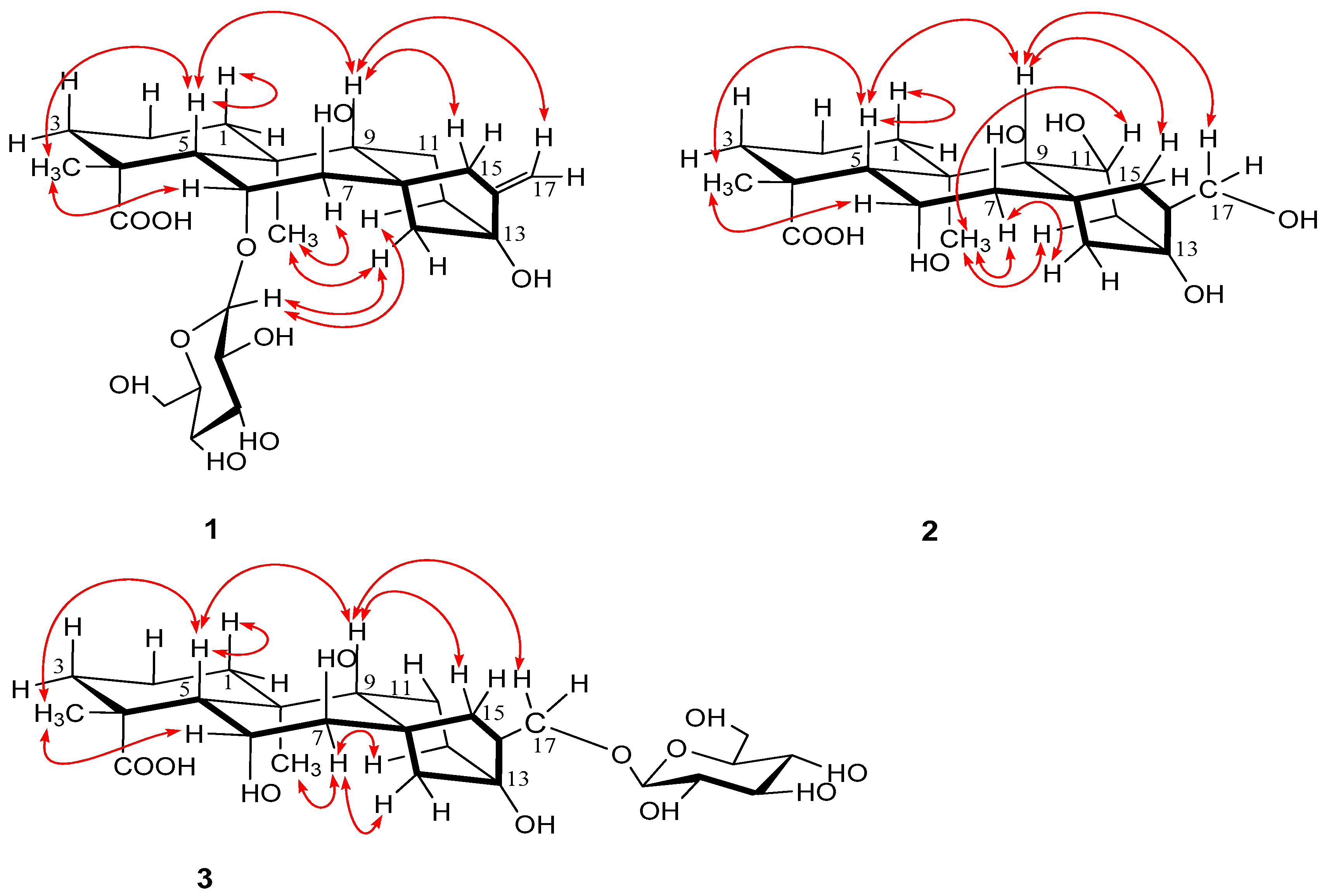
The coupling constants of H-6 (dd, J5,6 = 6.5 Hz and J6,7 = 6.5 Hz) and H-7 (d, J6,7 = 6.5 Hz) observed in the 1H-NMR spectrum were the same as those of H-6 (dd, J5,6 = 6.5 Hz and J6,7 = 6.5 Hz) and H-7 (d, J6,7 = 6.5 Hz) in 7β, 16α, 17-trihydroxy-ent-kauran-6α, 19-olide [21]. Thus, it was determined that H-6 was in the β-orientation and that H-7 was in the α-orientation. The relative stereochemistry of 1 was assigned by analysis of the NOESY spectrum. The NOESY correlations (Figure 3) of H-6/H-18, H-7/H-20 suggested that the configurations of C-18, C-20 were restricted as 18β, 20α, respectively. Likewise, the NOESY cross-peaks of H-19/H-5, H-5/H-9 and H-9/H-15 showed that the configurations of C-5, C-9 and C-15 were restricted as 5β, 9β and 15β, respectively. Thus, the structure of 1 was determined to be as shown (Figure 1) and elucidated as 7β, 13β-dihydroxy-6α-O-β-d-glucopyranosyl-ent-kauran-16-en-19-oic acid, named as arenterpenoid A (1).
Compound 2 possessed the molecular formula of C20H32O7 according to the HR-ESI-MS at m/z 385.2186 [M + H]+. 1H- and 13C-NMR spectra indicated that the structure of 2 was similar to that of 1, except for a 11-methylene group (δH 1.52 (m); δc 19.8 in 1), 16-olefinic proton group (δc 158.0 in 1) and a 17-olefinic proton group (δH 5.39 (br. s), 5.00 (br. s); δc 109.7 in 1), as well as presence of a 6β-hydroxy group (δH 4.54 (dd); δc 85.2 in 2), a 11β-hydroxy group (δH 3.95 (m); δc 65.5 in 2), a 16-homomethyl group (δH 2.03 (m), δc 43.1 in 2) and a 17-hydroxy group (δH 3.75 (d); δc 68.1 in 2). 13C-NMR and DEPT spectrum (Table 1) of 2 showed an ent-kauran-type diterpene skeleton [22]. The connectivity of these partial structures and the functional groups were investigated by analysis of HMBC of 2. As shown in Figure 2, long range correlations were observed between the following protons and carbon signals: H-18 (CH3) and C-3, C-4, C-5, C-19 (COOH); H-20 (CH3) and C-1, C-5, C-9, C-10; H-14 and C-7, C-8, C-9, C-13; H-15 and C-8, C-15; H-17 and C-13, C-15. Thus, structure 2 was confirmed as shown in Figure 2.
The relative stereochemistry of 2 was assigned by analysis of the NOESY spectrum. The 1H- and 13C-NMR spectra showed similar data for 1 and 2. Thus, it was determined that H-6 was in the β-orientation and that H-7 was in the α-orientation. The correlations of H-6/H-18 showed that they were cofacial and were arbitrarily assigned to be β-oriented. H-5, H-9 and H-15 were determined by their correlations with H-18. H-20 and H-11 were determined by their correlations with H-7. Thus, the structure of 2 was determined to be as shown (Figure 1) and elucidated as, 6α, 7β, 11β, 13β, 17-pentahydroxy-ent-kauran-19-oic acid, named as arenterpenoid B (2).
Compound 3 possessed the molecular formula of C26H42O11 according to the HR-ESI-MS at m/z 531.2769 [M + H]+. The acid hydrolysis of 2 liberated d-glucose, which was identified by HPLC analysis using an optical rotation detector. 1H- and 13C-NMR spectra indicated that the structure of 3 was similar to that of 2, except for a 11β-hydroxy group (δH 3.95; δc 65.5 in 2) and 17-hydroxy group (δH 3.75; δc 68.1 in 2), as well as the presence of a 11-methylene group (δH 1.36; δc 18.3 in 3), and 17-(β-glucopyranosyl oxy) group (δH 4.2, 3.46; δc 76.2 in 3). The connectivity of these partial structures and the functional groups were investigated by analysis of the HMBC of 3. As shown in Figure 2, long range correlations were observed between the following protons and carbon signals: H-18 (CH3) and C-3, C-4, C-5, C-19 (COOH); H-20 (CH3) and C-1, C-5, C-9, C-10; H-15 and C-7, C-8, C-9, C-16; H-7 and C-8, C-14, C-15; H-17 and C-13, C-16, C-1′. Thus, the structure of 3 was confirmed as shown in Figure 2.
The relative stereochemistry of 3 was assigned by analysis of the NOESY spectrum. The 1H- and 13C-NMR spectra showed similar data for 3 and 2. Likewise, it was determined that H-6 was in the β-orientation and that H-7 was in the α-orientation. The NOESY of 3 showed the cross-peaks between H-6 and H-18, H-5, H-9; H-7 and H-20. The results showed that the stereochemistry of 3 is similar to the stereochemistry of 2. Therefore, the structure of 3 was determined to be as shown (Figure 1) and elucidated as, 6α, 7β, 13β-trihydroxy-17-O-β-d-glucopyranosyl-ent-kauran-19-oic acid, named as arenterpenoid C (3). The other compounds were characterized as pseudaminic acid (4), 12α-(β-d-glucopyranosyl)-7β-hydroxy-kaurenolide (5), paniculoside (6), agittarioside b (7), orychoside B (8) by comparing their NMR spectroscopic data with the literature values (Figures S1–S5 and Table S1).
All these compounds are reported here for the first time in Arenga genus. The kaurane type diterpene is a kind of tetracyclic diterpene with hydrogenated phenanthrene as the mother nucleus [23]. According to the structural rule of the kauri-type diterpene, the compounds 1–8 are all C-20 unoxidized kauri-type. Most of such structures isolated and artificially synthesized in plants have significant biological activities, such as antimicrobial activity and cytotoxicity [24]. This study provides an experimental and scientific basis for drug design and discovery in A. pinnata fruits.
3. Experimental Section
3.1. General Experimental Procedures
NMR spectra were measured on a Bruker AV-400 spectrometer (Bruker Company, Waltham MA, USA) with TMS as an internal standard. High-resolution ESI-MS mass spectra were carried out on an AB SCEIX Triple-TOFTM 5600+ instrument (A.B. Company, Milwaukee, WI, USA). UV spectra were recorded on a PerkinElmer Lambda UV-365 instrument (PE Company, Waltham MA, USA). IR spectra were recorded on a PerkinElmer Spectrum Two spectrometer (PE Company, Waltham MA, USA) with KBr disks. Preparative HPLC (515-2414, Waters, Milford, CT, USA) was performed on 5C18 MS-II (10 μm, 20 × 250 mm, cat. no.: 38024-01, COSMOSIL, Tokyo, Japan). NH2 column (4.6 × 250 mm, cat. no.: S1119, Kasei Company, Tokyo, Japan), silica gel (200–300 mesh, Haiyang Co, Qingdao, China), Amberlite IRA-400 (OH-, Alfa Aesar, Heysham, UK) and ODS (50 µm, AAG12S50, YMC Company, Kyoto, Japan) were used for column chromatography. Detectors (2424, ELS, Waters) and (2998, PDA, Waters) were used in the HPLC. Precoated silica GF 254 plates (Haiyang Company, Qingdao, China) were used for TLC analysis. All the solvents were of analytical grade (Tianjinfuyu Company Ltd., Tianjin, China).
3.2. Plant Material
The A. pinnata fruits were collected from Guangxi in China during September 2017, and authenticated by Prof. Weiming Wang of the Heilongjiang Research institute of Chinese Medicine. The fruitage had been deposited at the Heilongjiang Research institute of Chinese Medicine.
3.3. Extraction and Isolation
The A. pinnata fresh fruits (30.0 kg) were extracted with 70% EtOH (200 L × 3 h × 3 times). The combined extract was concentrated under vacuum yielding a residue (3.0 kg) which was dissolved in H2O (12 L) and extracted sequentially with petroleum, chloroform, ethyl acetate and n-butanol (12 L × 3 h × 5 times). The eluate was separately concentrated in vacuo to give a petroleum syrup (109.0 g), chloroform syrup (123.0 g), ethyl acetate syrup (205.0 g), and an n-butanol syrup (380.0 g). In this study, we only separated the n-butanol layer. The n-butanol (380.0 g) extract was subject to column chromatography on silica gel (4460.0 g) and eluted with CH2Cl2/MeOH (20:1 (80.0 L), 10:1 (110.0 L), 5:1 (120.0 L), 3:1 (100.0 L), 2:1 (80.0 L) and 1:1 (60.0 L), v/v) to afford six fractions (fractions A (36.0 g), B (96.7 g), C (90.2 g), D (40.1 g), E (21.2 g), F (20.1 g). The TLC and HPLC were used to observe each of the fractions, and similar fractions were combined to afford A1-A6, B1-B5, C1-C6, D1-D6, E1-E10, F1-F6. Fraction B4 (21.7 g) was eluted by Rp-18 (600.0 g) (MeOH/H2O 2:8 (1.4 L)→3:7 (2.0 L)→4:6 (2.7 L)→5:5 (2.0 L)→6:4 (2.0 L)→7:3 (1.4 L)→8:2 (1.4 L)→9:1 (0.8 L)→1:0 (1.0 L), v/v) to afford nine subfractions (subfractions B4-1–B4-9). Subfraction B4–6 further purified by a preparative RP-HPLC (55% MeOH/H2O, flow rate 5 mL/min) to give 1 (100.12 mg, tR = 23 min). Subfractions B4–5 were further purified by a preparative RP-HPLC (55% MeOH/H2O, flow rate 5 mL/min) to give 2 (9.52 mg, tR = 42 min). Fraction C1 (8.0 g) was eluted by Rp-18 (600.0 g) (MeOH/H2O 2:8 (1.4 L)→3:7 (2.0 L)→4:6 (2.7 L)→5:5 (2.7 L)→6:4 (2.0 L)→7:3 (1.4 L)→8:2 (1.4 L)→9:1 (0.8 L)→1:0 (1.0 L), v/v) to afford nine subfractions (subfractions C1-1–C1-9). Subfractions C1–3 were further purified by a preparative RP-HPLC (20% MeOH/H2O, flow rate 5 mL/min) to give 4 (11.89 mg, tR = 30 min). Subfraction C1–5 were further purified by a preparative RP-HPLC (45% MeOH/H2O, flow rate 5 mL/min) to give 5 (8.50 mg, tR = 25 min). Fraction D3 (18.0 g) was eluted by Rp-18 (600.0 g) (MeOH/H2O 2:8 (1.4 L)→3:7 (2.0 L)→4:6 (2.7 L)→5:5 (2.7 L)→6:4 (2.7 L)→7:3 (1.4 L)→8:2 (1.4 L)→9:1 (0.8 L)→1:0 (1.0 L), v/v) to afford nine subfractions (subfractions D3-1–D3-9). Subfractions D3–6 were further purified by a preparative RP-HPLC (40% MeOH/H2O, flow rate 5 mL/min) to give 3 (10.61 mg, tR = 32 min). Subfraction D3–6 were further purified by a preparative RP-HPLC (45% MeOH/H2O, flow rate 5 mL/min) to give 8 (4.51 mg, tR = 41 min). Subfractions D3–4 were further purified by a preparative RP-HPLC (60% MeOH/H2O, flow rate 5 mL/min) to give 6 (70.67 mg, tR = 29 min). Subfractions D3–5 were further purified by a preparative RP-HPLC (60% MeOH/H2O, flow rate 5 mL/min) to give 7 (4.32 mg, tR = 37 min).
Arenterpenoids A (1). Yellow amorphous powder. Gave +10.0 (c = 1.76, MeOH); IR (KBr) 3436, 2946, 2835, 1719, 1635, 1467, 1231, and 1069 cm−1; 1H- and 13C-NMR (MeOH, 400, 100 MHz) data, see Table 1; HR-ESI-MS m/z 513.2651 [M + H]+ (calc. for C26H40O10, 513.2655) (Figure 1 and Figure 2).
3.4. Acid Hydrolysis and HPLC Analysis
The isolated compounds (1,3) (2.0 mg) were in 1.0 mL HCl and were each heated under reflux for 3 h. After cooling, the two mixtures were separately filtered with Amberlite IRA-400 to give a solution. Assigned with AcOEt to get two layers. The aqueous layer was evaporated to dryness under vacuum, and then subjected to HPLC analysis using an NH2 column and an optical-rotation detector. d-glucose was confirmed by comparison of the tR with that of an authentic sample (mobile phase: MeCN/H2O 85: 15 (v/v); flow rate: 0.8 mL/min; tR = 12.8 min (d-glucose, positive optical rotation))
4. Conclusions
As a traditional Chinese medicine, A. pinnata fruits were mainly used to treat rheumatism and bone pain. This study obtained eight diterpene compounds from A. pinnata fruits, including three new diterpenes and five known ones. This also reveals some structural characteristics of the chemical constituents in the A. pinnata fruit, which provides some clues for further clarifying the composition of the components and correlations of the relative plant species. In this study we have made this contribution to discover active ingredients and leading compounds and additionally provided an experimental and scientific basis of drug design and drug discovery of the A. pinnata fruits.
Supplementary Materials
The following are available online: Figures S1–S5 and Table S1: The 13C-NMR data of Compounds 4–8.
Author Contributions
W.-M.W. and L.-Q.H. designed the experiments; J.-F.L. and C.W. performed the experiments; J.-F.L. wrote the paper. W.-M.W., F.-J.L. and J.-H.H. modified the paper; All authors read and approved the manuscript.
Funding
This work was financed by key projects at the central government level: The ability establishment of sustainable use for valuable Chinese medicine resources (2060302) and Supported by China Agriculture Research System (CARS-21).
Conflicts of Interest
The authors declare no conflict of interest.
Abbreviations
The following abbreviations are used in this manuscript:
| HR-ESI-MS | High-resolution electrospray ionization mass spectrometry |
| NMR | Nuclear magnetic resonance |
| DEPT | Distortionless Enhancement by Polarization Transfer |
| HMBC | Heteronuclear multiple bond correlation |
| NOESY | Nclear overhauser effect spectroscopy |
| 1H-1H COSY | Correlation spectroscopy |
| HSQC | Heteronuclear multiple quantum coherence |
| tR | Retention time |
| UV | Ultraviolet |
| TMS | Tetramethylsilane |
| HPLC | High performance liquid chromatography |
| ODS | Octadecylsilyl |
| EtOH | Ethyl alcohol |
| TLC | Thin Layer Chromatography |
| MeOH | Methanol |
| MeCN | Methyl cyanide |
| AcOEt | Ethyl acetate |
References
- Editorial Board of Chinese Academy of Sciences. Flora of China; Science and Technology Publishing House: Beijing, China, 1991; Volume 3, p. 108. [Google Scholar]
- Lu, D.X.; Shang, Z.J. Kai Bao Ben Cao; Anhui Science and Technology Publishing House: Anhui, China, 1998. [Google Scholar]
- Ni, Z.M.; Zheng, J.S.; Qi, X.Y. Ben Cao Hui Yan; Shanghai Science and Technology Publishing House: Shanghai, China, 2005. [Google Scholar]
- Su, S. Ben Cao Tu Jing; Anhui Science and Technology Publishing House: Anhui, China, 1994. [Google Scholar]
- Li, S.Z. Compendium of Materia Medica; People’s Health Publishing House: Beijing, China, 2005; p. 31. [Google Scholar]
- Lin, Q.; Fu, L.G. Higher Plants of China; Qingdao Publishing House: Qingdao, China, 2009. [Google Scholar]
- Mu, X.N.; Yang, W.Q.; Wang, W.J. Chemical constituents from the fruits of Areca catechu. Chin. Med. Mat. 2014, 35, 56–60. [Google Scholar]
- Chen, Q.M.; Deng, Z.; Guo, F.W. Constituent Analysis of Volatile Organic Compounds in Three Palmae. J. Anhui Agric. 2017, 45, 152–154. [Google Scholar]
- Zeng, Q. Chemical Constituents from the Fruits of Areca catechu. Master’s Thesis, Central South University of Forestry and Technology, Changsha, China, 2013. [Google Scholar]
- Gui, Y.M. Studies on Diterpenes Constituents and Anti Tumor Activity of Rabdosia excisa. Ph.D. Thesis, Jilin University, Changchun, China, 2013. [Google Scholar]
- Guan, Y.G.; Hu, W.Z.; Shi, Y.S. Research Progress in Terpene Constituents and Biological Activities of Pteris. Chin. J. Exp. Tradit. Med. Form. 2018, 24, 219–227. [Google Scholar]
- Zhao, X.Y.; Cai, X.; Hu, Z.H. Research progress on biology, chemical constituents in Euphorbia kansui, and their pharmacological effects. Chin. Tradit. Herbal. Drugs 2014, 45, 3029–3033. [Google Scholar]
- Gong, X.L.; Chen, Z.H.; Liang, N.C. Isolation and identification of glycoside of compound 5F from Pterissemi pinnata L. and their antitumor activity. Chin. Med. Mat. 2010, 32, 257–260. [Google Scholar]
- Toshiyuki, M.; Kentarou, K.; Hisashi, M. Medicinal foodstuffs. XXII. Structures of oleanane-type triterpene oligoglycosides, pisumsaponins I and II, and kaurane-type diterpene oligoglycosides, pisumosides A and B, from green peas, the immature seeds of Pisum sativum L. Chem. Pharm. Bull. 2001, 49, 73–77. [Google Scholar]
- Takashi, K.; Hiromi, A.; Ken-Ichiro, M. Two new ent-kaurane-type diterpene glycosides from zucchini (Cucurbita pepo L.) seeds. Fitoterapia 2015, 107, 69–76. [Google Scholar]
- Cai, X.F.; Shen, G.; Dat, N.T. Inhibitory Effect of Kaurane Type diterpenes from Acanthopanax koreanum on TNF-α Secretion from Trypsin-Stimulated HMC-1 Cells. Arch. Pharm. Res. 2003, 26, 731–734. [Google Scholar] [CrossRef] [PubMed]
- Yoshikawa, M.; Yoshizumi, S.; Murakami, T. Medicinal foodstuffs. II. On the bioactive constituents of the tuber of Sagittaria trifolia L. (Kuwai, Alismataceae): Absolute stereostructures of trifoliones A, B, C, and D, sagittariosides a and b, and arabinothalictoside. Chem. Pharm. Bull. 1996, 44, 492–499. [Google Scholar] [CrossRef] [PubMed]
- Ohkoshi, E.; Kamo, S.; Makino, M. Ent-kaurenoic acids from Mikania hirsutissima (compositae). Phytochemistry 2004, 65, 885–890. [Google Scholar] [CrossRef] [PubMed]
- Zhang, G.; Cui, H.; Liu, S.; Dong, J. A new triterpenoid saponin and a diterpene glucoside from the seeds of Orychophragmus violaceus. Nat. Prod. Res. 2018, 46, 1–7. [Google Scholar] [CrossRef] [PubMed]
- Murakami, T.; Kohno, K.; Kishi, A.; Matsuda, H. Medicinal foodstuffs. XIX. Absolute stereostructures of canavalioside, a new Ent-kaurane-type diterpene glycoside, and gladiatosides A1, A2, A3, B1, B2, B3, C1, and C2, new acylated flavonol glycosides, from sword bean, the seeds of Canavalia gladiata. Chem. Pharm. Bull. 2000, 48, 1673–1680. [Google Scholar] [CrossRef] [PubMed]
- Kim, K.H.; Choi, S.U.; Lee, K.R. Diterpene glycosides from the seeds of pharbitis nil. J. Nat. Prod. 2009, 72, 1121–1127. [Google Scholar] [CrossRef] [PubMed]
- Li, R.J.; Wang, S.; Li, G.; Zhou, J.C. Four new Kaurane diterpenoids from the Chinese liverwort Jungermannia comata Nees. Chem. Biodivers. 2016, 13, 1685–1690. [Google Scholar] [CrossRef] [PubMed]
- Zhang, D.Y.; Li, Z.Y.; Shi, L.Y. Advances in Research on Cytotoxinic Activity of ent-Kaurance Diterpenoids. Chin. J. Ore Chem. 2008, 1911–1917. [Google Scholar]
- Du, M.J.; Lei, X.G. Advanced in the Synthesis of Kaurane Diterpenoids. Chin. J. Ore Chem. 2015, 2447–2464. [Google Scholar]
Sample Availability: Samples of the compounds are available from the authors. |
Figure 1.
Structures of compounds 1–8 from of Arenga pinnata (Wurmb.) Merr. ruits.

Figure 2.
Key HMBC and 1H-1H COSY correlations of compound 1–3.

Figure 3.
Key NOESY correlations of compound 1–3.


Table 1.
1H- and 13C-NMR Data of 1–3 (CD3OD).
| NO. | 1 | 2 | 3 | |||
|---|---|---|---|---|---|---|
| δH (J, Hz) | δC | δH (J, Hz) | δC | δH (J, Hz) | δC | |
| 1 | 1.53 (m) | 38.4 | 1.88 (m) | 37.1 | 1.06 (m) | 38.4 |
| 1.04 (m) | 1.73 (m) | 1.55 (m) | ||||
| 2 | 1.53 (m) | 18.4 | 1.41 (m) | 18.6 | 1.54 (m) | 18.3 |
| 3 | 2.01 (m) | 29.2 | 1.83 (m) | 24.1 | 2.00 (m) | 29.2 |
| 1.40 (m) | 1.65 (m) | 1.42 (m) | ||||
| 4 | 43.0 | 43.2 | 43.1 | |||
| 5 | 1.88 (d, 6.5) | 52.7 | 1.85 (d, 7.3) | 51.4 | 1.82 (d, 6.5) | 52.8 |
| 6 | 4.62 (dd, 6.5, 6.5) | 84.9 | 4.54 (dd, 7.3, 7.3) | 85.2 | 4.56 (dd, 6.5, 6.5) | 85.2 |
| 7 | 4.26 (d, 6.5) | 72.2 | 4.14 (d, 7.3) | 72.3 | 4.23 (d, 6.5) | 72.1 |
| 8 | 44.6 | 48.2 | 48.0 | |||
| 9 | 1.17 (dd, 4.9, 12.1) | 56.2 | 1.17 (dd, 4.7, 12.1) | 59.7 | 1.17 (m) | 58.8 |
| 10 | 35.2 | 37.2 | 35.4 | |||
| 11 | 1.52 (m) | 19.8 | 3.95 (m) | 65.5 | 1.36 (m) | 18.3 |
| 12 | 2.31 (dd, 7.5, 13) | 39.2 | 1.76 (m) | 49.7 | 1.79 (m) | 24.4 |
| 1.37 (m) | 1.68 (m) | |||||
| 13 | 85.4 | 86.0 | 85.4 | |||
| 14 | 2.05 (d, 10.9) | 39.9 | 2.12 (d, 13.0) | 47.5 | 2.17 (d, 13.2) | 47.6 |
| 1.74 (m) | 1.16 (d, 13.0) | 1.16 (d, 13.2) | ||||
| 15 | 2.81 (br. d, 14.9) | 42.0 | 2.05 (m) | 33.5 | 2.09 (m) | 33.6 |
| 1.78 (m) | 1.49 (d, 12.6) | 1.53 (m) | ||||
| 16 | 158.0 | 2.03 (m) | 43.1 | 2.05 (m) | 43.8 | |
| 17 | 5.39 (br. s) | 109.7 | 3.75 (d, 11.5) | 68.1 | 4.27 (d, 10.4) | 76.2 |
| 5.00 (br. s) | 3.58 (d, 11.5) | 3.46 (d, 10.4) | ||||
| 18 | 1.28 (s) | 25.9 | 1.34 (s) | 24.7 | 1.27(s) | 25.8 |
| 19 | 184.8 | 185.1 | 185.0 | |||
| 20 | 0.86 (s) | 20.9 | 0.98 (s) | 23.4 | 0.85 (s) | 20.9 |
| 1′ | 4.40 (d, 7.8) | 100.1 | 4.28 (d, 7.7) | 100.1 | ||
| 2′ | 3.16 (m) | 75.2 | 3.21 (m) | 75.2 | ||
| 3′ | 3.37 (m) | 78.1 | 3.37 (m) | 78.1 | ||
| 4′ | 3.28 (m) | 71.7 | 3.28 (m) | 71.7 | ||
| 5′ | 3.28 (m) | 78.0 | 3.28 (m) | 78.0 | ||
| 6′ | 3.83 (dd, 2.1, 12.0) | 62.9 | 3.88 (dd, 2.1, 12.0) | 62.9 | ||
| 3.59 (m) | 3.65 (m) | |||||
© 2018 by the authors. Licensee MDPI, Basel, Switzerland. This article is an open access article distributed under the terms and conditions of the Creative Commons Attribution (CC BY) license (http://creativecommons.org/licenses/by/4.0/).
Share and Cite
MDPI and ACS Style
Liu, J.-F.; Huo, J.-H.; Wang, C.; Li, F.-J.; Wang, W.-M.; Huang, L.-Q. New Diterpenes from Arenga pinnata (Wurmb.) Merr. Fruits. Molecules 2019, 24, 87. https://doi.org/10.3390/molecules24010087
AMA Style
Liu J-F, Huo J-H, Wang C, Li F-J, Wang W-M, Huang L-Q. New Diterpenes from Arenga pinnata (Wurmb.) Merr. Fruits. Molecules. 2019; 24(1):87. https://doi.org/10.3390/molecules24010087
Chicago/Turabian StyleLiu, Ji-Fei, Jin-Hai Huo, Chang Wang, Feng-Jin Li, Wei-Ming Wang, and Lu-Qi Huang. 2019. "New Diterpenes from Arenga pinnata (Wurmb.) Merr. Fruits" Molecules 24, no. 1: 87. https://doi.org/10.3390/molecules24010087