Article

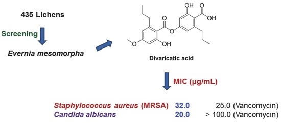
Antimicrobial Activity of Divaricatic Acid Isolated from the Lichen Evernia mesomorpha against Methicillin-Resistant Staphylococcus aureus

1
Department of Pharmacy, and Research Institute of Life Pharmaceutical Sciences, Sunchon National University, Suncheon 57922, Korea
2
Department of Agricultural Chemistry, Sunchon National University, Suncheon 57922, Korea
*
Author to whom correspondence should be addressed.
Molecules 2018, 23(12), 3068; https://doi.org/10.3390/molecules23123068
Submission received: 30 October 2018 / Revised: 20 November 2018 / Accepted: 23 November 2018 / Published: 23 November 2018
(This article belongs to the Section Natural Products Chemistry)

Abstract

:
One hundred and seventy seven acetone extracts of lichen and 258 ethyl acetate extracts of cultured lichen-forming fungi (LFF) were screened for antimicrobial activity against Staphylococcus aureus and Enterococcus faecium using a disk diffusion method. Divaricatic acid was isolated from Evernia mesomorpha and identified by LC-MS, 1H-, 13C- and DEPT-NMR. Purified divaricatic acid was effective against Gram + bacteria, such as Bacillus subtilis, Staphylococcus epidermidis, Streptococcus mutans, and Enterococcus faecium, with the minimum inhibitory concentration (MIC) values ranging from 7.0 to 64.0 μg/mL, whereas vancomycin was effective in the MICs ranging from 0.78 to 25.0 μg/mL. Interestingly, the antibacterial activity of divaricatic acid was higher than vancomycin against S. epidermidis and E. faecium, and divaricatic acid was active against Candida albicans. In addition, divaricatic acid was active as vancomycin against S. aureus (3A048; an MRSA). These results suggested that divaricatic acid is a potential antimicrobial agent for the treatment of MRSA infections.

Graphical Abstract

1. Introduction

Lichens are composite living organisms comprised of algae (photobiont) and fungi or cyanobacteria (mycobiont) [1,2], and produce numerous biologically active secondary metabolites with, for example, antiviral, antibiotic, antitumor, or anti-allergenic properties [3]. A huge number of secondary metabolites from more than 20,000 species of lichens have been investigated as potential scaffolds for drug development [2,4,5]. Usnic acid is probably the most investigated compound of these metabolites, and has antibiotic activity and has been used to develop pharmaceutics and cosmetics [6,7,8]. Recently, usnic derivatives were synthesized to enhance its antimicrobial activity [9].
Staphylococcus aureus is a Gram-positive bacterium found on skin and mucosal surfaces, and is a normal inhabitant of the healthy human body. However, in some individuals, such as immunocompromised patients, S. aureus can be pathogenic and cause a range of conditions from minor skin infections to life-threatening diseases, such as pneumonia, endocarditis, and sepsis [10]. Furthermore, S. aureus is related to nosocomial infections and methicillin-resistant S. aureus (MRSA) has become a major health problem [11,12]. Recently, antibiotic compounds of new classes were developed by two step or convenient synthesis to be active against MRSA [13,14]. Antibiotics for staphylococci target the cell envelope, ribosomes, and nucleic acids [15], and vancomycin has proven efficacy against MRSA infections and has been administered even other treatments have failed [16,17]. Enterococcus faecium is a Gram-positive bacterium found in the human gastrointestinal tract, causes diseases, such as neonatal meningitis or endocarditis, and has recently emerged as a therapeutically challenging pathogen [18,19].
Divaricatic acid has been reported in extracts of the lichens Ramalina hierrensis [20], Flavocetraria nivalis and Ophioparma ventosa [21], and Lepraria sp. [22], and the cyclohexane extracts four Algerian lichens, such as Cladonia rangiformis, Ramalina farinaceae, R. fastigiata, and Roccella phycopsis, were reported to be active against human pathogenic fungi [23]. Divaricatic acid isolated from Ramalina aspera was reported to have molluscicidal and antiparasitic activities [24], and that from Lecanora frustulosa had minimal inhibition concentration (MIC) values from 0.78 to 12.5 mg/mL [25].
In this study, we isolated divaricatic acid as the main constituent from an acetone extract of Evernia mesomorpha and investigated its antimicrobial activities, and its activities against MRSA and Candida albicans.

2. Results and Discussion

2.1. Antimicrobial Activity

2.1.1. Screening

Antimicrobial activities of 435 lichens were determined using 1.0 mg of extracts spotted onto paper disks. Based on halo sizes, samples tested were divided into three groups, i.e., low, moderate, and effective). None of the 258 EA extracts of cultured LFF were active against the tested bacteria, S. aureus and E. faecium. Of the 177 acetone extracts of lichens, 9 and 28 samples were effective against S. aureus and E. faecium, respectively, and 8 samples of them were effective against both strains. Total 29 samples were selected for further evaluation (Table 1).

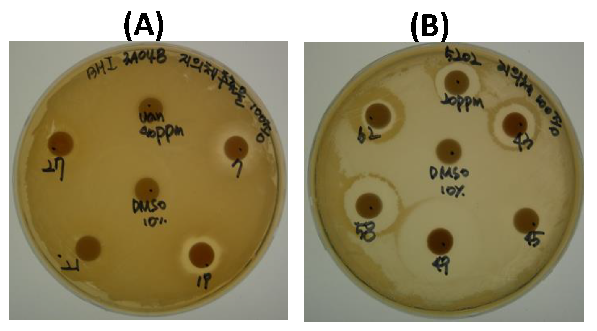
2.1.2. Assay of Antimicrobial Activity

In antimicrobial activity testing of these samples, halo sizes for S. aureus were smaller than those for E. faecium (Figure 1). The tests were carried out with duplicate experiments. Diameter of halo was measured including disk (8 mm). Halo size order of the samples for S. aureus was 419 > 407 = 431, and those for E. faecium was 449 > 407 > 458 = 517 (Table 1).

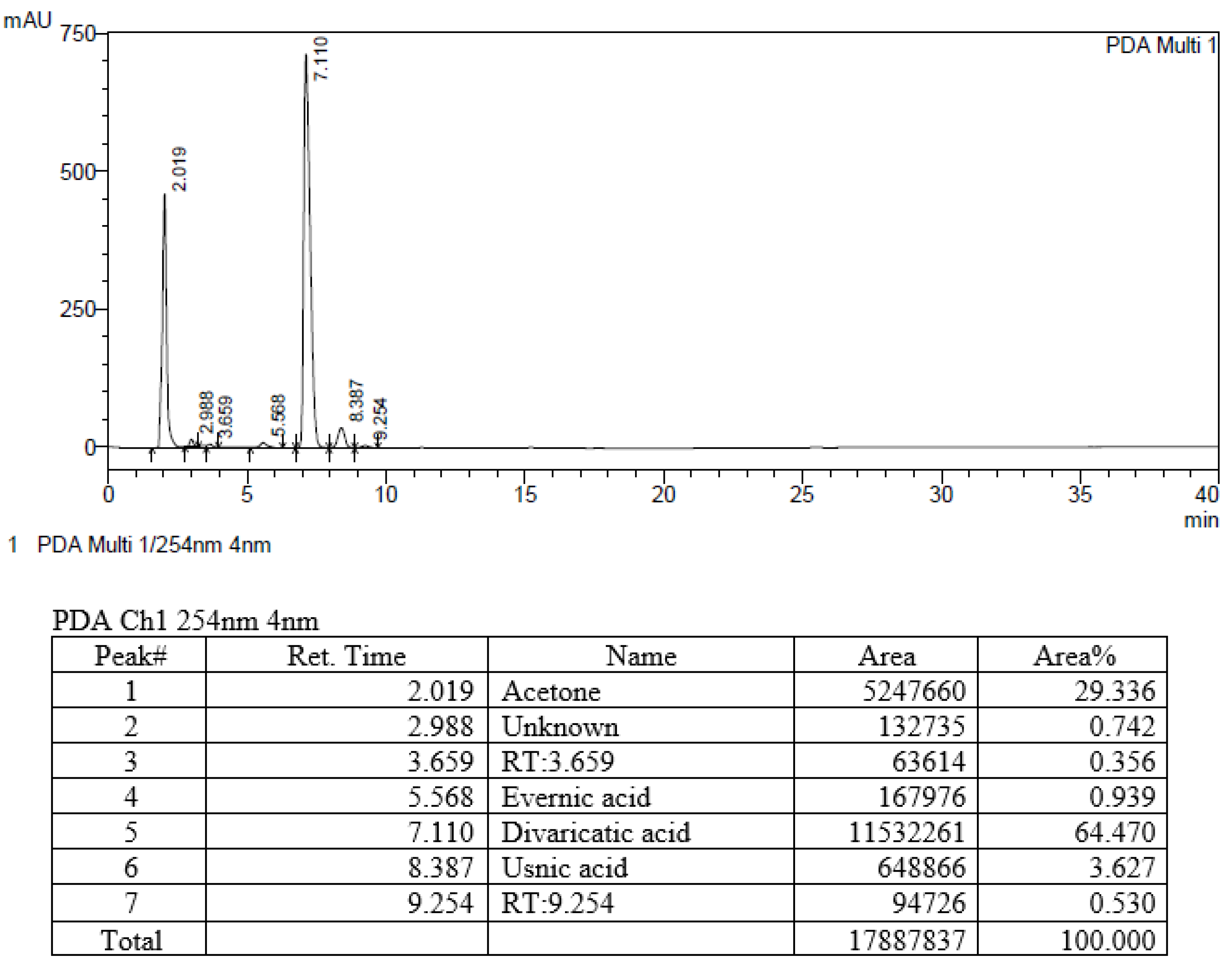
2.2. HPLC of the Selected Extracts

Prior to selecting extracts for further study, we searched literatures for information on the components and biological activities of the 34 extracts. Based on halo sizes and literature information, we analyzed 8 extracts by HPLC. Six peaks were observed in sample 458 (Evernia mesomorpha) (Figure 2), and other samples produced four to seven peaks (Figures S1–S7). Components were identified by comparing their retention times to standard data (Table 2). Of those compounds identified, usnic acid is well-known to have antimicrobial activity and galbinic acid, atranorin, and chloroatranorin have also been reported to be active [26,27]. We observed divaricatic acid was 17.8 times more abundant than usnic acid in E. mesomorpha extract, and that it exhibited high antimicrobial activity in the screening experiment. Usnea sp. showed the best antimicrobial activity, however, the major component was usnic acid; sample 458 showed less activity than Usnea sp., however, the major component was not well studied in its antimicrobial activity. In view of its antimicrobial activity, novelty, and availability, E. mesomorpha and divaricatic acid were selected for further study.

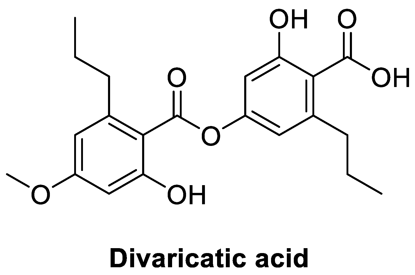
2.3. Structural Analysis of the Major Active Compound in E. mesomorpha

In the mass spectrometric analyses, the values of m/z ratios of the major MS peaks detected in negative ionization mode indicated the formation of [M − H] ions (C21H23O7-, m/z = 387) (Figure S8). The 1H-NMR data indicated the presence of one carboxyl group, two benzene ring protons, one methoxy group, and two propyl groups (Figure S9). We compared the MS and 1H-NMR data obtained with those reported for divaricatic acid [28]. Based on the twenty-one carbon peaks in 13C-NMR (Figure S10) and protonated carbon shifts from DEPT (distortionless enhancement by polarization transfer) (Figure S11), the compound was positively identified as divaricatic acid (Figure 3). Purity of the divaricatic acid by LC was 97.1% (Figure S12B), and the peak was corresponded to the third peak of acetone extract of sample 458 (E. mesomorpha) in the same analytical condition (Figure S12A).

2.4. MIC

The MIC values of E. mesomorpha extract against S. aureus and E. faecium were 64 and 16 μg/mL, respectively. The values coincided with the results of disk diffusion experiments. The purified divaricatic acid was found to be effective against S. aureus (32 μg/mL) and E. faecium (16 μg/mL) and against other Gram + bacteria, such as B. subtilis, S. epidermidis, Str. mutans, and E. faecium (5202) with MICs ranging from 7.0 to 64.0 μg/mL, whereas vancomycin had MICs ranging from 0.78 to 25.0 μg/mL (Table 3). Interestingly, the antibacterial activity of divaricatic acid was higher than that of vancomycin against S. epidermidis and E. faecium (5202). Furthermore, divaricatic acid was active against C. albicans (Table 3), whereas as vancomycin did not have antifungal properties [29], but slightly less effective than vancomycin against S. aureus (3A048; an MRSA). However, like vancomycin, divaricatic acid was not effective against Gram-bacteria. Divaricatic acid was more effective than cefotaxime, a broad-spectrum antibiotic, against E. faecium and the MRSA S. aureus (Table 3). MIC values of divaricatic acid against B. subtilis (7.0 μg/mL), S. aureus (64.0 μg/mL), and C. albicans (20.0 μg/mL) found in the present study were much lower than those (1.56, 1.56, 6.25 mg/mL, respectively) previously reported by Marijana et al. [25]. We attribute this discrepancy possibly to the types of strains tested, the culture conditions, the purity of the compound, and other unknown factors.
Structure of divaricatic acid, a depside, is quite different from those of well-known antibiotics. Usnic acid is a dibenzofuran having a structural unit similar to divaricatic acid and produced by various lichen species. It was reported that antibacterial activity of usnic acid was primarily caused by inhibition of DNA and RNA synthesis [30]. Based on the result, it might be suggested that divaricatic acid is related to the inhibition of nucleotide synthesis rather than cell-wall disruption. However, further study on the mechanism of divaricatic acid will be necessary. On the other hand, divaricatic acid showed a high degree of Escherichia coli RecA inhibition, relating to SOS response in bacteria [31]. The property might give an advantage in reduction of resistance induction. Toxicity of divaricatic acid to human cells was not reported, though gyrophoric acid, a depside (tridepside), showed no toxicity to human keratinocytes HaCaT cells [32]. The toxicity experiment should be needed in further study.

3. Materials and Methods

3.1. Chemicals and Media

Dimethyl sulfoxide (DMSO), ethyl acetate (EA), methanol (MeOH), chloroform, toluene, dioxane, dichloromethylene (DCM), acetic acid, and vancomycin were purchased from Sigma-Aldrich (St. Louis, MO, USA). Marine broth was obtained from MB Cell (Seoul, Korea). Nutrient broth, brain heart infusion (BHI), peptone, yeast extract, and agar were from Becton, Dickinson and Company (Sparks, MD, USA). Glucose was from Junsei (Tokyo, Japan) to prepare the GPYA (glucose-peptone-yeast extract-agar) medium.

3.2. Extracts of Lichen and Lichen-Forming Fungus Cultures

Acetone extracts of lichens (177 samples) and EA extracts of cultured lichen-forming fungi (LFF) (258 samples, 100 mg/mL) were obtained from the Korean Lichen Research Institute (KoLRI) in Sunchon National University, Korea. Samples were collected at various sites in Korea, China, Chile, Cameroon, Philippines, Romania, Vietnam, and the Arctic. Acetone extracts were dried and dissolved in DMSO (100 mg/mL).

3.3. Microbial Strains

S. aureus CCARM 3A048 (an MRSA), and E. faecium CCARM 5202, which were used for screening, were obtained from the Culture Collection of Antimicrobial Resistant Microbes (CCARM), Korea. To evaluate antimicrobial properties, we used five strains of Gram-positive bacteria (Bacillus subtilis KCTC 2189, Micrococcus luteus CCARM 0022, S. aureus CCARM 0027, S. epidermidis CCARM 3709, and Streptococcus mutans CCARM 0079), five strains of Gram-negative bacteria (Escherichia coli KCTC 2441, Pseudomonas aeruginosa CCARM 0057, Klebsiella pneumoniae CCARM 0015, Salmonella typhimurium CCARM 0240, and Vibrio vulnificus KCTC 2959), and C. albicans KCTC 27242 (a fungal strain).

3.4. Antibacterial Activity Assay

S. aureus CCARM 3A048 and E. faecium CCARM 5202 were grown at 37 °C for 24 h in BHI broth medium (calf brains 7.7 g, beef heart 9.8 g, protease peptone 10.0 g, dextrose 2.0 g, sodium chloride 5.0 g, disodium phosphate 2.5 g/L, pH 7.2–7.4) with shaking at 150 rpm. Cultures (0.1 mL) were then spread on BHI agar plates and paper disks (8 mm, Advantec, Toyo Roshi Kaisha. Ltd., Tokyo, Japan) sterilized with UV for 10 min, spotted with extracts (0.1 mL), and dried for 2–3 h, were placed on plates with spotted surfaces contacting the agar. For primary screening, aliquots of five extracts (1.0 mg each) were placed on a plate. Control of 10% EA or 10% DMSO, and vancomycin (20 or 40 ppm for E. faecium CCARM 5202 and S. aureus CCARM 3A048, respectively) were included. Plates were incubated at 37 °C for 12 h and the diameters of halos around the disks were observed or measured. Effective samples were selected by second screening (conducted in duplicate) using 0.5 mg of extract.

3.5. Analysis of Compounds in Extracts

Compounds in extracts with high antibacterial activity were analyzed at KoLRI by HPLC (LC-10AT, Shimadzu, Japan) equipped with a YMC-Pack ODS-A column using a solvent of methanol, water, and H3PO4 (80:20:1, v:v) as eluent [33], and putatively identified by comparing their retention times with a database, as previously described [34].

3.6. Isolation and Purification of Active Compounds and Structure Analysis

Compounds in the selected lichen (E. mesomorpha) extract were isolated by thin layer chromatography (TLC). The acetone extract was dissolved in 1 mL acetone in 1.5 mL EP tubes. For the preliminary study, this solution was spotted on Silica gel 60 F254 pre-coated plates (Merck Millipore, Darmstadt, Germany). The TLC plate was spotted with the sample and loaded in a twin trough chamber containing toluene, dioxane, and acetic acid (180:45:5, v/v), as previously described by Culberson [35]. Plate was removed from the chamber when the solvent front reached 15 cm from baseline and observed under 254 and 365 nm light. The plate was then sprayed with 10% aqueous sulfuric acid and heated for 5 min at 50 °C. For purification of the components, the lichen extract was separated by preparative TLC (PTLC) (silica gel 60 F254, 0.5 mm) using a toluene, dioxane, and acetic acid (90:25:4, v/v) mobile phase [24]. Spots were visualized at 254 nm and the major spot was removed. The silica gel obtained was then washed with MeOH and DCM (5:95, v/v), and the filtrate so obtained evaporated to dryness. Analysis of the compound was performed by a liquid chromatography–mass spectrometry (LC-MS) (LCMS-2020, Shimadzu, Kyoto, Japan) equipped with a reverse-phase column (Shim-pack VP-ODS/-C8/-Phenyl, 250 × 4.6 mm, Shimadzu). Elution was performed using combination of two solvents, that is, 1% formic acid in water (A) or acetonitrile (B). Linear gradient elution was performed using the following schedule: 5% B for 0–5 min; 5% to 30% B over 5–10 min; 30% B for 10–15min; 30 to 70% B over 15–20 min; and 70% B for 20–25 min. The structure of the compound was determined by 1H-NMR (JNM-ECZ400s/L1, JEOL, Tokyo, Japan).

3.7. Minimum Inhibitory Concentration (MIC)

Strains to be tested were grown at 37 °C for 24 h with shaking at 150 rpm. Nutrient agar was used for B. subtilis KCTC 2189 and E. coli KCTC 2441; marine agar for V. vulnificus KCTC 2959; and GPYA for C. albicans KCTC 27242. Other strains were grown in BHI medium. Cell densities were adjusted to an OD of 1.0 at 600 nm and diluted to 500 times. The isolated compound was dissolved in DMSO to produce a 10 mg/mL stock solution, then diluted to 256 μg/mL with BHI medium, then diluted serially twice from 1st to 10th well in a 96-well plate using 50 μL of BHI medium. Thereafter, 50 μL of diluted cells were added to 11th to 1st wells and incubated at 37 °C for 12 h. Each well contained 100 μL of liquid; 11th contained no compound or vancomycin, and 12th contained the medium only without the cell. MIC values were determined by comparing well turbidities with duplicate experiments and were presented as means ± standard errors.

4. Conclusions

In this study, divaricatic acid was isolated from the lichen E. mesomorpha and identified by LC-MS, 1H-, 13C- and DEPT-NMR. Divaricatic acid was found to be active against Gram + bacteria, such as B. subtilis, S. epidermidis, S. mutans, and E. faecium, and to show an antimicrobial activity against C. albicans. In addition, divaricatic acid was as effective as vancomycin against S. aureus (3A048; an MRSA). These results suggested that divaricatic acid may offer a means of treating MRSA infections.

Supplementary Materials

HPLC chromatograms of the selected lichens, and spectra of LC-MS, 1H-, 13C- and DEPT-NMR spectra of divaricatic acid are available in Supplementary Materials.

Author Contributions

J.M.O., Y.J.K. and H.-S.G. performed screening of antimicrobial activity and MIC test for the isolated compound; J.H. isolated the compound and analyzed the structure, and H.-H.H. supervised the experiments and wrote the part; H.K. supervised the study and wrote the paper.

Funding

This study was supported by the Suncheon Research Center for Natural Medicines, Republic of Korea.

Acknowledgments

We thank the Korean Lichen and Allied Bioresources Center at the Korean Lichen Research Institute in Sunchon National University, Republic of Korea, for the lichen samples.

Conflicts of Interest

The authors have no conflict of interest to declare.

References

  1. Ahmadjian, V. Lichens. Annu. Rev. Microbiol. 1965, 19, 1–20. [Google Scholar] [CrossRef] [PubMed]
  2. Ranković, B.; Kosanić, M. Lichens as a potential source of bioactive secondary metabolites. In Lichen Secondary Metabolites; Ranković, B., Ed.; Springer: Cham, Switzerland, 2015; pp. 1–26. [Google Scholar]
  3. Huneck, S. The significance of lichens and their metabolites. Naturwissenschaften 1999, 86, 559–570. [Google Scholar] [CrossRef] [PubMed]
  4. Müller, K. Pharmaceutically relevant metabolites from lichens. Appl. Microbiol. Biotechnol. 2001, 56, 9–16. [Google Scholar] [CrossRef] [PubMed]
  5. Zambare, V.P.; Christopher, L.P. Biopharmaceutical potential of lichens. Pharm. Biol. 2012, 50, 778–798. [Google Scholar] [CrossRef] [PubMed] [Green Version]
  6. Ingólfsdóttir, K. Usnic acid. Phytochemistry 2002, 61, 729–736. [Google Scholar] [CrossRef]
  7. Cocchietto, M.; Skert, N.; Nimis, P.L.; Sava, G. A review on usnic acid, an interesting natural compound. Naturwissenschaften 2002, 89, 137–146. [Google Scholar] [CrossRef] [PubMed]
  8. Cansaran, D.; Kahya, D.; Yurdakulola, E.; Atakol, O. Identification and quantitation of usnic acid from the lichen Usnea species of Anatolia and antimicrobial activity. Z. Naturforsch. C 2006, 61, 773–776. [Google Scholar] [CrossRef] [PubMed]
  9. Victor, K.; Boris, L.; Athina, G.; Anthi, P.; Marija, S.; Marina, K.; Oliver, R.; Marina, S. Design, synthesis and antimicrobial activity of usnic acid derivatives. MedChemComm 2018, 9, 870–882. [Google Scholar] [CrossRef] [PubMed]
  10. Dong, J.; Qiu, J.; Wang, J.; Li, H.; Dai, X.; Zhang, Y.; Wang, X.; Tan, W.; Niu, X.; Deng, X.; et al. Apigenin alleviates the symptoms of Staphylococcus aureus pneumonia by inhibiting the production of alpha-hemolysin. FEMS Microbiol. Lett. 2013, 338, 124–131. [Google Scholar] [CrossRef] [PubMed]
  11. Boucher, H.W.; Corey, G.R. Epidemiology of methicillin-resistant Staphylococcus aureus. Clin. Infect. Dis. 2008, 46, S344–S349. [Google Scholar] [CrossRef] [PubMed]
  12. Rahman, M.M.; Amin, K.B.; Rahman, S.M.M.; Khair, A.; Rahman, M.; Hossain, A.; Rahman, A.K.M.A.; Parvez, M.S.; Miura, N.; Alam, M.M. Investigation of methicillin-resistant Staphylococcus aureus among clinical isolates from humans and animals by culture methods and multiplex PCR. BMC Vet. Res. 2018, 14, 300. [Google Scholar] [CrossRef] [PubMed]
  13. Campanini-Salinas, J.; Andrades-Lagos, J.; Gonzalez Rocha, G.; Choquesillo-Lazarte, D.; Bollo Dragnic, S.; Faúndez, M.; Alarcón, P.; Silva, F.; Vidal, R.; Salas-Huenuleo, E.; et al. A new kind of quinonic-antibiotic useful against multidrug-resistant S. aureus and E. faecium infections. Molecules 2018, 23, 1776. [Google Scholar] [CrossRef] [PubMed]
  14. Su, J.C.; Huang, Y.T.; Chen, C.S.; Chiu, H.C.; Shiau, C.W. Design and synthesis of malonamide derivatives as antibiotics against methicillin-resistant Staphylococcus aureus. Molecules 2018, 23, 27. [Google Scholar] [CrossRef] [PubMed]
  15. Foster, T.J. Antibiotic resistance in Staphylococcus aureus. Current status and future prospects. FEMS Microbiol. Rev. 2017, 41, 430–449. [Google Scholar] [CrossRef] [PubMed]
  16. Sorrell, T.C.; Packham, D.R.; Shanker, S.; Foldes, M.; Munro, R. Vancomycin therapy for methicillin-resistant Staphylococcus aureus. Ann. Intern. Med. 1982, 97, 344–350. [Google Scholar] [CrossRef] [PubMed]
  17. Ellison, M.J. Vancomycin, metronidazole, and tetracyclines. Clin. Podiatr. Med. Surg. 1992, 9, 425–442. [Google Scholar] [PubMed]
  18. Amyes, S.G. Enterococci and streptococci. Int. J. Antimicrob. Agents 2007, 29, S43–S52. [Google Scholar] [CrossRef]
  19. Arias, C.A.; Murray, B.E. The rise of the Enterococcus: Beyond vancomycin resistance. Nat. Rev. Microbiol. 2012, 10, 266–278. [Google Scholar] [CrossRef] [PubMed]
  20. González, A.G.; Barrera, J.B.; Pérez, E.M.; Padrón, C.E. Chemical Constituents of the lichen Ramalina hierrensis. Planta Med. 1992, 58, 214–218. [Google Scholar] [CrossRef] [PubMed]
  21. Bjerke, J.W.; Lerfall, K.; Elvebakk, A. Effects of ultraviolet radiation and PAR on the content of usnic and divaricatic acids in two arctic-alpine lichens. Photochem. Photobiol. Sci. 2002, 1, 678–685. [Google Scholar] [CrossRef] [PubMed]
  22. Lendemer, J.C. A taxonomic revision of the North American species of Lepraria s.l. that produce divaricatic acid, with notes on the type species of the genus L. incana. Mycologia 2011, 103, 1216–1229. [Google Scholar] [CrossRef] [PubMed]
  23. Brakni, R.; Ahmed, M.A.; Burger, P.; Schwing, A.; Michel, G.; Pomares, C.; Hasseine, L.; Boyer, L.; Fernandez, X.; Landreau, A.; et al. UHPLC-HRMS/MS based profiling of Algerian lichens and their antimicrobial activities. Chem. Biodivers. 2018, 15, e1800031. [Google Scholar] [CrossRef] [PubMed]
  24. Silva, H.A.M.F.; Siqueira, W.N.; Sá, J.L.F.; Silva, L.R.S.; Martins, M.C.B.; Aires, A.L.; Amâncio, F.F.; Pereira, E.C.; Albuquerque, M.C.P.A.; Melo, A.M.M.A.; et al. Laboratory assessment of divaricatic acid against Biomphalaria glabrata and Schistosoma mansoni cercariae. Acta Trop. 2018, 178, 97–102. [Google Scholar] [CrossRef] [PubMed]
  25. Marijana, K.; Branislav, R.; Slobodan, S. Antimicrobial activity of the lichen Lecanora frustulosa and Parmeliopsis hyperopta and their divaricatic acid and zeorin constituents. Afr. J. Microbiol. Res. 2010, 4, 885–890. [Google Scholar]
  26. Türk, H.; Yilmaz, M.; Tay, T.; Türk, A.O.; Kivanç, M. Antimicrobial activity of extracts of chemical races of the lichen Pseudevernia furfuracea and their physodic acid, chloroatranorin, atranorin, and olivetoric acid constituents. Z. Naturforsch. C 2006, 61, 499–507. [Google Scholar] [CrossRef] [PubMed]
  27. Sultana, N.; Afolayan, A.J. A new depsidone and antibacterial activities of compounds from Usnea undulata Stirton. J. Asian Nat. Prod. Res. 2011, 13, 1158–1164. [Google Scholar] [CrossRef] [PubMed]
  28. Huneck, S.; Yoshimura, I. Identification of lichen substances. In Identification of Lichen Substances; Springer: Berlin, Germany, 1996; pp. 11–123. [Google Scholar]
  29. Ku, T.S.; Palanisamy, S.K.; Lee, S.A. Susceptibility of Candida albicans biofilms to azithromycin, tigecycline and vancomycin and the interaction between tigecycline and antifungals. Int. J. Antimicrob. Agents 2010, 36, 441–446. [Google Scholar] [CrossRef] [PubMed]
  30. Maciąg-Dorszyńska, M.; Węgrzyn, G.; Guzow-Krzemińska, B. Antibacterial activity of lichen secondary metabolite usnic acid is primarily caused by inhibition of RNA and DNA synthesis. FEMS Microbiol. Lett. 2014, 353, 57–62. [Google Scholar] [CrossRef] [PubMed] [Green Version]
  31. Bellio, P.; Di Pietro, L.; Mancini, A.; Piovano, M.; Nicoletti, M.; Brisdelli, F.; Tondi, D.; Cendron, L.; Franceschini, N.; Amicosante, G.; et al. SOS response in bacteria: Inhibitory activity of lichen secondary metabolites against Escherichia coli RecA protein. Phytomedicine 2017, 29, 11–18. [Google Scholar] [CrossRef] [PubMed]
  32. Nguyen, K.H.; Chollet-Krugler, M.; Gouault, N.; Tomasi, S. UV-protectant metabolites from lichens and their symbiotic partners. Nat. Prod. Rep. 2013, 30, 1490–1508. [Google Scholar] [CrossRef] [PubMed]
  33. Luo, H.; Yamamoto, Y.; Jeon, H.S.; Liu, Y.P.; Jung, J.S.; Koh, Y.J.; Hur, J.S. Production of anti-Helicobacter pylori metabolite by the lichen-forming fungus Nephromopsis pallescens. J. Microbiol. 2011, 49, 66–70. [Google Scholar] [CrossRef] [PubMed]
  34. Luo, H.; Yamamota, Y.; Kim, J.A.; Jung, J.S.; Koh, Y.J.; Hur, J.S. Lecanoric acid, a secondary lichen substance with antioxidant properties from Umbilicaria antarctica in maritime Antarctica (King George Island). Polar Biol. 2009, 32, 1033–1040. [Google Scholar] [CrossRef]
  35. Culberson, C.F. Improved conditions and new data for the identification of lichen products by a standardized thin-layer chromatographic method. J. Chromatogr. 1972, 72, 113–125. [Google Scholar] [CrossRef]
Sample Availability: Samples of the compounds are available from the authors.
Figure 1. Antimicrobial activity as determined by the disk diffusion method. (A) S. aureus; (B) E. faecium.
Molecules 23 03068 g001
Figure 2. HPLC chromatogram and peak details in sample 458 (Evernia mesomorpha).
Molecules 23 03068 g002
Figure 3. Structure of the compound identified as divaricatic acid.
Molecules 23 03068 g003
Table 1. Antimicrobial activity assay results for lichen extracts as determined by the disk diffusion method *.
Sample No.Lichen NameS. aureus (3A048)E. faecium (5202)
403Lobaria sp.-9.0 ± 1.41
407Usnea sp.11.8 ± 0.3523.0 ± 1.41
412Everniastrum sp.-13.0 ± 1.41
419Everniastrum sp.12.0 ± 2.8314.5 ± 0.71
421.-11.8 ± 0.35
422.-12.0 ± 2.83
431Rhizoplaca chrysoleuca11.8 ± 0.35-
433Evernia divaricata-13.0 ± 1.41
435Allocetraria ambigua-9.8 ± 0.35
442Everniastrum nepalense-10.5 ± 0.71
443Flavocetraria cucullata-12.5 ± 0.71
445Thamnolia vermicularis-11.0 ± 1.41
449Rhizoplaca chrysoleuca10.0 ± 0.0023.8 ± 0.35
458Evernia mesomorpha9.0 ± 0.5017.0 ± 1.41
462Ramalina sp.-9.8 ± 0.35
466Usnea sp.9.5 ± 0.7216.0 ± 2.83
470Ramalina sp.-15.0 ± 1.41
471Usnea sp.-15.0 ± 1.41
472Usnea sp.-15.0 ± 1.41
473Niebla ceruchoides-10.3 ± 0.35
493Ramalina sp.-14.5 ± 0.71
504Cladonia sp.-10.5 ± 0.71
511Usnea articulata-12.5 ± 0.71
514Parmotrema sp.-15.0 ± 2.83
517Ramalina sp.9.0 ± 0.5017.0 ± 1.41
518Ramalina sp.-16.3 ± 0.35
519Usnea cf. scabrida10.3 ± 0.3514.5 ± 0.71
523Usnea sp.9.75 ± 0.3513.5 ± 0.71
535Heterodermia sp.-11.5 ± 0.71
* The values were obtained from duplicate experiments and were expressed as mm; 0.5 mg of lichen extract was applied to the disks. ., unidentified; -, not detected.
Table 2. Components in selected lichen extracts as determined by HPLC.
No.Lichen NameCountryHPLC
412Everniastrum sp.VietnamGalbinic acid, Atranorin, Chloroatranorin
419Everniastrum sp.VietnamGalbinic acid, Atranorin, Chloroatranorin
421.ChinaUnknown, Usnic acid
422.ChinaGalbinic acid, Usnic acid
435Allocetraria ambiguaChinaUsnic acid
442Everniastrum nepalenseChinaGalbinic acid, Usnic acid
458Evernia mesomorphaChinaUnknown, Divaricatic acid, Usnic acid
473Niebla ceruchoidesChileUsnic acid
., unidentified.
Table 3. MICs of divaricatic acid against bacteria and Candida albicans *.
Strains Divaricatic AcidVancomycinCefotaxime
(μg/mL)(μg/mL)(μg/mL)
Bacillus subtilisGram +7.0 ± 2.00.78 ± 0.00.5 ± 0.0
Micrococcus luteus40.0 ± 16.025.0 ± 0.01.0 ± 0.0
Staphylococcus epidermidis16.0 ± 0.025.0 ± 0.00.5 ± 0.0
Streptococcus mutans32.0 ± 0.012.5 ± 0.00.5 ± 0.0
Staphylococcus aureus (0027)64.0 ± 0.025.0 ± 0.064.0 ± 0.0
Enterococcus faecium (5202)16.0 ± 0.025.0 ± 0.0>256.0
Escherichia coliGram –>256.0>100.00.5 ± 0
Pseudomonas aeruginosa128.0 ± 0.031.25 ± 12.532.0 ± 0
Klebsiella pneumoniae>256.0>100.0>100.0
Salmonella typhimurium>256.0>100.00.5 ± 0
Vibrio vulnificus>256.0>100.08.0 ± 0
Candida albicansFungus20.0 ± 8.0>100.0>256.0
Staphylococcus aureus (3A048)MRSA32.0 ± 0.025.0 ± 0.0>256.0
* The values obtained from duplicate experiments are presented as means ± standard errors.

