Article

A Simple and Effective Ratiometric Fluorescent Probe for the Selective Detection of Cysteine and Homocysteine in Aqueous Media

1
Department of Science of Pesticides, School of Resources and Environment, Anhui Agricultural University, No. 130 Changjiang West Road, Hefei 230036, China
2
Collaborative Innovation Center of Henan Grain Crops, National Key Laboratory of Wheat and Maize Crop Science, College of Plant Protection, Henan Agricultural University, Wenhua Road No. 95, Zhengzhou 450002, China
3
Department of Environmental Engineering, School of Resources and Environment, Anhui Agricultural University, No. 130 Changjiang West Road, Hefei 230036, China
*
Authors to whom correspondence should be addressed.
Molecules 2016, 21(8), 1023; https://doi.org/10.3390/molecules21081023
Submission received: 18 July 2016 / Revised: 2 August 2016 / Accepted: 2 August 2016 / Published: 5 August 2016
(This article belongs to the Special Issue Molecular Imaging Probes)

Abstract

:
Biothiols such as cysteine (Cys) and homocysteine (Hcy) are essential biomolecules participating in molecular and physiological processes in an organism. However, their selective detection remains challenging. In this study, ethyl 2-(3-formyl-4-hydroxyphenyl)-4-methylthiazole-5-carboxylate (NL) was synthesized as a ratiometric fluorescent probe for the rapid and selective detection of Cys and Hcy over glutathione (GSH) and other amino acids. The fluorescence intensity of the probe in the presence of Cys/Hcy increased about 3-fold at a concentration of 20 equiv. of the probe, compared with that in the absence of these chemicals in aqueous media. The limits of detection of the fluorescent assay were 0.911 μM and 0.828 μM of Cys and Hcy, respectively. 1H-NMR and MS analyses indicated that an excited-state intramolecular proton transfer is the mechanism of fluorescence sensing. This ratiometric probe is structurally simple and highly selective. The results suggest that it has useful applications in analytical chemistry and diagnostics.

Graphical Abstract

1. Introduction

Biothiols such as cysteine (Cys), homocysteine (Hcy), and glutathione (GSH) are essential biomolecules and participate in molecular and physiological processes in living systems. Recently, they have received much research attention [1,2]. Cys is an important amino acid that not only plays a pivotal role in many physiological processes including protein synthesis, detoxification, and metabolism but is also very closely related to many serious diseases such as slow growth in children, liver damage, muscle and fat loss, skin lesions, and weakness [3,4,5]. Hcy has been proposed to be closely associated with the risk of Alzheimer’s disease [6], inflammatory bowel disease, and osteoporosis [7]. Therefore, the detection of Cys and Hcy is very important for potential disease diagnosis.
Various detection techniques such as fluorescence assays, high-performance liquid chromatography, capillary electrophoresis, immunoassays, and colorimetry [8,9] have been developed. Fluorescence detection is particularly attractive because of its high selectivity, high sensitivity, low cost, and its great potential for bioimaging with fluorescent probes [4,10,11,12,13,14,15,16,17,18,19,20]. Distinguishing Cys and Hcy from other biothiols, particularly GSH, remains a challenge because of their similar structures and reactivity [21,22]. Furthermore, the intracellular concentration of Cys (30–200 µM) is more than 10 times lower than that of GSH (1–10 mM), which makes the detection of Cys more difficult [23,24,25]. Many fluorescent probes have been developed for the detection of Cys/Hcy [4,26,27,28,29,30], however, most of these Cys/Hcy probes were based on fluorescence measurements at a single wavelength, which may be limited by many factors such as the instrument detection efficiency and environmental conditions. In contrast, ratiometric fluorescent probes allow the measurement of emission intensities at two different wavelengths, which provides a built-in correction of environmental effects and can also increase the dynamic range of the fluorescence measurement. Therefore, there is a need for ratiometric fluorescent probes for selective detection of Cys/Hcy to be developed.
In the present study, we synthesized ethyl 2-(3-formyl-4-hydroxyphenyl)-4-methylthiazole-5-carboxylate (NL) (Figure 1) that can readily discriminate Cys/Hcy from other biothiols in approximately 2.5 h of detection. This probe undergoes an excited-state intramolecular proton transfer (ESIPT) process, which is confirmed by 1H-NMR and MS spectra. The NL probe is easy to synthesize (Supplementary Materials), versatile, and further structural modifications can improve its properties for biothiol detection.

2. Results and Discussion

NL was characterized and optimized as a fluorescent probe via studies of its UV-Vis absorption, fluorescence selectivity, reaction time of biothiols, quantification of Cys/Hcy, effect of pH on detection of Cys, as well as its detection mechanisms.

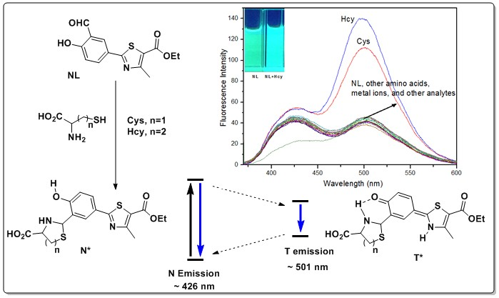
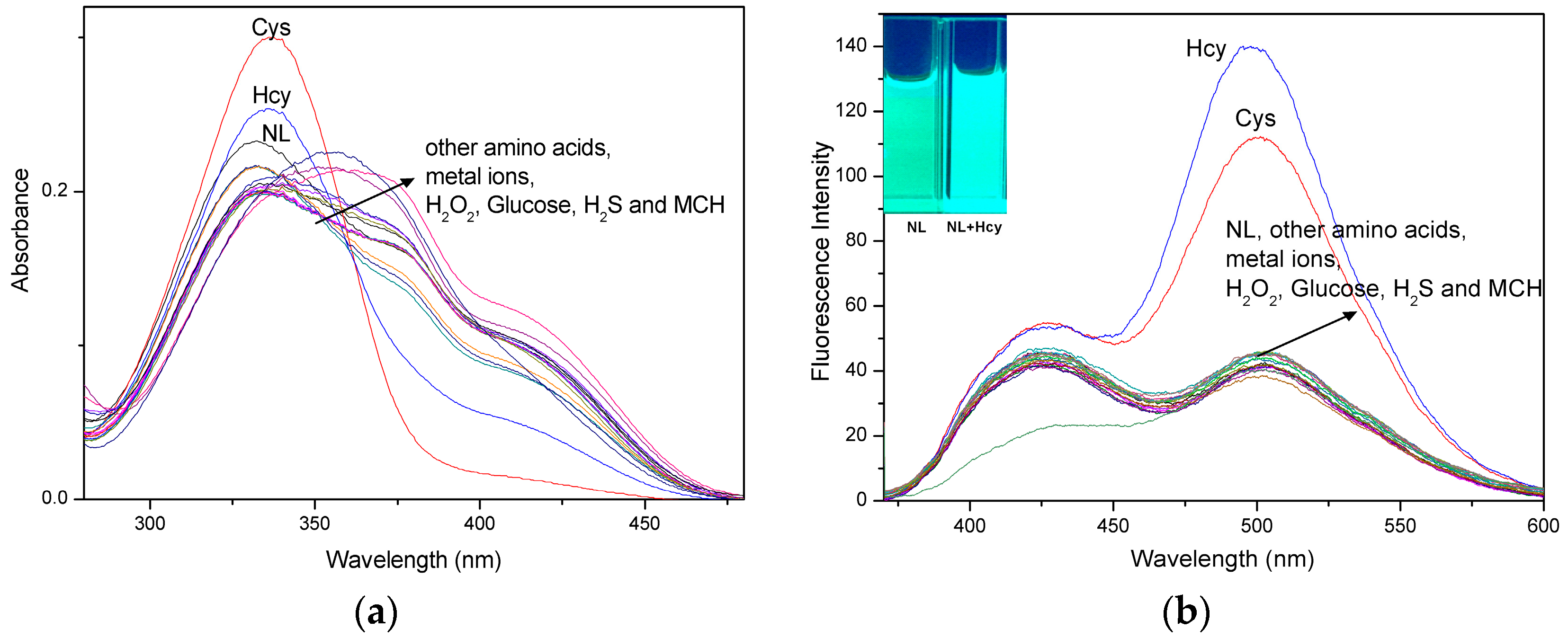
2.1. UV-Vis Absorption and Fluorescence Selectivity Studies of the NL Probe

First, the effect of dimethylsulfoxide (DMSO) content on the fluorescence intensity of the free NL probe and the behavior of the probe towards Cys were investigated. The fluorescence intensity of NL increased with increasing content of DMSO (Figure S3). In the presence of a polar aprotic solvent, the proton of the hydroxyl group is most likely strongly bonded with the nearest carbonyl group (Scheme 1). Buffer solutions were optimized based on the fluorescence signal-to-background ratio of NL with or without Cys in aqueous DMSO. The optimized solution was DMSO in 4-(2-hydroxyethyl)-1-piperazineethanesulfonic acid (HEPES) buffer (8:2, v/v, pH 7.4) and was used in the subsequent experiments.
The selectivity of NL for Cys/Hcy measurements was evaluated with the DMSO/HEPES solution containing each of the 20 interference chemicals, in addition to the analytes of Cys and Hcy. The interference chemicals were GSH, arginine, aspartic acid, glutamic acid, glycine, histidine, lysine, proline, threonine, tryptophan, tyrosine, K+, Ca2+, Na+, Mg2+, Zn2+, H2O2, H2S, HSCH2CH2OH (MCH), and glucose, each at a concentration of 20-fold compared to the NL probe. Cys/Hcy increased the absorbance of the solutions at 327 nm, while the other chemicals did not increase the absorbance of the solutions (Figure 2a). In addition, fluorescence intensity at λem 501 nm was markedly enhanced in the presence of Cys/Hcy (Figure 2b). The other analytes caused negligible effects on the fluorescence intensity of NL. In particular, GSH did not affect the UV-Vis absorbance and fluorescence intensity of the probe (Figure 2a,b, Figures S4 and S5). The presence of a wide range of interferences had little effect on the detection of Cys/Hcy. Therefore, NL possesses good selectivity toward Cys/Hcy in the presence of other amino acids and metal ions as well as GSH.

2.2. Reaction Time of Biothiols

To investigate the reaction time of NL with biothiols, the fluorescence intensity at λem 501 nm was recorded at varying periods of time after additon of Cys, Hcy, and GSH to the NL solutions (Figure S6). The reaction of NL with Cys or Hcy was nearly completed within 2.5 h at ambient temperature. These reactions are relatively fast compared with those reported in the literature [31,32]. In contrast, there was no obvious change in the fluorescence intensity of the solution containing GSH during the measurement time. Some reported probes can detect biothiols rapidly but with large amounts of biothiols, such as 30 equiv. or even more than 1000 equiv. of the probe [33,34,35,36]. In this study, we used 20 equiv. to achieve an accurate measurement of the biothiols.

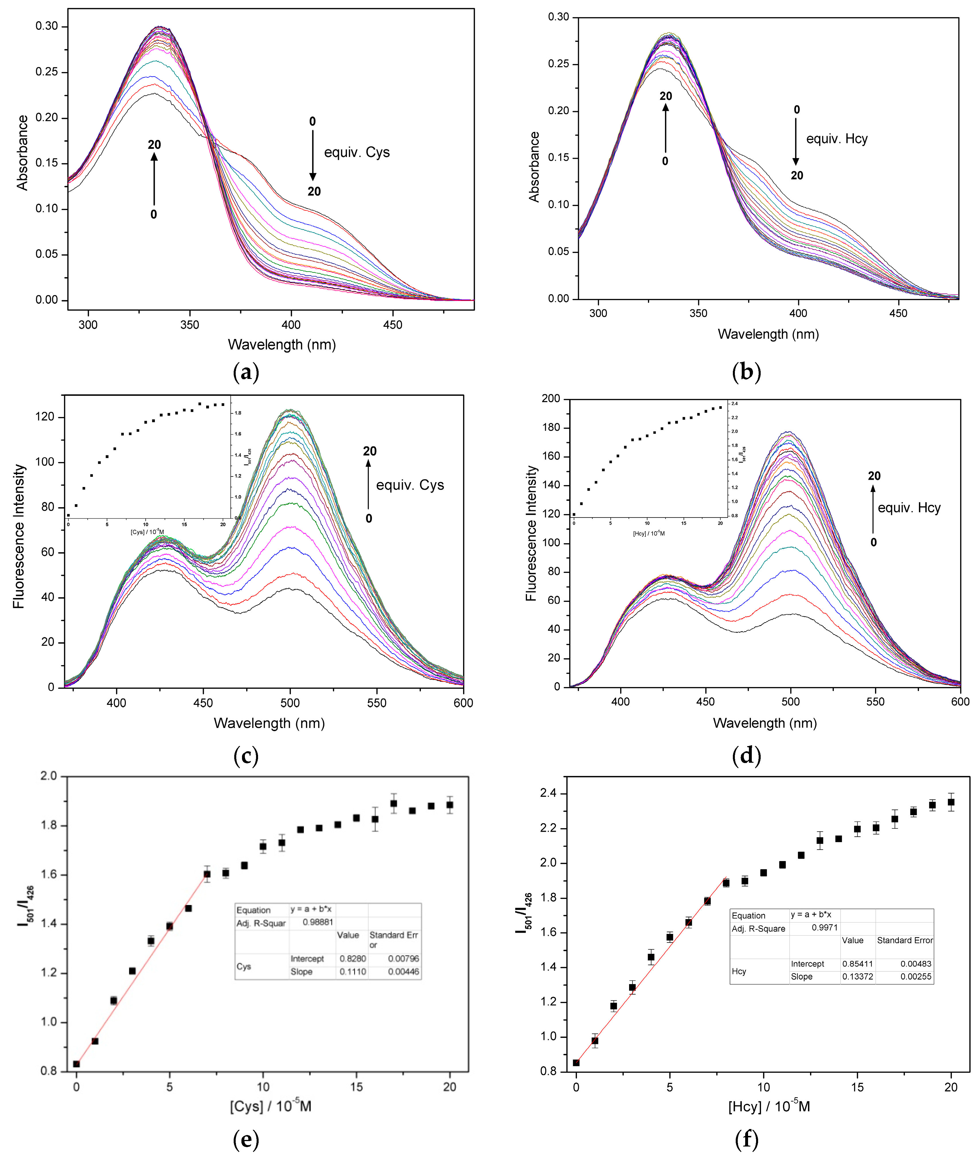
2.3. Quantification of Cys/Hcy

Figure 3a,b show the change in the UV-Vis absorbance of the solutions containing the NL probe with increasing content of Cys/Hcy. The absorbance intensity at 327 nm increased, but the intensity at 426 nm decreased with increasing Cys/Hcy concentration. Figure 3c,d reveal that the fluorescence intensity of the probe solution increased at λem 501 nm with increasing content of Cys/Hcy. When Cys/Hcy was at 20 equiv. of NL, the fluorescence intensity and the ratio of the absorption intensities at 501 and 426 nm (I501/I426) were almost at their maximum values, which is a superior performance in comparison with some other probes [33,37]. In addition, we found a linear correlation (R2 = 0.9888) between the fluorescence intensity and Cys concentration from 1–7 µM. The limit of detection (LOD) of Cys was 0.911 µM (Figure 3e), which was calculated by S/N = 3. A similar response of the NL probe to Hcy was also observed, as shown in Figure 3f. In this case, the LOD was 0.828 µM based on the linear relationship (R2 = 0.9971) between the fluorescence intensity and Hcy concentration in the range of 1–8 µM. In comparison with the previously reported probes [20,38,39,40], NL provides a much lower LOD of Cys/Hcy for qualitative analysis of Cys/Hcy despite the presence of GSH (Figure S7), making it attractive for use in human plasma samples [28].

2.4. Effect of pH on Detection of Cys

The effect of pH on the response of NL to Cys is illustrated in Figure S8. In the presence or absence of Cys, NL was stable under acidic conditions. With increasing pH from 2.0–8.0, the fluorescence intensity of the solution increased until it reached its maximum value at pH 8.0. Weak alkaline conditions possibly promoted the reaction between Cys and NL. With a further increase of the pH, the fluorescence intensity of the solution decreased until it was quenched, which might be caused by the hydrolysis of ester groups or the opening of the thiazolidine ring under strong alkaline conditions. Therefore, we chose pH 7.4 as a suitable pH for the detection experiments. Unlike the early and even recently reported probes in the literature that require use at pH ≥ 9.0 [28,41,42], NL can function in the physiological pH range.

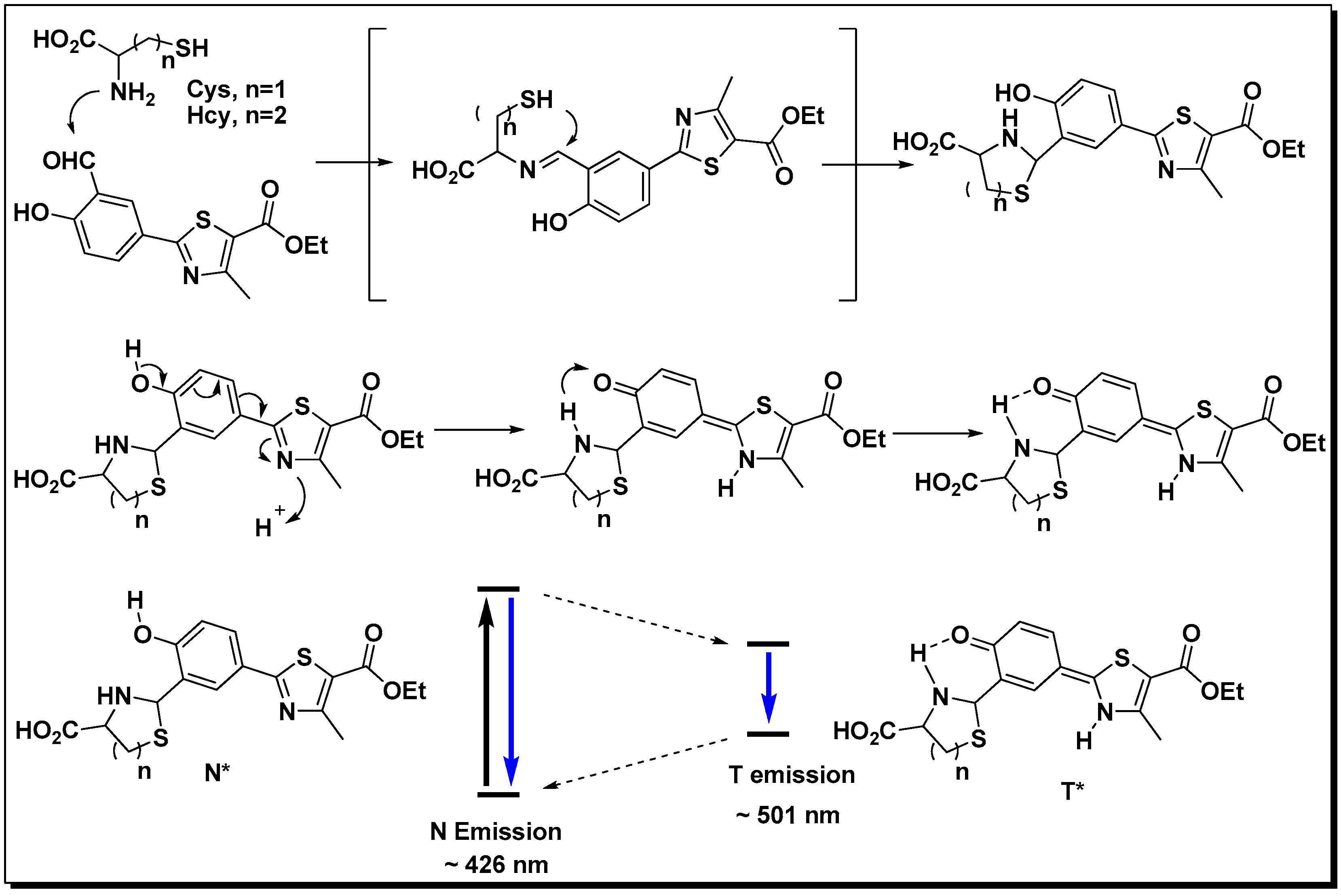
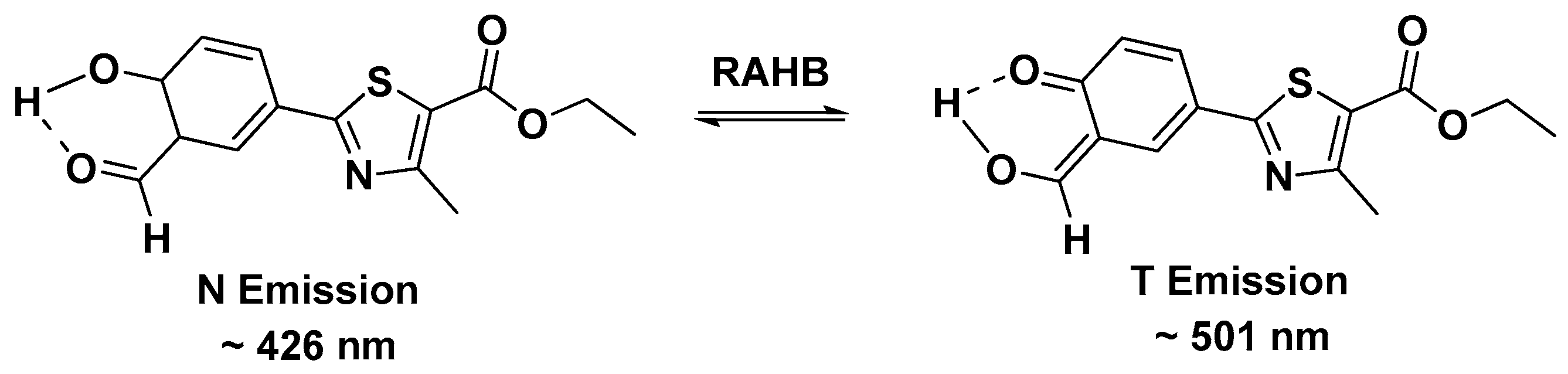
2.5. Mechanism of NL in Detection of Cys/Hcy

To verify the detection mechanism between NL and Cys/Hcy as shown in Scheme 2, we investigated the 1H-NMR spectrum of NL upon the addition of Cys and compared it with that of the probe itself. Under the same conditions, Hcy also reacted similarly with NL and formed the six-membered thiazinane ring. After the reaction of NL with Cys/Hcy in DMSO-d6 for 4 h, the reaction mixture was analyzed with NMR, which indicated that the aldehyde proton at around δ 10.3 ppm had disappeared, and that the thiazolidine methane proton at around δ 5.5 ppm had newly appeared (Figure S2). Furthermore, UPLC-MS/MS were carried out to confirm that the thiazolidine ring was formed by the reaction of Cys/Hcy with the aldehyde group of NL. The mass spectrum (ESI MS) of the NL probe shows a peak at m/z 292.09 (NL + H)+. The peak of the product of NL + Cys is 394.93, which also proves a single mononuclear addition of Cys to NL. A peak at m/z 408.92 corresponding to the product of NL + Hcy was also observed (Figure S2).
NL at λex 327 nm produced two emission peaks at ~426 nm and ~501 nm. Adding Cys/Hcy to NL enhanced the ESIPT process to shift the emission signal to a longer wavelength. As a result, the fluorescence intensity at λem 501 nm, which was attributed to the tautomer (T* emission) of the Cys/Hcy-products, was enhanced strongly, and the normal isomer (N* emission) of the products caused the fluorescence intensity at 426 nm to increase slowly. The two emission peaks can be used for the ratiometric fluorescent measurement to determine the presence of the analytes more accurately with the minimization of the background signal [43].

3. Experimental Section

3.1. General Methods and Materials

Commercial reagents were used as obtained from Sigma-Aldrich (St. Louis, MO, USA), Alfa Aesar (Ward Hill, MA, USA), and J&K (Beijing, China). Twice-distilled water was used throughout all the experiments. Nuclear magnetic resonance (NMR) spectra were recorded with a 600-MHz spectrometer (Agilent Technologies, Inc., Santa Clara, CA, USA) and mass spectrometry (MS) was performed with a UPLC/XevoTQ MS/MS spectrometer (Waters, Milford, MA, USA). All fluorescence measurements were carried out on a 970CRT fluorescence spectrophotometer (Inesa, Shanghai, China) equipped with a xenon lamp source. UV-Vis spectra were measured on a UV-1800 spectrophotometer (Shimadzu, Kyoto, Japan). Quartz cuvettes with a 1-cm path length and 3-mL volume were used for all measurements. All pH measurements were completed with a PHS-25 digital pH meter (Shanghai REX Instrument Factory, Shanghai, China).

3.2. Optical Studies of the NL Probe upon Addition of Various Analytes

A stock solution of the NL probe with a concentration of 12.5 µM was prepared in DMSO. Amino acids (Cys, Hcy, GSH, arginine, aspartic acid, glutamic acid, glycine, histidine, lysine, proline, threonine, tryptophan, and tyrosine), cations (K+, Ca2+, Na+, Mg2+ and Zn2+), H2S, and HSCH2CH2OH (MCH) were all dissolved in distilled water at a concentration of 5 mM for the absorption and fluorescence spectra analyses. The stock solutions of the analytes were diluted to various concentrations with HEPES buffer. Test solutions were prepared by adding 2400 µL of the NL stock solution (the final concentration of NL was 10 µM) and an appropriate aliquot of test analyte into a 3-mL volumetric cuvette. Each solution was diluted to 3 mL using a mixture of DMSO and HEPES buffer (8:2, v/v) at pH 7.4. The resulting solution was shaken well and incubated for 2.5 h at room temperature. Fluorescent spectra were then recorded using an excitation wavelength of 327 nm and scan speed of 450 nm·min−1.

4. Conclusions

In order to find a simple method to distinguish Cys/Hcy from other amino acids at physiological pH, an effective ratiometric fluorescent NL probe was developed. The fluorescence intensity of the probe was enhanced 3-fold upon addition of Cys/Hcy at a concentration of 20 equiv. of the probe, and its absorbance in buffer solution was also increased. NL can detect Cys at concentrations of 0.911 to 20 µM and Hcy at concentrations of 0.828 to 20 µM.

Supplementary Materials

Supplementary materials can be accessed at: https://www.mdpi.com/1420-3049/21/8/1023/s1.

Acknowledgments

This work was supported by the National High Technology Research and Development Program of China (2013AA102804B), the National Natural Science Foundation of China (NSFC) (No. 31272062), the Anhui Agricultural University Youth Fund Project (2014zr003, yj2015-25), the Natural Science Foundation of Anhui Province of China (1408085MKL36), and the Foundation for the Excellent Youth Scholars of Anhui Province (2013SQRL016ZD).

Author Contributions

This research was carried on by all the authors. Risong Na, Yi Wang, and Rimao Hua designed the theme of the study and carried out the probe synthesis as well as the manuscript preparation. Meiqing Zhu, Shisuo Fan, Zhen Wang, Xiangwei Wu, and Jun Tang participated in UV-Vis and fluorescence experiments. Jia Liu contributed greatly to the manuscript revision.

Conflicts of Interest

The authors declare no conflict of interest.

References

  1. Dai, X.; Wu, Q.H.; Wang, P.C.; Tian, J.; Xu, Y.; Wang, S.Q.; Miao, J.Y.; Zhao, B.X. A simple and effective coumarin-based fluorescent probe for cysteine. Biosens. Bioelectron. 2014, 59, 35–39. [Google Scholar] [CrossRef] [PubMed]
  2. Zhou, X.; Jin, X.; Sun, G.; Wu, X. A sensitive and selective fluorescent probe for cysteine based on a new response-assisted electrostatic attraction strategy: The role of spatial charge configuration. Chemistry 2013, 19, 7817–7824. [Google Scholar] [CrossRef] [PubMed]
  3. Shahrokhian, S. Lead phthalocyanine as a selective carrier for preparation of a cysteine-selective electrode. Anal. Chem. 2001, 73, 5972–5978. [Google Scholar] [CrossRef] [PubMed]
  4. Wang, W.; Rusin, O.; Xu, X.; Kim, K.K.; Escobedo, J.O.; Fakayode, S.O.; Fletcher, K.A.; Lowry, M.; Schowalter, C.M.; Lawrence, C.M.; et al. Detection of homocysteine and cysteine. J. Am. Chem. Soc. 2005, 127, 15949–15958. [Google Scholar] [CrossRef] [PubMed]
  5. Paulsen, C.E.; Carroll, K.S. Cysteine-mediated redox signaling: Chemistry, biology, and tools for discovery. Chem. Rev. 2013, 113, 4633–4679. [Google Scholar] [CrossRef] [PubMed]
  6. Seshadri, S.; Beiser, A.; Selhub, J.; Jacques, P.F.; Rosenberg, I.H.; D’Agostino, R.B.; Wilson, P.W.F.; Wolf, P.A. Plasma homocysteine as a risk factor for dementia and alzheimer’s disease. N. Engl. J. Med. 2002, 346, 476–483. [Google Scholar] [CrossRef] [PubMed]
  7. Van Meurs, J.B.J.; Dhonukshe-Rutten, R.A.M.; Pluijm, S.M.F.; van der Klift, M.; de Jonge, R.; Lindemans, J.; de Groot, L.C.P.G.M.; Hofman, A.; Witteman, J.C.M.; van Leeuwen, J.P.T.M.; et al. Homocysteine levels and the risk of osteoporotic fracture. N. Engl. J. Med. 2004, 350, 2033–2041. [Google Scholar] [CrossRef] [PubMed]
  8. Refsum, H.; Smith, A.D.; Ueland, P.M.; Nexo, E.; Clarke, R.; McPartlin, J.; Johnston, C.; Engbaek, F.; Schneede, J.; McPartlin, C.; et al. Facts and recommendations about total homocysteine determinations: An expert opinion. Clin. Chem. 2004, 50, 3–32. [Google Scholar] [CrossRef] [PubMed]
  9. Nekrassova, O.; Lawrence, N.S.; Compton, R.G. Analytical determination of homocysteine: A review. Talanta 2003, 60, 1085–1095. [Google Scholar] [CrossRef]
  10. Dai, X.; Wang, Z.Y.; Du, Z.F.; Cui, J.; Miao, J.Y.; Zhao, B.X. A colorimetric, ratiometric and water-soluble fluorescent probe for simultaneously sensing glutathione and cysteine/homocysteine. Anal. Chim. Acta 2015, 900, 103–110. [Google Scholar] [CrossRef] [PubMed]
  11. Niu, L.-Y.; Chen, Y.-Z.; Zheng, H.-R.; Wu, L.-Z.; Tung, C.-H.; Yang, Q.-Z. Design strategies of fluorescent probes for selective detection among biothiols. Chem. Soc. Rev. 2015, 44, 6143–6160. [Google Scholar] [CrossRef] [PubMed]
  12. Tang, Y.; Lee, D.; Wang, J.; Li, G.; Yu, J.; Lin, W.; Yoon, J. Development of fluorescent probes based on protection-deprotection of the key functional groups for biological imaging. Chem. Soc. Rev. 2015, 44, 5003–5015. [Google Scholar] [CrossRef] [PubMed]
  13. Kowada, T.; Maeda, H.; Kikuchi, K. Bodipy-based probes for the fluorescence imaging of biomolecules in living cells. Chem. Soc. Rev. 2015, 44, 4953–4972. [Google Scholar] [CrossRef] [PubMed]
  14. Daly, B.; Ling, J.; de Silva, A.P. Current developments in fluorescent pet (photoinduced electron transfer) sensors and switches. Chem. Soc. Rev. 2015, 44, 4203–4211. [Google Scholar] [CrossRef] [PubMed]
  15. Peng, H.; Chen, W.; Cheng, Y.; Hakuna, L.; Strongin, R.; Wang, B. Thiol reactive probes and chemosensors. Sensors 2012, 12, 15907–15946. [Google Scholar] [CrossRef] [PubMed]
  16. Chen, X.; Pradhan, T.; Wang, F.; Kim, J.S.; Yoon, J. Fluorescent chemosensors based on spiroring-opening of xanthenes and related derivatives. Chem. Rev. 2012, 112, 1910–1956. [Google Scholar] [CrossRef] [PubMed]
  17. Anand, T.; Sivaraman, G.; Mahesh, A.; Chellappa, D. Aminoquinoline based highly sensitive fluorescent sensor for lead(ii) and aluminum(iii) and its application in live cell imaging. Anal. Chim. Acta 2015, 853, 596–601. [Google Scholar] [CrossRef] [PubMed]
  18. Zhu, L.; Younes, A.H.; Yuan, Z.; Clark, R.J. 5-arylvinylene-2,2′-bipyridyls: Bright “push-pull” dyes as components in fluorescent indicators for zinc ions. J. Photochem. Photobiol. A Chem. 2015, 311, 1–15. [Google Scholar] [CrossRef] [PubMed]
  19. Yuan, Z.; Younes, A.H.; Allen, J.R.; Davidson, M.W.; Zhu, L. Enhancing the photostability of arylvinylenebipyridyl compounds as fluorescent indicators for intracellular zinc(ii) ions. J. Org. Chem. 2015, 80, 5600–5610. [Google Scholar] [CrossRef] [PubMed]
  20. Liu, B.; Wang, J.; Zhang, G.; Bai, R.; Pang, Y. Flavone-based esipt ratiometric chemodosimeter for detection of cysteine in living cells. ACS Appl. Mater. Interfaces 2014, 6, 4402–4407. [Google Scholar] [CrossRef] [PubMed]
  21. Zheng, C.; Pu, S.; Liu, G.; Chen, B.; Dai, Y. A highly selective colorimetric sensor for cysteine and homocysteine based on a new photochromic diarylethene. Dyes Pigment. 2013, 98, 280–285. [Google Scholar] [CrossRef]
  22. Mei, J.; Wang, Y.; Tong, J.; Wang, J.; Qin, A.; Sun, J.Z.; Tang, B.Z. Discriminatory detection of cysteine and homocysteine based on dialdehyde-functionalized aggregation-induced emission fluorophores. Chem. A Eur. J. 2013, 19, 613–620. [Google Scholar] [CrossRef] [PubMed]
  23. Park, S.; Imlay, J.A. High levels of intracellular cysteine promote oxidative DNA damage by driving the fenton reaction. J. Bacteriol. 2003, 185, 1942–1950. [Google Scholar] [CrossRef] [PubMed]
  24. Hwang, C.; Sinskey, A.; Lodish, H. Oxidized redox state of glutathione in the endoplasmic reticulum. Science 1992, 257, 1496–1502. [Google Scholar] [CrossRef] [PubMed]
  25. Liu, Y.; Yu, D.; Ding, S.; Xiao, Q.; Guo, J.; Feng, G. Rapid and ratiometric fluorescent detection of cysteine with high selectivity and sensitivity by a simple and readily available probe. ACS Appl. Mater. Interfaces 2014, 6, 17543–17550. [Google Scholar] [CrossRef] [PubMed]
  26. Goswami, S.; Manna, A.; Paul, S.; Das, A.K.; Nandi, P.K.; Maity, A.K.; Saha, P. A turn on esipt probe for rapid and ratiometric fluorogenic detection of homocysteine and cysteine in water with live cell-imaging. Tetrahedron Lett. 2014, 55, 490–494. [Google Scholar] [CrossRef]
  27. Zhang, M.; Yu, M.; Li, F.; Zhu, M.; Li, M.; Gao, Y.; Li, L.; Liu, Z.; Zhang, J.; Zhang, D.; et al. A highly selective fluorescence turn-on sensor for cysteine/homocysteine and its application in bioimaging. J. Am. Chem. Soc. 2007, 129, 10322–10323. [Google Scholar] [CrossRef] [PubMed]
  28. Rusin, O.; St. Luce, N.N.; Agbaria, R.A.; Escobedo, J.O.; Jiang, S.; Warner, I.M.; Dawan, F.B.; Lian, K.; Strongin, R.M. Visual detection of cysteine and homocysteine. J. Am. Chem. Soc. 2004, 126, 438–439. [Google Scholar] [CrossRef] [PubMed]
  29. Han, C.; Yang, H.; Chen, M.; Su, Q.; Feng, W.; Li, F. Mitochondria-targeted near-infrared fluorescent off-on probe for selective detection of cysteine in living cells and in vivo. ACS Appl. Mater. Interfaces 2015, 7, 27968–27975. [Google Scholar] [CrossRef] [PubMed]
  30. Yin, K.; Yu, F.; Zhang, W.; Chen, L. A near-infrared ratiometric fluorescent probe for cysteine detection over glutathione indicating mitochondrial oxidative stress in vivo. Biosens. Bioelectron. 2015, 74, 156–164. [Google Scholar] [CrossRef] [PubMed]
  31. Das, P.; Mandal, A.K.; Reddy G, U.; Baidya, M.; Ghosh, S.K.; Das, A. Designing a thiol specific fluorescent probe for possible use as a reagent for intracellular detection and estimation in blood serum: Kinetic analysis to probe the role of intramolecular hydrogen bonding. Org. Biomol. Chem. 2013, 11, 6604–6614. [Google Scholar] [CrossRef] [PubMed]
  32. Wang, S.-Q.; Wu, Q.-H.; Wang, H.-Y.; Zheng, X.-X.; Shen, S.-L.; Zhang, Y.-R.; Miao, J.-Y.; Zhao, B.-X. A novel pyrazoline-based selective fluorescent probe for detecting reduced glutathione and its application in living cells and serum. Analyst 2013, 138, 7169–7174. [Google Scholar] [CrossRef] [PubMed]
  33. Jung, H.S.; Ko, K.C.; Kim, G.-H.; Lee, A.-R.; Na, Y.-C.; Kang, C.; Lee, J.Y.; Kim, J.S. Coumarin-based thiol chemosensor: Synthesis, turn-on mechanism, and its biological application. Org. Lett. 2011, 13, 1498–1501. [Google Scholar] [CrossRef] [PubMed]
  34. Yuan, L.; Lin, W.; Xie, Y.; Zhu, S.; Zhao, S. A native-chemical-ligation-mechanism-based ratiometric fluorescent probe for aminothiols. Chem. A Eur. J. 2012, 18, 14520–14526. [Google Scholar] [CrossRef] [PubMed]
  35. Long, L.; Zhou, L.; Wang, L.; Meng, S.; Gong, A.; Du, F.; Zhang, C. A coumarin-based fluorescent probe for biological thiols and its application for living cell imaging. Org. Biomol. Chem. 2013, 11, 8214–8220. [Google Scholar] [CrossRef] [PubMed]
  36. Zhu, B.; Zhao, Y.; Zhou, Q.; Zhang, B.; Liu, L.; Du, B.; Zhang, X. A chloroacetate-caged fluorescein chemodosimeter for imaging cysteine/homocysteine in living cells. Eur. J. Org. Chem. 2013, 2013, 888–893. [Google Scholar] [CrossRef]
  37. Yue, Y.; Guo, Y.; Xu, J.; Shao, S. A bodipy-based derivative for selective fluorescence sensing of homocysteine and cysteine. New J. Chem. 2011, 35, 61–64. [Google Scholar] [CrossRef]
  38. Su, D.; Teoh, C.L.; Sahu, S.; Das, R.K.; Chang, Y.-T. Live cells imaging using a turn-on fret-based bodipy probe for biothiols. Biomaterials 2014, 35, 6078–6085. [Google Scholar] [CrossRef] [PubMed]
  39. Tian, M.; Guo, F.; Sun, Y.; Zhang, W.; Miao, F.; Liu, Y.; Song, G.; Ho, C.-L.; Yu, X.; Sun, J.Z.; et al. A fluorescent probe for intracellular cysteine overcoming the interference by glutathione. Org. Biomol. Chem. 2014, 12, 6128–6133. [Google Scholar] [CrossRef] [PubMed]
  40. Murale, D.P.; Kim, H.; Choi, W.S.; Kim, Y.; Churchill, D.G. Extremely selective fluorescence detection of cysteine or superoxide with aliphatic ester hydrolysis. RSC Adv. 2014, 4, 46513–46516. [Google Scholar] [CrossRef]
  41. Wei, M.; Yin, P.; Shen, Y.; Zhang, L.; Deng, J.; Xue, S.; Li, H.; Guo, B.; Zhang, Y.; Yao, S. A new turn-on fluorescent probe for selective detection of glutathione and cysteine in living cells. Chem. Commun. 2013, 49, 4640–4642. [Google Scholar] [CrossRef] [PubMed]
  42. Xu, Y.; Li, B.; Han, P.; Sun, S.; Pang, Y. Near-infrared fluorescent detection of glutathione via reaction-promoted assembly of squaraine-analyte adducts. Analyst 2013, 138, 1004–1007. [Google Scholar] [CrossRef] [PubMed]
  43. Domaille, D.W.; Zeng, L.; Chang, C.J. Visualizing ascorbate-triggered release of labile copper within living cells using a ratiometric fluorescent sensor. J. Am. Chem. Soc. 2010, 132, 1194–1195. [Google Scholar] [CrossRef] [PubMed]
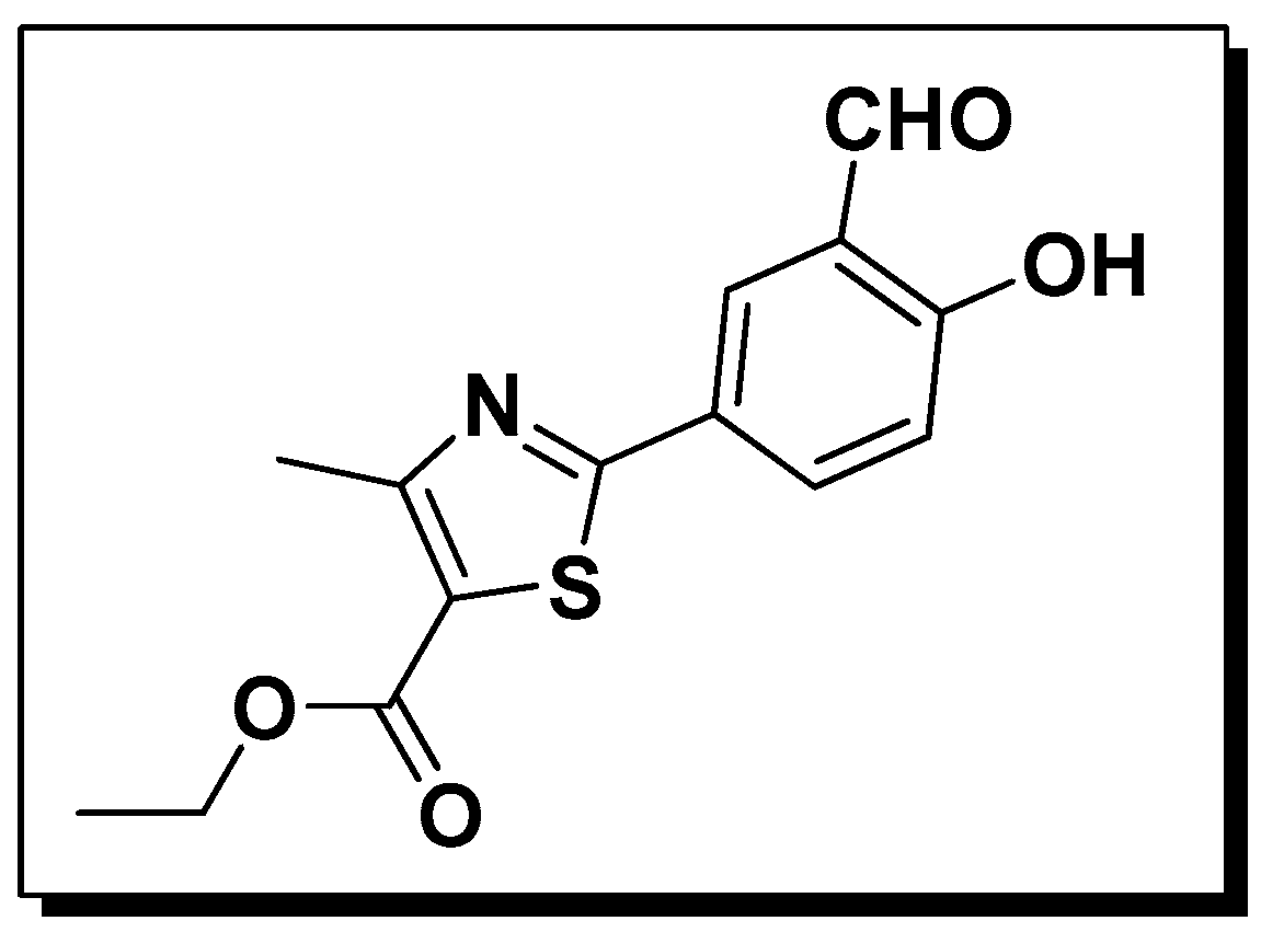
  • Sample Availability: Samples of the ethyl 2-(3-formyl-4-hydroxyphenyl)-4-methylthiazole-5-carboxylate (NL) are available from the authors.
Figure 1. Structure of the fluorescent probe NL.
Molecules 21 01023 g001
Scheme 1. Probable isomers of NL.
Molecules 21 01023 sch001
Figure 2. (a) UV-vis absorption spectra of the NL probe (10 µM) with various analytes (200 µM) in HEPES buffer solution (DMSO/HEPES = 8:2, pH 7.4); (b) Fluorescence spectra of NL (10 µM) with various analytes (200 µM) in HEPES buffer solution (DMSO/HEPES = 8:2, pH 7.4, λex = 327 nm, slit: 2.0 nm/2.0 nm). Inset is a photograph of NL without and with Hcy upon excitation at 365 nm.
Molecules 21 01023 g002
Figure 3. (a,b) UV-vis absorption spectra of NL (10 µM) with Cys/Hcy (0, 1, 2, 3, 4, 5, 6, 7, 8, 9, 10, 11, 12, 13, 14, 15, 16, 17, 18, 19 and 20 equiv.) in HEPES buffer solution (DMSO/HEPES = 8:2, pH 7.4); (c,d) Fluorescence spectra of NL (10 µM) with Cys/Hcy (0, 1, 2, 3, 4, 5, 6, 7, 8, 9, 10, 11, 12, 13, 14, 15, 16, 17, 18, 19 and 20 equiv.) in HEPES buffer solution (DMSO/HEPES = 8:2, pH 7.4, λex= 327 nm, slit: 2.0 nm/2.0 nm). Inset is the corresponding ratiometric response (I501/I426) of NL to the addition of Cys/Hcy; (e,f) Plot of the ratiometric response (I501/I426) of NL (10 µM) against Cys/Hcy equiv.; data are mean ± standard error (bars) (n = 3).
Molecules 21 01023 g003
Scheme 2. Proposed reaction mechanism of NL and Cys/Hcy.
Molecules 21 01023 sch002

