Communication

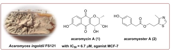
Secondary Metabolites from the Deep-Sea Derived Fungus Acaromyces ingoldii FS121

1
State Key Laboratory of Applied Microbiology Southern China, Guangdong Provincial Key Laboratory of Microbial Culture Collection and Application, Guangdong Open Laboratory of Applied Microbiology, Guangdong Institute of Microbiology, Guangzhou 510070, China
2
College of Pharmacy, Guangdong Pharmaceutical University, Guangzhou 510006, China
*
Author to whom correspondence should be addressed.
These authors contributed equally to this work.
Molecules 2016, 21(4), 371; https://doi.org/10.3390/molecules21040371
Submission received: 2 February 2016 / Revised: 8 March 2016 / Accepted: 14 March 2016 / Published: 29 March 2016
(This article belongs to the Section Metabolites)

Abstract

:
Activity-guided isolation of the fermentation broth of the deep-sea derived fungus Acaromyces ingoldii FS121, which was obtained from the China South Sea, yielded a new naphtha-[2,3-b]pyrandione analogue, acaromycin A (1) and a new thiazole analogue, acaromyester A (2), as well as the known compound (+)-cryptosporin (3). Their structures, including absolute configurations, were determined by extensive spectroscopic analysis and electronic circular dichroism (ECD) spectra. Compounds 13 were evaluated for in vitro growth inhibitory activities against four tumor cell lines (MCF-7, NCI-H460, SF-268 and HepG-2), wherein compounds 1 and 3 exhibited considerable growth inhibitory effects, with IC50 values less than 10 µM.

Graphical Abstract

1. Introduction

Marine organisms live in a biologically competitive environment with unique environmental conditions, therefore marine-derived fungi have been proven to be a rich source of structurally unique and biologically active secondary metabolites [1,2,3,4,5,6,7,8,9,10]. In spite of the fact that the investigations of fungal metabolites and their derivatives have not yet led to a clinical cancer drug, significant research efforts have revealed a large number of fungi-derived natural products with promising anticancer activity [11,12,13]. Such chemical entities not only provide insight into the biogenetic landscape, revealing new structure classes and new biosynthetic pathways, but also enlighten the approach to biomimetic synthesis and further bioactivity investigations, suggesting potential candidates for the development of new pharmaceutical agents [14].
In the course of our search for new bioactive natural products from marine-derived fungi, we found that a culture broth of Acaromyces ingoldii FS121 showed potent cell growth inhibitory effects against SF-268, MCF-7, NCI-H460 and HepG-2 human tumor cell lines [15]. Further chemical investigation of this strain led to the isolation of two new secondary metabolites—acaromycin A (1) and acaromyester A (2)—along with the known compound (+)-cryptosporin (3) (Figure 1). Herein, details of the isolation, structure elucidation, and cell growth inhibition effects of these compounds are described.

2. Results

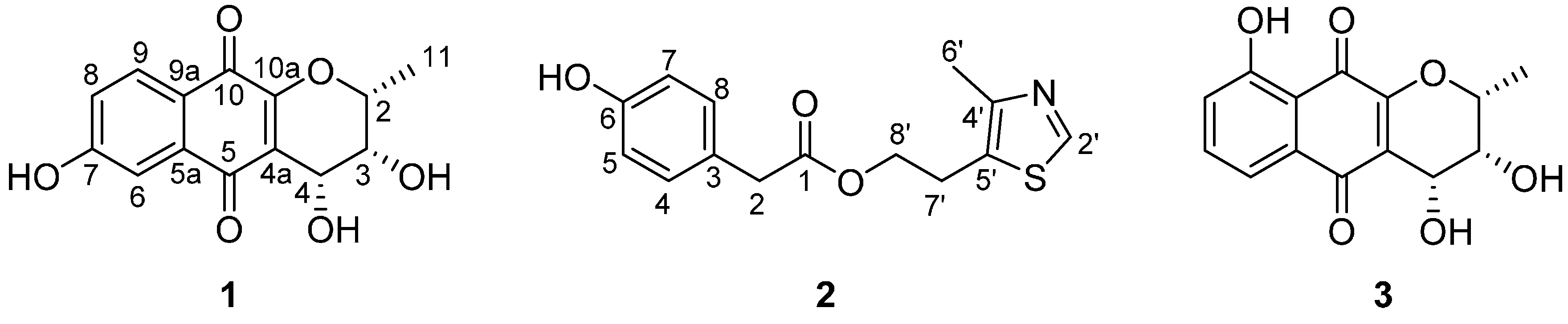
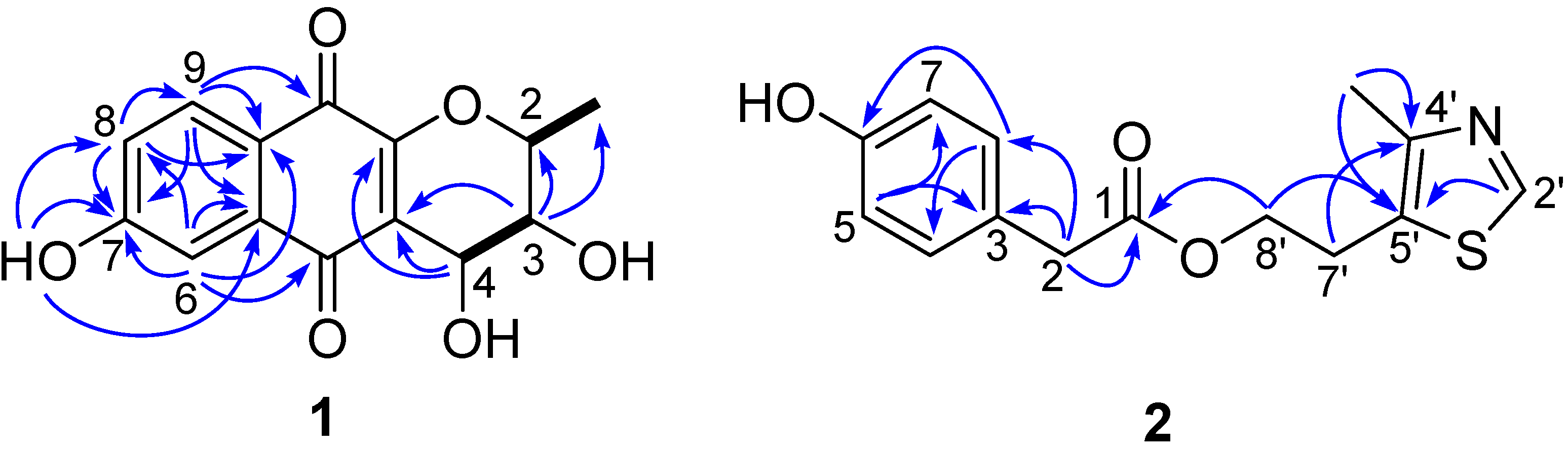
2.1. Structural Elucidation of New Compounds

Acaromycin A (1) was isolated as an orange amorphous powder with the molecular formula of C14H12O6 as determined by HRESIMS (m/z 299.0533, [M + Na]+, calcd. for 299.0532), corresponding to nine degrees of unsaturation. The IR spectrum exhibited absorption bands for OH (3309 cm−1) and carbonyl (1647 and 1625 cm−1) groups. The 1H-NMR data (Table 1) exhibited signals for a methyl proton δH 1.65 (H3-11, d, J = 6.7 Hz), three aromatic protons δH 7.61 (H-9, d, J = 1.9 Hz), 7.60 (H-6, s), 7.23 (H-8, dd, J = 5.6, 4.0 Hz), three methine protons δH 4.96 (H-4, d, J = 4.3, 0.9 Hz), 4.30 (H-2, q, J = 6.7, 1.0 Hz), 4.03 (H-3, dd, J = 4.3, 1.1 Hz), and a hydrogen-bonded phenolic hydroxy group at δH 11.68 (7-OH, s). The 13C-NMR and DEPT spectra of 1 revealed 14 carbon resonances attributable to two conjugated carbonyl groups [δC 186.5 (C-5) and 183.8 (C-10)], five sp2 quaternary carbons δC 162.2 (C-7), 155.2 (C-10a), 132.1 (C-9a), 118.8 (C-4a), 114.1 (C-5a), three sp2 methines δC 137.2 (C-6), 124.7 (C-8), 119.3 (C-9), three sp3 methines δC 76.2 (C-2), 66.9 (C-3), 65.1 (C-4), and one methyl δC 16.7 (C-11). As six of nine degrees of unsaturation account for four double bonds (eight sp2 carbons), and two carbonyl groups, the remaining degrees of unsaturation requires that 1 be a tricyclic system. The HMBC correlations (Figure 2) from H-6 to C-5, C-8, C-9a, and C-7, H-8 to C-7 and C-9a, H-9 to C-7 and C-10, and 7-OH to C-7 and C-8 suggested the presence of a 6-hydroxy-1,4-naphthoquinone moiety [16]. The location of the hydroxyl at C-7 was supported by HMBC correlations from 7-OH (δH 11.68) to C-5a, C-7 and C-8. Furthermore, the 1H-1H COSY spectrum (Figure 2) of 1 indicated the following fragments H3-11/H-2 and H-3/H-4, together with the HMBC correlations from H-3 to C-2, C-4a and C-11, and H-4 to C-4a, C-10a implied the remaining monocyclic was a 2H-pyran ring with a methyl at C-2 and OH at C-3 and C-4. The abovementioned information was similar to those of the co-isolated known compound (+)-cryptosporin (3), except for the OH at C-9 in 3 was locked at C-7 in 1. Therefore, the planar structure of 1 was confirmed.
The relative configuration of 1 was established by the comparison of its 1H-NMR coupling constants [H-2 (J = 6.7, 1.0 Hz), H-3 (J = 4.3, 1.1 Hz), H-4 (J = 4.3, 0.9 Hz)] with those of 3 [H-2 (J = 6.5, 1.4 Hz), H-3 (J = 4.6, 2.1 Hz), H-4 (J = 4.6, 1.0 Hz)]. This indicated that these protons were cofacial and designated as β-oriented, whereas the Me-11 was α-oriented in 1. The absolute configuration of 1 was determined by the CD spectrum. The absolute configuration of (+)-cryptosporin (3) was established by synthesis and its CD spectra data was reported in the literature [17]. The CD spectrum (Figure S19) of 1 showed a positive Cotton effect at 287, 314, 370 nm and a negative Cotton effect at 239, 425 nm, which was similar to that of 3. Thus, the absolute stereochemistry of 1 was established as 2R,3R,4R and given the trivial name as acaromycin A.
Compound 2 was obtained as a light pink oil. The HRESIMS (m/z 278.0845, [M + H]+, calcd. for C14H16NO3S, 278.0851) allowed the molecular formula C14H15NO3S to be assigned to 2, corresponding to eight degrees of unsaturation. The IR spectrum exhibited absorption bands for OH (3309 cm−1) and carboxyl groups (1732 and 1716 cm−1). The 1D NMR data of 2 (Table 1), in combination with the HSQC experiment revealed 14 carbon resonances, which showed representative signals for a 4-hydroxyphenylacetic acid moiety [δC 173.7, 157.6, 131.3, 126.1, 116.3, 41.2; δH 7.02 (2H, d, J = 8.6 Hz), 6.70 (2H, d, J = 8.6 Hz), 3.51 (s)]. This assumption was further confirmed by the HMBC correlations from H-2 to C-1, C-3 and C-8, H-5 to C-3 and C-7, and H-8 to C-6 and C-4. Besides, the 1H-NMR of 2 also exhibited a methyl signal at δH 2.24 (3H, s), two methylene protons at δH 3.02 (2H, t, J = 7.1 Hz) and 3.84 (2H, m), and an olefinic proton due to the thiazole ring at δH 8.61 (1H, s), suggesting the presence of a 4-methyl-5-thiazoleethanol substructure in 2, which has been reported in the literature [18]. Furthermore, the key COSY correlations of H-7′/H-8′ and HMBC correlations from H-2′ to C-4′ and C-5′, as well as Me-6′ to C-4′, and C-5′ also supported this deduce (Figure 2). Finally, the two substructures was connected by the HMBC correlations between H-8 to C-1 and C-5′. Thus, compound 2 was determined as depicted and given the trivial name as acaromyester A.
The known compound (+)-cryptosporin (3) was identified by comparison of its NMR data with those in the literature [19].

2.2. In Vitro Growth Inhibition Assay

All of the isolates 13 were tested for in vitro growth inhibitory activities against the MCF-7, NCI-H460, SF-268 and HepG-2 tumor cell lines at an initial concentration of 100 µM. Then, compounds with inhibition greater than 50% were further analyzed to determine their corresponding IC50 values. Both 1 and 3 exhibited considerable inhibitory activities against the growth of all four cell lines (Table 2). Especially, the inhibitory effects of 1 and 3 against the MCF-7 cells were found to be comparable to that of cisplatin with IC50 values of 6.7 and 4.1 µM, respectively. The growth inhibitory curves of 1, 3 and cisplatin against MCF-7 were presented in Figure 3.

3. Experimental Section

3.1. General Procedures

Optical rotations were measured on an MCP-500 spectropolarimeter (Anton Paar, Graz, Austria). Circular dichroism (CD) measurements were carried out under N2 gas on a Chirascan circular dichroism spectrometer (Applied Photophysics Ltd., Surrey, UK). IR spectra were recorded on a IR Affinity-1 spectrophotometer (Shimadzu, Kyoto, Japan). UV spectra were measured on a Shimadzu UV-2600 UV-visible spectrophotometer. NMR spectra were recorded on a Bruker Avance-500 spectrometer (Bruker Corporation, Fremont, CA, USA) with the signal of tetramethylsilane as an internal standard at 500/125 MHz, respectively. HRESIMS were measured on a Thermo MAT95XP high resolution mass spectrometer (Thermo Fisher Scientific, Bremen, Germany) and ESIMS on a 1290-6430A Triple Quad LC/MS (Agilent Technologies, (Agilent Technologies Inc., California, CA, USA). Column chromatography (CC) was performed on silica gel (200–300 mesh, Qingdao Haiyang Chemical Group Co., Qingdao, China), Lichroprep C18 reversed-phase (RP-18) silica gel (40–63 µm, Merck, Darmstadt, Germany) and Sephadex LH-20 (Pharmacia Fine Chemical Co. Ltd., Uppsala, Sweden). Thin-Layer Chromatography (TLC) was conducted with precoated glass plates GF-254 (Qingdao Haiyang Chemical Group Co.). TLC spots were visualized under UV light and by dipping into 10% H2SO4 in alcohol, followed by heating. Solvents were of the industrial purity and distilled prior to use.

3.2. Fungal Material

The marine fungal strain FS121 was isolated from a marine sediment sample, which was collected in the South China Sea (18°44.606′ N, 119°44.263′ E; 3415 m depth) in September 2011. The strain was identified by the sequence analysis of the rDNA ITS (internal transcribed spacer) region. The sequence of the ITS region of the strain FS121 has been submitted to GenBank (Accession No. KT989306). By using BLAST (nucleotide sequence comparison program) to search the GenBank database, FS121 has 99.8% similarity with Acaromyces ingoldii CBS 10536 (Accession No. AM991023). The strain is preserved at the Guangdong Provincial Key Laboratory of Microbiology Culture Collection and Application, Guangdong Institute of Microbiology.

3.3. Fermentation, Extraction and Compound Isolation

The fungal strain FS121 was maintained on potato dextrose agar (PDA) medium at 28 °C for 7 days, and then three pieces (0.5 × 0.5 cm2) of mycelial agar plugs were inoculated into 20 × 500 mL Erlenmeyer flasks, each containing 250 mL potato dextrose broth (potato 20%, glucose 2%, K2HPO4 0.3%, MgSO4•7H2O 0.15%, vitamin B1 10 mg/L, sea salt 1.5%). After 6 days of incubation at 28 °C on a rotary shaker at 120 r/m, 20 mL seed cultures were aseptically transferred into each of a total of 220 flasks (1000 mL) containing 500 mL of potato dextrose broth. The liquid cultivation that followed was kept for 7 days at 28 °C and 120 r/m on a rotary shaker. The culture (110 L) was centrifuged to give the broth and mycelia. The broth was exhaustively extracted with EtOAc four times, then the EtOAc layers were combined and evaporated under reduced pressure at a temperature not exceeding 40 °C to yield a dark brown gum (35.8 g). The crude extract was subjected to silica gel (200–300 mesh) column chromatography (CC) with a gradient system of increasing polarity (petroleum ether/EtOAc, 50:1→1:2) to afford 33 fractions, Fr.1–Fr.33. Fr.22 eluting with petroleum ether/EtOAc (1:2) was further fractionated by CC on Sephadex LH-20 (CH2Cl2/MeOH, 1:1) to yield three subfractions, Frs. 22-1, 22-2 and 22-3. Then, Fr. 22-1 was followed by CC over RP-18 silica gel (MeOH/H2O, 1:4) to yield Fr.22-1-3, then followed by preparative TLC on a silica gel plate (20 × 20 cm) developed with CH2Cl2/MeOH (10:1) to obtain compound 1 (2.6 mg). Fr.16 eluting with petroleum ether/EtOAc (2:1) was subjected to CC on Sephadex LH-20 (CH2Cl2/MeOH, 1:1) to afford compound 2 (22 mg). Fr.20 was subjected to CC on Sephadex LH-20 (CH2Cl2/MeOH, 1:1) to give three subfractions: Fr.20-1, Fr.20-2 and Fr.20-3. Fr.20-1 was purified by CC over RP-18 silica gel (MeOH/H2O, 1:1) to yield compound 3 (3.8 mg).

3.4. Compound Characterization

Acaromycin A (1): orange amorphous powder; [ α ] D 25 +102.0 (c 0.1, MeOH); UV (MeOH) λmax (log ε) 194 (3.69), 204 (4.05), 286 (3.67) nm; CD (c 1.8 × 10⁻³, MeOH) λ(Δε) 474 (0), 425 (−0.25), 405 (0), 370 (0.76), 314 (0.19), 287 (2.1), 257 (0), 239 (−0.67), 221 (0) nm; IR νmax 3498, 1647, 1625, 1257, 1026, 792 cm−1; 1H- and 13C-NMR, see Table 1; HRESIMS m/z [M + Na]+ 299.0533 (calcd for C14H12O6Na, 299.0532).
Acaromyester A (2): light pink oil; UV (MeOH) λmax (log ε) 202 (4.24), 225 (4.08), 277 (3.44) nm; IR νmax 3309, 3178, 1732, 1716, 1516, 1236, 1145, 804, 783 cm−1; 1H- and 13C-NMR, see Table 1; HRESIMS m/z [M + H]+ 278.0845 (calcd for C14H16NO3S, 278.0851).

3.5. In Vitro Growth Inhibition Assay

Cell growth inhibitory effects of compounds 13 were tested against four tumor cell lines, including MCF-7 (human breast adenocarcinoma cell line), NCI-H460 (human non-small cell lung cancer cell line) and SF-268 (human glioma cell line), HepG-2 (human hepatomacarcinoma cell line) by the SRB method [20]. Cells (180 μL) with a density of 3 × 10⁴ cells/mL of media were seeded onto 96-well plates and incubated for 24 h at 37 °C, 5% CO2. Various concentrations of compounds (20 μL) were added to the plate wells, and plates were further incubated for 72 h. After incubation, cell monolayers were fixed with 50% (w/v) trichloroacetic acid (50 μL) and stained for 30 min with 0.4% (w/v) SRB dissolved in 1% acetic acid. Unbound dye was removed by washing repeatedly with 1% acetic acid. The protein-bound dye was dissolved in 10 mM Tris base solution (200 µL) for OD determination at 570 nm using a microplate reader. Cisplatin was used as a positive control. All data were obtained in triplicate and are presented as means ± S.D. IC50 values were calculated with the SigmaPlot 10.0 software (San Jose, California, CA, USA) using a nonlinear curve-fitting method.

4. Conclusions

In summary, we have described herein the isolation and structural elucidation of a new 3,4-dihydro-2H-naphtho-[2,3-b]pyran-5,10-quinone and a new heterodimeric compound combining a hydroxyphenylacetate and a 4-methyl-5-thiazoleethanol substructure, which were isolated from the fermentation broth of Acaromyces ingoldii FS121. It merits attention that compounds 1 and 3 contained a 3,4-dihydro-2H-naphtho-[2,3-b]pyran-5,10-quinone core framework, which exhibited a wide range of biological activities [21]. The cell growth inhibitory effects of compounds 13 were evaluated, wherein compound 1 and 3 showed considerable activities against MCF-7, NCI-H460, SF-268 and HepG-2 tumor cell lines.

Supplementary Materials

Supplementary materials can be accessed at: https://www.mdpi.com/1420-3049/21/4/371/s1.

Acknowledgments

Financial support for this research was provided by the National Natural Science Foundation of China (31272087); the National High Technology Research and Development Program of China (863 Program, 2012AA092104); the Guangdong Provincial Project for Science and Technology (2014A020217012); the Guangzhou Project for Science and Technology (2013J4100067); and the Guangdong Provincial Innovative Development of Marine Economy Regional Demonstration Projects (GD2012-D01-002).

Author Contributions

Wei-Min Zhang and Yu-Zhi Tan conceived and designed the experiments and reviewed the manuscript; Xiao-Wei Gao and Yu-Chan Chen performed the experiments; Hong-Xin Liu and Zhang-Hua Sun analyzed the data; Xiao-Wei Gao, Hong-Xin Liu, and Zhang-Hua Sun wrote the paper. All authors read and approved the final manuscript.

Conflicts of Interest

The authors declare no conflict of interest.

References

  1. Bhadury, P.; Mohammad, B.T.; Wright, P.C. The current status of natural products from marine fungi and their potential as anti-infective agents. J. Ind. Microbiol. Biot. 2006, 33, 325–337. [Google Scholar] [CrossRef] [PubMed]
  2. Blunt, J.W.; Copp, B.R.; Munro, M.H.G.; Northcote, P.T.; Prinsep, M.R. Marine natural products. Nat. Prod. Rep. 2003, 20, 1–48. [Google Scholar] [CrossRef] [PubMed]
  3. Blunt, J.W.; Copp, B.R.; Munro, M.H.G.; Northcote, P.T.; Prinsep, M.R. Marine natural products. Nat. Prod. Rep. 2004, 21, 1–49. [Google Scholar] [CrossRef] [PubMed]
  4. Debbab, A.; Aly, A.H.; Lin, W.H.; Proksch, P. Bioactive compounds from marine bacteria and fungi. Microb. Biotechnol. 2010, 3, 544–563. [Google Scholar] [CrossRef] [PubMed]
  5. Faulkner, D.J. Marine natural products. Nat. Prod. Rep. 2002, 19, 1–48. [Google Scholar]
  6. Jiang, W.; Ye, P.; Che, C.-T.A.; Wang, K.; Liu, P.; He, S.; Wu, X.; Gan, L.; Ye, Y.; Wu, B. Two novel hepatocellular carcinoma cycle inhibitory cyclodepsipeptides from a hydrothermal vent crab-associated fungus Aspergillus clavatus C2WU. Mar. Drugs 2013, 11, 4761–4772. [Google Scholar] [CrossRef] [PubMed]
  7. Saleem, M.; Ali, M.S.; Hussain, S.; Jabbar, A.; Ashraf, M.; Lee, Y.S. Marine natural products of fungal origin. Nat. Prod. Rep. 2007, 24, 1142–1152. [Google Scholar] [CrossRef] [PubMed]
  8. Sun, L.; Li, D.L.; Tao, M.H.; Chen, Y.C.; Dan, F.J.; Zhang, W.M. Scopararanes C-G: new oxygenated pimarane diterpenes from the marine sediment-derived fungus Eutypella scoparia FS26. Mar. Drugs 2012, 10, 539–550. [Google Scholar] [CrossRef] [PubMed]
  9. Wu, B.; Wu, X.; Sun, M.; Li, M. Two novel tyrosinase inhibitory sesquiterpenes induced by CuCl2 from a marine-derived fungus Pestalotiopsis sp. Z233. Mar. Drugs 2013, 11, 2713–2721. [Google Scholar] [CrossRef] [PubMed] [Green Version]
  10. Yamazaki, H.; Rotinsulu, H.; Kaneko, T.; Murakami, K.; Fujiwara, H.; Ukai, K.; Namikoshi, M. A new dibenz[b,e]oxepine derivative, 1-hydroxy-10-methoxy-dibenz[b,e]oxepin-6,11-dione, from a marine-derived fungus, Beauveria bassiana TPU942. Mar. Drugs 2012, 10, 2691–2697. [Google Scholar] [CrossRef] [PubMed]
  11. Evidente, A.; Kornienko, A.; Cimmino, A.; Andolfi, A.; Lefranc, F.; Mathieu, V.; Kiss, R. Fungal metabolites with anticancer activtity. Nat. Prod. Rep. 2014, 31, 617–627. [Google Scholar] [CrossRef] [PubMed]
  12. Gomes, N.G.M.; Lefranc, F.; Kijjoa, A.; Kiss, R. Can some marine-derived fungal metabolites become actual anticancer agents. Mar. Drugs 2015, 13, 3950–3991. [Google Scholar] [PubMed]
  13. Kornienko, A.; Evidente, A.; Vurro, M.; Mathieu, V.; Cinmmino, A.; Evidente, M.; Otterlo, W.A.L.; Dasari, R.; Lefranc, F.; Kiss, R. Toward a cancer drug of fungal origin. Med. Res. Rev. 2015, 35, 937–967. [Google Scholar] [CrossRef] [PubMed]
  14. Mander, L.N.; Liu, H.W. (Eds.) In natural product diversity from marine fungi. In Comprehensive Natural Products II; Elsevier Ltd.: Amsterdam, The Netherlands, 2010; pp. 223–262.
  15. Yang, X.L.; Chen, Y.C.; Li, H.H.; Zhang, W.M. Molecular identification of 23 marine fungal strains and their activties against plant pathogenic fungi and cytotoxic activities. Biotechnol. Bull. 2014, 8, 131–137. [Google Scholar]
  16. Huang, L.; Zhao, J.; Guo, S.; Zhang, C.; Ma, J. Bodipy derivatives as organic triplet photosensitizers for aerobic photoorganocatalytic oxidative coupling of amines and photooxidation of dihydroxylnaphthalenes. J. Org. Chem. 2013, 78, 5627–5637. [Google Scholar] [CrossRef] [PubMed]
  17. Brade, W.; Vasella, A. Synthesis of naphtho[2,3-b]pyrandiones: (‒)-cryptosporin. Helv. Chim. Acta 1989, 72, 1649–1657. [Google Scholar] [CrossRef]
  18. Park, J.D.; Kim, M.W.; Yoo, S.J.; Wee, J.J. A thiazole and two β-carboline constituents from Panax ginseng. Arch. Pharm. Res. 1988, 11, 52–55. [Google Scholar] [CrossRef]
  19. Closse, A.; Sigg, H.P. Isolation and structure elucidation of cryptosporin. Helv. Chim. Acta 1973, 56, 619–625. [Google Scholar] [CrossRef] [PubMed]
  20. Skehan, P.; Storeng, R.; Scudiero, D.; Monks, A.; McMahon, J.; Vistica, D.; Warren, J.T.; Bokesch, H.; Kenney, S.; Boyd, M.R. New colorimetric cytotoxicity assay for anticancer-drug screening. J. Natl. Cancer. Inst. 1990, 82, 1107–1112. [Google Scholar] [CrossRef] [PubMed]
  21. Gupta, R.B.; Franck, R.W. The total synthesis of (‒)-cryptosporin. J. Am. Chem. Soc. 1989, 111, 7668–7670. [Google Scholar] [CrossRef]
  • Sample Availability: Samples of the compounds of 13 are available from the authors.
Figure 1. Chemical structures of compounds 13.
Molecules 21 00371 g001
Figure 2. 1H, 1H-COSY and key HMBCs of 1 and 2.
Molecules 21 00371 g002
Figure 3. Growth inhibitory curves of compounds 1, 3 and cisplatin (positive control) against MCF-7.
Molecules 21 00371 g003
Table 1. 1H- (500 MHz) and 13C- (125 MHz) NMR data of 1 in CDCl3 and 2 in CD3OD.
Position1Position2
δCδH (J in Hz)δCδH (J in Hz)
276.1, CH4.30, q (6.7, 1.0)1173.7, C
366.9, CH4.03, dd (4.3, 1.1)241.2, CH23.51, s
465.1, CH4.96, d (4.3, 0.9)3126.1, C
4a118.8, C 4131.3, CH7.02, d (8.6)
5186.5, C 5116.3, CH6.70, d (8.6)
5a114.1, C 6157.6, C
6137.2, CH7.60, s7116.3, CH6.70, d (8.6)
7162.2, C 8131.3, CH7.02, d (8.6)
8124.7, CH7.23, dd (5.6, 4.0)2′152.4, CH8.75, s
9119.3, CH7.61, d (1.9)4′150.6, C
9a132.1, C 5′129.1, C
10183.9, C 6′14.5, CH32.33, s
10a155.2, C 7′26.5, CH23.12, t (6.3)
1116.7, CH31.65, d (6.7)8′65.5, CH24.26, t (6.3)
3-OH 2.92, brs
4-OH 5.02, s
7-OH 11.68, s
Table 2. Growth-inhibition effects of compounds 1 and 3.
CompoundsIC50 (µM) ( x ¯ ± s , n = 3 )
MCF-7NCI-H460SF-268HepG-2
16.7 ± 1.610.0 ± 0.17.8 ± 0.57.3 ± 1.8
34.1 ± 0.36.0 ± 0.15.7 ± 0.15.7 ± 0.6
Cisplatin *5.8 ± 0.41.3 ± 0.11.9 ± 0.11.7 ± 0.1
* Positive control.

