Article

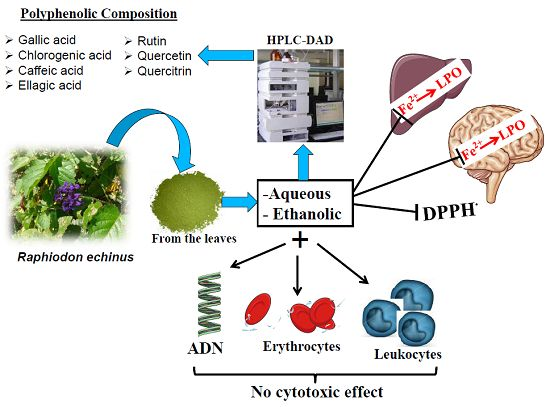
Polyphenolic Composition and Evaluation of Antioxidant Activity, Osmotic Fragility and Cytotoxic Effects of Raphiodon echinus (Nees & Mart.) Schauer

by
Antonia Eliene Duarte
1,2,
Emily Pansera Waczuk
2,
Katiane Roversi
3,
Maria Arlene Pessoa Da Silva
4,
Luiz Marivando Barros
1,2,
Francisco Assis Bezerra Da Cunha
1,2,
Irwin Rose Alencar De Menezes
5,
José Galberto Martins Da Costa
6,
Aline Augusti Boligon
7,
Adedayo Oluwaseun Ademiluyi
2,8,
Jean Paul Kamdem
2,9,
João Batista Teixeira Rocha
2,* and
Marilise Escobar Burger
3,*
1
Centro de Ciências Biológicas e da Saúde-CCBS, Departamento de Ciências Biológicas, Universidade Regional do Cariri (URCA), Pimenta, Crato CEP 63.100-000, CE, Brazil
2
Programa de Pós-Graduação em Bioquímica Toxicológica, Departamento de Bioquímica e Biologia Molecular, Universidade Federal de Santa Maria, Santa Maria 97105-900, RS, Brazil
3
Departamento de Fisiologia e Farmacologia, Universidade Federal de Santa Maria, Santa Maria 97105-900, RS, Brazil
4
Laboratório de Botânica Aplicada, Departamento de Ciências Biológicas, Universidade Regional do Cariri (URCA), Pimenta, Crato CEP 63.100-000, CE, Brazil
5
Laboratório de Farmacologia e Química Molecular, Departamento de Química Biológica, Universidade Regional do Cariri, Pimenta, Crato CEP 63.100-000, CE, Brazil
6
Laboratório de Pesquisas de Produtos Naturais, Departamento de Química Biológica, Universidade Regional do Cariri, Crato CEP 63.105.000, CE, Brazil
7
Laboratório de Fitoquímica, Departamento de Farmácia Industrial, Universidade Federal de Santa Maria, Santa Maria 97105-900, RS, Brazil
8
Functional Foods and Nutraceutical Unit, Department of Biochemistry, Federal University of Technology, P.M.B. 704, Akure 340001, Nigeria
9
Departamento de Bioquímica, Instituto de Ciências Básica da Saúde, Universidade Federal do Rio Grande do Sul, Porto Alegre CEP 90035-003, RS, Brazil
*
Authors to whom correspondence should be addressed.
Molecules 2016, 21(1), 2; https://doi.org/10.3390/molecules21010002
Submission received: 25 October 2015 / Revised: 8 December 2015 / Accepted: 9 December 2015 / Published: 29 December 2015
(This article belongs to the Section Natural Products Chemistry)

Abstract

:
Raphiodon echinus (R. echinus) is used in Brazilian folk medicine for the treatment of inflammation, coughs, and infectious diseases. However, no information is available on the potential antioxidant, cytotoxicity and genotoxicity of this plant. In this study, the polyphenolic constituents, antioxidant capacity and potential toxic effects of aqueous and ethanolic extracts of R. echinus on human erythrocytes and leukocytes were investigated for the first time. R. echinus extracts showed the presence of Gallic, chlorogenic, caffeic and ellagic acids, rutin, quercitrin and quercetin. Aqueous and ethanolic extracts of R. echinus exhibited antioxidant activity in DPPH radical scavenging with IC50 = 111.9 μg/mL (EtOH extract) and IC50 = 227.9 μg/mL (aqueous extract). The extracts inhibited Fe2+ (10 μM) induced thiobarbituric acid reactive substances (TBARS) formation in rat brain and liver homogenates. The extracts (30–480 μg/mL) did not induce genotoxicity, cytotoxicity or osmotic fragility in human blood cells. The findings of this present study therefore suggest that the therapeutic effect of R. echinus may be, in part, related to its antioxidant potential. Nevertheless, further in vitro and in vivo studies are required to ascertain the safety margin of its use in folk medicine.

Graphical Abstract

1. Introduction

Some medicinal plants used in folk medicine can cause toxicity to humans and also exhibit carcinogenicity and genotoxicity [1,2]. Therefore, toxicological studies of plant extracts used in traditional medicine are highly recommended, as it is part of the procedures that contribute to the standardization of phytopharmaceuticals [3,4].
The genus Raphiodon (Lamiaceae) is represented by only one species, Raphiodon echinus (R. echinus), which is common to Eastern Brazil and typical of the “caatinga” (semi-arid vegetation) [5]. It is a prostrate herb with aromatic leaves and long pedunculate spherical heads with bright purple flowers, found in the states of Bahia, Pernambuco, Paraíba, Ceará and Minas Gerais. The infusion of the leaves of R. echinus is used in Brazilian folk medicine for the treatment of inflammation, coughs and infectious diseases. Studies have shown that R. echinus exhibits antimicrobial [6], anti-inflammatory and analgesic activities [7]. These biological properties are generally attributed (at least in part) to the antioxidant activity. However, to the best of our knowledge, there are no reports on the antioxidant activity of this plant extract.
Free radicals are thought to be important mediators of tissues injury under pathological conditions [8]. Byproducts of lipid peroxidation (LPO) have been shown to decrease cell membrane fluidity, inactivation of membrane-bound enzymes and loss of essential fatty acids [9], resulting in increased osmotic fragility of the cell [10] Consequently, the use of plant extracts or compounds that can act as “physical barriers” to prevent free radicals generation from important sites (e.g., cell membranes), or able to inhibit the propagation of LPO are of utmost importance.
Given that there is limited literature information regarding the biological activities of R. echinus, especially its antioxidant activity, and no information on its potential toxic effects to human blood cells, the present study, therefore, aimed at investigating for the first time the antioxidant capacity, iron chelating activity, cytotoxicity, and genotoxicity of R. echinus leaf extracts (aqueous and ethanolic) in human leukocytes, as well as its effect on osmotic fragility in human erythrocytes. Furthermore, polyphenolic constituents that may be at least, in part, responsible for the beneficial effects of R. echinus extracts were characterized using high performance liquid chromatography coupled to diode array detector (HPLC-DAD). This study is particularly important in view of the fact that it provides supportive information on the use of this plant in traditional medicine.

2. Results and Discussion

The lack of scientific evidence for the biological activities and safety profile of plant extracts used in traditional medicine have generated considerable concern in the scientific community. In fact, it is imperative to isolate those plants that can represent serious public health problem. In the present study, we investigated for the first time the potential antioxidant activity of R. echinus leaves extracts as well as its potential cytotoxic and genotoxic effects in human leukocytes. In addition, the influence of R. echinus on human erythrocytes and the polyphenolic constituents of the leaves extracts were characterized and reported for the first time.

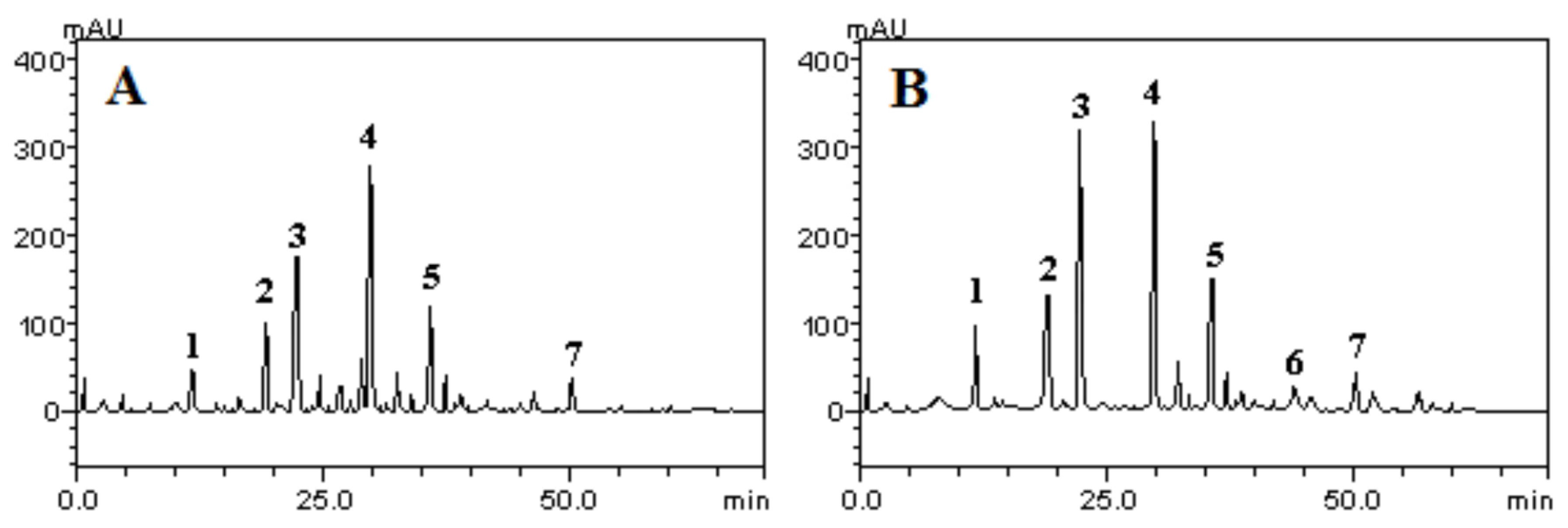
2.1. HPLC Characterization of the Polyphenolic Constituents of Aqueous and Ethanolic Extracts of R. echinus Leaves

The HPLC profile of the aqueous and ethanolic extracts of the leaves of R. echinus revealed the presence of polyphenolic constituents, which appeared with the following elution profile/order: Gallic acid (Rt = 7.12 min, peak 1), chlorogenic acid (Rt = 19.34 min, peak 2), caffeic acid (Rt = 22.61 min, peak 3), ellagic acid (Rt = 29.73 min, peak 4), rutin (Rt = 36.12 min, peak 5), quercitrin (Rt = 43.95 min, peak 6) and quercetin (Rt = 50.03 min, peak 7) (Figure 1A,B). However, quercitrin appeared to be absent in the aqueous extract (Figure 1A, Table 1) and present in the ethanolic extract of R. echinus (Figure 1B, Table 1). The identification of these constituents was made by comparing their retention time and UV spectra of the peaks in the samples with those of authentic reference samples or isolated compounds. Based on our results, ellagic acid appeared to be the major component of both extracts with 79.13 and 63.18 mg/g in ethanolic and aqueous extract, respectively. In contrast, quercetin (9.87 mg/g; representing about 0.98% of the aqueous extract) and quercitrin (7.12 mg/g; representing 0.81% of the ethanolic extract) were the two components detected in the smallest quantities in R. echinus extracts (Table 2).
Figure 1. HPLC-DAD chromatograms of aqueous (A) and ethanolic (B) extracts of the leaves of Raphiodon echinus (R. echinus): Gallic acid (peak 1), chlorogenic acid (peak 2), caffeic acid (peak 3), ellagic acid (peak 4), rutin (peak 5), quercitrin (peak 6) and quercetin (peak 7). Calibration curve for Gallic acid: y = 12574x + 1307.8 (r = 0.9999); chlorogenic acid: y = 11953x + 1278.2 (r = 0.9995); caffeic acid: y = 11976x + 1187.0 (r = 0.9996); ellagic acid: y = 13169x + 1346.8 (r = 0.9999); quercitrin: y = 12473x + 1187.5 (r = 0.9991); rutin: y = 12814x + 1189.3 (r = 0.9999) and quercetin: y = 12537x + 1375.6 (r = 0.9994). All chromatography operations were carried out at ambient temperature and in triplicate.
Molecules 21 00002 g001
Table 1. Schedule of evaluation of oxidation or chelation of Fe2+/Fe3+ by plant extracts.
TimeSequence of AdditionReading at 510 nm
0 minExtract (30–120 μg/mL)-
FeSO4 (110 μM)
10 minOrtho-phenanthroline (0.25%)-
10 minImmediately after mixing with First reading (0 min)
Ortho-phenanthroline
20 min-Second reading (10 min)
30 min-Third reading (20 min)
30 minAscorbic acid (AA, final concentration, 5 mM)-
35 min-First reading after AA (25 min)
40 min-Second reading after AA (30 min)
50 min-Third reading after AA (40 min)
Table 2. Phenolics and flavonoids composition of R. echinus extracts.
CompoundsAqueous Extract/mg·g−1 (%)Ethanolic Extract/mg·g−1 (%)LOD/μg·mL−1LOQ/μg·mL−1
Gallic acid11.59 ± 0.01 a (1.15)25.03 ± 0.01 a (2.50)0.0110.037
Chlorogenic acid25.07 ± 0.02 b (2.50)31.94 ± 0.02 b (3.19)0.0090.035
Caffeic acid40.19 ± 0.02 c (4.01)76.45 ± 0.02 c (7.64)0.0260.090
Ellagic acid63.18 ± 0.01 d (6.31)79.13 ± 0.03 c (7.91)0.0170.056
Rutin26.50 ± 0.03 b (2.65)35.84 ± 0.02 b (3.58)0.0240.080
Quercitrin-7.12 ± 0.01 d (0.81)0.0350.118
Quercetin9.87 ± 0.03 a (0.98)12.37 ± 0.01 e (1.23)0.0190.063
Results are expressed as mean ± standard deviations (SD) of three determinations. Different letters in the same column indicate significant difference by Tukey test at p < 0.01. LOD: limit of detection, LOQ: limit of quantification.

2.2. Antioxidant Activity

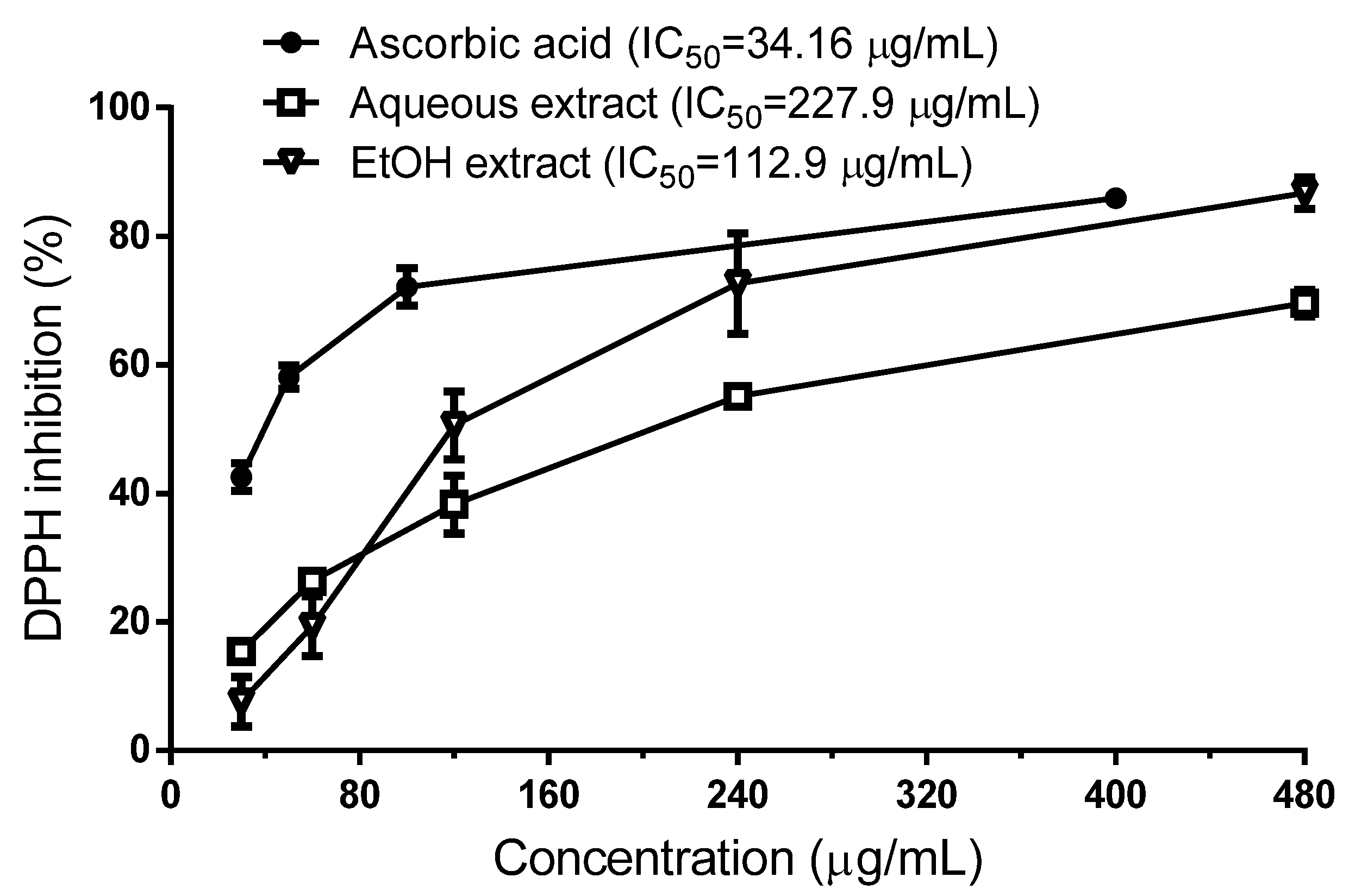
2.2.1. Scavenging Effect of R. echinus Extracts on DPPH Radical

Aqueous and ethanolic (EtOH) extracts of R. echinus exhibited antioxidant activity against DPPH radical in a concentration-dependent manner (Figure 2). According to the calculated IC50 values, the ethanolic extract exhibited stronger DPPH radical scavenging activity than aqueous extract, which was about two times higher than that of aqueous extract (Figure 2).
Figure 2. Quenching of DPPH radicals by aqueous and ethanolic extracts from the leaves of R. echinus. Data are expressed as mean ± SEM of n = 4 independent experiments.
Molecules 21 00002 g002
In this assay, the bleaching of the DPPH coloration is an indication of the free radical scavenging capacity of the samples. Our results revealed that the ethanolic extract showed greater antioxidant activity than aqueous extract (IC50 = 112.9 μg/mL vs. 227.9 μg/mL). However, this activity was three times lower than that of ascorbic acid (IC50 = 34.16 μg/mL), used as a standard. The higher antioxidant capacity of the ethanolic extract compared to the aqueous extract can be explained by its higher polyphenolic contents. In line with this, the total phenolic content of both extracts assayed by the Folin–Ciocalteu method revealed that ethanolic extract exhibit higher total phenolic content (TPC) than aqueous extract (Table 3). Numerous studies have described a positive correlation between the antioxidant activity and phenolic content [11,12]. Although we did not perform such correlation calculations, the results obtained here clearly demonstrate that ethanolic extract, which exhibited high TPC, has a stronger antioxidant activity by DPPH radical scavenging.
Table 3. Total phenolic content of R. echinus extracts.
R. echinus
Aqueous ExtractEthanolic Extract
Total phenolics (mg GAE/g dry extract)173.0 ± 0.07389.1 ± 0.04
Results are expressed in milligram Gallic acid equivalent (GAE) per gram of dry extract; n = 3.

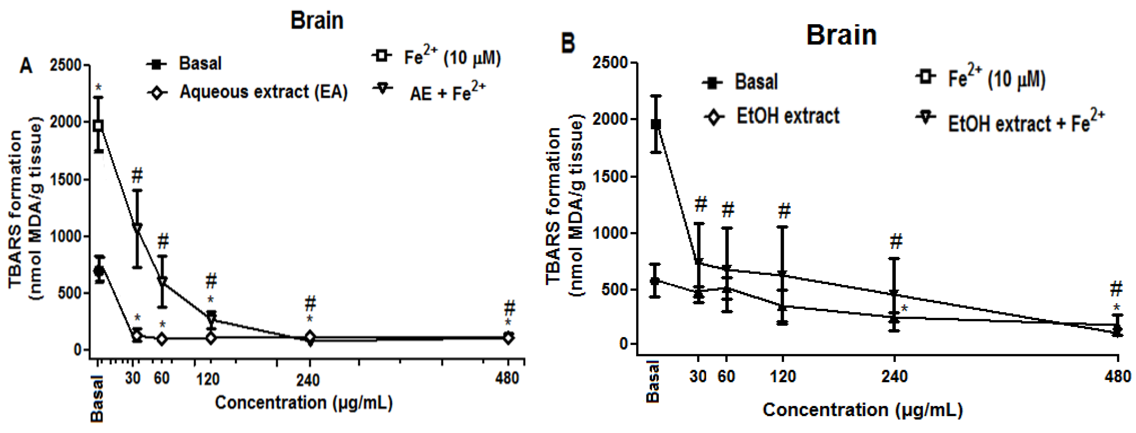
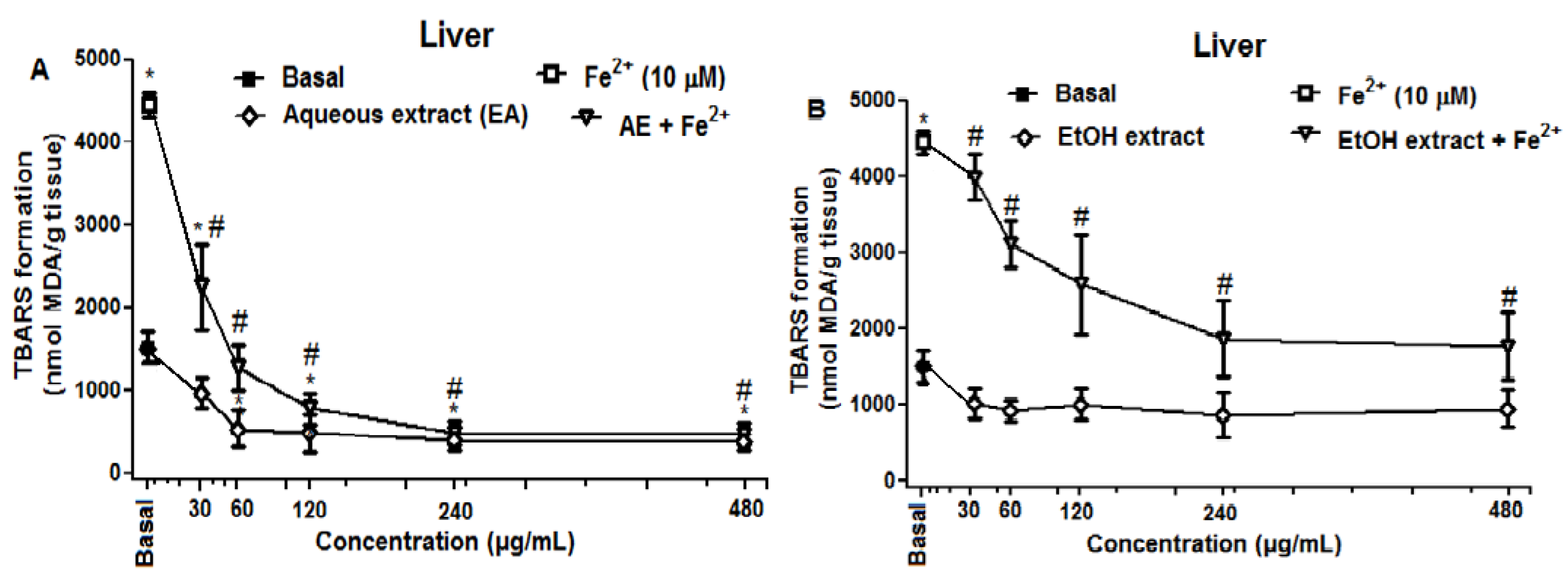
2.2.2. Effect of R. echinus Extracts on Fe2+ Induced Lipid Peroxidation (LPO) in the Rat Brain and Liver Homogenates

Fe2+ induced a significant increase in TBARS production in brain homogenates (p < 0.05; Figure 3). However, the aqueous (Figure 3A) and ethanolic (Figure 3B) extracts of R. echinus significantly reduced the LPO in concentration-dependent manner both under basal and iron-stimulated conditions (Figure 3A,B).
Figure 3. Effect of aqueous (A) and ethanolic (B) extracts of the leaves of R. echinus on lipid peroxidation induced by iron in rat brain homogenates. The homogenate was incubated for 1 h with Fe2+ (10 μM) in the presence or absence of different concentrations of the extracts. Values represent the mean of n = 3 independent experiments performed in duplicate ± SEM. *: p < 0.05 vs. basal and #: p < 0.05 vs. Fe2+.
Molecules 21 00002 g003
Similar to that observed with brain homogenates, crude extracts of R. echinus inhibited LPO induced by Fe2+ in rat liver homogenates (Figure 4). In contrast, ethanolic extract (EtOH extract) did not inhibited TBARS formation under basal condition (Figure 4B), while aqueous extract (AE) inhibited TBARS production under basal conditions (Figure 4A).
Figure 4. Effect of aqueous (A) and ethanolic (B) extracts of the leaves of R. echinus on lipid peroxidation induced by iron in rat liver homogenates. The homogenate was incubated for 1 h with Fe2+ (10 μM) in the presence or absence of different concentrations of the extracts. Values represent the mean of n = 3 independent experiments performed in duplicate ± SEM. *: p < 0.05 vs. basal and #: p < 0.05 vs. Fe2+.
Molecules 21 00002 g004
The results demonstrated that aqueous and ethanolic extracts from the leaves of R. echinus exhibited protective effects against Fe2+ induced lipid peroxidation (LPO) in rat brain homogenates. Although iron (II) is an essential element for life, free Fe2+ in biological systems, however, can be toxic [13] and its levels have been shown to be increased in many neurological disorders including Alzheimer’s and Parkinson’s diseases [14,15,16]. The results of the current study demonstrated that both extracts showed antioxidant activity against Fe2+ induced LPO in rat brain and liver homogenates at all the concentrations tested. Nevertheless, the ability of these extracts to inhibit LPO could be attributed (at least in part) to the capacity of their chemical constituents to chelate/inactivate Fe2+, thereby, preventing or reducing reactive oxygen species generation. The HPLC fingerprint of these extracts revealed the presence of phenolic acids (Gallic, ellagic, chlorogenic and caffeic acids) and flavonoids (quercetin, quercitrin and rutin), compounds that are known scavengers and inhibitors of LPO [17,18]. Of particular importance, is the finding which revealed that chlorogenic and caffeic acids could inhibit free radicals formation through several mechanisms including the reaction complex formation with iron ions in the reactions of hydrogen peroxide with iron (II), hydrogen peroxide with ferric iron and 3-hydroxyanthranilic acid [19]. In both tissues, aqueous extract showed highest antioxidant activity against Fe2+ induced LPO by significantly reducing TBARS formation at much lower concentrations when compared to the ethanolic extract. At basal conditions, ethanolic extract did not have any effect on TBARS formation in the liver, while it did in the brain at the higher concentrations tested (240 and 480 μg/mL).

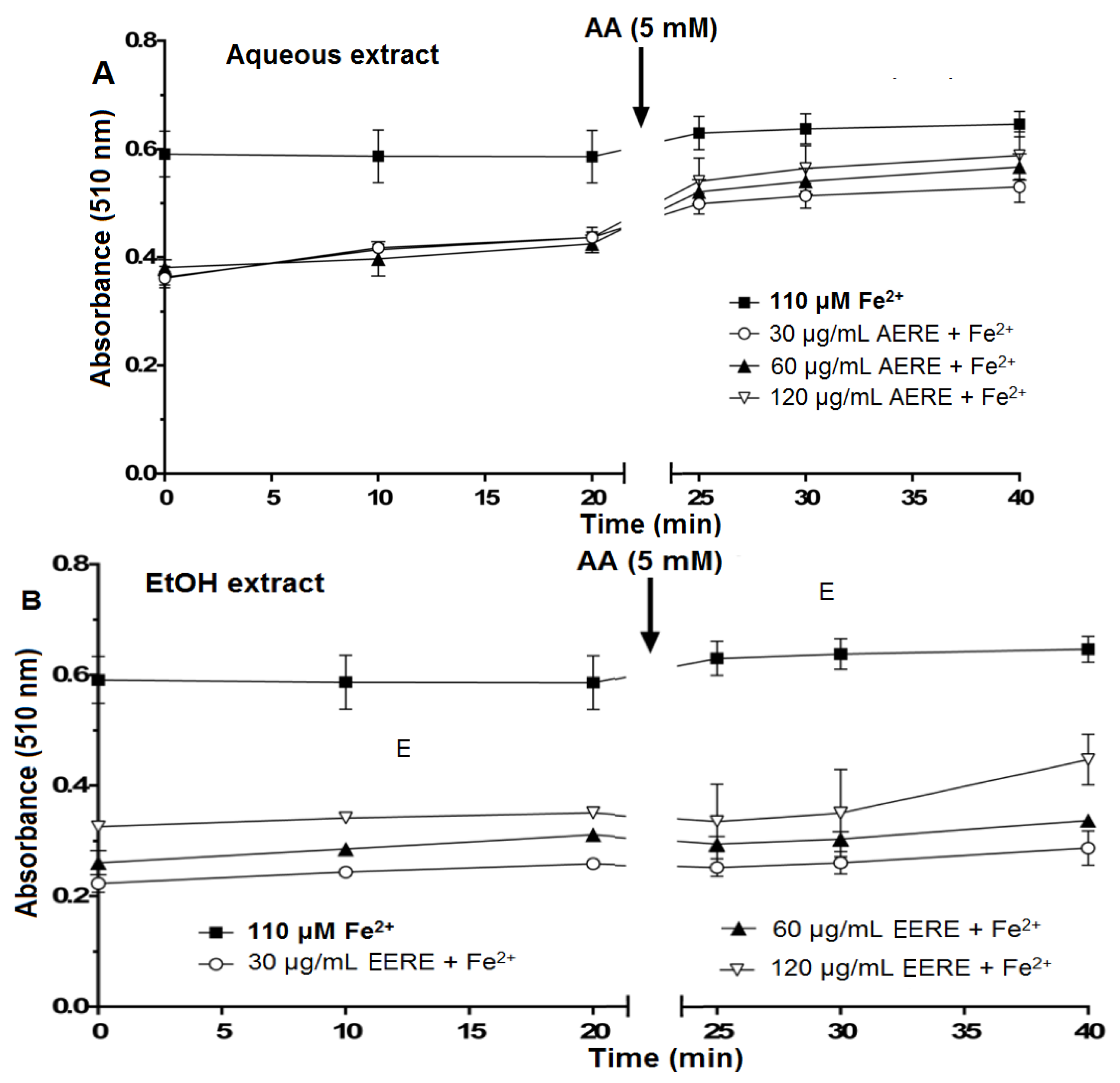
2.2.3. Iron Chelating Potential of R. echinus Extracts

The incubation of aqueous and ethanolic extracts with Fe2+ caused a decreased in the absorbance at 510 nm with the effect most apparent from 30 to 120 μg/mL (Figure 5A,B). The addition of ascorbic acid caused only a modest increase in the absorbance at 510 nm and this was a little more apparent for the aqueous extract (Figure 5A). The most plausible interpretation of these results is that the extracts can chelate Fe2+ and accelerate the oxidation of Fe2+ to Fe3+. However, this Fe3+ was only partially or not released from the complex, as otherwise ascorbic acid should have reduced Fe3+ to Fe2+, resulting in the formation of the complex between Fe2+ and ortho-phenanthroline. However, this occurred only to a limited extent.
Figure 5. Oxidation of Fe2+ by aqueous (A) and ethanolic (B) extracts from the leaves of R. echinus (1–60 μg/mL). The extracts (30–120 μg/mL) were incubated with FeSO4 (110 μM) for 10 min. Then, ortho-phenanthroline was added and the absorbance of the reaction mixture was measured at 0, 10 and 20 min following its addition. After the last reading (at 20 min), 5 mM ascorbic acid (AA) was added to the reaction mixture, and the absorbance was read again after 5 min (at 25 min), 10 min (at 30 min) and 20 min (at 40 min) (see Table 1 for details). Values represent the mean ± SEM of three independent experiments performed in duplicate. AERE, aqueous extract of R. echinus; EERE, ethanolic extract of R. echinus.
Molecules 21 00002 g005
To verify whether the decrease in the absorbance in the presence of aqueous (Figure 5A) or ethanolic (Figure 5B) extracts was caused by the chelation or oxidation of Fe2+, ascorbic acid (AA) was added to the reaction medium after 20 min of incubation of Fe2+ with the extracts and ortho-phenanthroline. The objective was to reduce Fe3+ that could have being formed during the reaction back to Fe2+. In the presence of aqueous extract (Figure 5A) or ethanolic extract (Figure 5B), the addition of AA to the reaction mixture caused a partial increase in the absorbance after 5, 10 and 20 min of incubation, which is an indication of stimulated oxidation of Fe2+ by the extracts.
Earlier reports have shown the ability of antioxidants to chelate or deactivate transition metals, thus preventing them from initiating lipid peroxidation and oxidative stress via metal-catalyzed reaction [20]. Chelation of transition metals such as iron can be viewed as a preventive antioxidant mechanism. In the present study, both extracts chelate Fe2+ partially and possibly converted it to Fe3+. However, Fe3+ was only partially released from the plant extract-iron complex as evidenced by the small increase in the absorbance after addition of the reducing agent, ascorbic acid (AA) to the reaction medium. Furthermore, if there was an appreciable free Fe3+ pool, this should have been reduced back to Fe2+ in the presence of ascorbic acid. However, this was very modest. Consequently, it is possible to presume that the observed inhibition of Fe2+ induced LPO in the presence of the extracts was a result of both direct interaction with free radicals and via chelation of Fe2+/Fe3+ species.

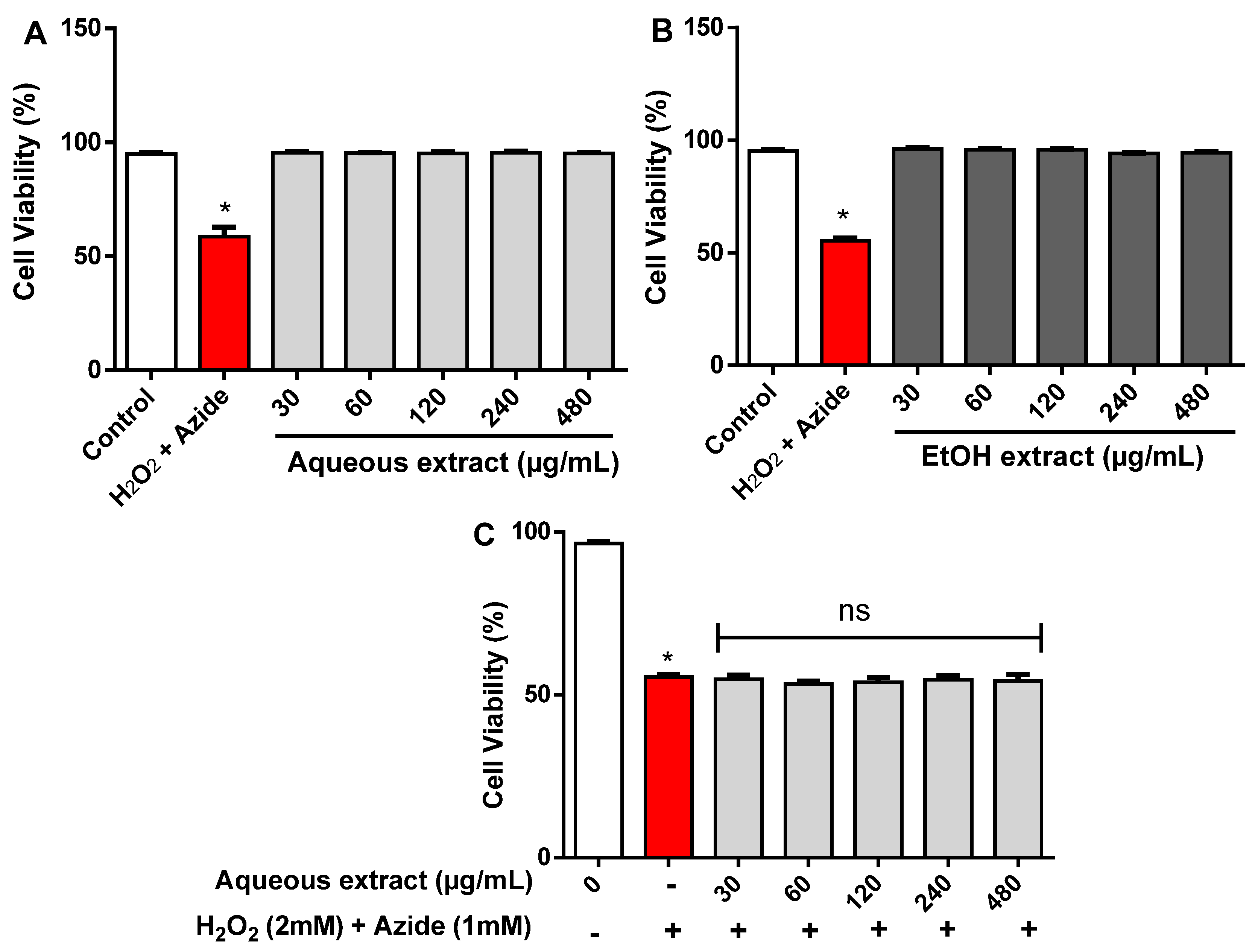
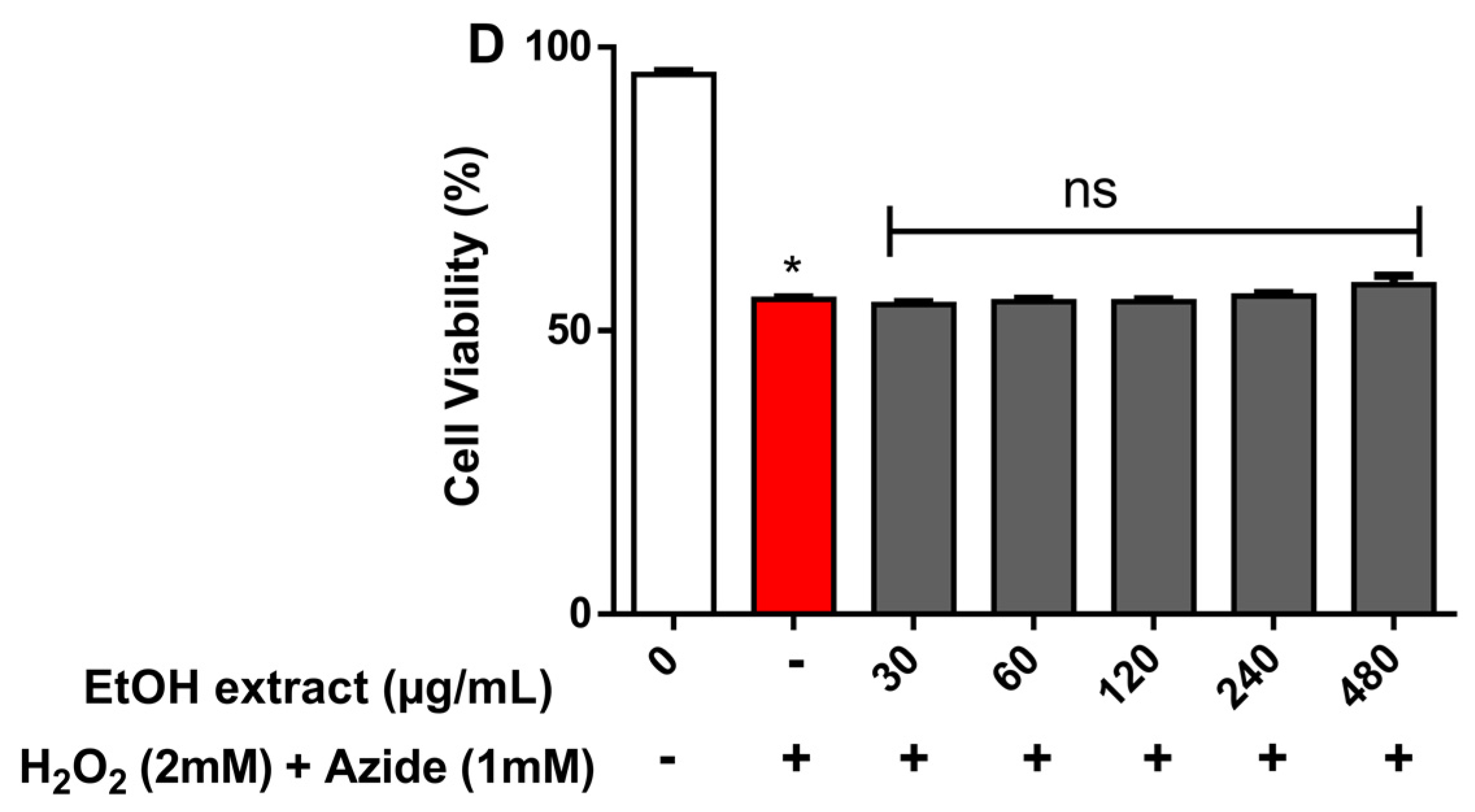
2.3. Cytotoxicity Effect of R. echinus Extracts on Human Leukocytes

The cytotoxicity of R. echinus extracts was investigated in human leukocytes using the Trypan blue exclusion method. In this method, dead cells allow passage of trypan blue into the cytoplasm due to loss of membrane selectivity [9,21]. Here, visual analysis of the cells using a Neubauer chamber allowed counting the number of dead (trypan blue positive) and living (trypan blue negative) cells. Exposure for 3 h of human leukocytes to aqueous and ethanolic extracts of R. echinus did not modify cell viability, when compared to the control (Figure 6A,B). Cell viability of treated leukocytes was generally greater than 90%, confirming the absence of cytotoxicity of the crude extracts of R. echinus. The potential protective effects of both extracts against H2O2 + azide induced cytotoxicity was also investigated. H2O2 (2 mM) + azide (1 mM) significantly decreased leukocytes viability when compared to control (p < 0.05; Figure 6C,D). However, aqueous (Figure 6C) and ethanolic (Figure 6D) extracts of R. echinus did not blunt the cytotoxicity caused by H2O2 + azide (Figure 6C,D).
Figure 6. Effect of R. echinus leaves extracts on human leukocytes in the absence (A,B) and presence (C,D) of H2O2 (2 mM) + Azide (1 mM). The signs (−) and (+) indicate the absence and presence of the mixture H2O2 (2 mM) + Azide (1 mM), respectively. Results are expressed as percentage of control. Values are the means of n = 3 independent experiments performed in triplicate ± SEM. *: p < 0.05 vs. control.
Molecules 21 00002 g006aMolecules 21 00002 g006b

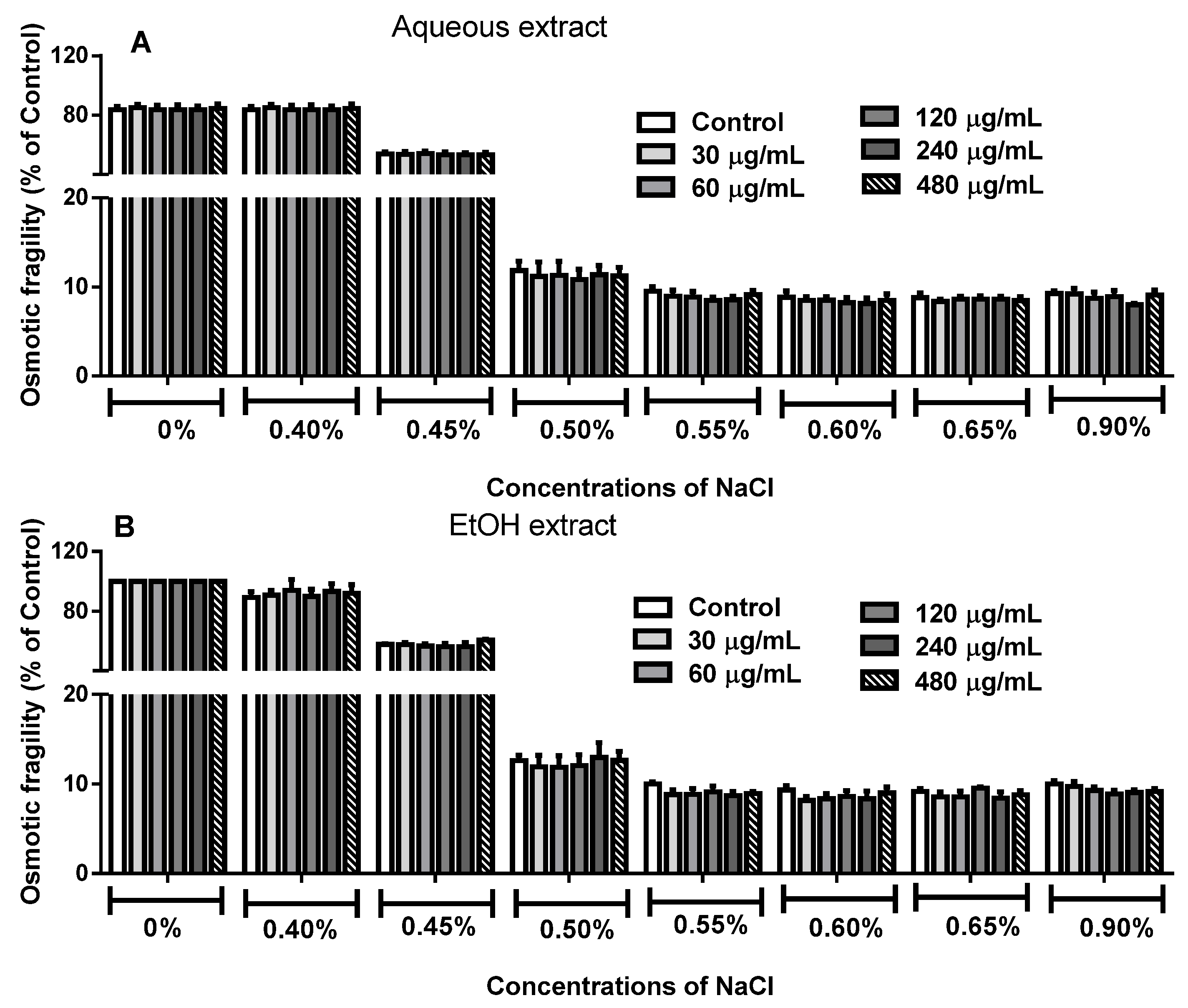
2.4. Effect of R. echinus Extracts on Osmotic Fragility of Human Erythrocytes

The influence of aqueous and EtOH extracts from the leaves of R. echinus on human erythrocytes osmotic fragility at different salt concentrations (0%–0.9%) is depicted in Figure 7. The results demonstrated no significant difference between treated and untreated erythrocytes at different salt concentrations in comparison to their respective control (p > 0.05; Figure 7A,B).
Figure 7. Osmotic fragility of human erythrocytes treated with aqueous (A) and ethanolic (B) extracts of the leaves of R. echinus. Untreated and treated erythrocytes were added to different concentrations of NaCl (0%–0.9%) and incubated for 20 min. The absorbance of the supernatant was measured and the hemolysis in each tube was expressed as percentage of control. Each bar represents the mean of n = 3 independent experiments performed in triplicate ± SEM.
Molecules 21 00002 g007
Osmotic fragility assay is widely used to verify the toxicity of chemicals (and plant extracts) and environmental pollutants on membrane integrity of erythrocytes [9,22,23]. Despite the fact that erythrocytes are well equipped with several biological mechanisms to defend against free radical induced LPO, erythrocyte membrane, however, is prone to oxidative stress, particularly because of the high oxygen tension in the blood and high polyunsaturated fatty acid content [24,25]. Here, the cytotoxic effect of the leaves extracts of R. echinus was investigated on human erythrocytes, so as to provide primary information on the interaction between their phytoconstituents and erythrocytes membrane [26,27]. Our results demonstrated that 3 h treatment of human erythrocytes with R. echinus extracts did not affect erythrocytes membrane integrity when compared to the control. These results indicate that the chemical constituents of the extracts did not cause gross changes in membrane protein and lipid structure of the cells.

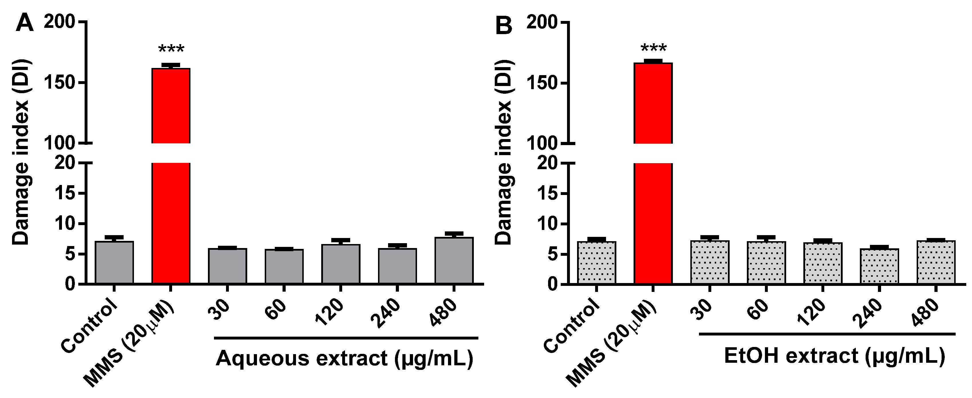
2.5. Effect of R. echinus Extracts on DNA Damage

As depicted in Figure 8, methyl methanesulfonate (MMS), used as positive control, caused a dramatic increase in DNA damage when compared with the control (p < 0.001). However, no significant difference was found in the DNA damage index (DI) between human leukocytes treated with aqueous (Figure 8A) and EtOH (Figure 8B) extracts, when compared to control (p > 0.05).
Figure 8. DNA damage of leukocytes treated with the aqueous (A) and ethanolic (B) leaves extracts of R. echinus. Methyl methanesulfonate (MMS) was used as positive control. The results are expressed as means ± SEM of n = 3 independent experiments. ***: p < 0.001 vs. control.
Molecules 21 00002 g008
Comet assay (single-cell gel electrophoresis) is widely used to access DNA damage and repair in individual cells. Studies have indicated that comet assay is a useful tool for the detection of genotoxicity thus, making this assay an important biomarker in toxicological studies [27,28]. In the present study, the potential genotoxic effect of the extracts components at the level of DNA was evaluated in human leukocytes. Our data showed that the leaf extracts of R. echinus are not genotoxic. This suggests that the phytoconstituents of these extracts do not/or have not interacted with the DNA. According to Galloway et al. [29,30], a situation where DNA damage occurs without a concomitant cytotoxicity is of greater concern. Here, we found no DNA damage and no cytotoxicity. Although it is difficult to extrapolate in vitro findings to in vivo exposure; nevertheless, we can speculate that the popular use of both extracts may not cause overt toxicity.

3. Experimental Section

3.1. Chemicals

All chemicals used including solvents were of analytical grade. Methanol, acetic acid, Gallic acid, caffeic acid, ellagic acid and chlorogenic acid were purchased from Merck (Darmstadt, Germany). Gallic, chlorogenic, caffeic, and ellagic acids, quercetin, rutin, and quercitrin, were acquired from Sigma Chemical Co. (St. Louis, MO, USA). 1,1-diphenyl-2-picrylhydrazyl (DPPH), ascorbic acid, malonaldehydebis-(dimethyl acetal) (MDA), thiobarbituric acid (TBA), sodium azide and hydrogen peroxide (H2O2) were purchased from Sigma Chemical Co.

3.2. Plant Material

The leaves R. echinus (Nees and Mart) Shauer were collected in Padre Cicero, Crato-Ceará (7°22′S; 39°28′W, 492 m above sea level), Brazil, in January 2014. The plant material was identified by Maria Arlene Pessoa da Silva, and deposited in the Herbarium Caririense Dárdano de Andrade—Lima, Universidade Regional do Cariri (URCA), with the number 7347. To obtain the aqueous extract of R. echinus, 300 g of crushed leaves was mixed with 2000 mL of hot water and the mixture was allowed to stand for three days. The mixture was filtered and the filtrate was lyophilized to obtain a dark green solid (3.3% yield). However, for the ethanolic extract, the fresh crushed leaves (300 g) were macerated with 1500 mL of 92% ethanol for three days. On the third day, the suspension was filtered and the filtrate containing the solvent was evaporated under reduced pressure and lyophilized to obtain 1.04% yield of the dry material. The prepared extracts (aqueous and ethanolic) were stored in the freezer until being tested. It should be stressed that the aqueous and ethanolic extracts of R. echinus were used in this study on the basis of its popular use in the form of infusion, and also because the roots of the plant are soaked in ethanol prior to use.

3.3. Quantification of Compounds by HPLC-DAD

Reverse phase chromatographic analyses were carried out under gradient conditions using C18 column (4.6 mm × 150 mm) packed with 5 μm diameter particles; the mobile phase was water containing 1% formic acid (A) and methanol (B), and the composition gradient was: 13% of B until 10 min and changed to obtain 20%, 30%, 50%, 60%, 70%, 20% and 10% B at 20, 30, 40, 50, 60, 70 and 80 min, respectively, following the method described by Pereira et al. [31], with slight modifications. Aqueous and ethanolic extracts of R. echinus as well as the mobile phase were filtered through 0.45 μm membrane filter (Millipore, Billerica, MA, USA) and then degassed by ultrasonic bath prior to use. The extracts were analyzed at concentration of 20 mg/mL for the presence or absence of Gallic, chlorogenic, caffeic and ellagic acids, and rutin, quercitrin and quercetin. These compounds were identified by comparing their retention time and UV absorption spectra with those of the commercial standards. The flow rate was 0.7 mL/min, injection volume 40 μL and the wavelength were 257 nm for Gallic acid, 327 nm for chlorogenic, caffeic and ellagic acids, and 365 nm for quercetin, quercitrin and rutin. Stock solutions of standards references were prepared in the HPLC mobile phase at a concentration range of 0.030–0.250 mg/mL for quercetin, quercitrin and rutin; and 0.035–0.300 mg/mL for ellagic, Gallic, caffeic and chlorogenic acids. Chromatography peaks were confirmed by comparing its retention time with those of reference standards and by DAD spectra (200 to 600 nm). All the chromatography operations were carried out at ambient temperature and in triplicates. The limit of detection (LOD) and limit of quantification (LOQ) were calculated based on the standard deviations of the responses and their slopes using three independent analytical curves. LOD and LOQ were calculated as 3.3 and 10 σ/S, respectively, where σ is the standard deviation of the response and S is the slope of the calibration curve.

3.4. Determination of Total Phenolics

The determination of total phenolic content was performed by the Folin–Ciocalteu method, as described by Kamdem et al. [32], with some modifications [33]. Briefly, 0.5 mL of 2 N Folin–Ciocalteu reagent was added to a 1 mL of ethanolic or aqueous extract of R. echinus (0.15 mg/mL) and the mixture was allowed to stand for 5 min before the addition of 2 mL of 20% Na2CO3. The tubes were then allowed to stand at room temperature for 10 min and the absorbance was measured against water at 730 nm using Shimadzu-UV-1201 (Shimadzu, Kyoto, Japan) spectrophotometer. The phenolic content was expressed in milligram of Gallic acid equivalent per gram of dry extract (mg GAE/g of dry extract). The standard curve obtained with Gallic acid was Y = 29.315 × X − 0.04 (r2 = 0.9997).

3.5. Antioxidant Activity

3.5.1. DPPH Radical Scavenging Activity

The radical scavenging ability of the aqueous and ethanolic extracts of R. echinus was performed using the stable free radical DPPH (1,1-diphenyl-2-picrylhydrazyl) as described by Kamdem et al. [32], with some modifications. Briefly, 50 μL of aqueous and ethanolic extracts at different concentrations (30–480 μg/mL) were mixed with 100 μL of freshly prepared DPPH solution (0.3 mM in ethanol). Then, the plate was kept in the dark at room temperature for 30 min. The reduction in the DPPH radical was measured by monitoring the decrease of absorption at 517 nm using a microplate reader (SpectraMax, Sunnyvale, CA, USA). Ascorbic acid was used as standard compound (i.e., positive control). The DPPH radical scavenging capacity was measured using the following equation:
% inhibition = 100 – (Asample − Ablank)/Acontrol × 100
where Asample is the absorbance of the tested sample with DPPH; Ablank, the absorbance of the test tube without adding the DPPH and Acontrol, is the absorbance of the DPPH solution.

3.5.2. Production of TBARS from Animal Tissues

The production of thiobarbituric acid reactive substances (TBARS) was measured as described by Ohkawa et al. [34], and modified by Barbosa-Filho et al. [35]. The rats were killed by decapitation. The whole brain and the liver were quickly removed, placed on ice and weighed. The tissues were immediately homogenized in cold 10 mM Tris-HCl, pH 7.4 (1:10, w/v for the liver and 1:5, w/v for brain) and centrifuged at 3600 rpm for 10 min. The pellet was discarded and the supernatant was used to perform the assay. Aliquots of brain or liver homogenates (20 μL) was incubated with 10 μM FeSO4 in the presence or absence of extracts (30–480 μg/mL) at 37 °C for 1 h, to induce lipid peroxidation. Subsequently, 40 μL of sodium dodecyl sulphate (8.1%), 100 μL of acetic acid/HCl (pH 3.4) and 100 μL of 0.6% thiobarbituric acid (TBA) were added and incubated at 100 °C for 1 h. After cooling, the samples were centrifuged for 2 min at 6000 rpm and the absorbance of the supernatant was read at 532 nm using an ELISA plate reader (SpectraMax). This work was carried out in accordance with the Guidelines of the Ethical Committee of UFSM and approved by the institutional review board of UFSM (076.2012-2).

3.5.3. Iron Chelating Activity of R. echinus Extracts

The chelating capacity of aqueous and ethanolic extracts of the leaves of R. echinus was determined according to the modified method of Kamdem et al. [27]. The reaction mixture containing 58 μL of saline solution (0.9%, w/v), 45 μL Tris-HCl (0.1 M, pH, 7.5), 27 μL of extracts (30–120 μg/mL) and 36 μL of 110 μM FeSO4 was incubated for 10 min at 37 °C. Then, 34 μL of 1,10-phenanthroline (0.25%, w/v) was added and the absorbance of the orange coloured complex formed was measured at 0, 10 and 20 min at 510 nm (against blank solutions of the samples) using microplate reader (SpectraMax). The same procedure was performed for the control (i.e., Fe2+), but without the extract. To ascertain the chelating potential of the extracts, we determined the potential reduction of any Fe3+ (that might be formed during the incubation periods) by adding the reducing agent, ascorbic acid (to give a final concentration of 5 mM) o the reaction mixture. The absorbance was then determined after 5, 10 and 20 min following ascorbic acid addition. This is because the extracts could be oxidizing Fe2+ to Fe3+, leading to a decrease in absorbance that was not related to Fe2+ chelation. To summarize, the schedule for the evaluation of Fe2+ chelation or oxidation by the extracts is presented in Table 1.

3.6. Blood Collection and Preparation of Human Leukocytes and Erythrocytes

Heparinized venous blood was obtained from healthy volunteer donors at the hospital of the Federal University of Santa Maria (UFSM), Santa Maria-RS, Brazil (age 26 ± 9). The study was approved by the Ethical Committee of UFSM and registered under the protocol number 0089.0.243.000-07. Human leukocytes and erythrocytes were obtained as previously reported [27,35]. The leukocytes were separated by differential sedimentation rate using 5% dextran and subsequent adjustment of samples to 2 × 10⁶ leukocytes/mL with Hank’s buffered saline solution (HBSS)/heparin (5.4 mM KCl, 0.3 mM Na2HPO4, 0.4 mM KH2PO4, 4.2 mM NaHCO3, 1.3 mM CaCl2, 0.5 mM MgCl2, 0.6 mM MgSO4, 137 mM NaCl, 10 mM d-glucose, 10 mM Tris-HCl, and heparin 15 IU/mL, adjusted to pH 7.4). However, the erythrocytes were separated by centrifuging the blood sample at 2000 rpm for 5 min at room temperature. The plasma was aspirated and the cell pellet was washed three times with phosphate buffered saline (6.1 mM, pH 7.4, containing 150 mM NaCl).

3.7. Determination of Erythrocytes Osmotic Fragility

The influence of the extracts on osmotic fragility of erythrocytes was estimated by measuring their resistance to hemolysis in increasing concentrations of salt solutions as modified by Barbosa-Filho et al. [35]. Five hundred microliters of erythrocytes, 100 μL of various concentrations of aqueous and ethanolic extracts (30–480 μg/mL) and 900 μL of phosphate buffer saline (PBS) (6.1 mM, pH 7.4, containing 150 mM NaCl) were pre-incubated for 3 h at 37 °C. After incubation, samples were mixed and centrifuged at 2500 rpm for 10 min and the supernatant was discarded. The erythrocytes were washed twice with PBS, centrifuged at 2500 rpm for 2 min and the supernatant discarded. Treated and untreated erythrocytes (7.5 μL) were then incubated with 1.5 mL of varying concentrations (0%–0.9%) of NaCl, pH 7.4 for 20 min. The samples were homogenized and centrifuged at 2000 rpm for 5 min. The supernatant obtained from each Eppendorf was transferred to microplate and the lysis of erythrocytes was monitored by measuring the absorbance of hemoglobin content in the supernatants at 540 nm using microplate reader (SpectraMax). The results were expressed as percentage of the control.

3.8. Genotoxicity by the Comet Assay

The procedure described by Collins [36] was used with some modifications as described by Kamdem et al. [27]. Leukocytes were isolated as described earlier and treated with different concentrations of aqueous and ethanolic extracts of R. echinus (30–480 μg/mL) for 3 h. The comet assay was performed according to the following steps: (1) 15 μL of leukocyte suspension (2 × 10⁶ leukocytes/mL) was mixed with low-melting agarose; (2) added to 90 mL of 0.75% LMP agarose (w/v), mixed, and placed on a microscope slide precoated with normal melting point agarose (1% w/v); (3) a coverslip was added and the samples allowed to solidify at 4 °C; (4) coverslips were removed and slides were placed in a lysis solution (2.5 M NaCl; 100 mM EDTA; 8 mM Tris-HCl; 1% Triton X-100, pH 10–10.5), where they remained for 24 h protected from light; (5) after lysis, the slides were placed in bucket containing neutralizing solution (400 mM Tris-HCl; pH 7.5) for 15 min; (6) then, they were incubated in electrophoretic solution (300 mM NaOH, 1 mM EDTA, pH 13.5) for 20 min at 4 °C under the condition of 25 V, 300 mA, 7W; (7) the slides were washed three times in distilled water and allowed to dry at room temperature; (8) the slides were rehydrated for 3 min in distilled water and fixed for 10 min in 15% trichloroacetic acid, 5% zinc sulfate, and 5% glycerol, then, washed three times in distilled water and allowed to dry at room temperature; (9) the slides were stained with 5% sodium carbonate, 0.1% ammonium nitrate, 0.1% silver nitrate, 0.25% tungstosilicico acid and 0.15% formaldehyde; (10) staining was stopped with 1% acetic acid and air dried sheets; and, finally, (11) slides were visualized and scored according to tail length into five class (from class 0: undamaged, without a tail; to class 4: maximum damage, comet with no heads) under blind conditions by at least two individuals. DNA damage was presented as DNA damage index (DI), which is based on the length of migration. The DI was calculated from cells in different damage classes as follows:
DI = 1 × n1 + 2 × n2 + 3 × n3 + 4 × n4
where, n1–n4 indicates the number of cells with level 1–4 of damage. Methyl methanesulfonate (MMS) (20 μM) was used as positive control and the negative control with distilled water.

3.9. Cytotoxicity by the Trypan Blue Exclusion Method (Cell Viability)

The cell viability was determined using the Trypan blue exclusion method, which assumes that nonviable cells are stained blue [37]. Briefly, 2.5 μL of the extracts (30–480 μg/mL) was added to 497.5 μL of leukocytes suspension and incubated in the presence or absence hydrogen peroxide (2mM) + azide (1 mM), at 37 °C for 3 h. The azide was used to inhibit the activity of catalase in the cell and consequently detect the toxicity induced by H2O2 used as positive control. Thereafter, 50 μL leukocytes was mixed with 50 μL of 0.4% Trypan blue and allowed to stand for 5 min at room temperature. From the mixture, an aliquot of 10 μL was checked microscopically for viability using a hemocytometer. The viability was as expressed as viable cells in percent of the total cells.

3.10. Statistical Analysis

Values were expressed as mean ± standard error of mean (S.E.M). All the analyses were performed using one-way analysis of variance (ANOVA), except for iron chelation, where data were treated with two-way ANOVA followed by Bonferroni post-test. The results were considered significantly different when p < 0.05.

4. Conclusions

In conclusion, the results presented in this study demonstrated for the first time the in vitro antioxidant activity of extracts from the leaves of R. echinus, which was evidenced by their potential to scavenge the DPPH radical and inhibit lipid peroxidation in rat brain and liver homogenates. R. echinus extracts was neither cytotoxic nor genotoxic to human leukocytes and erythrocytes, respectively, which give indirect support of the popular use of this plant in traditional Brazilian medicine. However, in vivo investigation to ascertain the safety of this plant and its extracts needs to be conducted.

Acknowledgments

Supported by CAPES, FAPERGS, CNPq, FINEP and Vitae Fundation. A.E.D. is particularly grateful to CAPES-DINTER for financial support. A.E.D. is a beneficiary of the CAPES Postgraduate (Doctoral) fellowship. J.P.K. acknowledges CAPES, TWAS and CNPq-TWAS for the financial support.

Author Contributions

M.E.B., J.B.T.R. and J.P.K. designed the research; J.G.M.C., I.R.A. and M.A.P.S. helped in the preparation, concentration and lyophilization of extracts; A.E.D., E.P.W., L.M.B. and K.R. performed the experiments and contributed to data collection; F.A.B.C. assisted in the statistical analysis; A.A.B. performed the HPLC analysis; A.O.A. determined the total phenolic content and edited the English; and A.E.D. and J.P.K. wrote the manuscript. J.B.T.R. and M.E.B. contributed to the revisions of the manuscript. All authors have read and approved the final version of the manuscript.

Conflicts of Interest

The authors declare no conflict of interest.

References

  1. Schmeiser, H.H.; Stiborova, M.; Arlt, V.M. Chemical and molecular basis of the carcinogenicity of Aristolochia plants. Curr. Opin. Drug Discov. Dev. 2009, 12, 141–148. [Google Scholar]
  2. Ekor, M. The growing use of herbal medicines: Issues relating to adverse reactions and challenges in monitoring safety. Front. Pharmacol. 2014, 4. [Google Scholar] [CrossRef] [PubMed]
  3. Anantha, N.D. Approaches to pre-formulation R and d for phytopharmaceuticals emanating from herb based traditional Ayurvedic processes. J. Ayurveda Integr. Med. 2013, 4, 4–8. [Google Scholar] [CrossRef] [PubMed]
  4. Waczuk, E.P.; Kamdem, J.P.; Abolaji, A.O.; Meinerz, D.F.; Caeran Bueno, D.; do Nascimento Gonzaga, T.K.S.; do Canto Dorow, T.S.; Boligon, A.A.; Athayde, M.L.; da Rocha, J.B.T.; et al. Euphorbia tirucalli aqueous extract induces cytotoxicity, genotoxicity and changes in antioxidant gene expression in human leukocytes. Toxicol. Res. 2015, 4, 739–748. [Google Scholar]
  5. Vásquez, G.D.; Harley, R.M. Flora de Grão-Mogol, Minas Gerais: Labiatae. Bol. Bot. Univ. São Paulo 2004, 22, 193–204. [Google Scholar] [CrossRef]
  6. Souza, A.A.; Rodrigues, S.A. Antimicrobial activity of essential oil of Raphiodon echinus (Nees. e Mart) Shauer. Rev. Biol. Farm. 2012, 7, 12–17. [Google Scholar]
  7. Menezes, F.S.; Kaplan, M.A.K.; Cardoso, G.L.C.; Pereira, N.A. Phytochemical and pharmacological studies on Rhaphiodon echinus. Fitoterapia 1998, 69, 459–460. [Google Scholar]
  8. Kehrer, J.P.; Klotz, L. Free radicals and related reactive species as mediators of tissue injury and disease: Implications for health. Crit. Rev. Toxicol. 2015, 45, 765–798. [Google Scholar] [CrossRef] [PubMed]
  9. Ambali, S.F.; Abubakar, M.; Shittu, M.; Yaqub, L.S.; Anafi, S.B.; Abdullahi, A. Chlorpyrifos induced alteration of haematological parameters in Wistar rats: Ameriolative effect of zinc. Res. J. Environ. Toxicol. 2010, 4, 55–66. [Google Scholar]
  10. Fetoui, H.; Gdoura, R. Synthetic pyrethroid increases lipid and protein oxidation and induces glutathione depletion in the cerebellum of adult rats: Ameliorative effect of vitamin C. Hum. Exp. Toxicol. 2012, 31, 1151–1160. [Google Scholar] [CrossRef] [PubMed]
  11. Cheung, L.M.; Cheung, P.C.K.; Ooi, V.E.C. Antioxidant activity and total phenolics of edible mushroom extracts. Food Chem. 2003, 81, 249–255. [Google Scholar] [CrossRef]
  12. Kotássková, E.; Sumczynski, D.; Mlcek, J.; Valásek, P. Determination of free and bound phenolics using HPLC-DAD antioxidante activity and in vitro digestibility of Eragrostis tef. J. Food Compos. Anal. 2016, 46, 15–21. [Google Scholar] [CrossRef]
  13. Eugene, D.W. The hazards of iron loading. Metallomics 2010, 2, 732–740. [Google Scholar]
  14. Zecca, L.; Casella, L.; Albertini, A.; Bellei, C.; Zucca, F.A.; Engelen, M.; Zadlo, A.; Szewczyk, G.; Zareba, M.; Sarna, T. Neuromelanin can protect against iron-mediated oxidative damage in system modeling iron overload of brain aging and Parkinson’s disease. J. Neurochem. 2008, 106, 1866–1875. [Google Scholar] [PubMed]
  15. Becerril-Ortega, J.; Bordji, K.; Fréret, T.; Rush, T.; Buisson, A. Iron overload accelerates neuronal amyloid-β production and cognitive impairment in transgenic mice model of Alzheimer’s disease. Neurobiol. Aging 2014, 35, 2288–2301. [Google Scholar] [CrossRef] [PubMed]
  16. Hassan, H.; Oliveira, C.S.; Noreen, H.; Kamdem, J.P.; Nogueira, C.W.; Rocha, J.B.T. Organoselenium compounds as potential neuroprotective therapeutic agents. Curr. Org. Chem. 2016, 20, 218–231. [Google Scholar] [CrossRef]
  17. Chikezie, A.R.; Akuwudike, C.M. Membrane stability and methaemoglobin content of human erythrocytes incubated in aqueous leaf extract of Nicotiana tabacum. Free Radic. Antioxid. 2012, 2, 56–61. [Google Scholar] [CrossRef]
  18. Pistón, M.; Machado, I.; Branco, C.S.; Cesio, V.; Heinzen, H.; Ribeiro, D.; Fernandes, E.; Chisté, R.C.; Freita, M. Infusion, decoction and of leaves from artichoke are effective scavengers of physiologically relevant ROS and RNS. Food Res. Int. 2014, 64, 150–156. [Google Scholar] [CrossRef]
  19. Iwahashi, H. Inhibitory effects of caffeic acid on free-radical formation. In Coffee in Health and Disease prevention; Preedy, V.R., Ed.; Elsevier Inc.: Atlanta, GA, USA, 2014; Volume 88, pp. 803–812. [Google Scholar]
  20. Oboh, G.; Puntel, R.L.; Rocha, J.B.T. Hot pepper (Capsicum annuum, tepin & Capsicum Chinese, habanero) prevents Fe2+-induced lipid peroxidation in brain—In vitro. Food Chem. 2007, 102, 178–185. [Google Scholar]
  21. Stoddart, M.J. Cell viability assays: Introduction. Methods Mol. Biol. 2011, 740, 1–6. [Google Scholar] [PubMed]
  22. He, J.; Lin, J.; Li, J.; Zhang, J.H.; Sun, X.M.; Zeng, C.M. Dual effects of Ginkgo biloba leaf extract on human red blood cells. Basic Clin. Pharmacol. Toxicol. 2009, 104, 138–144. [Google Scholar] [CrossRef] [PubMed]
  23. Ghorbel, L.; Maktouf, S.; Kallel, C.; Ellouze Chaabouni, S.; Boudawara, T.; Zeghal, N. Disruption of erythrocyte antioxidant defense system, hematological parameters, induction of pro-inflammatory cytokines and DNA damage in liver of co-exposed rats to aluminiun and acrylamide. Chem. Biol. Interact. 2015, 236, 31–40. [Google Scholar] [CrossRef] [PubMed]
  24. Perrone, S.; Tataranno, M.L.; Stazzoni, G.; del Vecchio, A.; Buonocore, G. Oxidative injury in neonatal erythrocytes. J. Matern. Fetal. Neonatal. Med. 2012, 25, 104–108. [Google Scholar] [CrossRef] [PubMed]
  25. Lang, E.; Lang, F. Mechanisms and pathophysiological significance of eryptosis, the suicidal erythrocyte death. Semin. Cell. Dev. Biol. 2015, 39, 35–42. [Google Scholar] [CrossRef] [PubMed]
  26. Sharma, P.; Sharma, J.D. In vitro hemolysis of human erythrocytes by plant extracts with antiplasmodial activity. J. Ethnopharmacol. 2001, 74, 239–243. [Google Scholar] [CrossRef]
  27. Kamdem, J.P.; Adeniran, A.; Boligon, A.A.; Klimaczewski, C.V.; Elekofehinti, O.O.; Hassan, W.; Ibrahim, M.; Waczuk, E.P.; Meinerz, D.F.; Athayde, M.L. Antioxidant activity, genotoxicity and cytotoxicity evaluation of lemon balm (Melissa officinalis L.) ethanolic extract: Its potential role in neuroprotection. Ind. Crop. Prod. 2013, 51, 26–34. [Google Scholar] [CrossRef]
  28. Roy, P.; Mukherjee, A.; Giri, S. Evaluation of genetic damage in tobacco and arsenic exposed population of Southern Assam, India using buccal cytome and comet assay. Ecotoxicol. Environ. Saf. 2015, 124, 169–176. [Google Scholar] [CrossRef] [PubMed]
  29. Galloway, S.M.; Miller, J.E.; Armstrong, M.J.; Bean, C.L.; Skopek, T.R.; Nichols, W.W. DNA synthesis inhibition as an indirect mechanism of chromosome aberrations: Comparison of DNA-reactive and non-DNA-reactive clastogens. Mutat. Res. 1998, 400, 169–186. [Google Scholar] [CrossRef]
  30. Galloway, S.M. Cytotoxicity and chromosome aberrations in vitro: Experience in industry and the case for an upper limit on toxicity in the aberration assay. Environ. Mol. Mutagen. 2000, 35, 191–201. [Google Scholar] [CrossRef]
  31. Pereira, R.P.; Boligon, A.A.; Appel, A.S.; Fachinetto, R.; Ceron, C.S.; Tanus-Santos, J.E.; Athayde, M.L.; Rocha, J.B.T. Chemical composition, antioxidant and anticholinesterase activity of Melissa officinalis. Ind. Crops Prod. 2014, 53, 34–45. [Google Scholar] [CrossRef]
  32. Kamdem, J.P.; Stefanello, S.T.; Boligon, A.A.; Wagner, C.; Kade, I.J.; Pereira, R.P.; Preste, A.S.; Roos, D.H.; Waczuk, E.P.; Appel, A.S.; et al. In vitro antioxidant activity of stem bark of Trichilia catigua Adr. Juss. Acta Pharm. 2012, 62, 371–382. [Google Scholar] [CrossRef] [PubMed]
  33. Boligon, A.A.; Pereira, R.P.; Feltrin, A.C.; Machado, M.M.; Janovik, V.; Rocha, J.B.T.; Athayde, M.L. Antioxidant activities of flavonol derivatives from the leaves and stem bark of Scutia buxifolia Reiss. Bioresour. Technol. 2009, 100, 6592–6598. [Google Scholar] [CrossRef] [PubMed]
  34. Ohkawa, H.; Ohishi, N.; Yagi, K. Assay for lipid peroxides in animal tissues by thiobarbituric acid reaction. Anal. Biochem. 1979, 95, 351–358. [Google Scholar] [CrossRef]
  35. Barbosa-Filho, V.M.; Waczuk, E.P.; Kamdem, J.P.; Lacerda, S.R.; Martin da Costa, J.C.; Alencar de Menesez, I.R.; Abolaji, A.O.; Boligon, A.A.; Athayde, M.L.; Rocha, J.B.T.; et al. Phytochemical constituents, antioxidant activity, cytotoxicity and osmotic fragility effects of caju (Anacardium. microcarpum). Ind. Crop. Prod. 2014, 55, 280–288. [Google Scholar] [CrossRef]
  36. Collins, A.R. The comet assay for DNA damage and repair. Principles, application, and limitations. Mol. Biotechnol. 2004, 26, 249–261. [Google Scholar] [CrossRef]
  37. Mischell, B.B.; Shiingi, S.M. Selected Methods in Cellular Immunology; WH Freeman Company: New York, NY, USA, 1980; pp. 1–469. [Google Scholar]
  • Sample Availability: Samples of the compounds are not available from the authors.

