Communication

Novel Steroidal Glycosides from the Bulbs of Lilium pumilum

School of Pharmacy, Tokyo University of Pharmacy and Life Sciences, 1432-1 Horinouchi, Hachioji, Tokyo 192-0392, Japan
*
Author to whom correspondence should be addressed.
These authors contributed equally to this work.
Molecules 2015, 20(9), 16255-16265; https://doi.org/10.3390/molecules200916255
Submission received: 25 June 2015 / Revised: 25 August 2015 / Accepted: 31 August 2015 / Published: 8 September 2015
(This article belongs to the Section Natural Products Chemistry)

Abstract

:
Examination of the bulbs of Lilium pumilum (Liliaceae) led to the isolation of four novel steroidal glycosides (14) with a 2,3,4-trisubstituted β-d-glucopyranosyl unit. In 1 and 3, the α-l-arabinopyranosyl moiety is linked to C-3 of the inner trisubstituted β-d-glucopyranosyl group and is present as an usual 4C1 conformation. In contrast, in 2 and 4, the α-l-arabinopyranosyl moiety, which is attached to C-4 of the inner trisubstituted β-d-glucopyranosyl group, is present as a 1C4 conformation. The structures of the new steroidal glycosides were determined based on the results of spectroscopic analyses, including two-dimensional (2D) NMR data and hydrolysis.

Graphical Abstract

1. Introduction

Lilium pumilum D.C. (Liliaceae) is described in the Japanese Pharmacopoeia (16th edition) as a plant from which the crude drug Lilium Bulb is derived. Lilium Bulb has long been used as an antitussive and anti-inflammatory agent in traditional Chinese medicine [1]. The bulbs of L. pumilum (L. tenuifolium Fisch. ex Hook.) contain phenylpropanoid derivatives, such as regalosides A and D, and sterol glycosides, such as tenuifoliosides A and B [2]. Although plants belonging to the family Liliaceae are rich sources of bioactive steroidal glycosides, such as OSW-1 or galtonioside A with cytotoxic activities against tumor cells [3,4], few studies have focused on steroidal glycosides of L. pumilum [5]. Our preliminary analysis of the MeOH extract of L. pumilum bulbs suggests that it contains more steroidal glycosides. Here, we report four novel steroidal glycosides (14) with a trisubstituted β-d-glucopyranosyl unit isolated from the bulbs of L. pumilum. The structures of the new steroidal glycosides were determined based on the results of spectroscopic analysis, including two-dimensional NMR data, and hydrolysis followed by chromatographic and spectroscopic analyses.

2. Results and Discussion

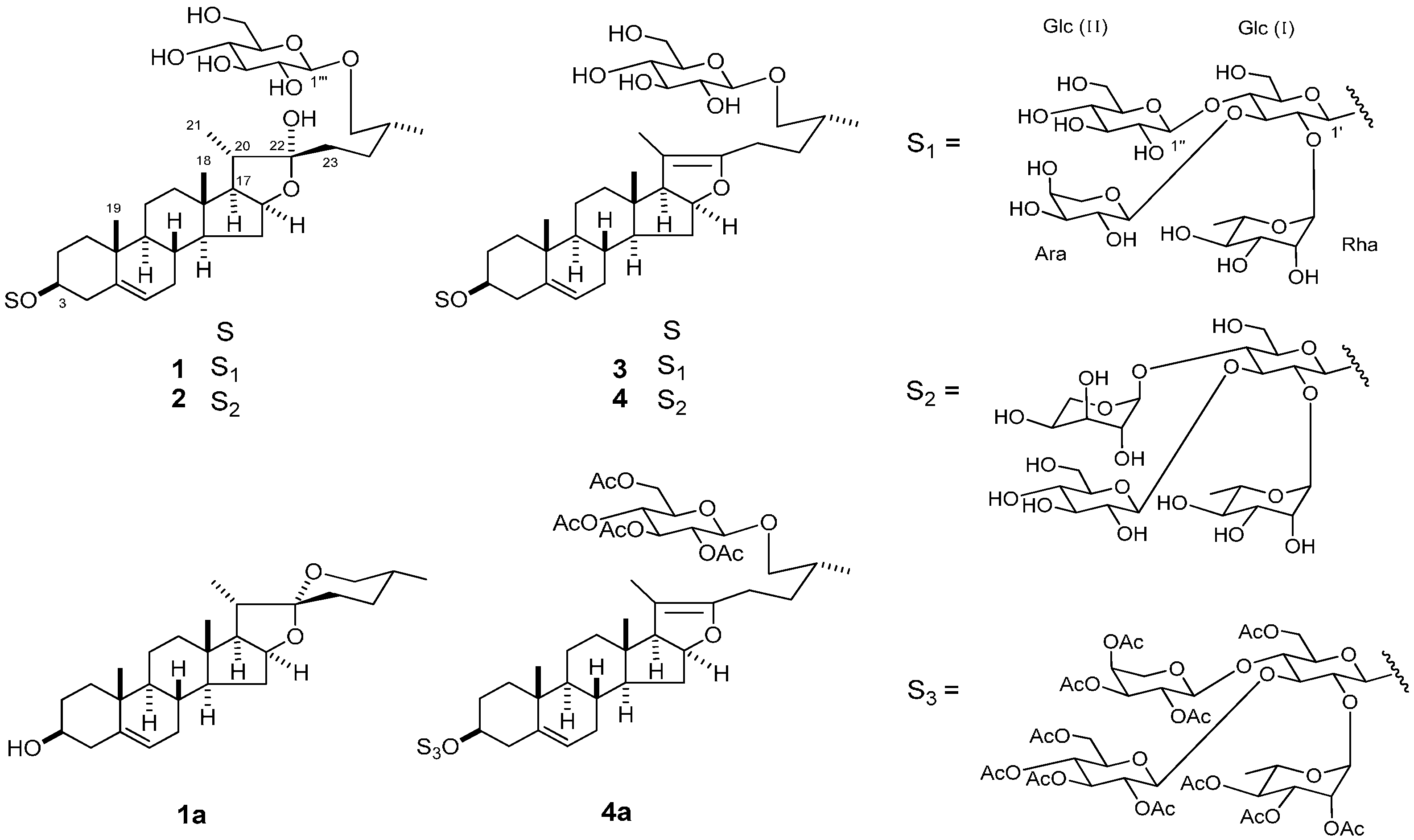
The bulbs of L. pumilum (1.4 kg fr. wt) were extracted with MeOH. The MeOH extract (65 g) was passed through a porous-polymer polystyrene resin (Diaion HP-20) column, and the MeOH-eluted fraction (1.8 g) was subjected to silica gel and octadecylsilanized (ODS) silica gel column chromatography (CC), giving 14 (Figure 1).
Figure 1. Steroidal glycosides from Lilium pumilum.
Molecules 20 16255 g001
Compound 1 was obtained as an amorphous solid, and its molecular formula was assigned as C56H92O27 based on data from high-resolution electrospray ionization time-of-flight mass spectrometry (HR-ESI-TOF-MS) (m/z 1219.5674 [M + Na]+, calcd for 1219.5724) and 13C-NMR (56 carbon signals) spectra. The 1H-NMR spectrum of 1 showed two singlet signals for tertiary methyl groups at δH 1.04 and 0.89 (each s), two doublet signals for secondary methyl groups at δH 1.34 (d, J = 6.9 Hz) and 1.00 (d, J = 6.7 Hz), and five anomeric proton signals at δH 6.07 (br s), 5.56 (d, J = 5.4 Hz), 5.39 (d, J = 7.8 Hz), 4.92 (d, J = 7.6 Hz), and 4.82 (d, J = 7.8 Hz). The 13C-NMR spectrum showed a signal for a hemiacetal carbon at δC 110.6, signals for five anomeric carbon at δC 104.9, 103.2, 103.0, 102.4, and 99.8, and signals for four steroid methyl groups at δC 19.4, 17.4, 16.4, and 16.3 as shown in Table 1, which were characteristic of 22-hydroxyfurostanol glycosides [6]. Acid hydrolysis of 1 with 1 M HCl yielded 1a as the aglycone and arabinose, glucose and rhamnose as the carbohydrate moieties. The monosaccharides and their absolute configurations were identified by direct HPLC analysis of the hydrolysate; l-arabinose, d-glucose and l-rhamnose, respectively. The aglycone (1a) was identified as (25R)-spirost-5-en-3β-ol (diosgenin) from its physical and spectroscopic data [7]. These NMR data, the chemical evidence, and the positive color reaction with Ehrlich’s reagent suggested that 1 was a 22-hydroxyprotodiosgenin bisdesmoside, the sugar moieties of which consisted of five monosaccharides. The 1H-1H COSY and 1D TOCSY spectra of 1 allowed the 1H-NMR chemical shifts, signal multiplet patterns, and coupling constants of the sugar moieties to be assigned as shown in Table 2. The 1H-NMR signals were associated with the corresponding one-bond coupled carbons using the HMQC and HSQC-TOCSY spectra, leading to the assignments of all the 13C-NMR chemical shifts of the sugar moieties. Comparing the 13C-NMR chemical shifts of each monosaccharide and reference methyl glycosides indicated the presence of a substituted β-d-glucopyranosyl (4C1) unit (Glc (I)), two β-d-glucopyranosyl (4C1) units (Glc (II) and Glc (III)), an α-l-arabinopyranosyl (4C1) unit (Ara), and an α-l-rhamnopyranosyl (1C4) unit (Rha) as the terminal glycosyl groups [8]. The 13C-NMR shifts of the inner Glc (I) moiety (δC 99.8, 78.9, 79.8, 72.5, 78.5, and 60.9) suggested that its C-2, C-3, and C-4 hydroxy groups were substituted with the other sugar moieties. The anomeric configurations of the Ara and Glc groups were ascertained as α and β, respectively, from the relatively large J values of their anomeric protons (5.4–7.8 Hz) [7]. For the Rha group, the 13C-NMR chemical shifts (δC 102.4, 72.4, 72.8, 73.8, 70.0, and 18.6) suggested an α-pyranoid anomeric form. The HMBC correlations between the anomeric proton (H-1) of Ara at δH 5.56 and C-3′ of Glc (I) at δC 79.8, between H-1′′ of Glc (II) at δH 5.39 and C-4′ of Glc (I) at δC 72.5, between H-1 of Rha at δH 6.07 and C-2′ of Glc (I) at δC 78.9, and between H-1′ of Glc (I) at δH 4.92 and C-3 of the aglycone at δC 77.9, indicated that the tribranched oligosaccharide of O-α-l-arabinopyranosyl-(1→3)-O-[β-d-glucopyranosyl-(1→4)]-O-[α-l-rhamnopyranosyl-(1→2)]-β-d-glucopyranosyl was linked to C-3 of the aglycone. A β-d-glucopyranosyl group (Glc (III)) linkage to the C-26 hydroxy group of the aglycone was confirmed by an HMBC correlation between H-1′′′ of Glc (III) at δH 4.82 (d, J = 7.8 Hz) and C-26 of the aglycone at δC 75.2. The NOE correlations between the signals of the H-20 proton at δH 2.21 and the H2-23 protons at δH 2.03 (2H) confirmed the C-22α configuration [9]. In the 1H-NMR spectrum, the difference chemical shifts of H2-26 geminal protons at δH 3.95 and δH 3.60, Δδ = 0.35 < 0.48 ppm, provided evidence for the (25R)-furostanol [10]. Thus, 1 was assigned as (25R)-26-[(β-d-glucopyranosyl)oxy]-22α-hydroxyfurost-5-en-3β-yl O-α-l-arabinopyranosyl-(1→3)-O-[β-d-glucopyranosyl-(1→4)]-O-[α-l-rhamnopyranosyl-(1→2)]-β-d-glucopyranoside.
Table 1. 13C-NMR spectral assignments for the aglycone moiety of 14 in C5D5N.
Position1234
137.537.137.537.6
229.929.929.929.9
377.978.077.877.9
439.739.738.638.6
5140.8140.8140.8140.7
6121.8121.8121.8121.6
732.432.432.432.2
831.631.631.431.5
950.350.350.350.2
1037.137.137.137.1
1121.021.121.221.2
1239.939.939.639.7
1340.740.743.443.4
1456.556.554.954.9
1532.132.234.434.5
1681.381.284.584.4
1763.864.164.564.5
1816.416.414.114.3
1919.419.319.419.5
2040.640.6103.6103.6
2116.316.411.711.7
22110.6110.6152.3152.4
2337.137.023.623.8
2428.328.331.431.5
2534.234.233.533.6
2675.275.275.275.3
2717.417.417.317.3
Table 2. 1H- and 13C-NMR spectral assignments for the sugar moieties of 14 in C5D5N.
PositionδHJ (Hz)δCPositionδHJ (Hz)δC
12
Glc (I)1′4.92d7.699.8Glc (I)1′4.85d8.099.8
2′4.17dd7.9, 7.678.92′4.47dd9.4, 8.079.4
3′4.57dd7.9, 7.679.83′4.33dd9.4, 8.174.2
4′4.58dd7.9, 7.972.54′4.12dd8.1, 8.179.4
5′4.20m78.55′3.72br d8.177.9
6′4.62br d12.460.96′4.79br d11.960.5
4.33br d12.44.35br d11.9
Rha16.07br s102.4Rha15.94br s102.6
24.75br d3.472.424.83br d3.672.3
34.53dd9.2, 3.472.834.51dd9.7, 3.672.7
44.35dd9.2, 9.273.844.56dd9.7, 9.773.7
54.86dq9.2, 6.370.054.82dq9.7, 6.370.0
61.76d6.318.661.75d6.318.6
Ara15.56d5.4103.2Glc (II)1′′5.49d7.8103.9
24.31dd6.6, 5.473.02′′4.15dd8.5, 7.874.8
34.19dd7.9, 6.675.53′′4.24dd8.8, 8.578.5
44.21m70.44′′4.20dd8.8, 8.771.6
54.67dd12.0, 3.565.65′′3.84br d8.878.5
3.69dd12.0, 4.16′′4.40m(2H)61.4
Glc (II)1′′5.39d7.8103.0Ara15.74d1.2102.3
2′′4.10dd7.9, 7.874.924.72dd4.2, 1.271.0
3′′4.19dd7.9, 7.777.934.35dd4.2, 4.273.3
4′′4.25dd7.7, 7.171.644.65m65.6
5′′3.87m78.455.02dd11.3. 10.562.1
6′′4.45br d12.862.03.77dd11.3. 4.2
4.35overlapping
Glc (III)1′′′4.82d7.8104.9Glc (III)1′′′4.81d7.8104.9
2′′′4.05dd8.0, 7.875.22′′′4.03dd8.3, 7.875.1
3′′′4.24dd8.0, 7.778.53′′′4.26dd9.1, 8.378.6
4′′′4.22dd8.9, 7.771.74′′′4.22dd9.1, 9.171.7
5′′′3.96m78.65′′′3.95br d9.178.7
6′′′4.58br d12.962.86′′′4.52br d12.962.8
4.38br d12.94.39br d12.9
34
Glc (I)1′4.91d7.699.8Glc (I)1′4.85d7.799.7
2′4.15dd7.9, 7.678.92′4.46dd9.5, 7.779.3
3′4.55dd8.1, 7.979.83′4.32dd9.5, 7.974.3
4′4.57dd8.1, 8.172.64′4.11dd7.9, 7.979.4
5′4.19m78.75′3.71m77.9
6′4.59br d12.761.46′4.78br d11.760.4
4.33br d12.74.33br d11.7
Rha16.05br s102.4Rha15.92br s102.6
24.77br d3.572.324.78br d3.872.4
34.52dd9.6, 3.572.834.49dd9.7, 3.872.7
44.34dd9.6, 9.273.844.55dd9.8, 9.773.7
54.81dq9.2, 6.370.054.80dd9.8, 6.270.0
61.75d6.318.661.73d6.218.6
Ara15.55d5.8103.2Glc (II)1′′5.47d7.9103.9
24.29dd6.5, 5.873.02′′4.13dd8.5, 7.974.7
34.21dd8.0, 6.575.53′′4.24dd9.5, 8.578.4
44.23m70.44′′4.22dd9.5, 9.571.7
54.66dd12.2, 3.965.55′′3.82m78.5
3.70br d12.26′′4.41m(2H)61.4
Glc (II)1′′5.37d7.9103.0Ara15.72br s102.2
2′′4.09dd7.9, 7.474.924.70dd4.6, 1.770.9
3′′4.19dd7.9, 7.477.934.54overlapping73.2
4′′4.24dd7.4, 7.172.344.64m65.7
5′′3.86m78.454.40dd11.5, 11.562.1
6′′4.34m(2H)62.03.76dd11.5, 4.5
Glc (III)1′′′4.82d7.8104.9Glc (III)1′′′4.83d7.9104.9
2′′′4.05dd8.1, 7.875.12′′′4.03dd7.9, 7.775.0
3′′′4.24dd8.1, 7.878.43′′′4.26dd9.4, 7.778.6
4′′′4.22dd8.1, 7.871.74′′′4.21dd9.4, 9.471.7
5′′′3.96m78.65′′′3.97m78.7
6′′′4.58br d12.962.86′′′4.56br d11.962.8
4.38br d12.94.40overlapping
Compound 2 had the same molecular formula as 1 of C56H92O27, based on the HR-ESI-TOF-MS and 13C-NMR (56 carbon signals) data. Two singlet signals for tertiary methyl groups at δH 1.04 and 0.89, two doublet signals for secondary methyl groups at δH 1.33 (d, J = 6.9 Hz) and 1.00 (d, J = 6.8 Hz), and five signals for anomeric protons at δH 5.94 (br s), 5.74 (d, J = 1.2 Hz), 5.49 (d, J = 7.8 Hz), 4.85 (d, J = 8.0 Hz), and 4.81 (d, J = 7.8 Hz) were observed in the 1H-NMR spectrum of 2. The 13C-NMR spectrum contained a signal for an acetal carbon at δC 110.6, and signals for four steroid methyl groups at δC 19.3, 17.4, 16.4, and 16.4. These spectroscopic data for 2 were analogous to those of 1, and suggested that 2 shared the same fundamental furostanol skeleton as 1. Acid hydrolysis of 2 gave 1a, l-arabinose, d-glucose, and l-rhamnose. Although the 1H- and 13C-NMR spectra of 2 indicated that the sugar moieties of 2 also consisted of two terminal β-d-glucopyranosyl units (Glc (II) and Glc (III)), a terminal α-l-arabinopyranosyl unit (Ara), a terminal α-l-rhamnopyranosyl unit (Rha), and a 2,3,4-trisubstituted inner β-d-glucopyranosyl moiety (Glc (I)), it was assumed that the linkage positions of the terminal sugar units to the inner Glc (I) moiety and the conformation of the Ara unit were different from those of 1. The 13C-NMR chemical shifts of C-1 to C-5 for Ara were assigned as δC 102.3, 71.0, 73.3, 65.6, and 62.1, respectively, by combining the 1H-1H COSY, HMQC, and HSQC-TOCSY spectra. Furthermore, the anomeric proton of Ara at δH 5.74 (d, J = 1.2 Hz) exhibited three-bond coupled strong HMBC correlations with the C-3 and C-5 carbons. These spectral features were consistent with the 1C4 conformation of Ara with an α-orientation of the anomeric center [11]. In the HMBC spectrum, correlation peaks between the H-1 of Ara at δH 5.74 and C-4′ of Glc (I) at δC 79.4, between H-1′′ of Glc (II) at δH 5.49 and C-3′ of Glc (I) at δC 74.2, between H-1 of Rha at δH 5.94 and C-2′ of Glc (I) at δC 79.4, and between H-1′ of Glc (I) at δH 4.85 and C-3 of the aglycone at δC 78.0 showed that Glc (II) and Ara were attached to C-3′ and C-4′ of Glc (I), respectively, in 2, which was different from 1. A β-d-glucopyranosyl group (Glc (III)) linkage to the C-26 hydroxy group of the aglycone was ascertained by the HMBC correlation between the H-1′′′ of Glc (III) at δH 4.81 (d, J = 7.8 Hz) and C-26 of the aglycone at δC 75.2. The NOE correlations between the signals of the H-20 proton at δH 2.22 and the H2-23 protons at δH 2.04 (2H) confirmed the C-22α configuration. Thus, 2 was elucidated as (25R)-26-[(β-d-glucopyranosyl)oxy]-22α-hydroxyfurost-5-en-3β-yl O-α-l-arabinopyranosyl-(1→4)-O-[β-d-glucopyranosyl-(1→3)]-O-[α-l-rhamnopyranosyl-(1→2)]-β-d-glucopyranoside.
Compound 3 was analyzed as C56H90O26 based on the HR-ESI-TOF-MS (m/z 1201.5615 [M + Na]+) and 13C-NMR (56 carbon signals) data. This molecular formula was smaller than that of 1 by 18.0059 (H2O). The 1H- and 13C-NMR spectral features of 3 were similar to those of 1; however, the Me-21 doublet signal at δH 1.34 (d, J = 6.9 Hz) in the 1H-NMR spectrum of 1 was replaced by a methyl singlet signal at δH 1.64 in that of 3, and an olefinic functionality (δC 152.3 and 103.6) was observed in addition to the 5(6)-ene group in the 13C-NMR spectrum of 3. These spectroscopic data and the HMBC correlations from H-17 at δH 2.45, H2-23 at δH 2.22, and Me-21 at δH 1.64, to C-22 at δC 152.3 and C-20 at δC 103.6 indicated that 3 was the corresponding Δ20(22)-pseudo-furostanol glycoside of 1. This was supported by all other spectroscopic data and the results of acid hydrolysis. The structure of 3 was assigned as (25R)-26-[(β-d-glucopyranosyl)oxy]-furosta-5,20(22)-dien-3β-yl O-α-l-arabinopyranosyl-(1→3)-O-[β-d-glucopyranosyl-(1→4)]-O-[α-l-rhamnopyranosyl-(1→2)]-β-d-glucopyranoside.
Compound 4 was shown to have a molecular formula of C56H90O26 by the HR-ESI-TOF-MS (m/z 1201.5653 [M + Na]+, calcd. 1201.5618) and 13C-NMR (56 carbon signals) data, and its aglycone was identified as the same Δ20(22)-pseudo-furostanol derivative as 3, based on its spectroscopic data. Further spectroscopic analysis and acid hydrolysis of 4 indicated that the sugar moieties attached to C-3 and C-26 of the aglycone were in agreement with those of 2, in which the terminal α-l-arabinopyranosyl group was present as an unusual 1C4 conformation. It is notable that the 1C4 α-l-arabinopyranosyl moiety in 4 was converted to the 4C1 moiety on peracetylation (4a). Therefore, 4 was the corresponding Δ20(22)-pseudo-furostanol glycoside of 2, (25R)-26-[(β-d-glucopyranosyl)oxy]-furosta-5,20 (22)-dien-3β-yl O-α-l-arabinopyranosyl-(1→4)-O-[β-d-glucopyranosyl-(1→3)]-O-[α-l-rhamnopyranosyl-(1→2)]-β-d-glucopyranoside.
The isolated compounds 14 were evaluated for their cytotoxic activity against HL-60 cells. They were not cytotoxic to HL-60 cells at sample concentrations up to 15 μM.

3. Experimental Section

3.1. General Experimental Procedures

Optical rotations were measured by using an automatic digital polarimeter (P-1030, Jasco, Tokyo, Japan). IR spectra were recorded on a spectrophotometer (FT-IR 620, Jasco, Tokyo, Japan). 1H-NMR spectra were obtained at 500 MHz by using standard Bruker pulse programs at 300 K (DRX-500, Bruker, Karlsruhe, Germany). Chemical shifts are given as δ values relative to tetramethylsilane (TMS, Wako, Osaka, Japan) used as an internal standard. HR-ESI-TOF-MS data were recorded on an LCT mass spectrometer (Waters-Micromass, Manchester, UK). Diaion HP-20 (50 mesh, Mitsubishi-Chemical, Tokyo, Japan), BW-300 silica gel (200-300 mesh, Fuji Silysia Chemical, Aichi, Japan), and COSMOSIL 75C18-OPN ODS silica gel (75 μM, Nacalai Tesque, Kyoto, Japan) were used for CC. TLC was carried out on precoated silica gel 60 F254 (0.25 mm thick, Merck, Darmstadt, Germany) and RP18 F254S plates (0.25 mm thick, Merck), and the compounds were visualized by spraying the plates with 10% H2SO4 (aq.) and then heating. HPLC was performed with a system consisting of a CCPM pump (Tosoh, Tokyo, Japan), an RI-8021 detector (Tosoh), and a Rheodyne injection port (Rohnert Park, CA, USA). A TSK gel ODS-100Z column (10 mm i.d. × 250 mm, 5 μm, Tosoh) was used for preparative HPLC. NMR spectra are available in the Supplementary materials.

3.2. Plant Material

The L. pumilum bulbs were obtained from the Sakata Seed Corporation (Yokohama, Japan) in 2008 and were identified by Dr. Yutaka Sashida, professor emeritus of Tokyo University of Pharmacy and Life Sciences. We have retained a voucher specimen in our laboratory (KS-2008-002).

3.3. Extraction and Isolation

The bulbs of L. pumilum (1.4 kg fr. weight) were extracted with MeOH under reflux for 6 h. After removing the solvent, the MeOH extract (65 g) was passed through a Diaion HP-20 column (2000 g, 8.5 cm i.d. × 60 cm) and successively eluted with MeOH–H2O (3:7, 1:1), MeOH, EtOH, and EtOAc (6 L of each eluent). CC of the MeOH-eluted fraction (1.8 g) on silica gel (2000 g, 8.0 cm i.d. × 30 cm), eluted with a stepwise gradient mixture of CHCl3–MeOH–H2O (7:4:1) and finally with MeOH, provided 6 fractions (M-1 to M-6). Fraction M-4 (0.5 g) was chromatographed on ODS silica gel (700 g, 4.0 cm i.d. × 25 cm) eluted with MeOH–H2O (2:1) and finally with MeOH, provided 9 fractions (M-4-1–M-4-9). Fraction M-4-5 (148 mg) was separated by HPLC (1.0 cm i.d. × 25 cm) using MeCN–H2O (1:3) to afford 3 (14.3 mg) and 4 (15.0 mg). Fraction M-4-7 (94 mg) was separated by HPLC (1.0 cm i.d. × 25 cm) using MeCN–H2O (1:2) to afford 1 (20.6 mg) and 2 (20.6 mg).

3.4. Acid Hydrolysis of 1, 2, 3, or 4

A solution of 1 (14.5 mg), 2 (14.5 mg), 3 (5.0 mg), or 4 (5.0 mg) in 1 M HCl in dioxane–H2O (1:1; 2.0 mL) were heated at 90 °C for 2 h under an Ar atmosphere, respectively. The reaction mixture was neturalized by passing it through an Amberlite column (IRA-96SB, Organo, Tokyo, Japan; 16–50 mesh, 50 g, 1.5 cm i.d. × 15 cm). The mixture was then eluted through a Diaion HP-20 column (50 g, 1.5 cm i.d. × 15 cm) with MeOH–H2O (3:7) and EtOH–Me2CO (1:1). The EtOH–Me2CO (1:1) fraction was purified by CC with CHCl3–MeOH–H2O (7:4:1) to give diosgenin (1a; 1.9 mg; 0.2 mg; 1.1 mg; and 5.0 mg), respectively. The MeOH–H2O (3:7) fraction was analyzed by HPLC under the following conditions: Capcell Pak NH2 UG80 column (4.6 mm i.d. × 25 cm, 5 μm, Shiseido, Tokyo, Japan); mobile phase of MeCN–H2O (85:15); detection by refractive index and optical rotation; and a flow rate of 1.0 mL/min. l-Arabinose, d-glucose, and l-rhamnose were identified, respectively, by comparing their retention times (tR) and optical rotation with those of authentic samples (l-arabinose 7.00 min, positive optical rotation; d-glucose 12.28 min, positive optical rotation; l-rhamnose 6.65 min, negative optical rotation): l-arabinose (6.85 min; 6.89 min; 6.77 min; 8.00 min; positive optical rotation), d-glucose (12.25 min; 12.74 min; 12.33 min; 12.74 min; positive optical rotation), and l-rhamnose (6.56 min; 6.60 min; 6.60 min; 6.97 min; negative optical rotation).

3.5. Acetylation of 4

A mixture of 4 (8.0 mg) and Ac2O (1.0 mL) in pyridine (7.0 mL) was stirred at room temperature for 24 h. The reaction mixture was purified by CC with CHCl3–EtOAc (1:1) to give the corresponding peracetylation (4a, 3.2 mg).

3.6. Data for 14 and 4a

(25R)-26-[(β-d-Glucopyranosyl)oxy]-22α-hydroxyfurost-5-en-3β-yl O-α-L-arabinopyranosyl-(1→3)-O-[β-d-glucopyranosyl-(1→4)]-O-[α-l-rhamnopyranosyl-(1→2)]-β-d-glucopyranoside (1); An amorphous solid; [ α ] D 25 −54.2 (c 0.10, MeOH); HR-ESI-TOF-MS (m/z: 1219.5674 [M + Na]+, calcd for C56H92NaO27: 1219.5724); IR νmax (film) cm−1: 3388 (OH), 2931 (CH); 1H-NMR (500 MHz, C5D5N): for the aglycone moiety, 5.33 (1H, br s, H-6), 4.96 (1H, br d, J = 6.8 Hz, H-16), 3.95 (1H, dd, J = 9.5, 7.1 Hz , H-26a), 3.86 (1H, m, W1/2 = 21.6 Hz, H-3), 3.60 (1H, dd, J = 9.5, 7.8 Hz, H-26b), 1.34 (3H, d, J = 6.9 Hz, Me-21), 1.04 (3H, s, Me-19), 1.00 (3H, d, J = 6.7 Hz, Me-27), 0.89 (3H, s, Me-18). For the sugar moieties, Table 2; 13C-NMR (125 MHz, C5D5N): Table 1 and Table 2.
(25R)-26-[(β-d-Glucopyranosyl)oxy]-22α-hydroxyfurost-5-en-3β-yl O-α-l-arabinopyranosyl-(1→4)-O-[β-d-glucopyranosyl-(1→3)]-O-[α-l-rhamnopyranosyl-(1→2)]-β-d-glucopyranoside (2); An amorphous solid; [ α ] D 25 −59.6 (c 0.10, MeOH); HR-ESI-TOF-MS (m/z: 1219.5693 [M + Na]+, calcd for C56H92NaO27: 1219.5724); IR νmax (film) cm−1: 3388 (OH), 2926 (CH). 1H-NMR (500 MHz, C5D5N): for the aglycone moiety, 5.33 (1H, br s, H-6), 4.95 (1H, br d, J = 7.5 Hz, H-16), 3.96 (1H, dd, J = 8.5, 7.0 Hz , H-26a), 3.84 (1H, overlapping, H-3), 3.59 (1H, dd, J = 8.5, 4.0 Hz, H-26b), 1.33 (3H, d, J = 6.9 Hz, Me-21), 1.04 (3H, s, Me-19), 1.00 (3H, d, J = 6.8 Hz, Me-27), 0.89 (3H, s, Me-18). For the sugar moieties, Table 2; 13C-NMR (125 MHz, C5D5N): Table 1 and Table 2.
(25R)-26-[(β-d-Glucopyranosyl)oxy]-furosta-5,20(22)-dien-3β-yl O-α-l-arabinopyranosyl-(1→3)-O-[β-d-glucopyranosyl-(1→4)]-O-[α-l-rhamnopyranosyl-(1→2)]-β-d-glucopyranoside (3); An amorphous solid; [ α ] D 25 −42.6 (c 0.04, MeOH); HR-ESI-TOF-MS (m/z: 1201.5615 [M + Na]+, calcd for C56H90NaO26: 1201.5618); IR νmax (film) cm−1: 3326 (OH), 2925 (CH); 1H-NMR (500 MHz, C5D5N): for the aglycone moiety, 5.33 (1H, br d, J = 4.4 Hz, H-6), 4.75 (1H, m, H-16), 3.95 (1H, dd, J = 9.0, 6.9 Hz, H-26b), 3.84 (1H, m, W1/2 = 25.3 Hz, H-3), 3.66 (1H, br d, J = 9.0 Hz, H-26a), 1.64 (3H, s, Me-21), 1.05 (3H, s, Me-19), 1.02 (3H, d, J = 6.7 Hz, Me-27), 0.73 (3H, s, Me-18). For the sugar moieties, Table 2; 13C-NMR (125 MHz, C5D5N): Table 1 and Table 2.
(25R)-26-[(β-d-Glucopyranosyl)oxy]-furosta-5,20(22)-dien-3β-yl O-α-l-arabinopyranosyl-(1→4)-O-[β-d-glucopyranosyl-(1→3)]-O-[α-l-rhamnopyranosyl-(1→2)]-β-d-glucopyranoside (4); An amorphous solid; [ α ] D 25 −51.7 (c 0.03, MeOH); HR-ESI-TOF-MS (m/z: 1201.5653 [M + Na]+ calcd for C56H90NaO26, 1201.5618); IR νmax (film) cm−1: 3357 (OH), 2925 (CH); 1H-NMR (500 MHz, C5D5N): for the aglycone moiety, 5.33 (1H, br d, J = 4.3 Hz, H-6), 4.75 (1H, m, H-16), 3.94 (1H, dd, J = 9.0, 7.0 Hz, H-26a), 3.83 (1H, m, W1/2 = 27.5 Hz, H-3), 3.47 (1H, dd, J = 9.0, 6.1 Hz, H-26b), 1.63 (3H, s, Me-21), 1.04 (3H, s, Me-19), 1.01 (3H, d, J = 6.7 Hz, Me-27), 0.72 (3H, s, Me-18). For the sugar moieties, Table 2; 13C-NMR (125 MHz, C5D5N): Table 1 and Table 2.
Compound 4a; An amorphous solid; [ α ] D 25 −33.0 (c 0.16, MeOH); HR-ESI-TOF-MS (m/z: 1831.7181 [M + ONa]+, calcd for C86H120NaO41: 1831.7203); IR νmax (film) cm1: 2960 and 2924 (CH), 1747 (C=O); 1H-NMR (500 MHz, C5D5N): δH 5.50 (1H, br d, J = 1.5 Hz, H-6), 3.90 (1H, m, overlapping, H-3), 3.89 (1H, dd, J = 9.4, 5.9 Hz, H-26a), 3.47 (1H, dd, J = 9.4, 6.1 Hz, H-26b), 0.94 (3H, d, J = 6.6 Hz, Me-27), 1.11 (3H, s, Me-19), 0.78 (3H, s, Me-18), 5.71 (1H, br s, H-1 of Rha), 5.11 (1H, d, J = 6.5 Hz, H-1 of Ara), 5.18 (1H, d, J = 7.8 Hz, H-1′′ of Glc (II)), 4.92 (1H, d, J = 8.0 Hz, H-1′′′ of Glc (III)), 4.89 (1H, d, J = 7.7 Hz, H-1′ of Glc (I)), 1.47 (3H, d, J = 6.2 Hz, H-6 of Rha), 2.26, 2.24, 2.22, 2.20, 2.16, 2.06, 2.04 × 2, 2.03, 2.02 × 2, 2.00, 1.98 × 2, 1.91 (each 3H, s, Ac × 15); 13C-NMR (125 MHz, C5D5N) : δC 152.3 (C-22), 140.4 (C-5), 122.3 (C-6), 84.5 (C-16), 78.5 (C-3), 19.3 (C-19), 16.7 (C-27), 14.1 (C-18), 103.7 (C-20), 11.7 (C-21), 101.4 (C-1′′′ of Glc (III)), 101.2 (C-1 of Ara), 99.9 (C-1′′ of Glc (II)), 99.0 (C-1′ of Glc (I)), 97.8 (C-1 of Rha), 78.6 (C-3′ of Glc (I)), 77.6 (C-2′ of Glc (I)), 76.8 (C-4′ of Glc (I)), 63.1 (C-5 of Ara), 63.1 (C-6′ of Glc (I)), 62.4 (C-6′′′ of Glc (III)), 61.6 (C-6′′of Glc (II)), 17.5 (C-6 of Rha).

3.7. Cell Culture Assay

Cell growth was measured with an MTT reduction assay, as described in a previous paper [12].

4. Conclusions

In conclusion, four new steroidal glycosides (14) were isolated from the bulbs of L. pumilum, and they were classified into furostanol glycosides (1 and 2) and Δ20(22)-pseudo-furostanol glycosides (3 and 4). Compounds 14 are novel steroidal glycosides with a 2,3,4-trisubstituted β-d-glucopyranosyl moiety at C-3 of the aglycone. In general, α-l-arabinopyranosyl groups are stable in a 4C1 conformation in glycosides, except for those glycosylated at C-2 [13]. In 1 and 3, the α-l-arabinopyranosyl moiety is linked to C-3 of the inner trisubstituted β-d-glucopyranosyl group and is present as an usual 4C1 conformation. In contrast, in 2 and 4, the α-l-arabinopyranosyl moiety, which is attached to C-4 of the inner trisubstituted β-d-glucopyranosyl group, is present as a 1C4 conformation. It is notable that the 1C4 α-l-arabinopyranosyl moiety in 4 was converted to the 4C1 moiety on peracetylation (4a). These interesting steric behaviors of arabinopyranose cannot be explained by steric hindrance only. In a recent study, there are reports of the inner substituted 1C4 conformation of α-l-arabinopyranosyl moiety in triterpene glycoside [14,15]. Many more examples and data need to be collected in future work.

Supplementary Materials

Supplementary materials can be accessed at: https://www.mdpi.com/1420-3049/20/09/16255/s1.

Acknowledgments

We are grateful to C. Sakuma, Tokyo University of Pharmacy and Life Sciences, for the NMR spectra.

Author Contributions

Yoshihiro Mimaki conceived and designed the experiments. Reina Takaku performed the experiments. Reina Takaku and Yukiko Matsuo analyzed the spectroscopic data and determined the chemical structures. Yukiko Matsuo and Yoshihiro Mimaki wrote the paper.

Conflicts of Interest

The authors declare no conflict of interest.

References

  1. Sashida, Y. Lily. J. Toyaku 2011, 33, 32–37. [Google Scholar]
  2. Mimaki, Y.; Sashida, Y.; Shimomura, H. Lipid and steroidal constituents of Lilium auratum var. platyphyllum and L. tenuifolium. Phytochemistry 1989, 28, 3453–3458. [Google Scholar] [CrossRef]
  3. Kubo, S.; Mimaki, Y.; Terao, M.; Sashida, Y.; Nikaido, T.; Ohmoto, T. Acylated cholestane glycosides from the bulbs of Ornithogalum saundersiae. Phytochemistry 1992, 31, 3969–3973. [Google Scholar] [CrossRef]
  4. Kuroda, M.; Mimaki, Y.; Sashida, Y.; Yamori, T.; Tsuruo, T. Galtonioside A, a novel cytotoxic cholestane glycoside from Galtonia candicans. Tetrahedron Lett. 2000, 41, 251–255. [Google Scholar] [CrossRef]
  5. Zhou, Z.L.; Feng, Z.C.; Fu, C.Y.; Zhang, H.L.; Xia, J.M. Steroidal and phenolic glycosides from the bulbs of Lilium pumilum DC and their potential Na+/K+ ATPase inhibitory activity. Molecules 2012, 17, 10494–10502. [Google Scholar] [CrossRef] [PubMed]
  6. Matsuo, Y.; Shinoda, D.; Nakamaru, A.; Mimaki, Y. Steroidal glycosides from the bulbs of Fritillaria meleagris and their cytotoxic activities. Steroids 2013, 78, 670–682. [Google Scholar] [CrossRef] [PubMed]
  7. Agrawal, P.K.; Jain, D.C.; Gupta, R.K.; Thakur, R.S. Carbon-13 NMR spectroscopy of steroidal sapogenins and steroidal saponins. Phytochemistry 1985, 24, 2479–2496. [Google Scholar] [CrossRef]
  8. Agrawal, P.K. NMR spectroscopy in the structural elucidation of oligosaccharides and glycosides. Phytochemistry 1992, 31, 3307–3330. [Google Scholar] [CrossRef]
  9. Fattorusso, E.; Iorizzi, M.; Lanzotti, V.; Taglialatela-Scafati, O. Chemical composition of shallot (Allium ascalonicum Hort.). J. Agric. Food Chem. 2002, 50, 5686–5690. [Google Scholar] [CrossRef] [PubMed]
  10. Agrawal, P.K. Assigning stereodiversity of the 27-Me group of furostane-type steroidal saponins via NMR chemical shifts. Steroids 2005, 70, 715–724. [Google Scholar] [CrossRef] [PubMed]
  11. Ishii, H.; Kitagawa, I.; Matsushita, K.; Shirakawa, K.; Tori, K.; Tozyo, T.; Yoshimura, Y. The configuration and conformation of the arabinose moiety in platycodins, saponins isolated from platycodongrandiflorum, and mi-saponins from madhucalongifolia based on carbon-13 and hydrogen-1 NMR spectroscopic evidence: The total structures of the saponins. Tetrahedron Lett. 1981, 22, 1529–1532. [Google Scholar]
  12. Matsuo, Y.; Mimaki, Y. Lignans from Santalum album and their cytotoxic activities. Chem. Pharm. Bull. 2010, 58, 587–590. [Google Scholar] [CrossRef] [PubMed]
  13. Kuroda, M.; Mimaki, Y.; Ori, K.; Koshino, H.; Nukada, T.; Sakagami, H.; Sashida, Y. Lucilianosides A and B, two novel tetranor-lanostane hexaglycosides from the bulbs of Chionodoxa luciliae. Tetrahedron 2002, 58, 6735–6740. [Google Scholar] [CrossRef]
  14. Guo, T.; Liu, Q.; Zhang, L.; Wang, P.; Li, Y. Facile synthesis of four natural triterpene saponins with important antitumor activity. Synth. Commun. 2011, 41, 357–371. [Google Scholar] [CrossRef]
  15. Ren, L.; Liu, Y.X.; Lv, D.; Yan, M.C.; Nie, H.; Liu, Y.; Cheng, M.S. Facile synthesis of the naturally cytotoxic triterpenoid saponin patrinia-glycoside B-II and its conformer. Molecules 2013, 18, 15193–15206. [Google Scholar] [CrossRef] [PubMed]
  • Sample Availability: Samples of the compounds are not available.