Article

Chemical Constituents and Antibacterial Properties of Indocalamus latifolius McClure Leaves, the Packaging Material for “Zongzi”

State Forestry Administration Key Open Laboratory, International Centre for Bamboo and Rattan, Beijing 100102, China
*
Author to whom correspondence should be addressed.
These authors contributed equally to this work.
Molecules 2015, 20(9), 15686-15700; https://doi.org/10.3390/molecules200915686
Submission received: 27 July 2015 / Revised: 20 August 2015 / Accepted: 24 August 2015 / Published: 28 August 2015
(This article belongs to the Section Natural Products Chemistry)

Abstract

:
The glutinous rice dumpling named “Zongzi” in Chinese is a type of traditional food that is popular in East Asian countries. “Zongzi” is made of glutinous rice and wrapped in the leaves of Indocalamus latifolius McClure as the packaging material. Four new compounds, latifoliusine A (2), (7S,8R) syringylglycerol-8-O-4′-sinapyl ether 4-O-β-d-glucopyranoside (7), (7S,8S) syringylglycerol-8-O-4′-sinapyl ether 7-O-β-d-glucopyranoside (8), and (7R,8S) syringylglycerol-8-O-4′-sinapyl ether 7-O-β-d-glucopyranoside (10), along with six known compounds (1, 36 and 9) were isolated from I. latifolius McClure leaves. The structures and relative configurations of the compounds were determined by detailed spectroscopic analysis, high-resolution electrospray ionization mass spectroscopy (HRESIMS), heteronuclear single quantum correlation (HSQC), heteronuclear multiple bond correlation (HMBC), nuclear overhauser enhancement (NOE) and circular dichroism (CD). All of the isolated compounds were screened for their antibacterial activities in vitro. The results indicated that apigenin 6-C-α-l-arabinopyranosyl-8-C-β-d-glucopyranoside (5) and apigenin 7-O,8-C-di-glucopyranoside (6) have antibacterial activities against four bacterial strains (Staphylococcus aureus, Bacillus thuringiensis, Escherichia coli and Pseudomonas solanacearum).

1. Introduction

“Zongzi”, which is believed to have a history of more than 2000 years, is a type of famous Chinese food that is also popular in many Asian countries [1]. It is made of glutinous rice and wrapped in the large flat leaves of Indocalamus latifolius McClure. “Zongzi” has been characterized by a long shelf life since ancient times.
Indocalamus latifolius McClure is widely distributed and cultivated in Southern China [2]. It belongs to the same genus as Indocalamus nakai, which is reported to have polysaccharides [3,4], metal elements [1], flavonoids [5,6], and volatile components [7] in its leaf extracts and possess anticancer, antitumor, and antioxidative effects, as well as antibacterial activity [8,9].
In our previous research, several new compounds have been identified from the leaves of different bamboo species including the following: Three novel lignans were isolated from Bambusa tuldoides Munro [10]; a new polyketide derivative named Amarusine A was isolated from the leaves of Pleioblastus amarus [11]; two new compounds, xylitol 1-O-(6′-O-p-hydroxylbenzoyl)-glucopyranoside and bambulignan B, were isolated from the leaves of Pleioblastus amarus (Keng) keng f [12]; and four diastereoisomeric oxyneolignans were isolated and characterized from Bambusa tuldoides Munro [13]. In the present research, on the basis of our continuing research interest in the phytochemistry of bamboo, we examined the phytoconstituents of I. latifolius McClure leaves in detail and their antibacterial activities against two Gram-positive and two Gram-negative bacterial strains for the first time.

2. Results and Discussion

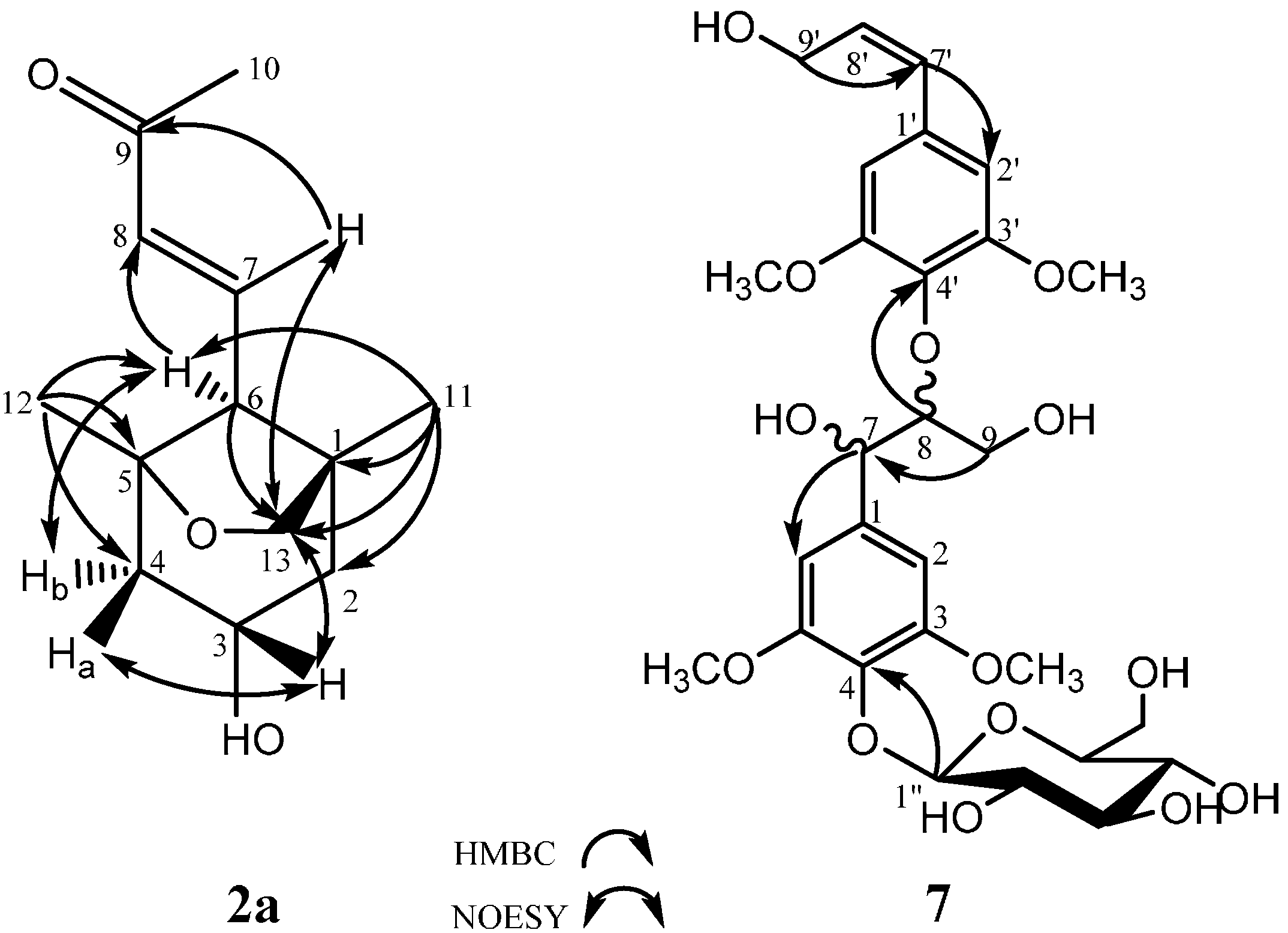
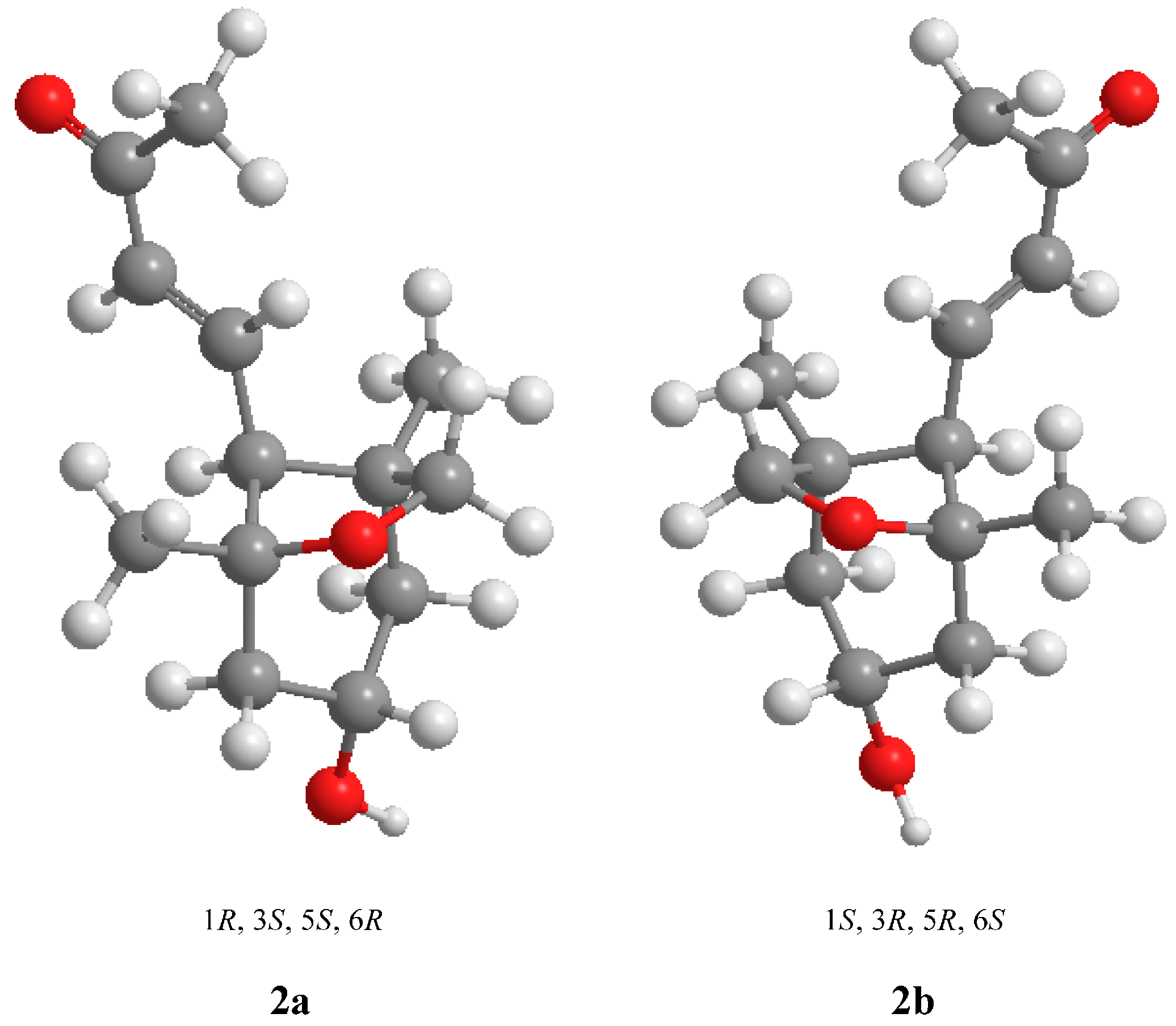
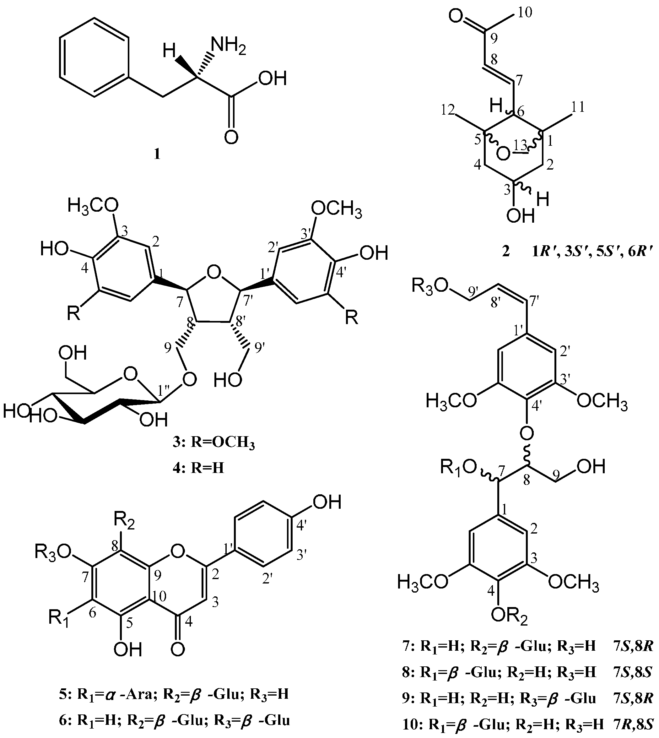
2.1. Structural Elucidation

Repeated chromatography over Sephadex LH-20, macroporous resin and Rp-18 columns as well as preparative HPLC of the 95% ethanol extract from I. latifolius McClure leaves led to the isolation of four new compounds, latifoliusine A (2), (7S,8R) syringylglycerol-8-O-4′-sinapyl ether 4-O-β-d-glucopyranoside (7), (7S,8S) syringylglycerol-8-O-4′-sinapyl ether 7-O-β-d-glucopyranoside (8) and (7R,8S) syringylglycerol-8-O-4′-sinapyl ether 7-O-β-d-glucopyranoside (10) along with six known compounds.
The six known compounds were identified (Figure 1) as L-phenylalanine (1) [14], dihydroxymethyl-bis(3,5-dimethoxy-4-hydroxyphenyl) tetrahydrofuran-9-O-β-d-glucopyranoside (3) [15], rel-(7R,8S,7′S,8′R)-4,9,4′,9′-tetrahydroxy-3,3′-dimethoxy-7,7′-epoxylignan 9-O-β-d-glucopyranoside (4) [16], apigenin 6-C-α-l-arabinopyranosyl-8-C-β-d-glucopyranoside (5) [17], apigenin 7-O,8-C-di-glucopyranoside (6) [18], and (7S,8S) syringylglycerol-8-O-4′-sinapyl ether 9′-O-β-d-glucopyranoside (9) [19] through comparing their spectroscopic and physical data with those of previous reports.
New compound 2 was purified as a yellowish oil ([α]D = +36.1°; c = 0.70, methanol), and its molecular formula, C13H20O3, was determined by positive HRESIMS (m/z 247.1313 [M + Na]+, calculated 247.1310) and suggests four degrees of unsaturation. The IR spectrum showed characteristic hydroxyl (3424 cm−1), methylene (2928 cm−1) and double bond (1670 cm−1) absorption bands. The 1H-NMR spectrum indicated the presence of one trans-double bond, as supported by hydrogen signals at δH 6.67 (1H, dd, J = 16.0, 11.0) and δH 6.12 (1H, d, J = 16.0). Additionally, one oxymethine at δH 3.91 (1H, m); one oxymethylene at δH 3.64 (2H, m); two methylenes at δHa 1.87 and δHb 1.31 (1H, dd, J = 12.5, 6.0) and at δHa 1.94 and δHb 1.36 (1H, dd, J = 12.5, 6.5); and three methylenes at δH 2.24 (3H, s), δH 1.02 (3H, s) and δH 0.85 (3H, s) were observed in the 1H-NMR spectrum, as well as active hydrogen signals at δH 4.58 (1H, s). The 13C-NMR spectrum revealed the resonances of thirteen carbons. Based on the DEPT spectrum, these resonances included one ketone carbon at δC 198.1; two olefinic carbons at δC 134.1 and δC 146.1; two quaternary carbons at δC 83.8 and δC 45.1; two methines at δC 64.7 and δC 60.6; three methylenes at δC 75.9, δC 48.7 and δC 47.8; and three methyls at δC 27.2, δC 23.9 and δC 20.6. Using the analysis of the degree of unsaturation, these data indicated that compound 2 contained two rings. In the HMBC spectrum (Figure 2), correlations of δ 0.85 (H-11) with δC 45.1 (C-1), δC 47.8 (C-2), δC 60.6 (C-6) and δC 75.9 (C-13), and δC 1.02 (H-12) with δC 48.7 (C-4), δC 83.8 (C-5) and δC 60.6 (C-6) were observed, suggesting that δC 45.1 (C-1) and δC 83.8 (C-5) were the bridgehead carbons of a furan ring (C-13, C-1, C-6 and C-5) and a six-membered ring (C-1, C-2, C-3, C-4, C-5 and C-6), respectively. Furthermore, the protons of the double-bond, δH 6.12 (H-7) and δH 6.67 (H-8), were correlated to δH 60.6 (C-6) and δH 198.1 (C-9), which confirmed that the butenone group was linked to C-6 (Figure 2). The relative configuration was further determined by NOESY correlations between δ 3.91 (H-3) and both δH 3.64 (H-13) and δH 1.94 (H-4a) and between δH 2.29 (H-6) and δH 1.36 (H-4b), confirming that H-3 and H-13 were on the same side of the molecule and that H-6 was positioned on the other side of the molecule (Figure 2). Based on these data, the pair of enantiomers of 1R′,3S′,5S′,6R′ (2a: 1R,3S,5S,6R and 2b: 1S,3R,5R,6S) was determined to be the relative configuration for compound 2 (Figure 3). Thus, the structure of compound 2 was elucidated as depicted and named latifoliusine A (Figure 1).
Figure 1. Compounds 110 isolated from Indocalamus latifolius McClure leaves.
Molecules 20 15686 g001
Figure 2. Significant HMBC and NOESY correlations of compounds 2a and 7.
Molecules 20 15686 g002
Figure 3. The relative configuration of compound 2.
Molecules 20 15686 g003
New compound 7 was obtained as a yellow amorphous powder ([α]D = −15.9°; c = 1.0, methanol). Its molecular formula, C28H38O14, was established by negative HRESIMS (m/z 597.2179 [M − H], calculated 597.2183). The IR spectrum showed absorption bands characteristic of hydroxyl groups (3381 cm−1), methylenes (2927 cm−1) and aromatic rings (1654 and 1451 cm−1). The 1H NMR spectrum exhibited signals for two 3,5-dimethoxy-4-hydroxyphenyl moieties, which included two aromatic hydrogen signals at δH 6.74 (2H, s) and δH 6.70 (2H, s); four methoxyl groups at δH 3.73 (6H, s) and δH 3.72 (6H, s); one trans double bond at δH 6.47 (1H, d, J = 16.0) and δH 6.34 (1H, dt, J = 16.0, 5.0); and one anomeric proton at δH 4.86 (1H, d, J = 7.5), indicating a β-glycosidic linkage for the d-glucose [10,20,21]. Moreover, there were other alkyl groups and signals attributed to a β-d-glucopyranosyl unit. The 13C-NMR spectra showed carbon signals corresponding to the chemical units described above and confirmed their presence. Furthermore, two oxymethines at δC 71.1 and δC 86.7 and an oxymethylene at δC 60.5 were attributed to an arylglyceryloxy unit. These spectral features indicated that compound 7 was an 8-O-4′-type neolignan glycoside formed by two phenylpropanoid glycosides. In the HMBC spectrum, the correlation between δH 4.09 (H-8) and δC 135.8 (C-4′) confirmed the neolignan structures, and the correlation between δH 4.86 (H-1′′) and δC 138.4 (C-4) implied that the O-glycoside was linked to δC 138.4 (C-4) (Figure 2). The erythro- configuration of 7 at C-7 and C-8 was determined by the J7,8-value (4.0 Hz) in the 1H-NMR spectrum [22,23]. The absolute configuration at C-7 and C-8 of compound 7 was assigned to be (7S,8R) based on the negative Cotton effect (ε243.5nm = −0.4727) in the CD spectrum [24,25]. Therefore, the structure of 7 was determined to be (7S,8R) syringylglycerol-8-O-4′-sinapyl ether 4-O-β-d-glucopyranoside (Figure 1).
New compound 8 was obtained as a yellow powder ([α]D = −3.11°; c = 1.0, methanol), and its molecular formula was determined to be C28H38O14 by negative HRESIMS (m/z 633.1952 [M + Cl], calculated 633.1950). The IR spectrum showed absorption bands characteristic for hydroxyl groups (3385 cm−1), methylenes (2927 cm−1) and aromatic rings (1584 and 1460 cm−1). The 1H NMR and 13C-NMR spectra of compound 8 also exhibited signals characteristic of an 8-O-4′-type neolignan glycoside. In the HSQC spectrum, an anomeric proton signal at δH 4.35 (1H, d, J = 7.5) correlated with a corresponding carbon signal at δC 102.6 in the 1H- and 13C-NMR spectra, respectively, which suggested that compound 8 had one terminal β-d-glucopyranosyl unit. In the HMBC analysis, the presence of a cross-peak between the anomeric proton at δH 4.35 (H-1″) and δC 79.0 (C-7) revealed the location of the glucosidic linkage at the C-7 position. In the 1H- and 13C-NMR spectra (Table 1), the signals of 8 were similar to the planar structure of 3-hydroxy-1-(4-hydroxy-3,5-dimethoxyphenyl)-2-[4-(3-hydroxy-1-(E)-propenyl)-2,6-dimethoxyphenoxy]propyl-β-d-glucopyranoside [26]. However, the J7,8 value was not directly applicable to distinguish the erythro- and threo- forms of 8-4′-oxyneolignan arylglycerol 7-O-β-d-glucopyranosides [13,27]. Therefore, the configuration of 8 will be further elucidated with the description of compound 10.
New compound 10 was also obtained as a yellow powder ([α]D = +4.55°; c = 1.0, methanol). The molecular formula of 10, C28H38O14, was confirmed by negative HRESIMS (m/z 633.1953 [M + Cl], calculated 633.1950) and coincided with that of 8. The IR and UV spectra of 8 and 10 showed similar absorption patterns. The 1H- and 13C-NMR spectra of 10 were very similar to those of 8, suggesting that the overall structure of 10 was the same as that of 8. Moreover, the HSQC and HMBC correlations of 10 corroborated the aforementioned deduction.
By comparing the 1H- and 13C-NMR spectral data of 8 and 10, there were several differences in the field shifts for C-1, C-7, C-8, and C-9 that could be observed (Table 1), which indicated compound 8 and 10 were chiral isomers at C-7 and C-8. The chemical shift difference between C-8 and C-7 (ΔδC8–C7) can distinguish the erythro- and threo-isomers. In DMSO-d6, the ΔδC8–C7 value of the threo-glycoside was larger than that of the erythro-glycoside by approximately 1 ppm [28,29,30]. Therefore, the ΔδC8–C7 value of the threo-glycoside 8 (5.6 ppm) was larger than that of the erythro- isomer 10 (4.2 ppm). Furthermore, the positive Cotton effect in the CD spectra of 8 (∆ε237.5nm = +2.0655) and 10 (∆ε231nm = +1.6611) indicated that the absolute configuration of 8 was (7S,8S) and that 10 was (7R,8S). Consequently, the structures of compounds 8 and 10 were determined to be (7S,8S) syringylglycerol-8-O-4′-sinapyl ether 7-O-β-d-glucopyranoside and (7R,8S) syringylglycerol-8-O-4′-sinapyl ether 7-O-β-d-glucopyranoside, respectively (Figure 1).
Detailed 1H, 13C, HSQC, HMBC, NOESY, IR, HRESIMS and UV spectra of compound (2) as well as 1H, 13C, HSQC, HMBC IR, HRESIMS, CD, and UV spectra of compounds 7, 8, and 10 are provided in the supplementary data.
Table 1. NMR spectroscopic data (measured at 500 MHz) of the isolated compounds 2, 7, 8, and 10 in DMSO from the leaves of Indocalamus latifolius McClure.
Compound 2 Compound 7Compound 8Compound 10
No.δCδH, J in HzNo.δCδH, J in HzδCδH, J in HzδCδH, J in Hz
145.1 1133.9 129.4 130.3
247.81.87, 1H, dd, 12.5, 6.0 (Ha)
1.31, 1H, dd, 12.5, 6.0 (Hb)
2105.56.74, 1H, s105.76.73, 1H, s106.36.70, 1H, s
3152.3 147.8 147.7
364.73.91, 1H, m4138.4 135.2 134.6
448.71.94, 1H, dd, 12.5, 6.5 (Ha)
1.36, 1H, dd, 12.5, 6.5 (Hb)
5152.3 147.8 147.7
6105.56.74, 1H, s105.76.73, 1H, s106.36.70, 1H, s
583.8 771.74.87, 1H, d, 4.079.05.12, 1H, d, 4.079.74.98, 1H, d, 6.5
660.62.29, 1H, d, 11.0886.74.09, 1H, m84.64.25, 1H, m83.94.07, m
7146.16.67, 1H, dd, 16.0, 11.0960.53.66, 3.28, 2H, m60.73.61, 3.20, 1H, m59.83.57, 3.15, 2H, m
8134.16.12, 1H, d, 16.01′132.8 135.9 135.2
9198.1 2′104.16.70, 1H, s104.16.75, 1H, s104.06.70, 1H, s
1027.22.24, 3H, s3′153.1 153.2 153.1
1120.60.85, 3H, s4′135.8 133.1 132.8
1223.91.02, 3H, s5′153.1 153.2 153.1
1375.93.64, 2H, m6′104.16.70, 1H, s104.16.75, 1H, s104.06.70, 1H, s
3-OH4.58, 1H, s7′128.96.47, 1H, d, 16.0129.06.50,1H, d, 16.0129.06.47, 1H, d, 16.0
8′130.66.34, 1H, dt, 16.0, 5.0130.86.37, 1H, dt, 16.0, 5.0130.66.34, 1H, dt, 16. 0, 5.0
9′61.84.10, 2H, m62.04.11, 2H, m61.94.10, 2H, m
3,5-OCH3 56.43.73, 6H, s56.53.78, 6H, s56.33.75, 6H, s
3′,5′-OCH3 56.83.72, 6H, s56.53.74, 6H, s56.43.72, 6H, s
4-O-glucose7′-O-glucose7′-O-glucose
1′′103.44.86, 1H, d, 7.5102.64.35, 1H, d, 7.5103.24.55, 1H, d, 8.0
2′′74.63.20, 1H, m74.63.08, 1H, m74.53.07, 1H, m
3′′76.93.20, 1H, m77.83.07,1H, m77.43.02, 1H, m
4′′70.43.16, 1H, m70.43.04, 1H, m70.72.99, 1H, m
5′′77.53.03, 1H, m76.93.15, 1H, m77.23.15, 1H, m
6′′61.33.60, 3.43, 2H, m61.43.61, 3.42, 2H, m61.73.64, 3.38, 2H, m

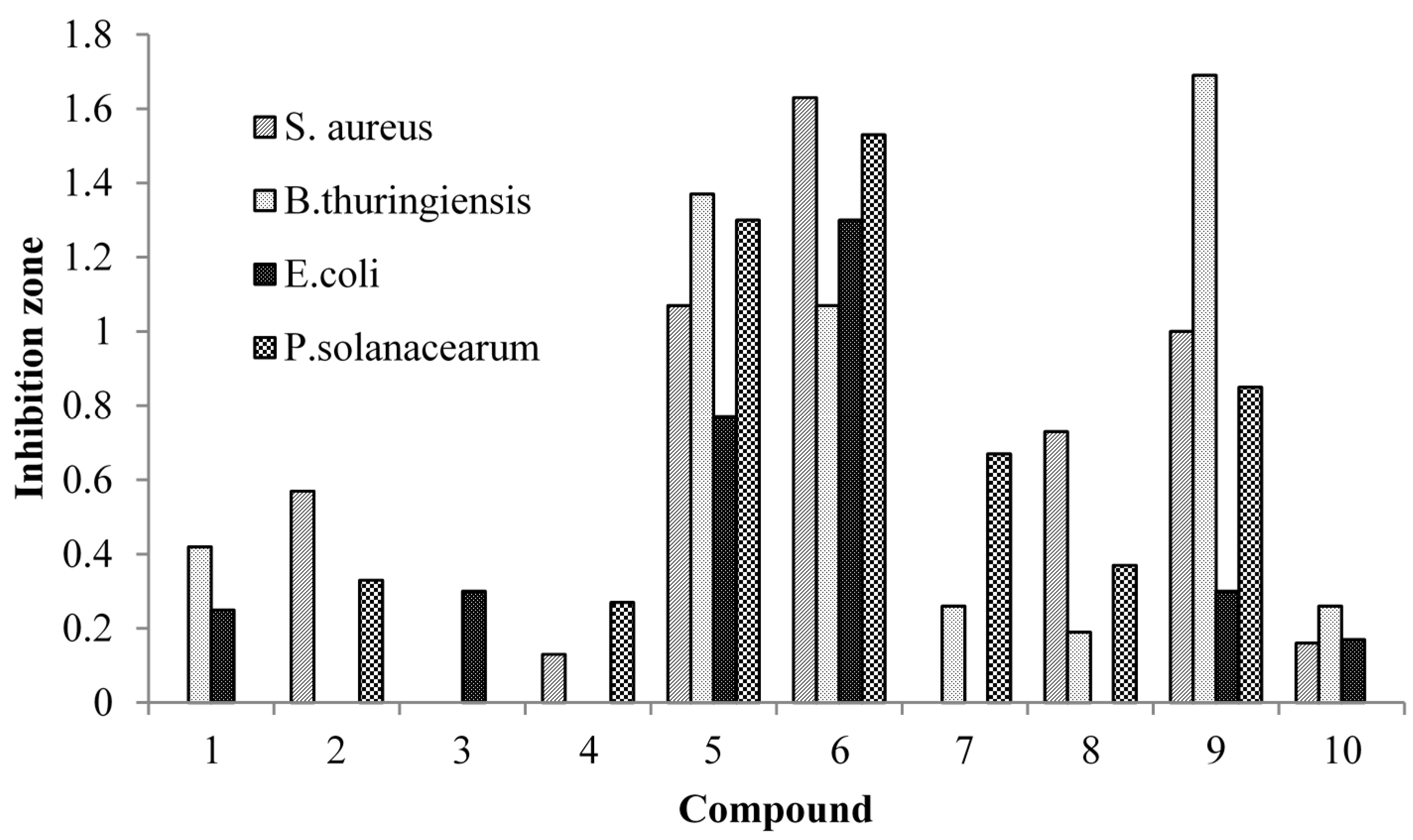
2.2. Antibacterial Activities of the Isolated Compounds

The agar-disk diffusion method is a traditional method for measuring the antibacterial activities of compounds, and their antibacterial effects can be visually observed [31,32,33].
The results of the antibacterial activity tests indicated that the 10 compounds had selective antibacterial properties. Figure 4 shows the zones of inhibition for each compound against the four test strains. All 10 compounds showed inhibition zones, which varied from 0.13 to 1.69 mm.
Compounds 5 and 6 had antibacterial activities against all four bacterial strains and, more notably, these two compounds showed strong antibacterial activities against S. aureus and E. coli, which are food-contaminating bacteria. Of the remaining two test strains, B. thuringiensis was most sensitive to compound 9, and P. solanacearum was most sensitive to compound 6.
Figure 4. Antibacterial activities of the compounds isolated from the leaves of Indocalamus latifolius McClure.
Molecules 20 15686 g004

2.3. Discussion

Consumers, nowadays, have a strong demand for greener food preservation techniques; hence there is great potential for developing naturally-derived antimicrobial agents. Extensive research has documented that compounds isolated from plants contain a large number of secondary metabolites and possess the capacity to inhibit the growth of bacteria and fungi [34]. The antimicrobial compounds in plants are a part of the self-defense mechanisms for combating harmful microbes in a natural environment [35]. Many of these compounds are under investigation and are not yet exploited commercially. Hao et al. [36] found the alcohol extracts of angelica root, banana purée, bay, caraway seed, carrot root, clove (eugenol), marjoram, pimento leaf, and thyme showed inhibition of A. hydrophila and L. monocytogenes in refrigerated poultry. Ahn et al. [37] also found grape seed extract and pine bark extract could control the growth of microorganisms in cooked beef. Kotzekidou et al. [38] tested plant extracts and essential oils with potent antimicrobial activities in chocolate at different temperatures and in dry or humidified environment, the most inhibitory action was observed by lemon flavor applied on chocolate inoculated with E. coli cocktail culture after storage at 20 °C for 9 days. Martinez-Romero et al. [39] reported that the vapor atmosphere of carvacrol could reduce the fungal growth in grape berries.
Another application of natural derived antimicrobials is in the bioactive packaging technologies for food preservation. Seydim [40] found the antimicrobial activity of some spice extracts could be expressed in a whey protein isolate (WPI)-based edible film; hence, they may act as releasable antimicrobial constituents in food packaging. Oussalah [41] studied milk protein-based edible films containing plant essential oils mix on beef muscle slices for controlling the growth of pathogenic bacteria during storage at 4 °C; the film containing oregano showed the most effective against two test bacteria. Nicholson [42] suggested naturally-occurring bio-preservatives could be applied in the food packaging system as part of a multiple hurdle technique, and should lead to increases in both the food safety and shelf-life of perishable foods.
Naturally-derived preservatives for food have been investigated for practical applications in the last 10 years; however, there are also challenges. Plant extracts, especially the EOs, always have strong odor/flavor and may transfer into the food. In this research, we investigated the compounds from a traditional natural packaging material, the leaves of Indocalamus latifolius McClure. In addition to the antibacterial capacity, we also found compound (2) has a pleasant smell. Thus, the isolated compounds in our research could act as an antimicrobial agent or as a component in antimicrobial packages, and also as an odor/flavor enhancer for packaged foods.
Whereas, the results and data obtained from laboratory in vitro experiments may not be applied to food products as foods are complex, the natural antimicrobial agents may offer exclusive advantages for food preservation, and the applications of naturally-derived antimicrobial agents in food will rise steadily in the future.

3. Experimental Section

3.1. Plant Material

I. latifolius McClure leaves were collected from the Century Garden of Bamboos in Yibin city, Sichuan, China. A voucher specimen was deposited in the State Forestry Administration Key Open Laboratory at the International Centre for Bamboo and Rattan in Beijing 100102, China.

3.2. Instrumental Equipment

Preparative HPLC was performed on a Shimadzu LC-6AD with an SPD-20A detector (Shimadzu, Kyoto, Japan) using a YMC-Pack ODS-A column (250 mm × 20 mm, 5 μm, YMC, Kyoto, Japan). HPLC-PAD analysis was performed using a Waters 2695-2996 system and a 2996 PDA detector (Waters, Milford, MA, USA) with a YMC-Pack ODS-AQ C18 column (250 mm × 4.6 mm, 5 μm, YMC, Kyoto, Japan). IR spectra were collected on a Thermo Nicolet FT-IR NEXUS 670 spectrophotometer (Thermo, Waltham, MA, USA) using KBr pellets, and NMR spectra were collected on Bruker 500 MHz spectrometers (Bruker, Zurich, Switzerland). HRESIMS spectra were obtained with an Agilent 6540 high resolution time-of-flight (Q-TOF) mass spectrometer (Agilent, Santa Clara, CA, USA). Circular dichroism (CD) spectra were recorded in methanol solutions using a JASCO J-815 CD spectrometer (JASCO, Tokyo, Japan). Antibacterial properties were determined by the filter agar-disk diffusion method [34].

3.3. Chemicals and Reagents

Column chromatography was performed with macroporous resin (Diaion HP-20, Mitsubishi Chemical Corp., Tokyo, Japan), Rp-18 (50 μm, YMC, Kyoto, Japan) and Sephadex LH-20 (Pharmacia Fine Chemicals, Uppsala, Sweden). All of the reagents and the nutrient agar were purchased from Beijing Chemical Works (Beijing, China) unless otherwise specified. HPLC-grade methanol (MeOH) and ethanol (EtOH) were purchased from Fisher Scientific (Pittsburgh, PA, USA).

3.4. Analytical Methods

HPLC analysis utilized a binary elution system consisting of solvent A (MeOH) and solvent B (water containing 0.2% acetic acid) with a YMC-PACK ODS-AQ C18 column. The flow rate was 1 mL/min, the column temperature was 30 °C and the injection volume was 10 μL. The PAD detection wavelength monitoring range was 210 to 400 nm. LC-MS analysis was performed to confirm the molecular weights of the compounds using an Rp-18 column and ESI-MS. The mobile phases were solvent A (MeOH) and solvent C (water containing 0.1% formic acid). The flow rate was 0.3 mL/min, the column temperature was 30 °C, and the effluent was monitored at either 220 or 270 nm. The 1H-, 13C-NMR, and 2D (HSQC, HMBC, and NOE) NMR spectra were recorded on 500 MHz Bruker spectrometers using DMSO-d6 as the solvent and tetramethylsilane (TMS) as the internal standard. The chemical shifts were expressed in δ (ppm), and the coupling constants were reported in Hertz. The concentration of the compound was 12 mg/mL, the NMR acquisition duration was 2 min for 1H-NMR and 5 h for 13C-NMR, and the widths of the NMR spectra were 0–14 ppm for 1H-NMR and 0–220 ppm for 13C-NMR.

3.5. Extraction, Isolation, and Purification of the Compounds from Indocalamus Latifolius McClure

Dried I. latifolius McClure leaves (7 kg) were extracted with 10 L of 95% aqueous ethanol for 24 h at room temperature three times. The solvent was removed under vacuum to collect the filtrates. The concentrated aqueous fraction was separated on a macroporous resin column using a step-wise gradient of water/ethanol (100:0, 85:15, 70:30, 50:50, 30:70, and 5:95) to yield six fractions. Medium-scale preparative performance liquid chromatography was applied to the 30% ethanol fraction (9.6 g) using a Rp-18 column, which was eluted using a step-wise water/methanol gradient (100:0, 80:20, 75:25, 70:30, 65:35, 60:40, 55:45, 50:50, 40:60, 30:70, and 5:95) to yield eleven fractions (1–11). Fraction 3 (128.4 mg) underwent additional column chromatography (CC) over Sephadex LH-20 with water elution, and preparative HPLC was performed with a methanol-water (15:85) elution, which yielded compound 1 (8.5 mg). Fraction 6 (286.3 mg), subjected to the same CC system with methanol/water (25:75) as the elution solvent, yielded compounds 2 (6.8 mg), 3 (52.6 mg) and 4 (27.9 mg). Fraction 7 (168.5 mg) was also subjected to the same two-step CC system and elution with methanol/water (30:70) yielded compounds 5 (12 mg) and 6 (5.8 mg). The two-step CC method was also performed on Fraction 8 (137.6 mg), and the methanol/water ratio used for elution was 35:65, yielding compounds 7 (8.7 mg), 8 (11.2 mg) and 9 (18.2 mg). The same CC was applied to Fraction 9 (320.4 mg), and the HPLC sample was eluted with methanol/water (40:60). This fraction was further precipitated with water, yielding compound 10 (79.0 mg).
L-phenylalanine (1). White amorphous powder; [α]D = −33.9° (c = 0.50, methanol). HRESIMS: C9H11NO2, m/z 164.0717 [M − H]+ (calculated 164.0712). IR (KBr) cm−1: νmax 2914, 1572, 1528, 1405. UV λmax (methanol) (log ε): 210 nm. 1H-NMR (500 MHz) (DMSO-d6): 7.43 (1H, s, H-4), 7.39 (2H, d, H-3,5), 7.30 (2H, d, H-2,6), 3.96 (1H, m, H-8), 3.22 (2H, m, H-7); 13C-NMR (125 MHz) (DMSO-d6): 173.9 (C-9), 135.2 (C-1), 129.4 (C-3,5), 129.1 (C-2, 6), 127.7 (C-4), 56.1 (C8), 36.4 (C-7).
Latifoliusine A (2). Yellowish oil; [α]D = +36.1° (c = 0.70, methanol). HRESIMS: C13H20O3, m/z 247.1313 [M + Na]+ (calculated 247.1310). IR (KBr) cm−1: νmax 3424, 2928, 1670, 1458, 1255. UV λmax (methanol) (log ε): 232.3 nm. 1H- and 13C-NMR (500 MHz) (DMSO-d6): see Table 1.
Dihydroxymethyl-bis(3,5-dimethoxy-4-hydroxyphenyl)tetrahydrofuran-9-O-β-d-glucopyranoside (3). White amorphous powder; [α]D = −25.4° (c = 1.0, methanol). HRESIMS: C28H38O14, m/z 597.2180 [M − H]+ (calculated 597.2183). IR (KBr) cm−1: νmax 3371, 2923, 1583, 1454. UV λmax (methanol) (log ε): 241, 271 nm. 1H-NMR (500 MHz) (DMSO-d6): 6.66 (2H, s, H-2,6 or 2′,6′), 6.66 (2H, s, H-2,6 or 2′,6′), 4.91 (1H, d, H-7′), 4.85 (1H, d, H-7), 4.16 (1H, d, H-1′′), 3.89, 3.56 (2H, m, H-9), 3.76 (12H, s, OCH3), 3.66, 3.44 (2H, m, H-6′′), 3.53, 3.48 (2H, m, H-9′), 3.18 (1H, m, H-5′′), 3.06 (1H, m, H-3′′), 3.03 (1H, m, H-4′′), 2.99 (1H, m, H-2′′), 2.12 (1H, m, H-8′), 2.32 (1H, m, H-8); 13C-NMR (125 MHz) (DMSO-d6): 148.4 (C-3,5 or 3′,5′), 135.1 (C-4 or 4′), 133.5 (C-1′), 133.3 (C-1), 104.3 (C-2,6 or 2′,6′), 103.6 (C-1′′), 82.6 (C-7′), 82.4 (C-7), 77.3 (C-5′′), 77.2 (C-3′′), 74.1 (C-2′′), 70.6 (C-4′′), 69.5 (C-9), 61.5 (C-6′′), 60.4(C-9′), 56.5 (OCH3), 53.7 (C-8), 50.7 (C-8′).
Rel-(7R,8S,7′S,8′R)-4,9,4′,9′-tetrahydroxy-3,3′-dimethoxy-7,7′-epoxylignan 9-O-β-d-glucopyranoside (4). White amorphous powder; [α]D = −23.9° (c = 1.0, methanol). HRESIMS: C26H34O12, m/z 537.1969 [M − H]+ (calculated 537.1972). IR (KBr) cm−1: νmax 3365, 2937, 1584, 1451. UV λmax (methanol) (log ε): 233, 279 nm. 1H-NMR (500 MHz) (DMSO-d6): 6.94 (1H, s, H-2), 6.93 (1H, s, H-2′), 6.75 (1H, d, H-6), 6.74 (1H, d, H-6′), 6.69 (1H, d, H-5), 6.68 (1H, d, H-5′), 4.87 (1H, d, H-7′), 4.86 (1H, d, H-7), 4.16 (1H, d, H-1′′), 3.86, 3.53 (2H, m, H-9), 3.76 (6H, s, OCH3), 3.66, 3.44 (2H, m, H-6′′), 3.53, 3.45 (2H, m, H-9′), 3.12 (1H, m, H-5′′), 3.07 (1H, m, H-3′′), 3.05 (1H, m, H-4′′), 2.97 (1H, m, H-2′′), 2.31 (1H, m, H-8), 2.15 (1H, m, H-8′); 13C-NMR (125 MHz) (DMSO-d6): 147.8 (C-3), 147.7 (C-3′), 146.2 (C-4), 146.1 (C-4′), 134.5 (C-1), 134.2 (C-1′), 119.3 (C-6), 119.0 (C-6′), 115.5 (C-5), 115.4 (C-5′), 111.1 (C-2), 110.9 (C-2′), 103.6 (C-1′′), 82.3 (C-7′), 82.2 (C-7), 77.3 (C-5′′), 77.2 (C-3′′), 74.0 (C-2′′), 70.5 (C-4′′), 69.3 (C-9), 61.5 (C-6′′), 60.4 (C-9′), 56.1 (OCH3), 53.6 (C-8), 50.7 (C-8′).
Apigenin 6-C-α-l-arabinopyranosyl-8-C-β-d-glucopyranoside (5). Yellow amorphous powder; HRESIMS: C26H28O14, m/z 563.1400 [M − H]+ (calculated 563.1401). IR (KBr) cm−1: νmax 3386, 2954, 1706, 1573, 1467. UV λmax (methanol) (log ε): 271, 334 nm. 1H-NMR (500 MHz) (DMSO-d6): 7.95 (2H, d, H-2′,6′), 6.93 (2H, d, H-3′,5′), 6.81 (1H, s, H-3), 4.81 (1H, d, H-1′′′), 4.62 (1H, d, H-1′′), 4.00 (1H, m, H-2′′), 3.89 (1H, m, H-2′′′), 3.79, 3.57 (2H, m, H-5′′), 3.77 (1H, m, H-4′′), 3.74, 3.52 (2H, m, H-6′′′), 3.42 (1H, m, H-3′′), 3.36 (1H, m, H-4′′′), 3.30 (1H, m, H-3′′′), 3.27 (1H, m, H-5′′′); 13C-NMR (125 MHz) (DMSO-d6): 180.8 (C-4), 162.8 (C-7), 161.5 (C-2), 160.6 (C-4′), 159.4 (C-5), 154.5 (C-9), 128.7 (C-2′,6′), 121.9 (C-1′), 115.7 (C-3′,5′), 110.1 (C-6), 104.5 (C-8), 102.0 (C-3), 100.1 (C-10), 81.5 (C-5′′′), 78.8 (C-3′′′), 74.1 (C-1′′), 74.0 (C-3′′), 73.8 (C-1′′′), 71.0 (C-2′′′), 70.4 (C-4′′′), 69.7 (C-5′′), 69.0 (C-2′′), 68.5 (C-4′′), 61.0 (C-6′′′).
Apigenin 7-O,8-C-di-glucopyranoside (6). Yellow amorphous powder; HRESIMS: C26H34O12, m/z 563.1405 [M − H]+ (calculated 563.1401). IR (KBr) cm−1: νmax 3331, 2835, 1725, 1544, 1486. UV λmax (methanol) (log ε): 271, 336 nm. 1H-NMR (500 MHz) (DMSO-d6): 7.93 (2H, d, H-2′,6′), 6.94 (2H, d, H-3′,5′), 6.79 (1H, s, H-3), 6.19 (1H, s, H-8), 4.88 (1H, d, H-1′′′), 4.64 (1H, d, H-1′′), 4.02 (1H, m, H-2′′), 3.79 (1H, m, H-4′′), 3.77, 3.55 (2H, m, H-5′′), 3.60, 3.44 (2H, m, H-6′′′), 3.43 (1H, m, H-3′′), 3.23 (1H, m, H-3′′′), 3.22 (1H, m, H-2′′′), 3.20 (1H, m, H-4′′′), 3.05 (1H, m, H-5′′′); 13C-NMR (125 MHz) (DMSO-d6): 181.0 (C-4), 164.6 (C-7), 162.2 (C-2), 160.7 (C-4′), 159.6 (C-5), 155.0 (C-9), 128.6 (C-2′,6′), 122.0 (C-1′), 115.9 (C-3′,5′), 110.4 (C-6), 105.3 (C-10), 103.4 (C-1′′′), 102.3 (C-3), 92.5 (C-8), 77.6 (C-5′′′), 77.0 (C-3′′′), 74.3 (C-1′′), 74.2 (C-3′′), 72.5 (C-2′′′), 70.4 (C-4′′′), 69.8 (C-5′′), 69.0 (C-4′′), 68.5 (C-2′′), 61.4 (C-6′′′).
(7S,8R) Syringylglycerol-8-O-4′-sinapyl ether 4-O-β-d-glucopyranoside (7). Yellow amorphous powder; [α]D = −15.9° (c = 1.0, methanol). HRESIMS: C28H38O14, m/z 597.2179 [M − H] (calculated 597.2183). IR (KBr) cm−1: νmax 3381, 2927, 1654, 1451, 1253. UV λmax (methanol) (log ε): 230 nm, 270 nm. CD (c 1.0 × 10⁻³, MeOH): ∆ε205nm +9.4440, ∆ε243.5nm −0.4727, ∆ε282.0nm +0.1389. 1H- and 13C-NMR (500 MHz) (DMSO-d6): see Table 1.
(7S,8S) Syringylglycerol-8-O-4′-sinapyl ether 7-O-β-d-glucopyranoside (8). Yellow amorphous powder; [α]D = −3.11° (c = 1.0, methanol). HRESIMS: C28H38O14, m/z 633.1952 [M + Cl] (calculated 633.1950). IR (KBr) cm−1: νmax 3385, 2927, 1584, 1460. UV λmax (methanol) (log ε): 230 nm, 269 nm. CD (c 1.0 × 10⁻³, MeOH): ∆ε212.5nm +6.6316, ∆ε237.5nm +2.0655, ∆ε285.0nm −0.5970. 1H- and 13C-NMR (500 MHz) (DMSO-d6): see Table 1.
(7S,8S) Syringylglycerol-8-O-4′-sinapyl ether 9′-O-β-d-glucopyranoside (9). White amorphous powder; [α]D = −9.5° (c = 1.0, methanol). HRESIMS: C28H38O14, m/z 597.2181 [M – H]+ (calculated 597.2183). IR (KBr) cm−1: νmax 3379, 2931, 1582, 1464. UV λmax (methanol) (log ε): 230, 270 nm. 1H-NMR (500 MHz) (DMSO-d6): 6.75 (2H, s, H-2′,6′), 6.60 (1H, s, H-2,6), 6.57 (1H, d, H-7′), 6.34 (1H, dt, H-8′), 4.81 (1H, dd, H-7), 4.41, 4.19 (1H, d, H-9′), 4.15 (1H, m, H-8), 4.21 (1H, d, H-1′′), 3.68, 3.40 (1H, m, H-9), 3.67, 3.44 (2H, m, H-6′′), 3.14 (1H, m, H-5′′), 3.09 (1H, m, H-3′′), 3.07 (1H, m, H-4′′), 3.05 (1H, m, H-2′′); 13C-NMR (125 MHz) (DMSO-d6): 152.8 (C-3′′,5′′), 147.6 (C-3,5), 135.5 (C-4′), 134.4 (C-4), 132.6 (C-1), 132.0 (C-1′), 131.4 (C-7′), 125.8 (C-8′), 104.4 (C-2,6), 103.9 (C-2′,6′), 102.2 (C-1′′), 86.4 (C-8), 77.0 (C-3′′), 76.9 (C-5′′), 73.6 (C-2′′), 72.4 (C-7), 70.2 (C-4′′), 68.7 (C-9′), 61.2 (C-6′′), 59.9 (C-9).
(7R,8S) Syringylglycerol-8-O-4′-sinapyl ether 7-O-β-d-glucopyranoside (10). Yellow amorphous powder; [α]D = +4.55° (c = 1.0, methanol). HRESIMS: C28H38O14, m/z 633.1953 [M + Cl] (calculated 633.1950). IR (KBr) cm−1: νmax 3387, 2929, 1585, 1461. UV λmax (methanol) (log ε): 230 nm, 271 nm. CD (c 1.0 × 10⁻³, MeOH): ∆ε 207.5 nm +4.6915, ∆ε 231.0 nm +1.6611, ∆ε 272.0 nm ‒1.7423. 1H- and 13C-NMR (500 MHz) (DMSO-d6): see Table 1.

3.6. Antibacterial Activity Assay

3.6.1. Microbial Strains

Two food contaminating bacteria Staphylococcus aureus (Gram (+)) and Escherichia coli (Gram (−)) were selected as test strains, another Gram -ositive bacteria (Bacillus thuringiensis) along with another Gram-negative bacteria (Pseudomonas solanacearum) were selected for testing the antibacterial selectiveness of isolated compounds, all four bacteria strains were obtained from the Agricultural Product Key Laboratory of Anhui Agriculture University, Hefei City, Anhui, China.

3.6.2. Antibacterial Screening

The concentrations of the compounds used for the antibacterial screening experiments were 6.2 mg/mL (1), 7.0 mg/mL (2), 30.0 mg/mL (3), 21.0 mg/mL (4), 20.0 mg/mL (5), 13.6 mg/mL (6), 6.92 mg/mL (7), 6.88 mg/mL (8), 21.0 mg/mL (9) and 20.0 mg/mL (10). The concentrations were set for simulating the content ratio in Indocalamus latifolius McClure Leaves, which were determined in our preliminary research. Briefly, 200 μL of a suspension containing 108 colony-forming units (CFU)/mL of bacteria was spread onto nutrient agar (NA). The disks (6 mm in diameter) were impregnated with 10 μL of different concentrations of the compounds (dissolved in water-ethanol) and placed on the inoculated agar. Negative controls were prepared using water and ethanol. Ampicillin sodium (5 μg/disc) was used as the positive control. The inoculated plates of bacteria were incubated at 37 °C for 24 h. The antibacterial activity was evaluated by measuring the zone of inhibition.

4. Conclusions

Since ancient times, the leaves of I. latifolius McClure have been used as a packaging material for food, and presently, they still play a unique role in producing “Zongzi” in China. The identification of the antibacterial compounds in the leaves of I. latifolius McClure is important for helping us to understand the long shelf life of “Zongzi” as well as for exploring the potential of I. latifolius McClure leaves as a natural, healthy, and eco-friendly alternative packaging material for other applications.

Supplementary Materials

1H, 13C, HSQC, HMBC and NOESY spectra of latifoliusine A (2), 1H, 13C, HSQC and HMBC spectra of (7S,8R) syringylglycerol-8-O-4′-sinapyl ether 4-O-β-d-glucopyranoside (7) and (7S,8S) syringylglycerol-8-O-4′-sinapyl ether 7-O-β-d-glucopyranoside (8), 1H and 13C spectra of (7R,8S) syringylglycerol-8-O-4′-sinapyl ether 7-O-β-d-glucopyranoside (10) in DMSO. IR, HRESIMS and UV spectra of latifoliusine A (2); IR, HRESIMS, CD and UV spectra of (7S,8R) syringylglycerol-8-O-4′-sinapyl ether 4-O-β-d-glucopyranoside (7), (7S,8S) syringylglycerol-8-O-4′-sinapyl ether 7-O-β-d-glucopyranoside (8), (7R,8S) syringylglycerol-8-O-4′-sinapyl ether 7-O-β-d-glucopyranoside (10). Supplementary materials can be accessed at: https://www.mdpi.com/1420-3049/20/09/15686/s1.

Acknowledgments

The authors are grateful for financial support from the National Science and Technology Infrastructure Program (No. 2012BAD23B03) and the Basic Science Research Fund Program of the International Centre for Bamboo and Rattan (ICBR) (1632013007). The authors are thankful to the staff from the analytical group of the State Forestry Administration Key Open Laboratory in ICBR and the Institute of Materia Medica at the Chinese Academy of Medical Sciences & Peking Union Medical College for measuring the spectroscopic data.

Author Contributions

J.Y. performed the experiments; J.S. and H.X. carried out the statistical analysis and drafted the manuscript; these three authors contributed equally. F.T. and Y.D.Y. conceived of the design of the study and provided feedback on the manuscript. X.F.G. assisted with data collection.

Conflicts of Interest

The authors declare no conflicts of interest.

References

  1. Qin, Y.; Zhang, Z.; Li, L.; Chen, C.; Shun, S.; Huang, Y. Inductively coupled plasma orthogonal acceleration time-of-flight mass spectrometry (ICP-oa-TOF-MS) analysis of heavy metal content in Indocalamus tesselatus samples. Food Chem. 2013, 141, 2154–2157. [Google Scholar] [CrossRef] [PubMed]
  2. Jiang, Z.H. Bamboo and Rattan in the World; China Forestry Publishing House: Beijing, China, 2003; pp. 334–347. [Google Scholar]
  3. Ding, Y.Q.; Li, Y.; Chen, C.Y.; Elmahadi, E.A.; Xu, H.B. Chromatographic analysis of polysaccharides extracted from Chinese Indocalamus tesselatus. Biomed. Chromatogr. 1998, 12, 86–88. [Google Scholar] [CrossRef]
  4. Chen, C.; Ding, Y.; Elmahadi, E.A.; Zhou, J.; Li, Y.; Xu, H. Study on the isolation, purification and physicochemical properties of polysaccharides from Indocalamus tesselatus. Biomed. Chromatogr. 1999, 13, 11–14. [Google Scholar] [CrossRef]
  5. Zou, Y.H. Analysis of flavonoids in bamboo leaves by high performance liquid chromatography. Chin. J. Anal. Chem. 1996, 24, 216–219. [Google Scholar]
  6. Cui, J.; Yue, Y.D.; Tang, F.; Wang, J. HPTLC analysis of the flavonoids in eight species of Indocalamus leaves. J. Planar Chromatogr. 2011, 24, 394–399. [Google Scholar] [CrossRef]
  7. Zhou, Y.; Tan, X.H.; Li, Q.M. GC-MS analysis of volatile oil extracted from Indocalamus latifolius leaves by simultaneous distillation and extraction. Food Sci. 2009, 30, 199–202. [Google Scholar]
  8. Li, S.F.; Dai, Y.; Li, J.J. Comparison of scavenging DPPH free radical and restraining bacteria of Indocalamus latifolius leaf’s extracts. Food Sci. Technol. 2010, 35, 174–177. [Google Scholar]
  9. Zhang, H.; Lin, H.P.; Sheng, E.H.; Li, L.; Hu, S.H.; Mao, S.F. Study on antibacterial activity of extracts from Indocalamus tessellatus leaves. J. Zhejian For. Sci. Technol. 2010, 30, 38–41. [Google Scholar]
  10. Sun, J.; Yu, J.; Zhang, P.C.; Tang, F.; Yue, Y.D.; Yang, Y.N.; Feng, Z.M.; Guo, X.F. Isolation and identification of lignans from Caulis Bambusae in Taenia with antioxidant properties. J. Agric. Food Chem. 2013, 61, 4556–4562. [Google Scholar] [CrossRef] [PubMed]
  11. Sun, J.; Zhang, P.C.; Wei, Q.; Xun, H.; Tang, F.; Yue, Y.D.; Li, L.; Guo, X.F.; Zhang, R. Amarusine A, a new dioxaspiro [4.4] nonane derivative with a butyrolactone ring from Pleioblastus amarus. Tetrahedron Lett. 2014, 55, 4529–4531. [Google Scholar] [CrossRef]
  12. Sun, J.; Tang, F.; Yue, Y.D.; Xun, H.; Guo, X.F. Two new compounds from the dry leaves of Pleioblastus amarus (Keng) keng f. J. Asian Nat. Prod. Res. 2014, 16, 930–935. [Google Scholar] [CrossRef] [PubMed]
  13. Sun, J.; Yu, J.; Zhang, P.C.; Yue, Y.D. Enantiomeric determination of four diastereoisomeric oxyneolignans from Bambusa tuldoides munro. Phytochem. Anal. 2015, 26, 54–60. [Google Scholar] [CrossRef] [PubMed]
  14. Wu, S.B.; Wen, Y.; Li, X.W.; Zhao, Y.; Zhao, Z.; Hu, J.F. Chemical constituents from the fruits of Sonneratia caseolaris and Sonneratia ovata (Sonneratiaceae). Biochem. Syst. Ecol. 2009, 37, 1–5. [Google Scholar] [CrossRef]
  15. Pretsch, E.; Bühlmann, P.; Affolter, C. Structure Determination of Organic Compounds Tables of Spectral Data; Springer-Verlag: Berlin, Germany, 2009; pp. 149–150. [Google Scholar]
  16. Machida, K.; Yamauchi, M.; Kurashina, E.; Kikuchi, M. Four new lignan glycosides from Osmanthus fragrans Lour. var. aurantiacus Makino. Helv. Chim. Acta 2010, 93, 2164–2175. [Google Scholar] [CrossRef]
  17. Kanchanapoom, T.; Kamel, M.S.; Kasai, R.; Yamasaki, K.; Picheansoonthon, C.; Hiraga, Y. Lignan glucosides from Acanthus ilicifolius. Phytochemistry 2001, 56, 369–372. [Google Scholar] [CrossRef]
  18. Xie, C.; Veitch, N.C.; Houghton, P.J.; Simmonds, M.S. Flavone C-glycosides from Viola yedoensis Makino. Chem. Pharm. Pull. 2003, 51, 1204–1207. [Google Scholar] [CrossRef]
  19. Niemann, G.J.; van Brederode, J. Separation of glycoflavones and their glycosides by high-performance liquid chromatography. J. Chromatogr. A 1978, 152, 523–527. [Google Scholar] [CrossRef]
  20. Feng, X.S.; Qu, Y.; Xu, L.; Wang, D.C.; Wu, L.J.; Meng, F.H.; Liu, Y.R. Two new neolignans from Syringa velutina Kom. Molecules 2009, 14, 953–958. [Google Scholar] [CrossRef] [PubMed]
  21. Buske, A.; Schmidt, J.; Porzel, A.; Adam, G. Alkaloidal, megastigmane and lignan glucosides from Antidesma membranaceum (Euphorbiaceae). Eur. J. Org. Chem. 2001, 18, 3537–3543. [Google Scholar] [CrossRef]
  22. Higuchi, H.; Fukui, K.; Kinjo, F.; Nohara, T. Four new glycosides from Albizziae Cortex. III. Chem. Pharm. Bull. 1992, 40, 534–535. [Google Scholar] [CrossRef]
  23. Huo, C.; Liang, H.; Zhao, Y.; Wang, B.; Zhang, Q. Neolignan glycosides from Symplocos caudata. Phytochemistry 2008, 69, 788–795. [Google Scholar] [CrossRef] [PubMed]
  24. Machida, K.; Sakamoto, S.; Kikuchi, M. Structure elucidation and NMR spectral assignments of four neolignan glycosides with enantiometric aglycones from Osmanthus ilicifolius. Magn. Reson. Chem. 2008, 46, 990–994. [Google Scholar] [CrossRef] [PubMed]
  25. Dellagreca, M.; Molinaro, A.; Monaco, P.; Previtera, L. Lignans from Arum italicum. Phytochemistry 1994, 35, 777–779. [Google Scholar]
  26. Liao, S.G.; Wu, Y.; Yue, J.M. Lignans from Wikstroemia hainanensis. Helv. Chim. Acta 2006, 89, 73–80. [Google Scholar] [CrossRef]
  27. Takara, K.; Matsui, D.; Wada, K.; Ichiba, T.; Nakasone, Y. New antioxidative phenolic glycosides isolated from Kokuto non-centrifuged cane sugar. Biosci. Biotechnol. Biochem. 2002, 66, 29–35. [Google Scholar] [CrossRef] [PubMed]
  28. Gan, M.; Zhang, Y.; Lin, S.; Liu, M.; Song, W.; Zi, J.; Yang, Y.; Fan, X.; Shi, J.; Hu, J.; et al. Glycosides from the root of Iodes cirrhosa. J. Nat. Prod. 2008, 71, 647–654. [Google Scholar] [CrossRef] [PubMed]
  29. Lin, S.; Wang, S.; Liu, M.; Gan, M.; Li, S.; Yang, Y.; Wang, Y.; He, W.; Shi, J. Glycosides from the stem bark of Fraxinus sieboldiana. J. Nat. Prod. 2007, 70, 817–823. [Google Scholar] [CrossRef] [PubMed]
  30. Takara, K.; Otsuka, K.; Wada, K.; Iwasaki, H.; Yamashita, M. 1,1-Diphenyl-2-picrylhydrazyl radical scavenging activity and tyrosinase inhibitory effects of constituents of sugarcane molasses. Biosci. Biotechnol. Biochem. 2007, 71, 183–191. [Google Scholar] [CrossRef] [PubMed]
  31. Millet, A.; Lamy, E.; Jonas, D.; Stintzing, F.; Mersch-Sundermann, V.; Merfort, I. Fermentation enhances the biological activity of Allium cepa bulb extracts. J. Agric. Food Chem. 2012, 60, 2148–2156. [Google Scholar] [CrossRef] [PubMed]
  32. Filippi, J.J.; Lanfranchi, D.A.; Prado, S.; Baldovini, N.; Meierhenrich, U.J. Composition, enantiomeric distribution, and antibacterial activity of the essential oil of Achillea ligustica All. from Corsica. J. Agric. Food Chem. 2006, 54, 6308–6313. [Google Scholar] [CrossRef] [PubMed]
  33. Xia, L.; Idhayadhulla, A.; Lee, Y.R.; Kim, S.H.; Wee, Y.J. Microwave-assisted synthesis of diverse pyrrolo [3, 4-c] quinoline-1, 3-diones and their antibacterial activities. ACS Comb. Sci. 2014, 16, 333–341. [Google Scholar] [CrossRef] [PubMed]
  34. Sokmen, A.; Gulluce, M.; Akpulat, H.A.; Daferera, D.; Tepe, B.; Polissiou, M.; Sokmen, M.; Sahin, F. The in vitro antimicrobial and antioxidant activities of the essential oils and methanol extracts of endemic Thymus spathulifolius. Food Control 2004, 15, 627–634. [Google Scholar] [CrossRef]
  35. Rauha, J.P.; Remes, S.; Heinonen, M.; Hopia, A.; Kahkonen, M.; Kujala, T.; Pihlaja, K.; Vuorela, H.; Vuorela, P. Antimicrobial effects of Finnish plant extracts containing flavonoids and other phenolic compounds. Int. J. Food Microbiol. 2000, 56, 3–12. [Google Scholar] [CrossRef]
  36. Hao, Y.Y.; Brackett, R.E.; Doyle, M.P. Efficacy of plant extracts in inhibiting Aeromonas hydrophila and Listeria monocytogenes in refrigerated cooked poultry. Food Microbiol. 1998, 15, 367–378. [Google Scholar] [CrossRef]
  37. Ahn, J.; Grun, I.U.; Mustapha, A. Effect of plant extracts on microbial growth, color change, and lipid oxidation in cooked beef. Food Microbiol. 2007, 24, 7–14. [Google Scholar] [CrossRef] [PubMed]
  38. Kotzekidou, P.; Giannakidis, P.; Boulamatsis, A. Antimicrobial activity of some plant extracts and essential oils against foodborne pathogens in vitro and on the fate of inoculated pathogens in chocolate. LWT Food Sci. Technol. 2007, 41, 119–127. [Google Scholar] [CrossRef]
  39. Martinez-Romero, D.; Guillen, F.; Valverde, J.M.; Bailen, G.; Zapata, P.; Serrano, M.; Castillo, S.; Valero, D. Influence of carvacrol on survival of Botrytis cinerea inoculated in table grapes. Int. J. Food Microbiol. 2007, 115, 144–148. [Google Scholar] [CrossRef] [PubMed]
  40. Seydim, A.C.; Sarikus, G. Antimicrobial activity of whey protein based edible films incorporated with oregano, rosemary and garlic essential oils. Food Res. Int. 2006, 39, 639–644. [Google Scholar] [CrossRef]
  41. Oussalah, L.; Caillet, S.; Salmieri, S.; Saucier, L.; Lacroix, M. Antimicrobial and antioxidant effects of milk protein-based film containing essential oils for the preservation of whole beef muscle. J. Agric. Food Chem. 2004, 52, 5598–5605. [Google Scholar] [CrossRef] [PubMed]
  42. Vermeiren, L.; Devlieghere, F.; van Beest, M.; de Kruijf, N.; Debevere, J. Developments in the active packaging of foods. Trends Food Sci. Technol. 1999, 10, 77–86. [Google Scholar] [CrossRef]
  • Sample Availability: Samples of the compounds 110 are available from the authors.

