Synergistic Radiation Protective Effect of Purified Auricularia auricular-judae Polysaccharide (AAP IV) with Grape Seed Procyanidins

Abstract
:1. Introduction
2. Results and Discussion
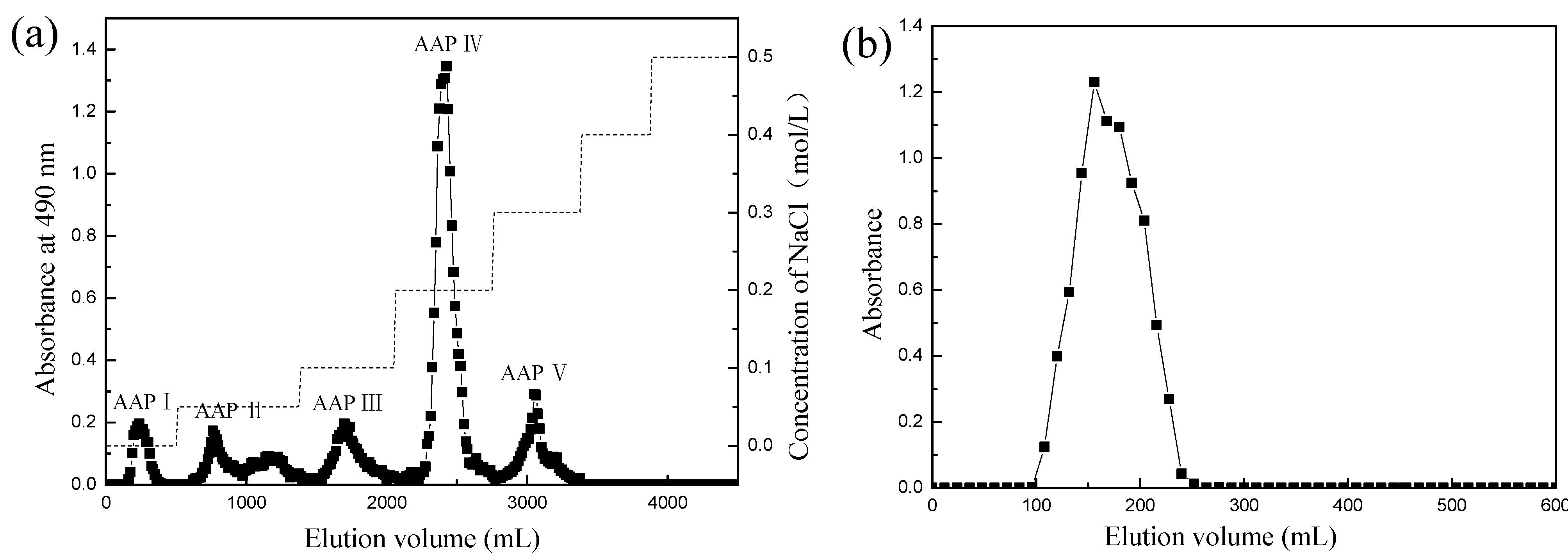
2.1. Purification of the AAP
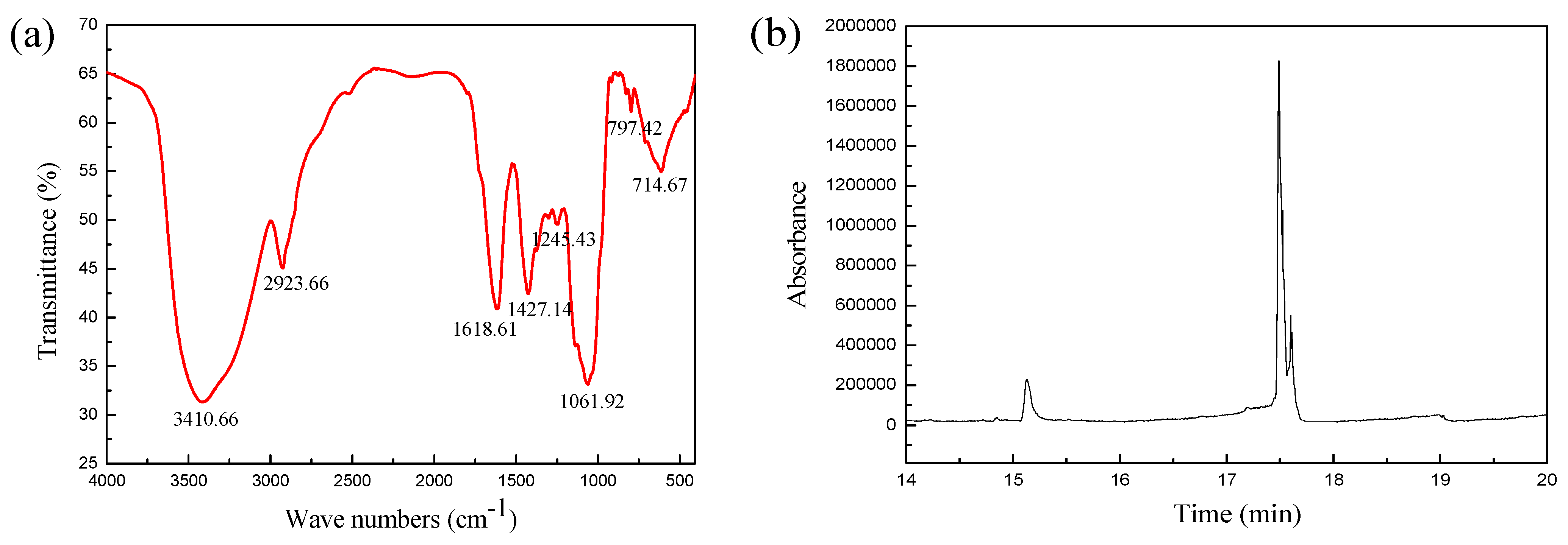
2.2. Primary Structure Analysis of the AAP IV

| Absorption at Region (cm−1) | Vibration Type | Functional Group |
|---|---|---|
| 3410.66 | Stretching vibration of O-H | O-H |
| 2923.66 | Stretching vibration of C-H | -CH2 |
| 1618.61 | Variable angle vibration of N-H | -CONH |
| 1427.14 | Stretching vibration of C-O | -COOH |
| 1000–1250 | Stretching vibration of C-O-C and C-O-H | C-O |
| 797.42 | Symmetric stretching vibration of C-O-C | d-glucose pyranose |
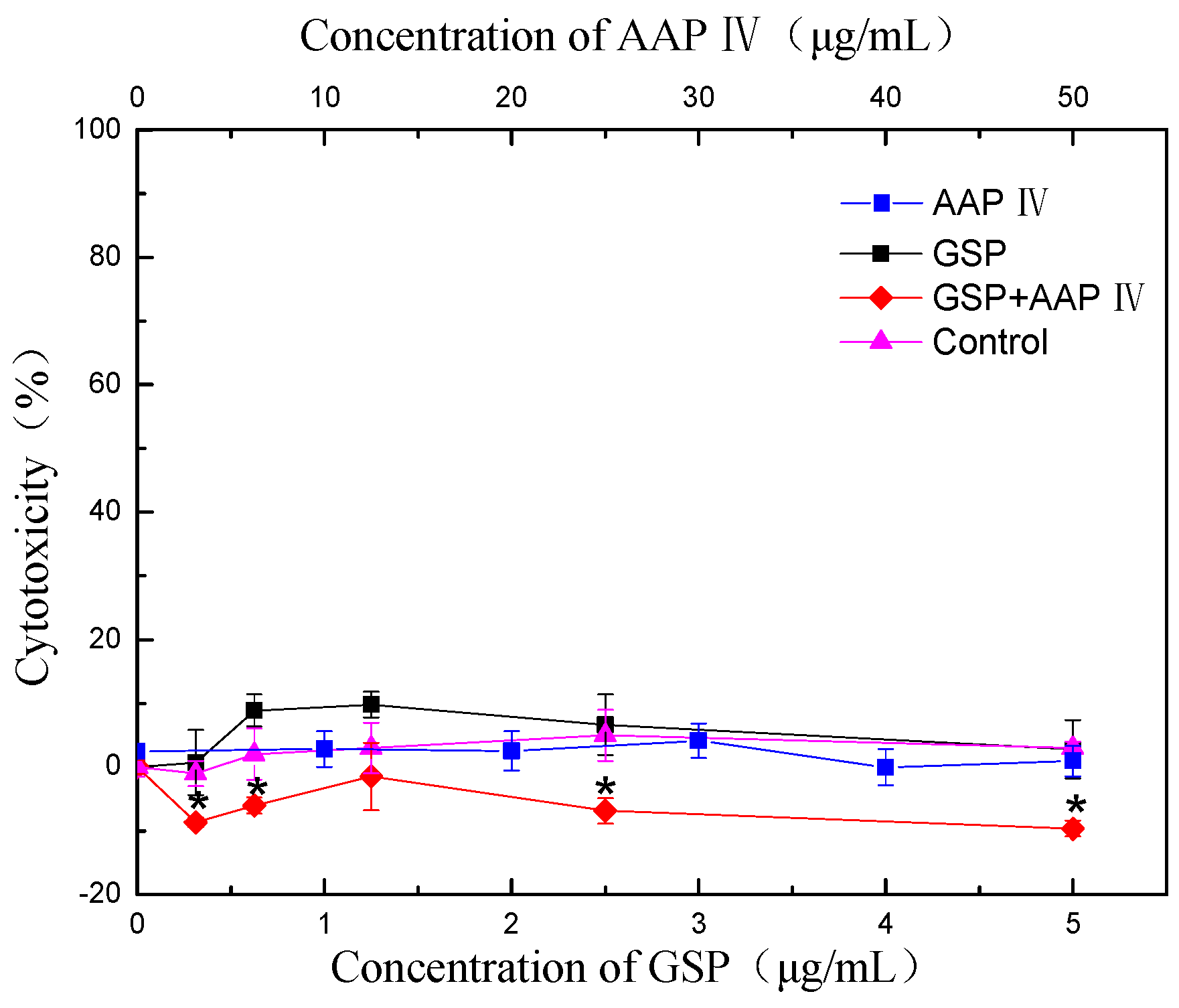
2.3. Cytotoxicity
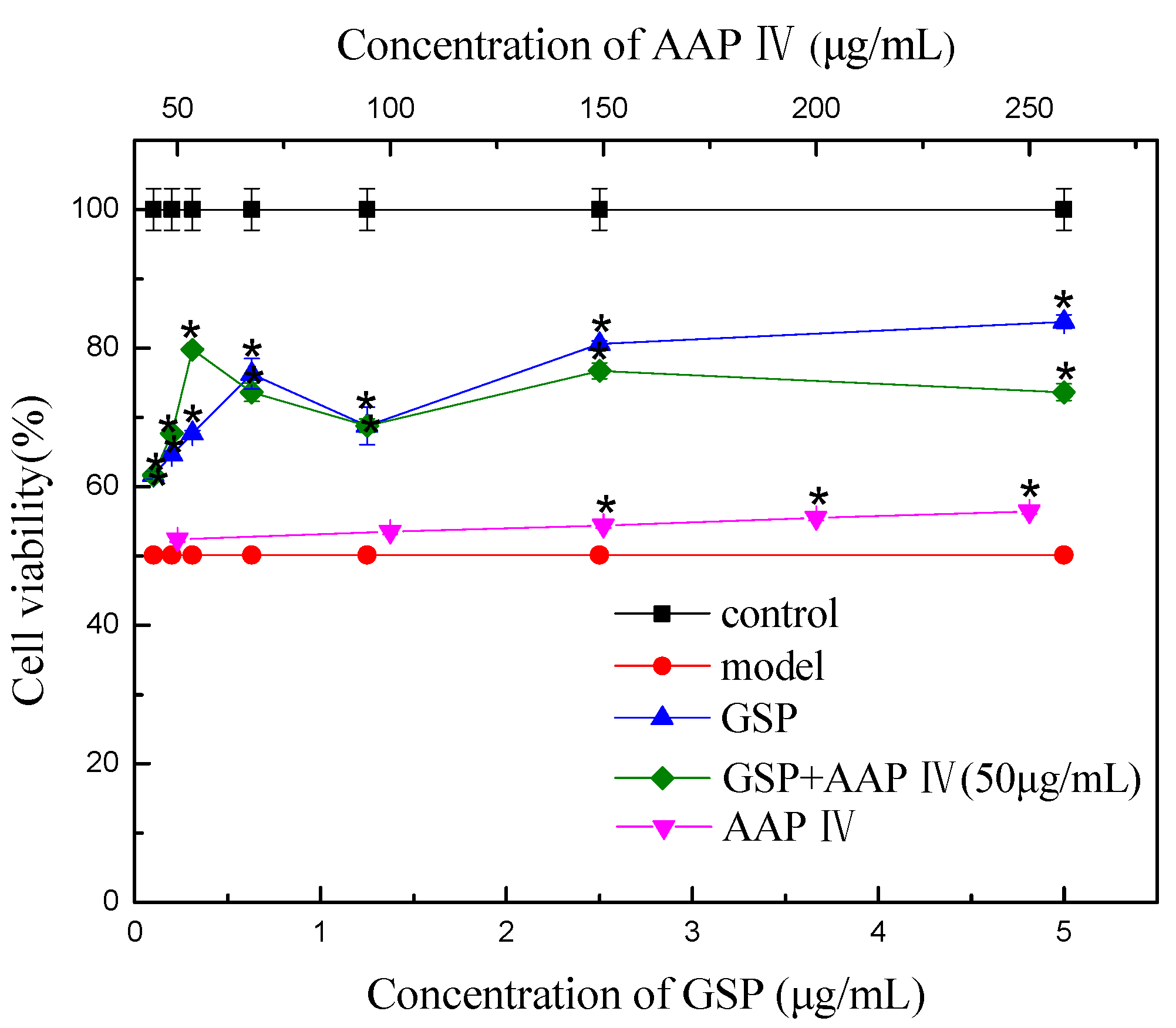
2.4. Splenocyte Viability
| Combination | Effect | CI | |
|---|---|---|---|
| GSP(μg/mL) | AAP IV(μg/mL) | ||
| 0.10 | 50 | 0.18 | 1.02 |
| 0.20 | 50 | 0.35 | 0.65 |
| 0.31 | 50 | 0.60 | 0.11 |
| 0.63 | 50 | 0.47 | 0.64 |
| 1.25 | 50 | 0.37 | 3.60 |
| 2.50 | 50 | 0.53 | 1.43 |
| 5.00 | 50 | 0.47 | 5.09 |
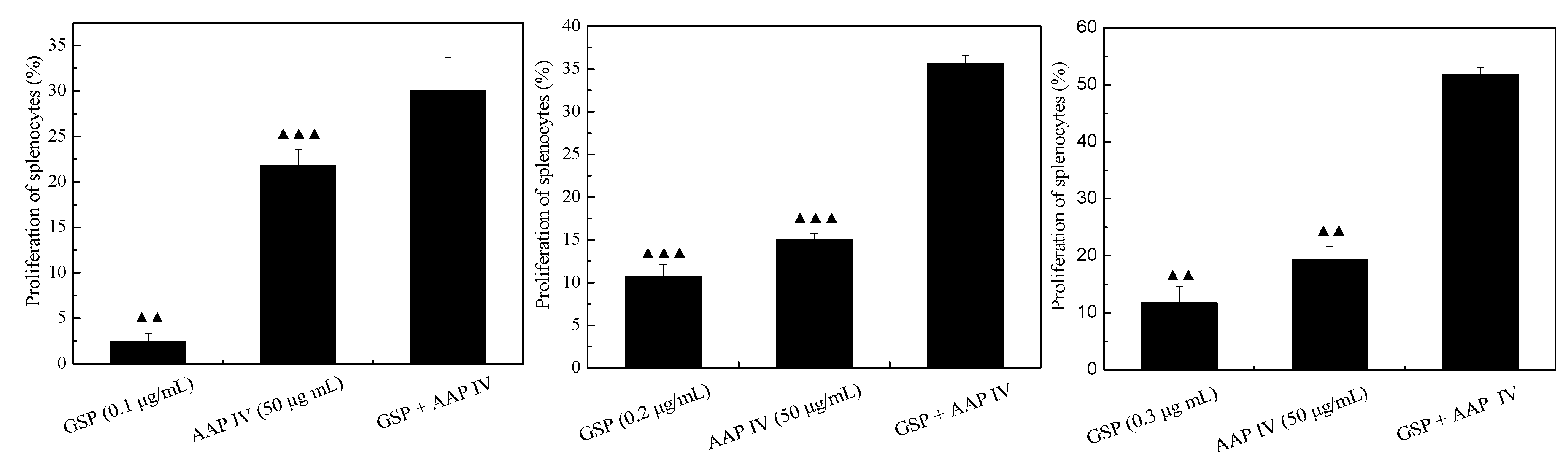
2.5. Proliferation of Splenocytes
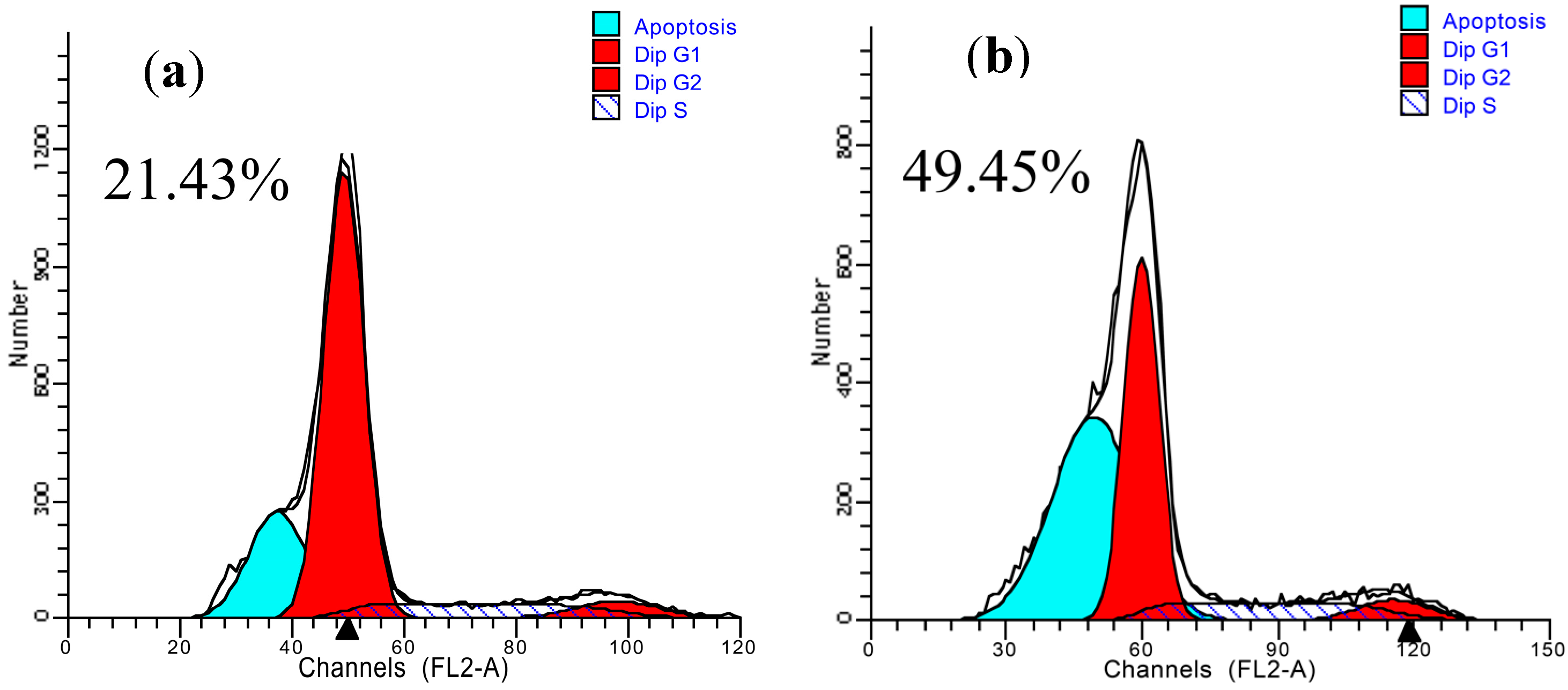
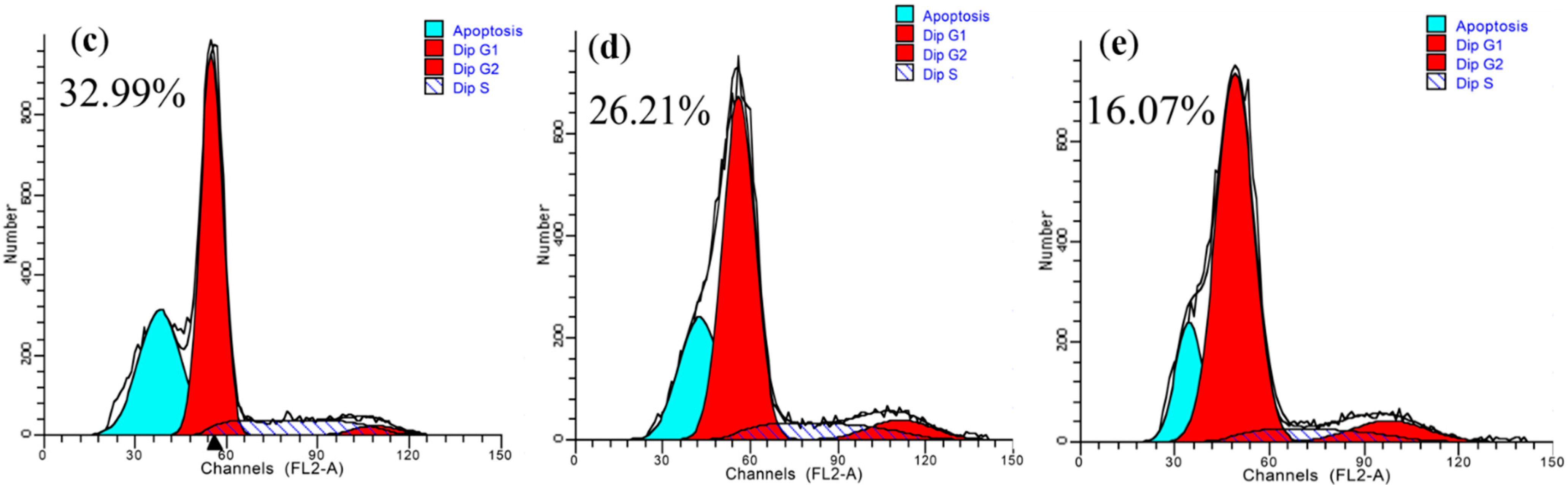
2.6. Apoptosis of Splenocytes
2.7. Product of Splenocytes
| Groups | ROS | MDA (nmol/mg) | GSH (mg/g) |
|---|---|---|---|
| Normal Control | 32.49 ± 1.66 | 7.93 ± 0.30 | 80.00 ± 2.12 |
| RT model group | 47.07 ± 1.59 ▲▲ | 17.84 ± 0.45 ▲▲▲ | 57.03 ± 3.76 ▲▲ |
| RT + GSP (0.1 μg/mL) | 46.16 ± 1.68 | 16.19 ± 0.90 | 61.61 ± 3.83 |
| RT + GSP (0.2 μg/mL) | 45.77 ± 1.70 | 14.45 ± 0.78 # | 61.30 ± 4.01 |
| RT + GSP (0.3 μg/mL) | 44.38 ± 1.60 | 12.50 ± 0.55 ## | 64.19 ± 5.55 |
| RT + AAP IV (50 μg/mL) | 42.18 ± 1.33 | 15.97 ± 0.65 # | 60.65 ± 6.25 |
| RT + AAP IV (100 μg/mL) | 40.65 ± 1.84 # | 14.31 ± 0.58 # | 61.53 ± 2.35 ## |
| RT + GSP (0.1 μg/mL) + AAP IV (50 μg/mL) | 41.92 ± 1.58 ## (1.01) | 13.56 ± 0.50 ## (0.81) | 68.36 ± 5.34 ### (0.23) |
| RT + GSP (0.2 μg/mL) + AAP IV (50 μg/mL) | 39.16 ± 1.66 ###(0.38) | 10.51 ± 0.56 ###(0.50) | 63.64 ± 5.04 ### (0.94) |
| RT + PGS (0.3 μg/mL) + AAP IV (50 μg/mL) | 38.78 ± 1.78 ###(0.38) | 8.95 ± 0.45 #### (0.32) | 73.80 ± 0.13 ### (0.13) |
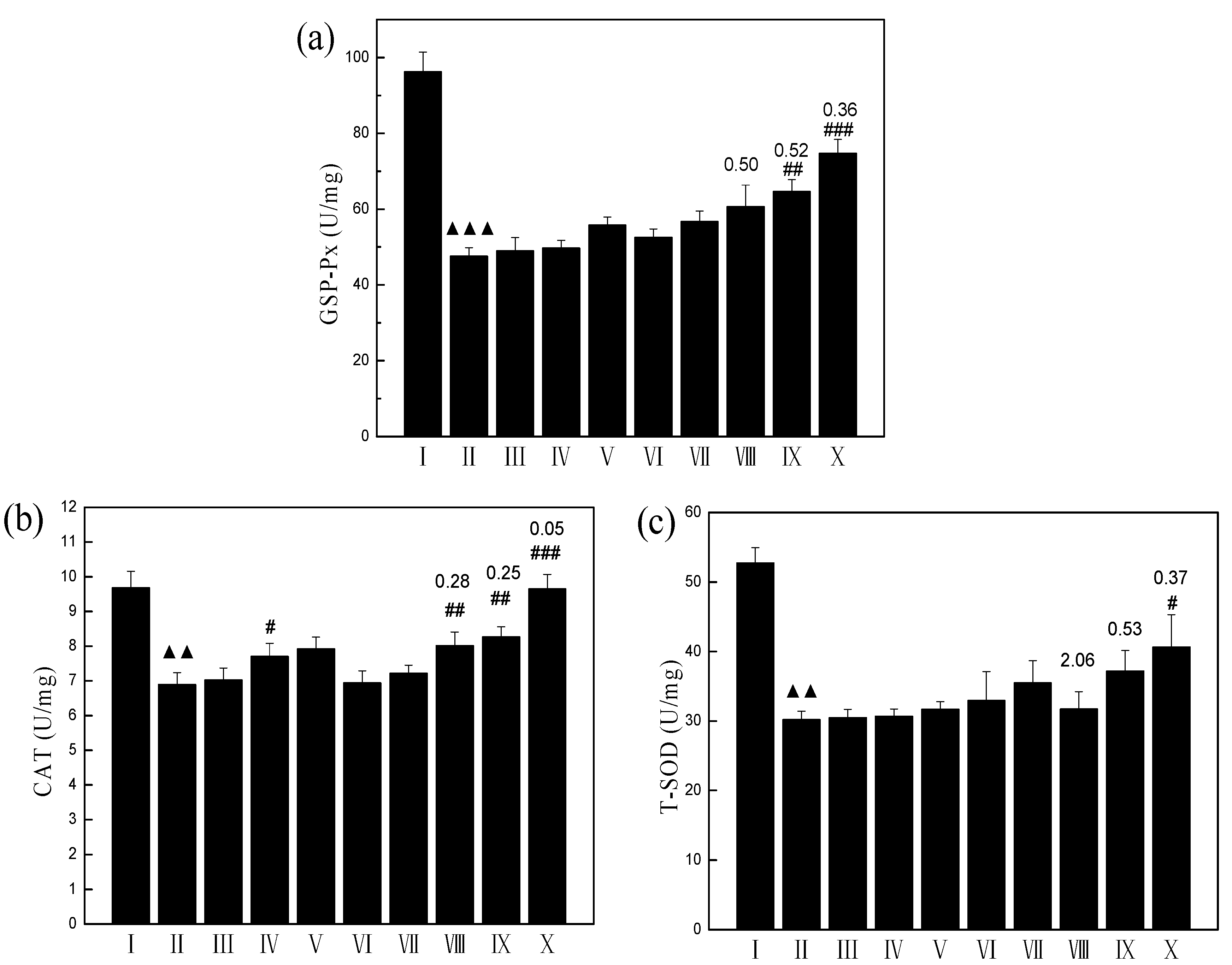
2.8. Enzyme Levels of Splenocytes
2.9. Spleen Indices
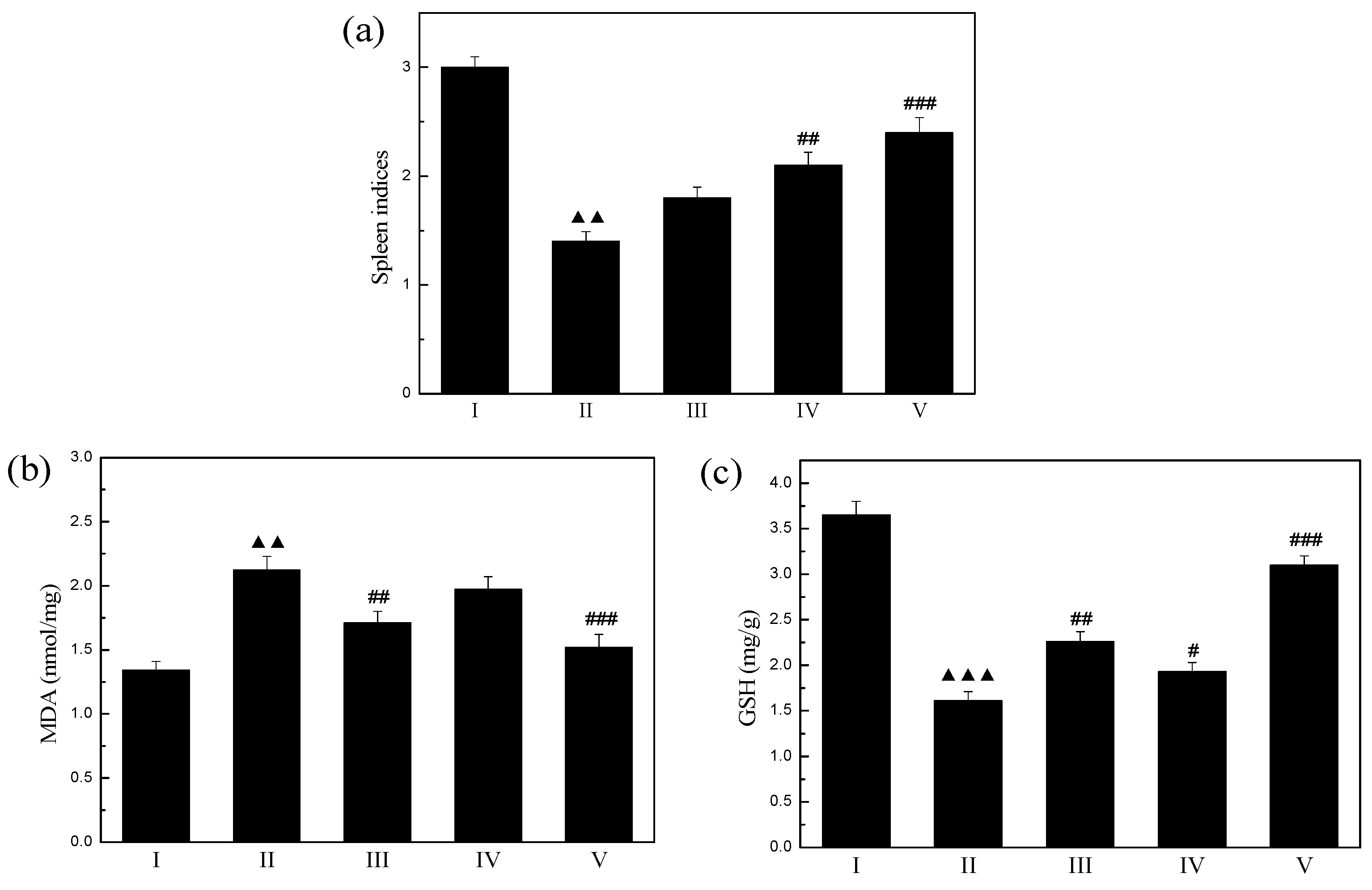
2.10. Spleen Products
3. Experimental Section
3.1. Chemicals and Reagents
3.2. Preparation and Purification of the AAP
3.3. Infrared Spectroscopy Analysis
3.4. Analysis of Monosaccharide Composition by GC-MS
3.4.1. Hydrolysis
3.4.2. Derivatization
3.4.3. GC-MS Analysis
3.5. Preparation of Splenocytes
3.6. Irradiation with 60Coγ-Rays
3.7. Splenocytes Viability Assay
3.8. Proliferation of Splenocytes
3.9. PI (Propidium Iodide) Staining Assay
3.10. 2'-7'-Dichlorofluorescein Diacetate (DCF-DA) Assay
3.11. Animals
3.12. Experimental Design and Oral Administration
- Group I: Normal mice (Normal group)
- Group II: Radiation (RT Model group)
- Group III: Radiation + GSP (1.2 mg/kg BW/D)
- Group IV: Radiation + AAP IV (200 mg/kg BW/D)
- Group V: Radiation + GSP (1.2 mg/kg BW/D) + AAP IV (200 mg/kg BW/D)
3.13. Analysis of Spleen Indices
3.14. Determination of Biological Parameters of Splenocytes and Spleen
3.15. Combination Index for Determining Addition, Synergism, and Antagonism
3.16. Statistical Analysis
4. Conclusions
Acknowledgments
Author Contributions
Conflicts of Interest
References
- Park, S.J.; Ahn, G.; Lee, N.H.; Park, J.W.; Jeon, Y.J.; Jee, Y. Phloroglucinol (PG) purified from Ecklonia cava attenuates radiation-induced apoptosis in blood lymphocytes and splenocytes. Food Chem. Toxicol. 2011, 49, 2236–2242. [Google Scholar] [CrossRef] [PubMed]
- Anderson, R.E.; Warner, N.L. Ionizing Radiation and the Immune Response. In Advances in Immunology; Frank, J.D.A.H., Ed.; Academic Press: Pittsburgh, PA, USA, 1976; Volume 24, pp. 215–335. [Google Scholar]
- Nair, C.K.K.; Parida, D.K.; Nomura, T. Radioprotectors in Radiotherapy. J. Radiat. Res. 2001, 42, 21–37. [Google Scholar] [CrossRef] [PubMed]
- Paul, P.; Bansal, P.; Nayak, P.G.; Pannakal, S.T.; Priyadarsini, K.I.; Unnikrishnan, M.K. Polyphenolic fraction of Pilea microphylla (L.) protects Chinese hamster lung fibroblasts against γ-radiation-induced cytotoxicity and genotoxicity. Environ. Toxicol. Pharmacol. 2012, 33, 107–119. [Google Scholar]
- Dixit, A.K.; Bhatnagar, D.; Kumar, V.; Chawla, D.; Fakhruddin, K.; Bhatnagar, D. Antioxidant potential and radioprotective effect of soy isoflavone against gamma irradiation induced oxidative stress. J. Funct. Foods 2012, 4, 197–206. [Google Scholar] [CrossRef]
- Shih, P.; Yeh, C.; Yen, G. Anthocyanins induce the activation of phase II enzymes through the antioxidant response element pathway against oxidative stress-induced apoptosis. J. Agric. Food Chem. 2007, 55, 9427–9435. [Google Scholar] [CrossRef] [PubMed]
- Park, E.; Hwang, I.; Song, J.Y.; Jee, Y. Acidic polysaccharide of Panax ginseng as a defense against small intestinal damage by whole-body gamma irradiation of mice. Acta Histochem. 2011, 113, 19–23. [Google Scholar] [CrossRef] [PubMed]
- Iglesias, J.; Pazos, M.; Torres, J.L.; Medina, I. Antioxidant mechanism of grape procyanidins in muscle tissues: Redox interactions with endogenous ascorbic acid and α-tocopherol. Food Chem. 2012, 134, 1767–1774. [Google Scholar] [CrossRef] [PubMed]
- Zuo, Y.M.; Wang, X.H.; Gao, S.; Zhang, Y. Oligomerized Grape Seed Proanthocyanidins Ameliorates Isoproterenol-induced Cardiac Remodeling in Rats: Role of Oxidative Stress. Phytother. Res. 2011, 25, 732–739. [Google Scholar] [PubMed]
- Yin, J.; Hu, Y.X.; Hu, Y.M.; Liu, X.Y.; Ma, Z. Effect of the grape seed procyanidins ionizing radiation in mice. Food Chem. 2007, 7, 1285–1286. [Google Scholar]
- Tong, H.B.; Song, X.F.; Sun, X.; Sun, G.R.; Du, F.F. Immunomodulatory and Antitumor Activities of Grape Seed Proanthocyanidins. J. Agric. Food Chem. 2011, 59, 11543–11547. [Google Scholar] [CrossRef] [PubMed]
- Mantena, S.K.; Katiyar, S.K. Grape seed proanthocyanidins inhibit UV-radiation-induced oxidative stress and activation of MAPK and NF-κB signaling in human epidermal keratinocytes. Free Radic. Biol. Med. 2006, 40, 1603–1614. [Google Scholar] [CrossRef] [PubMed]
- Matito, C.; Agell, N.; Sanchez-Tena, S.; Torres, J.L.; Cascante, M. Protective Effect of Structurally Diverse Grape Procyanidin Fractions against UV-Induced Cell Damage and Death. J. Agric. Food Chem. 2011, 59, 4489–4495. [Google Scholar] [CrossRef] [PubMed]
- Ma, J.; Qiao, Z.; Xiang, X. Optimisation of extraction procedure for black fungus polysaccharides and effect of the polysaccharides on blood lipid and myocardium antioxidant enzymes activities. Carbohydr. Polym. 2011, 84, 1061–1068. [Google Scholar] [CrossRef]
- Zhang, H.; Wang, Z.; Zhang, Z.; Wang, X. Purified Auricularia auricular-judae polysaccharide (AAP I-a) prevents oxidative stress in an ageing mouse model. Carbohydr. Polym. 2011, 84, 638–648. [Google Scholar] [CrossRef]
- Fan, L.S.; Gong, C.R.; Zhang, S.H. Study on Radio-Protective Effects of the Polysaccharide from Auricular auriculain Mice. Acta Nutr. Sin. 2005, 27, 525–526. [Google Scholar]
- Wagner, H.; Ulrich-Merzenich, G. Synergy research: Approaching a new generation of phytopharmaceuticals. Phytomedicine 2009, 16, 97–110. [Google Scholar] [CrossRef] [PubMed]
- Seed, T.M. Radiation protectants: Current status and future prospects. Health Phys. 2005, 89, 531–545. [Google Scholar] [CrossRef] [PubMed]
- Bai, H.; Liu, R.; Chen, H.; Zhang, W.; Wang, X.; Zhang, X.; Li, W.; Hai, C. Enhanced antioxidant effect of caffeic acid phenethyl ester and Trolox in combination against radiation induced-oxidative stress. Chem.-Biol. Interact. 2014, 207, 7–15. [Google Scholar] [CrossRef] [PubMed]
- Fan, Y.; Lin, M.; Luo, A.; Chun, Z.; Luo, A. Characterization and Antitumor Activity of a Polysaccharide from Sarcodia ceylonensis. Molecules 2014, 19, 10863–10876. [Google Scholar] [CrossRef] [PubMed]
- He, J.; Ru, Q.; Dong, D.; Sun, P. Chemical Characteristics and Antioxidant Properties of Crude Water Soluble Polysaccharides from Four Common Edible Mushrooms. Molecules 2012, 17, 4373–4387. [Google Scholar] [CrossRef] [PubMed]
- Mao, J.P.; Fang, J.; Zhou, Y.; Cui, Y.F.; Jiang, Z.J.; Du, L.; Ma, Q. Immunomodulator, immunosuppression of radiation and immune reconstruction. Bull. Acad. Mil. Med. Sci. 2010, 34, 480–484. [Google Scholar]
- Potten, C.S. The relationship between ionizing radiation-induced apoptosis and stem cells in the small and large intestine. Br. J. Cancer 1998, 78, 993–1003. [Google Scholar] [CrossRef] [PubMed]
- Park, E.; Ahn, G.; Lee, N.H.; Kim, J.M.; Yun, J.S.; Hyun, J.W.; Jeon, Y.; Wie, M.B.; Lee, Y.J.; Park, J.W.; et al. Radioprotective properties of eckol against ionizing radiation in mice. Febs Lett. 2008, 582, 925–930. [Google Scholar]
- Park, E.; Ahn, G.; Yun, J.S.; Kim, M.J.; Bing, S.J.; Kim, D.S.; Lee, J.; Lee, N.H.; Park, J.W.; Jee, Y. Dieckol rescues mice from lethal irradiation by accelerating hemopoiesis and curtailing immunosuppression. Int. J. Radiat. Biol. 2010, 86, 848–859. [Google Scholar] [PubMed]
- Qi, L.; Liu, C.Y.; Wu, W.Q.; Gu, Z.L.; Guo, C.Y. Protective effect of flavonoids from Astragalus complanatus on radiation induced damages in mice. Fitoterapia 2011, 82, 383–392. [Google Scholar] [CrossRef] [PubMed]
- Yang, Y.; Zhang, Z.; Li, S.; Ye, X.; Li, X.; He, K. Synergy effects of herb extracts: Pharmacokinetics and pharmacodynamic basis. Fitoterapia 2014, 92, 133–147. [Google Scholar] [CrossRef] [PubMed]
- Yan, F.; Zhang, Q.Y.; Jiao, L.; Han, T.; Zhang, H.; Qin, L.P.; Khalid, R. Synergistic hepatoprotective effect of Schisandrae lignans with Astragalus polysaccharides on chronic liver injury in rats. Phytomedicine 2009, 16, 805–813. [Google Scholar] [CrossRef] [PubMed]
- Li, M.H.; Xu, Y.; Yang, W.J.; Li, J.K.; Xu, X.Y.; Zhang, X.; Chen, F.T.; Li, D.P. In vitro synergistic anti-oxidant activities of solvent-extracted fractions from Astragalus membranaceus and Glycyrrhiza uralensis. LWT-Food Sci. Technol. 2011, 44, 1745–1751. [Google Scholar] [CrossRef]
- Boath, A.S.; Stewart, D.; Mcdougall, G.J. Berry components inhibit α-glucosidase in vitro: Synergies between acarbose and polyphenols from black currant and rowanberry. Food Chem. 2012, 135, 929–936. [Google Scholar] [CrossRef]
- Bai, H.N.; Wang, Z.Y.; Li, H.; Wang, L. Effect of five berry polyphenols and auricularia auricular polysaccharides combination on radiation protection. Sci. Technol. Food Ind. 2013, 34, 117–120. [Google Scholar]
- Sreeja, S.; Santhosh Kumar, T.R.; Lakshmi, B.S.; Sreeja, S. Pomegranate extract demonstrate a selective estrogen receptor modulator profile in human tumor cell lines and in vivo models of estrogen deprivation. J. Nutr. Biochem. 2012, 23, 725–732. [Google Scholar] [CrossRef] [PubMed]
- Leung, M.Y.K.; Liu, C.; Koon, J.C.M.; Fung, K.P. Polysaccharide biological response modifiers. Immunol. Lett. 2006, 105, 101–114. [Google Scholar] [CrossRef] [PubMed]
- John, C.M.; Sandrasaigaran, P.; Tong, C.K.; Adam, A.; Ramasamy, R. Immunomodulatory activity of polyphenols derived from Cassia auriculata flowers in aged rats. Cell. Immunol. 2011, 271, 474–479. [Google Scholar] [CrossRef] [PubMed]
- Hushmendy, S.; Jayakumar, L.; Hahn, A.B.; Bhoiwala, D.; Bhoiwala, D.L.; Crawford, D.R. Select phytochemicals suppress human T-lymphocytes and mouse splenocytes suggesting their use in autoimmunity and transplantation. Nutr. Res. 2009, 29, 568–578. [Google Scholar] [CrossRef] [PubMed]
- Lee, S.; Lillehoj, H.S.; Cho, S.; Chun, H.; Park, H.; Lim, C.; Lillehoj, E.P. Immunostimulatory effects of oriental plum (Prunus salicina Lindl). Comp. Immunol. Microbiol. Infect. Dis. 2009, 32, 407–417. [Google Scholar] [CrossRef] [PubMed]
- Lin, J.; Tang, C. Determination of total phenolic and flavonoid contents in selected fruits and vegetables, as well as their stimulatory effects on mouse splenocyte proliferation. Food Chem. 2007, 101, 140–147. [Google Scholar] [CrossRef]
- Manosroi, A.; Saraphanchotiwitthaya, A.; Manosroi, J. Immunomodulatory activities of fractions from hot aqueous extract of wood from Clausena excavata. Fitoterapia 2004, 75, 302–308. [Google Scholar] [CrossRef] [PubMed]
- Tu, J.; Sun, H.; Ye, Y. Immunomodulatory and antitumor activity of triterpenoid fractions from the rhizomes of Astilbe chinensis. J. Ethnopharmacol. 2008, 119, 266–271. [Google Scholar] [CrossRef] [PubMed]
- Sung, N.; Yang, M.; Song, D.; Byun, E.; Kim, J.; Park, J.; Song, B.; Lee, J.; Park, S.; Park, H.; et al. The procyanidin trimer C1 induces macrophage activation via NF-kappa B and MAPK pathways, leading to Th1 polarization in murine splenocytes. Eur. J. Pharmacol. 2013, 714, 218–228. [Google Scholar]
- Waselenko, J.K.; Blakely, W.F.; Wiley, A.L.; Tsu, H.; Coleman, C.N.; Lowry, P.; Dainiak, N. Medical management of the acute radiation syndrome: Recommendations of the Strategic National Stockpile Radiation Working Group. Ann. Intern. Med. 2004, 140, 1037–1051. [Google Scholar] [CrossRef] [PubMed]
- Neta, R.; Stiefel, S.M.; Finkelman, F.; Herrmann, S.; Ali, N. IL-12 protects bone marrow from and sensitizes intestinal tract to ionizing radiation. J. Immunol. 1994, 153, 4230–4237. [Google Scholar] [PubMed]
- Kadiiska, M.B.; Gladen, B.C.; Baird, D.D.; Dikalova, A.E.; Sohal, R.S.; Hatch, G.E.; Jones, D.P.; Mason, R.P.; Barrett, J.C. Biomarkers of oxidative stress study: Are plasma antioxidants markers of CCl4 poisoning? Free Radic. Biol. Med. 2000, 28, 838–845. [Google Scholar] [CrossRef] [PubMed]
- Archana, P.R.; Rao, B.N.; Rao, B. Modulation of Gamma Ray-Induced Genotoxic Effect by Thymol, a Monoterpene Phenol Derivative of Cymene. Integr. Cancer Ther. 2011, 10, 374–383. [Google Scholar] [CrossRef] [PubMed]
- Srinivasan, M.; Sudheer, A.R.; Pillai, K.R.; Kumar, P.R.; Sudhakaran, P.R.; Menon, V.P. Modulatory effects of curcumin on γ-radiation-induced cellular damage in primary culture of isolated rat hepatocytes. Environ. Toxicol. Pharmacol. 2007, 24, 98–105. [Google Scholar] [CrossRef] [PubMed]
- Yao, D.; Shi, W.; Gou, Y.; Zhou, X.; Yee Aw, T.; Zhou, Y.; Liu, Z. Fatty acid-mediated intracellular iron translocation: A synergistic mechanism of oxidative injury. Free Radic. Biol. Med. 2005, 39, 1385–1398. [Google Scholar] [CrossRef] [PubMed]
- Taysi, S.; Polat, F.; Gul, M.; Sari, R.A.; Bakan, E. Lipid peroxidation, some extracellular antioxidants, and antioxidant enzymes in serum of patients with rheumatoid arthritis. Rheumatol. Int. 2002, 21, 200–204. [Google Scholar] [CrossRef]
- Chen, Y.; Tang, J.; Wang, X.; Sun, F.; Liang, S. An immunostimulatory polysaccharide (SCP-IIa) from the fruit of Schisandra chinensis (Turcz.) Baill. Int. J. Biol. Macromol. 2012, 50, 844–848. [Google Scholar] [CrossRef]
- Zhang, H.; Wang, Z.; Yang, L.; Yang, X.; Wang, X.; Zhang, Z. In Vitro Antioxidant Activities of Sulfated Derivatives of Polysaccharides Extracted from Auricularia auricular. Int. J. Mol. Sci. 2011, 12, 3288–3302. [Google Scholar] [CrossRef] [PubMed]
- Limem, I.; Harizi, H.; Ghedira, K.; Chekir-Ghedira, L. Leaf extracts from Phlomis crinita Cav.subs. mauritanica Munby affect immune cell functions in vitro. Immunopharmacol. Immunotoxicol. 2011, 33, 309–314. [Google Scholar]
- Carmichael, J.; Degraff, W.G.; Gazdar, A.F.; Minna, J.D.; Mitchell, J.B. Evaluation of a tetrazolium-based semiautomated colorimetric assay: Assessment of chemosensitivity testing. Cancer Res. 1987, 47, 936–942. [Google Scholar] [PubMed]
- Ran, Y.; Wang, R.; Gao, Q.; Jia, Q.; Hasan, M.; Awan, M.U.F.; Tang, B.; Zhou, R.; Dong, Y.; Wang, X.; et al. Dragon’s blood and its extracts attenuate radiation-induced oxidative stress in mice. J. Radiat. Res. 2014, 55, 699–706. [Google Scholar]
- Berenbaum, M.C. What is synergy? Pharmacol. Rev. 1989, 41, 91–143. [Google Scholar]
- Chou, T.; Talalay, P. Quantitative analysis of dose-effect relationships: The combined effects of multiple drugs or enzyme inhibitors. Adv. Enzym. Regul. 1984, 22, 27–55. [Google Scholar] [CrossRef]
- Chou, T.C. Theoretical basis, experimental design, and computerized simulation of synergism and antagonism in drug combination studies. Pharmacol. Rev. 2006, 58, 621–681. [Google Scholar] [CrossRef] [PubMed]
- Sample Availability: Samples of the compounds AAP IV, GSP are available from the authors.
© 2014 by the authors. Licensee MDPI, Basel, Switzerland. This article is an open access article distributed under the terms and conditions of the Creative Commons Attribution license ( http://creativecommons.org/licenses/by/4.0/).
Share and Cite
Bai, H.; Wang, Z.; Cui, J.; Yun, K.; Zhang, H.; Liu, R.H.; Fan, Z.; Cheng, C. Synergistic Radiation Protective Effect of Purified Auricularia auricular-judae Polysaccharide (AAP IV) with Grape Seed Procyanidins. Molecules 2014, 19, 20675-20694. https://doi.org/10.3390/molecules191220675
Bai H, Wang Z, Cui J, Yun K, Zhang H, Liu RH, Fan Z, Cheng C. Synergistic Radiation Protective Effect of Purified Auricularia auricular-judae Polysaccharide (AAP IV) with Grape Seed Procyanidins. Molecules. 2014; 19(12):20675-20694. https://doi.org/10.3390/molecules191220675
Chicago/Turabian StyleBai, Haina, Zhenyu Wang, Jie Cui, Keli Yun, Hua Zhang, Rui Hai Liu, Ziluan Fan, and Cuilin Cheng. 2014. "Synergistic Radiation Protective Effect of Purified Auricularia auricular-judae Polysaccharide (AAP IV) with Grape Seed Procyanidins" Molecules 19, no. 12: 20675-20694. https://doi.org/10.3390/molecules191220675