Article

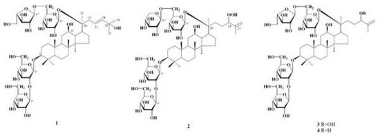
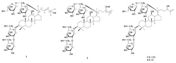
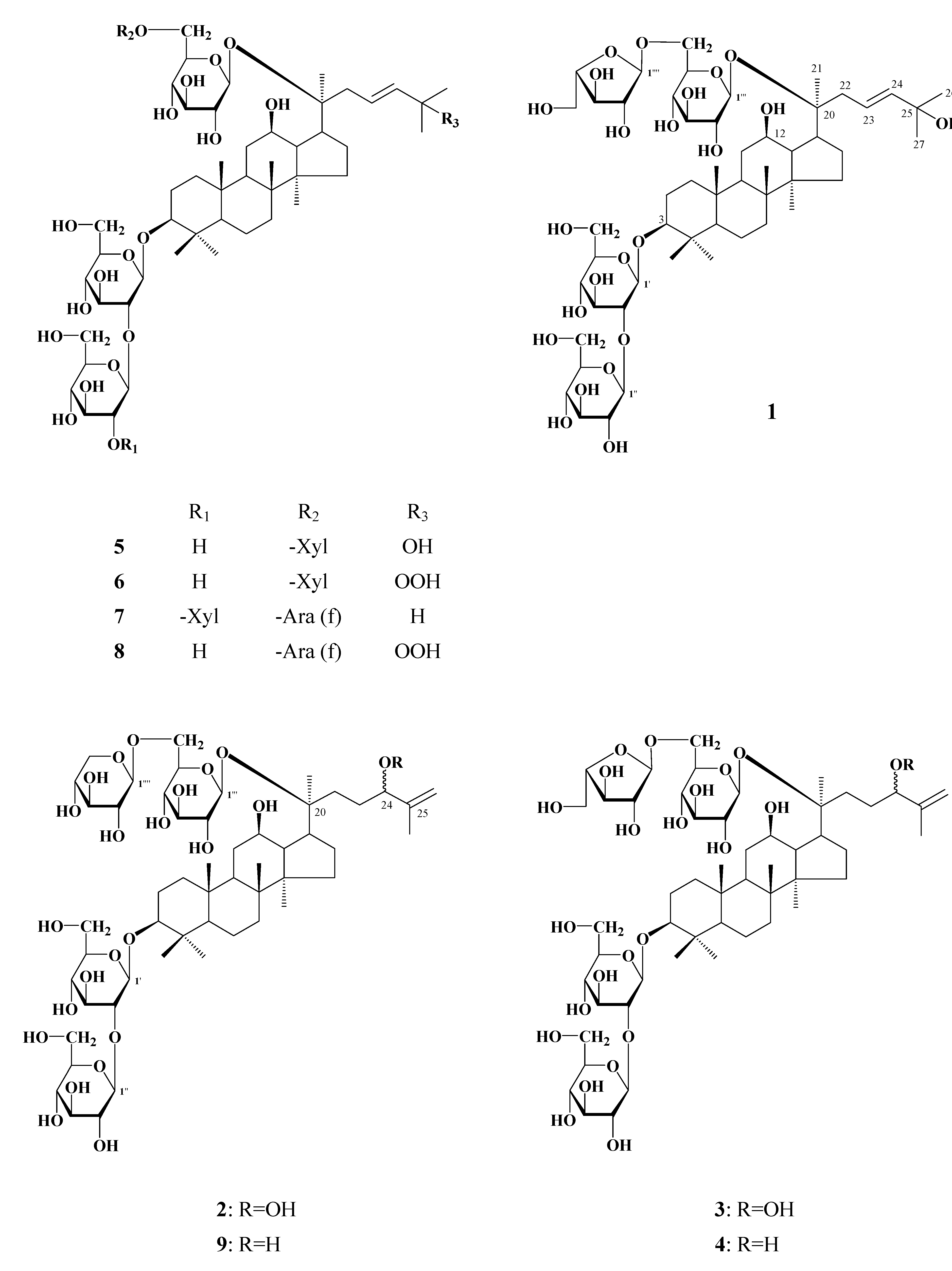
Dammarane-type Triterpene Saponins from the Flowers of Panax notoginseng

1
School of Chinese Medicine, Hong Kong Baptist University, Kowloon Tong, Hong Kong, China
2
Department of Natural Product Chemistry, Graduate School of Biomedical Sciences, Nagasaki University, Bunkyo-machi 1-14, Nagasaki 852-8521, Japan
*
Authors to whom correspondence should be addressed.
Molecules 2009, 14(6), 2087-2094; https://doi.org/10.3390/molecules14062087
Submission received: 11 May 2009 / Accepted: 1 June 2009 / Published: 10 June 2009

Abstract

:
Four new dammarane-type triterpene saponins named floranotoginsenosides A (1), B (2), C (3) and D (4), together with five known triterpene saponins, were isolated from the flowers of Panax notoginseng. Their structures were elucidated on the basis of spectral and chemical evidence.

Graphical Abstract

1. Introduction

Root of Panax notoginseng (Burk) F. H. Chen (Araliaceae), commonly referred to notoginseng and “San-Qi”, is a well-known medicinal herb, used historically in traditional Chinese medicine as a hemostatic agent that both invigorates and builds blood. The flowers of P. notoginseng have been used for treating hypertension, vertigo, tinnitus and laryngopharyngitis [1]. The saponin fraction of the notoginseng flowers has been proven to have hepatoprotective effect [2]. More than 60 dammarane-type saponins named ginsenosides, notoginsenosides and gypenosides have been hitherto isolated from this plant [3,4], among which about 20 saponins were obtained from its flower buds [5,6].
We herein report the isolation and structural elucidation of four new dammarane-type triterpene saponins 1-4, together with five known saponins 5-9 from the flowers of Panax notoginseng.

2. Results and Discussion

The water layer of methanolic extracts of notoginseng flowers was separated by MCI gel CHP 20P and ODS column chromatographies, and purified by preparative HPLC (ODS) to afford compounds 1-9. Compounds 5-9 were identified as gypenoside LXIX (5) [7], 3β,12,20S-trihydroxy-25-hydro-peroxydammar-23-ene-3-O-[β-D–glucopyranosyl(1→2)-β-D-glucopyranosyl]-20-O-[β-D-xylopyrano-syl(1→6)]-β-D-glucopyranoside (6) [8], notoginsenoside FP2 (7) [4], floraginsenoside O (8) [9] and gypenoside LXXI (9) [10], respectively, by comparison of their spectral data with those described in the literature. Compounds 7 and 8 were previously isolated from Panax notoginseng and P. ginseng, respectively, while compounds 5, 6 and 9 were isolated from Gynostemma pentaphyllum.
Compound 1, named floranotoginsenoside A, was obtained as a white powder, [α]D25: -4.93o (MeOH). The molecular formula of 1 was determined to be C53H90O23 on the basis of its negative FAB-MS (m/z 1,093 for [M-H]-) and high-resolution ESI-MS data (m/z 1,093.5790 for [M-H]-). The 1H-NMR spectrum of 1 showed signals assignable to eight singlet methyls (δH 0.86, 0.91, 0.93, 1.02, 1.07, 1.29 × 2, 1.33) together with four anomeric proton signals at δΗ 4.94 (s), 4.82 (d, J = 8 Hz), 4.66 (d, J = 8 Hz), 4.60 (d, J = 8 Hz). In the 13C-NMR spectrum of 1, 30 carbon signals due to the aglycone moiety were in good agreement with those of 5, especially the olefinic carbon signals (δC 123.8 and 141.9), and the oxygenated carbon signal (δC 71.5) of the side chain, suggesting that the aglycone of compound 1 is the same as that of 5, in which a double bond located at C-23, 24 and a hydroxyl group attached to C-25. The signals at δC 91.3 and 84.6 indicated that 1 is a bisdesmoside. Besides the aglycone’s 13C-NMR signals, the remaining carbon signals of compound 1 were found to be superimposable on those of compound 5 except for the presence of signals derived from an α-L-arabinofuranosyl moiety instead of β−D-xylopyranosyl moiety in 5, suggesting that oligosaccharide chain of α-L-arabinofuranosyl-(1→6)-β-D-glucopyranosyl attached to 20-OH of the aglycone. This was further supported by chemical shift comparison of these sugar signals with those of compound 8. On the basis of these results, the structure of floranotoginsenoside A was assigned to formula 1 (Figure 1).
Floranotoginsenoside B (2) was also obtained as a white powder, whose negative FAB-MS showed a quasimolecular ion peak at m/z 1,091 ([M-H-H2O]-). The molecular formula C53H90O24 was determined by high-resolution ESI-MS (m/z 1,109.5748 for [M-H]-). The 1H-NMR spectra of 2 showed four anomeric proton signals at δΗ 5.38 (d, J = 8 Hz), 5.05 (d, J = 8 Hz,), 5.00 (d, J = 8 Hz), 4.92 (d, J = 8 Hz), a vinyl methyl [δΗ 1.96 (s, H3-27)], and a methine proton bearing a hydroperoxyl group [δΗ 4.79 (dd-like, H-24)] which was supported by chemical shift comparison with those of related hydroperoxylated triterpenes [11]. The 1H-NMR and 13C-NMR of 2 are quite similar to those of notoginsenoside-C [12], except for the presence of signals for a β-D-xylopyranosyl moiety and absence of signals for a β-D-glucopyranosyl moiety. This suggested that 2 has the same aglycone and similar glycosylation manner with that of notoginsenoside-C with the only difference in the β-D-xylopyranosyl moiety as the terminal sugar of C-20 in 2. Reduction of 2 by triphenylphosphine afforded gypenoside LXXI (9), which strongly supports the proposed structure. Thus the structure of floranotoginsenoside B was characterized as shown for 2 (Figure 1).
Figure 1. Structures of compounds 1-9.
Molecules 14 02087 g001
Floranotoginsenoside C (3) was isolated as a white powder and has the same molecular formula as 2 on the basis of the [M-H-H2O]- peak at m/z 1,091 in the negative FAB-MS and [M-H]- at m/z 1,109.5747 in negative HR-ESI-MS. Its 1H-NMR and 13C-NMR spectral data again suggested a side chain with a hydroperoxyl group at C-24, as evidenced by the signals ascribed to the vinyl methyl [δΗ 1.97 (s, H3-27)], and methine group bearing hydroperoxyl group [δΗ 4.79 (dd-like, H-24), δC 90.1 (C-24)]. Comparison of the 1H-NMR and 13C-NMR spectra of 3 with those of 2 revealed that the only difference was the replacement of the terminal β-D-xylopyranosyl group in C-20 position in 2 by an α-L-arabinofuranosyl moiety in 3. Based on these evidences, structure 3 was assigned to floranoto-ginsenoside C (Figure 1).
Floranotoginsenoside D (4) has the molecular formula C53H90O23 which is one oxygen less than 3, on the basis of the [M-H]- peak at m/z 1,093 in the negative FAB-MS and m/z 1,093.5792 for [M-H]- in negative HR-ESI-MS spectra. It’s 1H-NMR and 13C-NMR spectra closely resembled those of 3, especially the signals arising from the sugar moiety and the rings A-D, indicating that 4 is a also dammaranes bidemoside. In comparison of the 13C-NMR spectra of 4 with that of 3, the upfield shift of C-24 (ΔδC -13.7) and C-26 (ΔδC -2.9), as well as the downfield shift of C-25 (ΔδC +3.4) were observed, indicating that a hydroxyl group is located at C-24 position in 4. This was supported by chemical shift comparison of H-24 (δH 4.42) and C-24 (δC 76.4) with that of 9 and related compounds with the same side chain structure [13]. When 3 was treated by triphenylphosphine, 4 was obtained. Therefore, the structure of floranotoginsenoside D was determined to be the formula 4 (Figure 1).
Table 1. 13C-NMR data for compounds 1-9.
1a)2c)3 c)4 c)5a)8a)9 c)
C-139.939.239.239.240.240.239.2
C-227.026.826.726.727.027.026.7
C-391.389.089.089.091.391.389.0
C-439.840.039.739.740.540.640.0
C-557.656.456.356.357.557.556.4
C-619.218.418.418.419.219.218.6
C-735.835.135.135.0 35.835.835.1
C-840.240.040.040.041.040.640.0
C-950.050.250.250.151.052.550.2
C-1037.936.936.936.937.937.936.9
C-1130.930.930.930.930.930.930.9
C-1271.371.170.370.471.572.071.1
C-1350.0 49.549.449.3 49.852.549.4
C-1451.051.451.451.452.451.051.4
C-1530.130.730.730.831.331.330.8
C-1627.226.626.626.727.227.226.7
C-1752.551.551.552.053.153.151.8
C-1816.416.016.0 15.816.716.416.0
C-1916.716.316.316.316.716.716.3
C-2084.683.483.483.584.684.683.4
C-2123.422.422.422.523.323.422.6
C-2239.932.732.732.740.041.032.7
C-23123.826.326.530.4123.8127.530.7
C-24141.990.190.176.4141.9138.176.7
C-2571.5146.2146.1149.571.582.6149.4
C-2631.4113.3113.4110.530.124.8110.0
C-2731.417.417.718.029.825.218.4
C-2828.428.128.128.128.428.428.1
C-2916.716.616.616.616.716.716.6
C-3017.317.417.317.217.317.317.3
Glc-1'105.4105.1105.1105.1105.3105.4105.1
2'81.183.483.383.381.083.283.7
3'78.578.378.378.278.478.378.2
4'71.971.671.671.671.871.571.6
5'78.178.178.378.278.078.178.1
6'62.862.962.962.863.062.862.8
Glc-1''104.5106.0106.0106.0104.4105.4106.0
2''77.777.177.977.977.677.677.1
3''77.978.178.178.977.877.878.1
4''71.971.672.072.071.671.971.7
5''78.378.378.878.978.278.178.2
6''63.162.762.762.762.863.062.6
Glc (Xyl)-1'''98.198.098.098.098.098.098.0
2'''75.375.074.975.075.375.375.9
3'''78.579.379.279.078.478.979.2
4'''71.571.671.671.671.171.571.6
5'''77.777.177.177.177.577.677.1
6'''68.570.068.368.370.163.069.8
Ara (Xyl, Glc)-1''''110.0105.6110.0110.0104.4110.0105.6
2''''83.374.883.483.574.883.274.8
3''''78.578.378.878.978.478.578.2
4''''85.771.185.985.971.185.671.1
5''''62.866.962.762.766.862.867.0
a) 75 MHz, CD3OD; b) 125 MHz, pyridine-d5; c) 75 MHz, pyridine-d5.

Experimental

General

Optical rotations were measured with a JASCO DIP-370 digital polarimeter. 1H- and 13C-NMR spectra were recorded on Varian Unity plus 500 and Varian Gemini 300 spectrometers. Coupling constants (J) are expressed in Hz, and chemical shifts are given on a δ (ppm) scale with tetramethylsilane as an internal standard. MS were recorded on a JEOL JMS DX-303 spectrometer, and glycerol was used as a matrix for FAB-MS measurement. HR-ESI-MS was preformed on a Q-TOF mass spectrometer (Bruker Daltonics, MA, USA). Column chromatographies were performed with Kieselgel 60 (70 - 230 mesh, Merck), MCI-gel CHP 20P (75 - 150 mm, Mitsubishi Chemical Co. Ltd., Japan), Chromatorex ODS (100 - 200 mesh, Fuji Silysia Chemical). Preparative HPLC (Shimadzu ODS) was performed on a Tosoh apparatus equipped with a CCPM solvent delivery system. Thin layer chromatography (TLC) was performed on precoated Kieselgel 60 F254 plates (0.2 mm thick, Merck), and spots were detected by ultraviolet (UV) illumination and by spraying 10% sulfuric acid reagent.

Extraction and Isolation

Air-dried flowers of Panax notoginseng (800 g) purchased from a herbs market in Kunming, Yunnan Province, P.R. China in August, 1997 were extracted three times with methanol at room temperature. After removal of the solvent by evaporation in vacuo, the extract was suspended in water and extracted successively with Et2O, EtOAc, n-BuOH. The H2O layer (40.8 g of 168.5 g) was subjected to column chromatography over MCI-gel CHP 20P eluted from 0 % to 100 % MeOH to afford four fractions. Fraction-2 (3.15 g) eluted by (50-60 % MeOH) was chromatographed on MCI-gel CHP 20P (eluted with 50 % to 80 % MeOH) and Chromatorex ODS (eluted from and 50 to 100 % MeOH) to afford a saponin fraction (0.89 g). This fraction were purified by preparative HPLC to give compounds 6 (37.7 mg), 2 (25.3 mg), 9 (32.6 mg), 5 (28.4 mg), 3 (28.4 mg), 4 (11.4 mg), 1 (15.6 mg), 8 (28.6 mg) and 7 (58.4 mg).
Floranotoginsenoside A (1): C53H90O23, white powder, [α]D25: -4.93º (MeOH). Negative FAB-MS (m/z):1,093 [M-H]-; Negative HR-ESI-MS (m/z): 1,093.5790 [M-H]- (calculated for C53H89O23, 1,093.5795); 1H-NMR (CD3OD, 300 MHz): δ 0.86, 0.91, 0.93, 1.02, 1.07, 1.33, (3H each, all s, H-19, 30, 18, 21, 29, 28), 1.29 (6H, s, H-26, 27), 4.60 (1H, d, J = 8 Hz, H-1'), 4.66 (1H, d, J = 8 Hz, H-1'''), 4.82 (1H, d, J = 8 Hz, H-1''), 4.94 (1H, s, H-1 '''') 5.71 (2H, m, H-23, 24); 13C-NMR data are listed in Table 1.
Floranotoginsenoside B (2): C53H90O24, white powder, [α]D25: -9.50º (MeOH); Negative FAB-MS (m/z):1,091[M-H-H2O]-; Negative HR-ESI-MS (m/z) 1,109.5748 [M-H]- (calculated. for C53H89O24, 1,109.5744); 1H-NMR (pyridine-d5, 300 MHz): δ0.81, 0.94, 0.96, 1.11, 1.28, 1.64, 1.96 ( 3H each, all s, H-19, 30, 18, 29, 28,21,27), 3.27 (1H, dd, J = 3, 11Hz, H-3 ), 4.74 (1H, dd-like, H-24 ), 4.92 ( 1H, d, J = 8 Hz, H-1' ), 5.00 ( 1H, d, J = 8 Hz, H-1'''' ), 5.05 ( 1H, d, J = 8 Hz, H-1''' ), 5.38 ( 1H, d, J = 8 Hz, H-1'' ); 13C-NMR data are listed in Table 1.
Floranotoginsenoside C (3): C53H90O24, white powder, [α]D25: -14.68º (MeOH); Negative FAB-MS (m/z):1,091[M-H-H2O]-; Negative HR-ESI-MS (m/z) 1,109.5747 [M-H]- (calculated. for C53H89O24, 1,109.5744); 1H-NMR (pyridine-d5, 300 MHz): δ 0.80, 0.94, 0.94, 1.11, 1.28, 1.62, 1.97,(3H each, all s, H-19, 30, 18, 29, 28, 21, 27), 3.27 (1H, dd, J = 3, 11 Hz, H-3), 4.79 (1H, dd-like, H-24), 4.91 (1H, d, J = 8 Hz, H-1' ), 5.10 (1H, d-like, H-1'''), 5.39 (1H, d, J = 8 Hz, H-1"), 5.66 (1H, s, H-1''''); 13C-NMR data are listed in Table 1.
Floranotoginsenoside D (4): C53H90O23, white powder, [α]D25: -14.04º (MeOH); Negative FAB-MS (m/z):1,093[M-H]-; Negative HR-ESI-MS (m/z) 1,093.5792 [M-H]- (calculated. for C53H89O23, 1,093.5795); 1H-NMR (pyridine-d5, 300 MHz): δ0.80, 0.93, 0.99, 1.10,1.28, 1.64, 1.95, (3H each, all s, H-19, 30, 29, 28, 21, 27), 3,25 (1H, dd, J = 3, 11 Hz, H-3), 4.90 (1H, d, J = 8 Hz, H-1'), 5.10 ( Overlapping, H-26 ), 5.09 (Overlapping, H-1'''), 5.40 (1H, d, J = 7.4 Hz, H-1"), 5.66 ( 1H, s, H-1''''); 13C-NMR data are listed in Table 1.
Reduction of 2 and 3: Triphenylphosphine (10 mg) was added to a solution of 2 (10 mg) in MeOH (5 ml). After stirring at room temperature for 4 hr, the solution was evaporated to dryness under reduced pressure, then subjected to silica gel chromatography (CHCl3-MeOH-Water: 8:2:0.2) to afford 9 (6.1 mg) whose 1H- and 13C-NMR spectral data are completely identical with those of gypenoside LXXI. Reduction of 3 (10 mg) in a manner similar to that described for 2 yielded floranotoginsenoside D (4, 5.6 mg).

Conclusions

Among four new darmmarane-type triterpene saponins isolated from notoginseng flowers, compounds 2 and 3 have a hydroperoxyl group. Although hydroperoxydammarane triterpene saponins have been previously isolated from the roots of Panax notoginseng [12], this was the first report of the isolation of this kind of saponins from the notoginseng flowers. These results may contribute to better understanding on the chemical characteristics of this medicinal herb.

References

  1. Zhonghuabencao Editorial Board. Zhonghuabencao; Shanghai Science and Technology Press: Shanghai, China, 1997. [Google Scholar]
  2. Yoshikawa, M.; Morikawa, T.; Kashima, Y.; Ninomiya, K.; Matsuda, H. Structures of new dammarane-type triterpene saponins from the flower buds of Panax notoginseng and hepatoprotective effects of principal ginseng saponins. J. Nat. Prod. 2003, 66, 922–927. [Google Scholar] [CrossRef]
  3. Yoshikawa, M.; Murakami, T.; Ueno, T.; Hirokawa, K.Y.; Murakami, N.; Yamahara, J.; Matsuda, H.; Saijoh, R.; Tanaka, O. Bioactive saponins and glycosides. IX. Notoginseng (2): structures of five new dammaranes-type triterpene oligoglycosides, notoginsenosides-E, -G,-H, -I, and -J, and a novel acetylenic fatty acid glycoside, notoginsenic acid β-sophoroside, from the dried root of Panax notoginseng (Burk.) F. H. Chen. Chem. Pharm. Bull. 1997, 45, 1056–1062. [Google Scholar] [CrossRef]
  4. Wang, X.Y.; Wang, D.; Ma, X.X.; Zhang, Y.J.; Yang, C.R. Two new dammarane-type bisdemosides from the fruit pedicels of Panax notoginseng. Helv. Chim. Acta 2002, 91, 60–66. [Google Scholar]
  5. Zuo, G.Y.; Wei, J.X.; Du, Y.C; Cao, S.M; Chen, Y.G. Saponins from flower-buds of Sanchi (Panax notoginseng (Burk) F. H. Chen). Nat. Prod. Res. Dev. 1991, 3, 24–30. [Google Scholar]
  6. Taniyasu, S.; Tanaka, O.; Yang, T.R.; Zhou, J. Dammarane saponins of flower buds of Panax notoginseng (Sanchi-Ginseng). Planta Med. 1982, 4, 124–5. [Google Scholar] [CrossRef]
  7. Yin, F.; Hu, L.H.; Pan, R.X. Novel Dammarane-type glycosides from Gynostemma pentaphyllum. Chem. Pharm. Bull. 2004, 52, 60–66. [Google Scholar]
  8. Yin, F.; Hu, L.H.; Lou, F.C.; Pan, R.X. Dammarane-type glycosides from Gynostemma pentaphyllum. J. Nat. Prod. 2004, 67, 942–952. [Google Scholar] [CrossRef]
  9. Yoshikawa, M.; Sugimoto, S.; Nakamura, S.; Sakumae, H.; Matsuda, H. Medicinal Flowers. XVI. New dammaranes-type tetraglycosides and gastroprotective principles from flower buds of Panax ginseng. Chem. Pharm. Bull. 2007, 55, 1034–1038. [Google Scholar] [CrossRef]
  10. Yoshikawa, K.; Takemoto, T.; Arihara, S. Studies on the constituents of Cucurbitaceae plants. XVL. On the saponin constituents of Gynostemma pentaphyllum Makino (II). Yakugaku Zasshi 1987, 107, 262–267. [Google Scholar]
  11. Jiang, Z.-H.; Fukuoka, R.; Aoki, F.; Tanaka, T.; Kouno, I. Dammarane-type triterpene glycosides from the leaves of Rhoiptelea chiliantha. Chem. Pharm. Bull. 1999, 47, 257–262. [Google Scholar] [CrossRef]
  12. Yoshikawa, M.; Murakami, T.; Ueno, T.; Yashiro, K.; Hirokawa, N.; Murakami, N.; Yamahara, J.; Matsuda, H.; Saijoh, R.; Tanaka, O. Bioactive saponins and glycosides. VIII. Notoginseng (1), New dammaranes-type triterpene oligoglycosides, notoginsenosides-A, -B,-C, and -D from the dried root of Panax notoginseng (Burk.) F. H. Chen. Chem. Pharm. Bull. 1997, 45, 1039–1045. [Google Scholar] [CrossRef]
  13. Duc, N.M.; Kasai, R.; Ohtani, K.; Ito, A.; Nham, N.T.; Yamasaki, K.; Tanaka, O. Saponins from Vietnamese ginseng, Panax vietnamensis Ha et Grushv collected in central Vietnam II. Chem. Pharm. Bull. 1994, 42, 115–122. [Google Scholar] [CrossRef]
  • Sample Availability: Samples of the compounds 1-4 are available from the authors.

