Review

Biological Water Dynamics and Entropy: A Biophysical Origin of Cancer and Other Diseases †

1
Internal Medicine Group Practice, PhyNet, Inc., 4002 Technology Center, Longview, TX 75605, USA
2
Independent Researcher, Houston, TX 77084, USA
3
Computer Science and Artificial Intelligence Laboratory, MIT, 32 Vassar Street, Cambridge, MA 02139, USA
*
Author to whom correspondence should be addressed.
Note added by the Publisher: The editors of the journal have been alerted to concerns over potential bias in opinions and bias in the choice of citation sources used in this article. We note that the authors stand by the content as published. Since the nature of the claims against the paper concern speculation and opinion, and not fraud or academic misconduct, the editors would like to issue an Expression of Concern to make readers aware that the approach to collating literature citations for this article was likely not systematic and may not reflect the spectrum of opinions on the issues covered by the article. Please refer to our policy regarding <a href='https://www.mdpi.com/about/controversial-articles'>possibly controversial articles.
Entropy 2013, 15(9), 3822-3876; https://doi.org/10.3390/e15093822
Submission received: 3 June 2013 / Revised: 26 August 2013 / Accepted: 30 August 2013 / Published: 13 September 2013
(This article belongs to the Section Entropy Reviews)

Abstract

:
This paper postulates that water structure is altered by biomolecules as well as by disease-enabling entities such as certain solvated ions, and in turn water dynamics and structure affect the function of biomolecular interactions. Although the structural and dynamical alterations are subtle, they perturb a well-balanced system sufficiently to facilitate disease. We propose that the disruption of water dynamics between and within cells underlies many disease conditions. We survey recent advances in magnetobiology, nanobiology, and colloid and interface science that point compellingly to the crucial role played by the unique physical properties of quantum coherent nanomolecular clusters of magnetized water in enabling life at the cellular level by solving the “problems” of thermal diffusion, intracellular crowding, and molecular self-assembly. Interphase water and cellular surface tension, normally maintained by biological sulfates at membrane surfaces, are compromised by exogenous interfacial water stressors such as cationic aluminum, with consequences that include greater local water hydrophobicity, increased water tension, and interphase stretching. The ultimate result is greater “stiffness” in the extracellular matrix and either the “soft” cancerous state or the “soft” neurodegenerative state within cells. Our hypothesis provides a basis for understanding why so many idiopathic diseases of today are highly stereotyped and pluricausal.
PACS Codes:
87.19.xj; 87.19.xr; 87.19.xv; 87.19.xw; 87.19.xb; 87.19.xp; 03.75.Hh; 03.75.Kk; 42.50.Gy; 87.15.B; 03.75.Lm; 87.15.N; 75.10.-b; 75.78.-n; 82.70.Uv; 36.40.Wa; 68.35.Rh; 74.25.fc

1. Introduction: Is Biomacromolecular Dysfunction a Cause or Biomarker of Disease?

The vast medical research literature contains extensive documentation of dysfunctional changes in biomacromolecular structure and activity seen in chronic and infectious diseases. Molecular-level phenomena described include inappropriate gene activation and protein synthesis driving uncontrolled division of cancer cells, diversion of this same gene expression process to proliferate infectious viruses, and beta-amyloid protein tangles characteristic of Alzheimer’s disease, to name just a few. Details for individual diseases vary, but the common feature of molecular mechanisms offered for all of them is emphasis on the roles of macromolecules and their non-aqueous substrates or ligands, with little or no attention given to water, the most abundant molecule in the body and the most essential for all forms of life. In this paper, we present an alternative view of disease etiology that places water at the center of the stage. We propose that a major cause of inflammation and disease is disruption of normal water structures between and within cells, which then gives rise to the pathological macromolecular changes reported in the literature. We provide a detailed hypothesis specifying a water-driven route to pathology and review recent advances in magnetobiology, nanobiology, and colloid and interface science that support our ideas.
The disruptions in water and biomolecular structure that we discuss here generally involve increases in entropy, where the word entropy is used in the conventional thermodynamic sense of “disorder” or “energy not available for useful work.” We direct our attention to molecular structural entropy of water and biomolecules, as opposed to systems entropy. Hence, we do not wish to confuse our use of the word entropy with the concept of biosemiotic entropy, as defined by Oller [1], although molecular structural entropy would logically be a component of a nested hierarchical model of biological organization, perhaps providing the means for both energy and information flow.
We begin our “water-based” view of the etiology of disease in Section 2 below, where we briefly discuss key developments in diagnostic and analytical instrumentation that have enabled scientists to measure properties of water essential to life, to obtain evidence for water’s crucial role in determining and maintaining normal macromolecular structure and function, and to detect differences between water structure in normal and diseased tissue. These findings show biological water structure disruptions as causes of pathology. Indeed, in an earlier review, we proposed exogenous interfacial water stress (EIWS), a pathological increase in water tension at biological interfaces such as cell surfaces, as the initial stage in a common pathway to inflammation and thrombohemorrhagic phenomena, including sudden death [2]. Building on our prior review [2], we describe in Section 3 our “central hypothesis” that EIWS causes a sequence of events in extracellular and intracellular space that can lead to a variety of pathological responses. Section 4, Section 5 and Section 6 survey the extensive literature that provides strong support for each step in our proposed path to oncologic, infectious, and neurologic disease states. Specifically, Section 4 presents recent findings on the structures and properties of biological water, as maintained by biological sulfates on membrane surfaces and by weak magnetic fields. Section 5 considers the water-disrupting effects of exogenous interfacial water stressors, with emphasis on the aluminum cation, which has been associated with breast cancer and Alzheimer’s disease. This section also deals with the ensuing extracellular and intracellular damage caused by the disconnection between the extracellular matrix and cellular cytoskeleton, as well as additional direct adverse effects of interfacial water stressors on the intracellular environment. Section 6 surveys the application of EIWS to specific diseases, including breast cancer, neurologic disease, and infectious disease. Section 7 contains our concluding remarks.

2. Historical Background: Advances in Measurement of Biologically Relevant Water Properties

The electrical conductivity of aqueous systems in the body such as blood plasma and neurons is a well-accepted phenomenon today. Also, it has been known since the 19th century that electrical currents generate magnetic fields, and, more recently the converse also has been found to be true: moving magnetic fields can give rise to electrical currents in nearby conductors. However, the science of magnetobiology did not gain credibility until the 1960s, when magnetometers of sufficient sensitivity were finally developed to enable measurement of the heart’s magnetic field (about a million times weaker than that of the Earth) and the even weaker magnetic fields of other organs and tissues [3,4]. The most widely-used and sensitive magnetometers today are superconducting quantum interference devices (SQUIDs), which contain Josephson junctions, consisting of two superconductors separated by a thin layer of insulating material through which quantum tunneling can take place. With the help of SQUIDs, researchers have been able to gain insight into how migrating animals navigate [5] and how pulsed electromagnetic therapy can help heal broken bones [3]. SQUIDs have also enabled studies of magnetized biological water as discussed in Section 4 below.
The development of SQUIDs to measure very weak, biologically-relevant magnetic fields has paralleled the development of magnetic resonance imaging (MRI), which utilizes much higher-energy magnetic fields in the radiofrequency range as a medical diagnostic tool. MRI depends on the high concentration of water in body tissues, as water is the main source of the 1H nuclei that align with or against the applied magnetic field. Differences of water protons’ relaxation times and spin density underlie the spatial and contrast resolution of MRI images. Since the invention of MRI in the early 1970’s [6], studies have validated its usefulness in distinguishing tumor cells from non-tumor cells, with water appearing less structured in tumor cells [7,8,9]. Proton magnetic resonance studies have also revealed significant changes in cell water structure during normal mitosis [10,11,12]. Despite the widespread use of MRI as a medical diagnostic tool, the changes in biological water structure indicated by the MRI measurements seem to be widely regarded as signs rather than possible causes of the diseases being investigated. This irony has been remarked upon by Oschman [3] but seems to have gone unnoticed by “mainstream” medical researchers.
In addition to the advances in weak magnetic field measurement and MRI diagnostics, recent developments in various spectroscopic techniques have enabled researchers to probe the properties of water molecules close to hydrophilic and hydrophobic surfaces, including inorganic and biological materials. Inelastic and quasielastic neutron scattering [13,14], sum-frequency generation spectroscopy [15], infrared photodissociation (IRPD) spectroscopy [16], and broadband dielectric spectroscopy [17] comprise a few examples of these newer analytical tools. The major results of these studies, which reveal substantial structural differences between interfacial and bulk liquid water, as well as the abundance of interfacial water in biological systems, are discussed in Section 4 of this paper.
The fourth analytical development worth noting here comprises techniques such as broadband dielectric spectroscopy, various neutron scattering modalities, and kinetic terahertz absorption spectroscopy (KITA) that enable study of biological systems on time scales down to picoseconds and can thus provide insight into the connection between water and biomolecular motions [18,19,20,21,22,23,24,25]. In a recent KITA investigation of peptide substrate binding to a human metalloproteinase, no conclusion was reached as to whether water motions preceded or followed enzyme motions [23]. In contrast, numerous studies of protein folding and of proteins and nucleic acids passing through their glass transition temperatures (the temperature below which the hydrated macromolecule shows highly restricted movement and little or no biological activity) have generally indicated that more-rapid changes in local water structure precede the slower, major conformational changes of the macromolecules [17,18,19,20,21,22,23,24,25,26,27,28,29,30,31,32,33,34,35,36].
Based on the data from the folding and glass transition temperature studies, some researchers have claimed that biological molecule motions are “slaved” to changes in water structure [17,36], although others believe that this term does not sufficiently acknowledge the influence of the macromolecule on the surrounding water [14,37]. The complicated nature and timing of this mutual water-macromolecule interaction is well illustrated in experiments by Fuxreiter et al. [38], who showed that distributions of hydration water near DNA display base sequence-dependent variations, which in turn control the number of water molecules released from a given sequence upon transformation from the loose to the tight complex. However, even the investigators who object to “slaving” as a descriptive term for these phenomena have noted that “there may be no “enslavement”, but the hydration water must be the driving force in the dynamic coupling” [14].
The evidence that changes in water structure drive or determine normal changes in protein and DNA structure leads naturally to the question of whether other changes in water structure could cause the pathological changes observed in these macromolecules during disease development. In the remainder of this paper, we consider the extensive evidence supporting the conclusion that disruption of biological water structure is indeed a cause rather than merely a biomarker of disease.

3. Central Thesis: EIWS Drives Extracellular and Intracellular Changes toward Disease

The central thesis is that exogenous interfacial water stress (EIWS), by disrupting biological water structure, initiates a series of events in extracellular and intracellular space leading toward disorder and disease, such as neuropathologies, infections, cancers, and fatalities. Disruptive changes occur in the aqueous interphase, the zone near a biomacromolecular surface where water structure and properties differ from those of bulk liquid water. (A detailed discussion of the interface/interphase distinction is provided by Geckeler et al. [39].) The proposed cascade toward pathology consists of the following steps:
(a)
Life-enabling water structures in the aqueous interphase, normally maintained by weak magnetic fields and the heparan sulfate proteoglycans (HSPGs) that decorate cell membrane surfaces, are disrupted by exogenous interfacial water stressors such as aluminum cations.
(b)
This disruption leads to localized water hydrophobicity, unwetting, increased water tension, and membrane “softening.” In addition, cationic aluminum ties up cell surface HSPGs by charge neutralization and thus breaks up the HSPG-membrane complex that connects extracellular matrix components to the intracellular cytoskeleton.
(c)
The resulting disconnection of the cytoskeleton from the plasma membrane has several adverse consequences, including impaired electrical conductivity of the cytoskeleton and microtubules and re-orientation of the cytoskeleton toward the cell nucleus, which can accelerate the pathological mitosis characteristic of cancer.
(d)
In addition, penetration of the interfacial water stressor (e.g., aluminum cations) into the cell disrupts intracellular water structure, leading to unfolded protein response, unfolded DNA response, and excess ROS production.
Emphasizing the central role of water, the most abundant molecule in the body, marks a departure from the typical molecular biological enzyme-substrate, protein-receptor, and genetic, Watson-Crick base pairing, “lock and key” approach to understanding cancer and other diseases. The following sections survey the extensive literature showing the special properties of magnetized water as found in biological systems and supporting each step in the proposed sequence from EIWS toward disorders, diseases, cancers, and fatalities.

4. Biological Water Structures in Extracellular and Intracellular Space

The first step in the EIWS journey toward increasingly severe pathologies is to consider recent research pertaining to the structure of biological water. In biological systems, arrangements of water molecules are affected by interaction with small solutes (ions, dissolved gases, small molecules), extended surfaces of large biomolecules or assemblies (proteins, cell membrane surfaces, etc.), and weak electromagnetic fields. To a first approximation, normal water structures are maintained largely by interactions with biomacromolecular surfaces and electromagnetic fields, which enable extended networks for electron and proton conductivity. However, as discussed in Section 5, aluminum cation and many other interfacial water stressors (e.g., mercury, lead, glyphosate, ammonia, formaldehyde, arsenic, fluoride, etc.) are small solutes which can disrupt extended networks for conductivity, so understanding how small solutes impact local water structure is a necessary starting point.

4.1. Interaction with Small Solutes

In 1888, Hofmeister reported the results of his experimental studies on the power of various salts to precipitate or solubilize proteins in aqueous solution [40]. Based on these and subsequent studies, many ions have been classified as “kosmotropes” or “chaotropes” depending on their inferred “structure-making” or “structure-breaking” effects on surrounding water molecules [41,42,43,44]. Ions with high charge density and low polarizability, such as Li+ and F, tend to be kosmotropes, while those with lower charge density and high polarizability, such as Cs+ and SCN, tend to be chaotropes.
In this paper, we focus on the effects of two strongly kosmotropic ions, SO4−2 and Al3+, on biological water structure. The adverse effects of Al3+, a quintessential exogenous interfacial water stressor, are covered extensively in Section 5 and Section 6 below. In contrast, as discussed earlier [2], sulfate’s beneficial lowering of surface tension and raising of the zeta potential of suspended molecules and cells in the bloodstream toward more-negative values are maximized at about 0.5 mM, the concentration of sulfate in blood plasma (the zeta potential of a colloidal particle, a quantity closely related to its net surface charge and the amount and types of ions present in the medium in which it is suspended, can be readily determined by measuring the mobility of the particle in the medium under the influence of an applied electric field (electrophoresis) [45]. Most biological colloids have a net negative surface charge; hence, a higher (more negative) zeta potential indicates a greater tendency for the particle to resist coagulation or agglomeration with other, similarly-charged particles).
Although Hofmeister ion effects on water structure are currently considered to be confined to the first one or two hydration layers [42,43,44], recent infrared photodissociation (IRPD) spectroscopy studies suggest that a sulfate ion may “order” up to ca. 36–43 water molecules, equivalent to at least three hydration layers [16]. However, it should be noted that no comparable IRPD data have yet been reported for other biologically-significant kosmotropic anions such as phosphate and carbonate. Organic polysulfates (complex sulfated molecules such as heparan sulfate, as shown in Figure 1) decorate the exterior of nearly all cells in the body, and they are essential to the function of the glycocalyx lining the luminal wall in all blood vessels [46]. The beneficial, longer-range effects of organic polysulfates such as these sulfated glycosaminoglycans on biological water structure are considered below.
Figure 1. Structural formula of a typical heparan sulfate unit.
Entropy 15 03822 g001
Like kosmotropic ions, small, nonionic hydrophobic solute molecules can also induce local ordering of surrounding molecules. With the small nonpolar solutes, however, this local water rearrangement involves a significant loss of entropy—with the term “entropy” used here in the classical thermodynamic sense of association with “disorder” or “energy unavailable for useful work.” Loss of entropy is thermodynamically disfavored and thus gives rise to the hydrophobic effect (low solubility of hydrophobes) at scales below ca. 1 nm. However, molecular dynamics simulations with hard spheres and graphene sheets indicate that hydrophobic interactions between larger surfaces are enthalpy- rather than entropy-driven [47,48], where “enthalpy” (another thermodynamic term) refers to the heat energy transferred in a constant-pressure process, such as a chemical reaction or other type of intermolecular interaction. The hydrophobic effect in biological systems will be discussed further below.

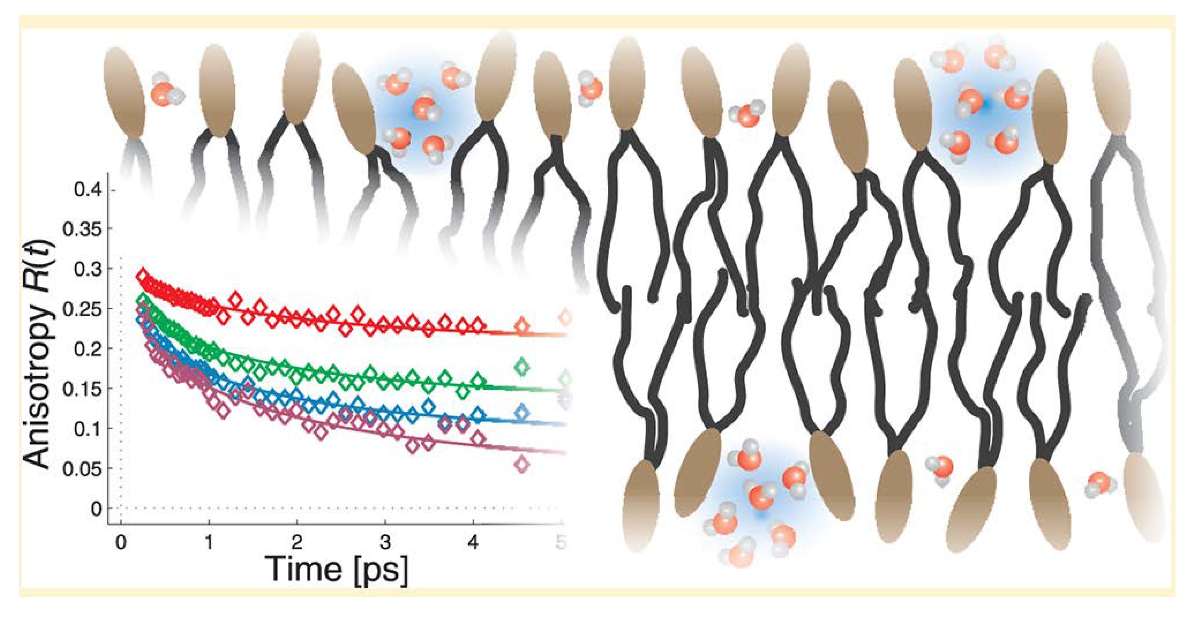
4.2. Interfacial Water: Interaction with Hydrophilic and Hydrophobic Surfaces

In biological systems, liquid water interacts not only with small solutes but also with many larger, extended hydrophilic and hydrophobic surfaces, such as those of proteins, nucleic acids, various organelles, and cell membranes. Results of inelastic incoherent neutron scattering studies of several cell and tissue types suggest that ca. 20%–30% of the total (intracellular plus extracellular) water in these systems is interfacial water, i.e. water located within 1–4 nm of these surfaces, with bulk water comprising the remaining 70%–80% [13]. Not surprisingly, experimental and computational studies reveal different changes in water properties at hydrophilic vs. hydrophobic surfaces at this nanoscale level. Interfacial water near hydrophilic surfaces like hydroxylated diamond or amorphous silica displays viscosity from about 2 up to 106 times greater than that of bulk water, while no significant water viscosity changes were seen near hydrophobic surfaces like methylated silica or hydrogenated diamond [49,50].
Most interestingly, exposure of a hydrophobic hydrogenated nanocrystalline diamond surface to 670 nm laser light gave evidence of hydrogen bond excitation and a resulting density decrease/volume increase in the interfacial water, whereas bulk liquid water is essentially transparent to this wavelength [51]. This result is consistent with predictions of Chandler et al. based on computational studies indicating that water near extended (> ca. 1 nm) hydrophobic surfaces shows less hydrogen bonding and behaves more like water near a liquid-vapor interface than bulk water [47,52,53,54,55,56,57]. The full significance of this observation and of the Lum-Chandler-Weeks theory for our central thesis of EIWS-driven disease etiology is discussed later in this paper.
Historically, the interaction of water with large biomolecules, especially their nonpolar domains, has been considered mainly from the macromolecule’s point of view and designated as the hydrophobic effect. Rezus and Bakker describe it as the tendency of apolar groups to associate in aqueous solution, thereby minimizing the total hydrophobic surface that is exposed to water [58]. Previously, in 2004, Despa, Fernandez, and Berry showed that water constrained by vicinal hydrophobes undergoes a librational dynamics effect that lowers the dielectric susceptibility and induces a “redshift” of the relaxation frequency in the hydration shell [59]. Subsequently, in 2006, Despa described how water in tissues and cells is confined and subject to structural effects not present in its bulk counterpart. Despa added that the structuring effect of confined water in tissues is also a source of polarization fields that contribute to the effective interactions between macromolecules. Dissimilar behavior of water molecules at hydrophilic sites versus hydrophobic sites was said to promote the anisotropy of the hydration shell of proteins. According to Despa, the anisotropy of the hydration shell is essential for enzyme function [60].
No definite consensus has yet been reached regarding the structure of water near the hydrophobic surfaces of biomolecules. While “clathrate-like” (lattices, sometimes layered sheets, or cage-like) structures of hydrophobic hydration have been proposed, studied, and seem reasonable, some studies suggest more-subtle restructuring effects involving the second hydration layer [61,62,63,64]. The subject of hydrophobic hydration structure is discussed in an excellent review article on water in cell biology written by Ball in 2008 [65]. Keutsch and Saykally presented a compilation of terahertz laser vibration-rotation-tunneling spectra and mid-IR laser spectra of several small nanoclusters of water, including a cyclic hexamer of water whose cage structure has a certain clathrate-like quality to its appearance [66]. Molecular dynamics simulations have shown that cyclic pentamers are a dominant topology in liquid water. Csajka and Chandler found that pentamer-like patterns are important in solvation of hydrophobic solutes and in the structures of clathrate hydrates [67]. Significantly, the most stable structure of the water hexamer determined in the gas phase resembles the basic unit in ice [66]. In 1995, Xantheas reported ab initio studies of cyclic water clusters (H2O)n, for n = 1–6 [68]. In 2000, Nauta and Miller identified a cyclic water hexamer in liquid helium which closely resembled the six membered ring forms found in crystalline ice forms of water [69].
According to Perez et al., the water hexamer is predicted by theory to be the smallest water cluster with a three-dimensional hydrogen-bonding network as its minimum energy structure. Previous experimental work provided evidence for cage, book, and cyclic isomers of hexameric water. Using broadband rotational spectroscopy in a pulsed supersonic expansion, this group unambiguously identified all three coexisting isomers. The cage was found to be the minimum energy structure. Rotational spectra consistent with heptamer and nonamer structures were also reported [70].
Evidence for water nanocluster formation near lipid surfaces has recently been reported by Piatkowski et al. [71]. Using ultrafast Forster Vibrational Energy Transfer, they found that water hydrating 1,2-dioleoyl-sn-glycero-3-phosphocholine (DOPC) membranes forms nanoclusters at low hydration levels with an average intermolecular distance of 3.4 Å. See Figure 2 below for the putative relative positioning of water molecules. While the density of the water nanoclusters increased with increasing hydration level, the average intermolecular distance did not [71].
Figure 2. The putative relative positioning of water molecules in 1,2-dioleoyl-sn-glycero-3-phosphocholine (DOPC) membranes obtained by measuring the rate of vibrational resonant (Forster) energy transfer between the water hydroxyl stretch vibrations. Reproduced here from Piatkowski et al. [71] with permission of the American Chemical Society.
Entropy 15 03822 g002
Like the polar headgroups of lipid membranes, biological polysulfates also present an extended hydrophilic surface that interacts with water. There is even some evidence for a longer-range effect of biological polysulfates on water structure. The findings are particularly relevant to the structure of extracellular water near cell membrane surfaces, which are heavily decorated with sulfate-rich HSPGs arranged in protruding structures called glycocalyces. Observation of diffusion patterns of dyes added to living blood vessels reveals a gel-like, impenetrable layer of water surrounding the interior, HSPG-rich capillary wall [72,73]. This result is reminiscent of in vitro studies reported by Pollack et al. on the behavior of water near the hydrophilic, highly-charged surface of tubes and sheets made of Nafion, a sulfonated fluoropolymer [74,75,76]. The viscous surface water layer, extending out up to 200-300 µm from the Nafion surface and containing almost no ions or other solutes, has been described by Pollack as an “exclusion zone” (EZ), or a 4th phase of water. EZ thickness can increase by a factor of two to four upon exposure to IR radiation [77]. Whether these empiric in vitro results are relevant to in vivo physiology remains to be determined.
The properties of EZs described by Pollack and coworkers overlap to some extent with those of water “coherence domains” (CDs) proposed by del Giudice et al. based on quantum field theory calculations [78]. A water CD is a ca. 0.1 µm collection of ca. one million liquid water molecules oscillating in tune with a self-trapped electromagnetic field at some well-defined frequency. Evidence for the existence of stable water clusters up to several microns in size, based on electric force microscopy, atomic force microcopy, and infrared and Raman spectroscopic studies of evaporation of very dilute aqueous NaCl solutions at room temperature and pressure, has been reported by Lo and coworkers [79]. EZs may be regarded as longer-range ensembles of CDs, and some researchers use these two terms interchangeably [78]. These CDs/EZs create a negative electrical potential of as much as -150 mV relative to adjacent, “normal” liquid water and a corresponding concentration of protons at the interface with “normal” water. The detailed physics of this type of water, as revealed by the in vitro studies of Pollack et al., is only recently being elucidated, and investigation of potential clinical significance is warranted.

4.3. Interaction with Electric and Magnetic Fields

In this section we survey recent literature relating to the effects of electric and magnetic fields on interfacial water structure and properties. We will also consider evidence pointing to the Ca2+ signaling system as the primary cellular target of magnetic fields, as this has important implications for the uptake of toxic xenobiotics, including interfacial water stressors such as Al3+, into the cell.
The available evidence points to differing effects of electric and magnetic fields on water structure. In 2008, Rai et al. reported results of density functional theory calculations indicating that applied electric field “opened up” circular- or ring-type water clusters to form linear, branched, or netlike structures by making the dipolar water monomers align along the field axis. In general, the number of hydrogen bonds in a cluster decreased with an increase in the electric field strength [80]. In 2011, Acosta-Gutierrez et al. performed additional computational studies of the physical properties of small water clusters in low and moderate electric fields. At low electric field strengths, the hydrogen bonds oriented the water permanent dipoles along the field, whereas larger field strengths induced more extensive structural reorganization, including hydrogen bond-breaking as the cluster stretched along the field direction, with “the larger clusters (N > 10) usually forming helical structures” [81].
In contrast with the computational studies suggesting that external electric fields break up small water clusters and cause water monomers to line up in the direction of the field, the results of molecular dynamics simulations by Chang and Weng imply that external magnetic fields increase the stability and hydrogen bond strength of supramolecular water clusters while decreasing the self-diffusion of individual water molecules [82]. Moreover, experimental data obtained by Pang et al. on the effects of external magnetic fields on water properties [83,84,85,86] support Pang’s earlier hypothesis that such fields promote formation of both linear and closed chains of hydrogen-bonded water molecules [87]. Applying magnetic fields ranging from 2000 to 4400 G (0.20–0.44 T), Pang and Deng found that the infrared and ultraviolet absorptions, Raman scattering and X-ray diffraction of magnetized water were greatly changed relative to those of unexposed water: infrared (IR) peak strengths increased, frequencies of some peaks shifted, and some new peaks occurred after water was magnetized [83,84]. Significant hysteresis effects were observed in the IR absorption spectrum of magnetized water as temperature was increased and then decreased over the range of 25 °C to 70 °C. Importantly, magnetized water displayed a lower contact angle (lower hydrophobicity, or increased ability to solvate hydrophobic surfaces) than non-magnetized water with copper, graphite, and muscovite surfaces. For each surface, the contact angle difference between magnetized and unmagnetized water was small, on the order of 0.4 to 1.4, but still outside the range of experimental error of the instrument. External magnetic fields increased the refractive index, dielectric constant, and electrical conductivity of water while decreasing its viscosity [84]. The longer the magnetization time, the more the viscosity of the magnetized water decreased, until a minimum was reached.
As noted above, the results of these experimental studies of magnetic field effects on water properties [83,84,85,86] are consistent with Pang’s earlier proposal that exposure of water to a magnetic field facilitates formation of linear and closed hydrogen-bonded water clusters, the latter of which can become ring electric-current or “molecular electric-current” elements with magnetism due to their proton conductivity under the action of the Lorentz force [87]. This enables magnetic interactions of these “molecular electric-current” elements with each other or with the externally applied magnetic field to change the distribution and features of water molecules and the “magnetization of water” [83]. Examples of the proposed linear (open) and circular (closed) hydrogen-bonded chains of water molecules are shown in Figure 3.
The magnetic field strengths of 0.20–0.44 T used by Pang and Deng were several orders of magnitude higher than that of the geomagnetic field at the earth’s surface (ca. 50 µT) and even higher than the ca. 10-10–10-15 T values measured for human organs [3,4]; hence, the relevance of their studies to water in biological systems may legitimately be questioned. However, a complementary mechanism of water magnetization, presented by Mohri [88,89], is based on experimental studies involving a more physiologically-relevant, 6 Hz, 10 µT pulsed magnetic field. Mohri’s hypothesis involves an assumption of cyclotron resonance of protonated water clusters (H3O+(H2O)n).
Figure 3. Illustration of potential linear and circular clusters of hydrogen-bonded water molecules induced by an external magnetic field as proposed by Pang 2006 [87]. Reproduced here from Pang (2006) [87] with permission of Springer-Verlag Berlin/Heidelberg.
Entropy 15 03822 g003
Cyclotron resonance refers to the phenomenon of energy transfer to a charged particle that is moving circularly, normal to the direction of an applied magnetic field, as a manifestation of the Lorentz force; the so-called “cyclotron resonance frequency” of this circular motion depends on the particle’s charge and mass and the strength of the magnetic field. According to Mohri, this cyclotron resonance effect activates proton transport in water under the geo-magnetic field, an effect described as “magneto-protonics” [89]. Formation of a string of such resonating water clusters can give rise to enhanced proton conductivity. This hypothesis is consistent with the decreased electric resistivity of magnetized water reported by Mohri in studies conducted with the weak, pulsed magnetic field described above [89].
In addition to influencing the properties of interfacial water, magnetic fields can also induce changes in the many biological Ca2+ signaling systems. In 2002, Mohri found enhanced phagocytic immune activity and elevated intracellular Ca2+ levels in neutrophils exposed to phosphate buffered saline (PBS) solution that had been subjected to a milliGauss ultra-low-frequency AC (mg ULF-AC) magnetic field prior to exposing the neutrophils to it [90]. In a subsequent study, Mohri reported a reliable method for decreasing the electric resistivity of highly purified water by applying a small magnetic field of several milliGauss in amplitude and twin cyclotron resonance frequencies of 7.0 Hz and 8.4 Hz to excite hexameric and pentameric hydromolecular clusters, respectively [88].
In 2005, Fukushima et al. reported another extraordinary finding: pure water exposed to a 10 mG, ultra-low frequency (6 Hz) AC magnetic field (generated by a Helmholtz coil under visible light) stimulated firefly luciferin-luciferase luminescence and induced intracellular Ca2+ elevation of Chinese hamster ovary (CHO) cells in the absence of ATP [91]―suggesting that exposure to the magnetic field increased signaling activity without taxing normal energy sources. Thus, the luciferase-catalyzed luminescence of luciferin, which normally requires ATP in untreated water, occurred without any added ATP in water that had been treated with the magnetic field and light. Indeed, the luminescence activity of the luciferin-luciferase complex in water that was exposed to the magnetic field and light was several-fold higher than that obtained in light-shielded conditions. It should be noted that these experiments were conducted at 40°C, close to the normal human body temperature of 37 °C and near the optimum temperature for most enzymatic reactions. The 6 Hz frequency of the applied magnetic field corresponds to the cyclotron resonance frequency of the protonated hexameric water cluster H3O+(H2O)5 under the influence of the geomagnetic field (ca. 500 mG) and to the alpha-wave frequency of the brain [88,91]. The authors speculated that the magnetic energy applied to pure water was stored in a water cluster with stable hydrogen bond resonance and transferred to the luciferin-luciferase complex, with resultant formation of oxy-luciferin and luminescence in the absence of ATP [91].
While the results of Fukushima and coworkers [91] may seem surprising, recent work indicating an ability of low-entropy sunlight to impart long-range order in bulk as well as surface water [92] provides a plausible route by which water CD's may provide energy catalyzing chemical reactions not only at enzymes but indeed near many hydrophobic or hydrophilic surfaces [93,94], as well as for actual diffusion of enzymes through bulk aqueous solution toward areas of high local substrate concentration [95,96,97].
The results obtained by Fukushima and coworkers [91] are consistent with those of Gartzke et al. (2002), who pointed to the Ca2+ signaling system as the primary cellular target of magnetic fields. Specifically, the ion-conducting actin filament bundle within microvilli was proposed as the cellular target for magnetic fields. This target combines physiological relevance for Ca2+ signaling with unusual electrical properties capable of explaining the effect of low-energy magnetic fields on biological systems [98]. This target was previously shown to exhibit nonlinear, cable-like cation conduction through arrays of condensed ion clouds. Stochastic resonance and/or the Brownian motor hypothesis were employed to explain how the interaction of ion clouds with periodically applied electromagnetic fields results in cation pumping through a cascade of potential barriers within polyelectrolytes [98]. The proposed interaction mechanism was in accord with the postulated extreme sensitivity for excitation by very low field energies within specific amplitude and frequency windows. Thus, instead of a disturbing role, thermal “noise” itself became an essential and necessary signaling component. Microvillar cation transduction by F-actin bundles shielded by a lipid membrane amplify coherent signals on cation transduction and reduce stochastic (thermal) noise. The weak coherent signals are thought to be amplified by thermal noise via stochastic resonance which occurs upon application of a very low energy periodically-applied field, resulting in unidirectional cation transport along F-actin bundles.
An important implication of this proposed systematization is the synergistic action of magnetic fields on the uptake of xenobiotics into the cell. Toxic compounds can enter the cell more readily under the influence of electromagnetic fields, activating the Ca2+ signaling pathway. Lange pointed out that maintenance of intact microvillar surfaces is essential in providing the natural barrier function of epithelial cells [99]. Any disorganization of microvillar surface morphology was shown to severely accelerate the entrance of ionic and lipophilic xenobiotics into the cytoplasm. This point is discussed further in Section 5 and Section 6 below when we consider the adverse effects of aluminum cation and other exogenous interfacial water stressors on biological systems.

4.4. Life-Enabling Properties of Water at the Interphase

We propose that the main systems by which structured interfacial water promotes life-enabling biological processes include:
(A)
Promoting electrical conductivity at biological interfaces, thereby facilitating metabolism and voltage differences maintained by intracellular organelles;
(B)
Absorbing, storing, and emitting electromagnetic energy, enabling storage and transmission of energy and information;
(C)
Overcoming the kT or “thermal diffusion” problem; and
(D)
Solving the intracellular crowding and molecular self-assembly problems by way of chirality (handedness of molecules) and magnetization.
These interactions are discussed below, along with supporting data.

4.4.1. Promoting Electrical Conductivity at Biological Interfaces

Nanomolecular ensembles of water CDs at the aqueous interphase can provide an extended, long-range, scaffolding for protomeric and electromeric transfer on a mesoscopic, supramolecular scale, which supports energy metabolism in vivo. We have mentioned previously a role for external HSPGs in connecting the cytoskeleton to the plasma membrane. The cytoskeleton also plays a central role in caveolin-based lipid transport between the Golgi apparatus and the plasma membrane [100]. We hypothesize that the cytoskeleton also facilitates the transport of both electrons and protons, taking advantage of the water CDs to induce a magnetic field promoting proton and electron currents, thus sustaining the cell’s membrane voltage gradient. Similar ion transport to and from cytoplasmic organelles such as mitochondria (which must maintain a highly basic pH) and lysosomes (which must maintain a highly acidic pH) is likely also maintained by the cytoskeleton. The actin cytoskeleton has been shown to be integrally linked to both lysosomes [101] and mitochondria [102]. If these organelles are unable to maintain their extreme pH values, they will fail to function and the cell will be disabled.
Figure 3 above depicts some possible water nanoclusters that could promote proton conductivity [87]. Three additional proposed water cluster arrangements for enhanced proton transfer are shown in Figure 4, Figure 5, Figure 6 and Figure 7 [103,104,105,106].
Figure 4. The Eigen-Zundel-Eigen (EZE) proton mobility phenomenon [103,104]. Reproduced here from Markovitch et al. (2008) [105] with permission of the American Chemical Society.
Entropy 15 03822 g004
Figure 5. Protomeric ensembles acting as substrates for Grotthuss phenomenon. Reproduced here from Verdel et al. (2011) [106] with permission of MDPI AG.
Entropy 15 03822 g005
Verdel et al. [106,107] attributed increased proton transfer deduced from conductivity measurements to the “autothixotropic” phenomenon (weak gel-like behavior) of water, which supposedly develops spontaneously with time, where ions and hydrophilic surfaces seem to play an important role. Voth et al. [105,108,109] have shown that sulfonate groups in the sulfonated fluoropolymer Nafion influence excess proton solvation, as well as the proton hydration structure, by stabilizing a more Zundel-like (H5O2+) structure in their first solvation shells [110]. The sulfonate groups were also found to affect the proton hopping directions. These findings suggest how biosulfates function in living organisms.
Studies with Nafion, used in proton exchange membrane-based fuel cells [108,109,111], and with carbon nanotubes (CNTs) functionalized with CF3SO3H groups [112], have provided additional insights. At low water content, the sulfonated side chains of Nafion form isolated hydrophilic regions. As the water content increases, these domains expand and eventually form spanning water channels which are capable of efficiently transmitting protons. It is likely that eukaryotic cells use such a system for efficient proton transport. In ab initio molecular dynamic studies with the fluorosulfonated CNTs, decreasing the distance between sulfonate groups increased proton dissociation and interactions between water molecules. As sulfonate-sulfonate distance increased, connectivity among the water molecules decreased as they formed more isolated clusters around the sulfonate groups. Sulfonate-sulfonate distance and geometry were the most dominant factors in proton dissociation; however, the hydrophobic environment and nanoscale confinement became more important as distance between sulfonate groups increased [112].
Martin Chaplin, a preeminent expert in water structure and properties, has recently argued that both proton and electron delocalization constitute the normal state of affairs in liquid water molecule networks, as illustrated schematically in Figure 6 below [113,114]. Thus, if he is correct, electron (as well as proton) conductivity is enhanced in ensembles of water CDs. Czerlinski and Ypma proposed that electrons move statistically in electromeric domains like a dipole, initiating similar behavior in other domains by resonance [115,116]. When water networks are exposed to ionizing radiation, the structure is modified so as to give mobility to both protons and the hydroxyl radicals [117].
Theoretical physicist, Herbert Frölich, originally proposed in 1968 that coherent electrical polar oscillations and the generation of electromagnetic fields play important roles in living cells, and their disturbances occur in cancer cells [118,119]. Experimental support for Frölich’s ideas continues to accumulate. In 2013, Pokorný et al. reviewed the current biophysical literature pertaining to cancer transformation [120], wherein measurements performed on living cells have disclosed electric and electromagnetic oscillations, including dielectrophoretic forces of the cellular oscillating electric field. The resulting attraction of dielectric particles depends on their permittivity [121]. We refer the reader to [120] for descriptions of the experimental and theoretical research of the cellular electromagnetic activity, which today, points strongly to microtubules as major sources of electromagnetic interactions. Evidence for a key role of EIWS-induced cytoskeleton disruption in cancer causation is discussed in Section 6.1 below.
An additional route to enhanced electrical conductivity in biological systems could be ion-radical separation converting mesomeric [122] water nanoclusters into superconductors. Being mesomeric enables the delocalization of protons (making these nanoclusters protomeric) as well as free radical electrons (making them also electromeric). Such properties enable catalysis of oxidation-reduction reactions, propagation of electric currents, and generation of magnetic fields. Mesomeric systems, along with other stable water clusters, can theoretically also serve as vehicles for storing incident radiant energy as entropy loss and charge separation, as theorized by Chai, Yoo, and Pollack in 2009 [76]. Figure 7 (below) illustrates a hypothetical cyclic bipolaron―an electromeric and protomeric ensemble of structured water. Such a complex could assist in maintaining membrane potentials and enabling cellular cytoskeletal conduction. Stable cyclic hexamers of water have been studied spectroscopically and theoretically by Saykally [66], Pang [84,85,86,87], and Mitsui [123]. The electron movement in these extended coherence domains seems to resemble the free electron movement in metals or even superconductors [115,124,125,126]. Other models for electron capture in water―for example, electrons in p-orbital-like water cavities―have been proposed and verified experimentally [127].
Figure 6. Neither the protons nor the electrons are pinned to individual molecules. Reproduced here from Chaplin (2013) [113] with permission of the Institute of Science in Society.
Entropy 15 03822 g006
Figure 7. A hypothetical radical-cation cyclic water hexamer accounting for protomerism and electromerism.
Entropy 15 03822 g007
Electrical current which depends on the presence of water has been detected in association with cellulose [128], proteins [129,130], microtubules [131], and DNA [132,133]. In 1987, Careri et al. demonstrated direct current (DC) protonic conductivity of powders of lysozyme for varied levels of hydration [134,135] and suggested that hydration-induced protonic conduction and enzymatic activity corresponds to the formation of a percolation network of absorbed water molecules on the surface of the macromolecule. Computer simulation studies reported in 2006 suggest that hydration water percolation on DNA surfaces drives polymorphic transitions and DNA conductivity [136]. In 2012, Sontz et al. proposed a mechanism for charge transport mediation by duplex DNA [133]. Are these currents dependent on the presence of nanomolecular ensembles of water CDs? The research literature suggests that they are. Czerlinski and Ypma have provided much of the theoretical support for the electromerism (electron conductivity) of nanomolecular water CDs [115,116,137,138]. Given the Josephson effect in physics and the fact that the overlapping base pairs of the double helix have a certain metallic quality, almost like sheets of graphite [126,133], it is not surprising that DNA has been experimentally associated with electrical current.
We propose that dynamical nanomolecular ensembles of water CDs represent structural entropy-consuming [139,140,141,142,143] nano-engines which trap, transduce, and conduct the energy to induce conformational changes in both DNA and proteins. Nanoclusters of magnetized water and DNA, then, may act in concert to provide a supramolecular scaffolding acting to transmit both energy and information over long distances. Empirical evidence already cited shows that interfacial water stress (IWS) provides a supramolecular basis for both the formation and stability of rings of circular DNA [144], microDNAs [145], non-B-conformation DNA [146], and Z-DNA [147]. IWS and nanoclusters of magnetized water also provide a supramolecular basis for modulating DNA structural stability in both health, e.g., normal cell division, and disease, e.g., oncogenesis. This topic is explored in greater depth in Section 6 below.

4.4.2. Absorbing, Storing, and Emitting Electromagnetic Energy

In addition to their ability to enhance electrical conductivity, relevant research and sound theory suggest that water CDs can absorb and emit electromagnetic energy, thus storing and transmitting both information and energy [148,149,150]. EZ water absorbs light at 270 nm and fluoresces when excited at this wavelength [151]. Based in part on Preparata’s application of quantum electrodynamic field theory [152,153,154], Marchettini, Del Giudice, Fuchs, Vitiello, and Voeikov proposed in 2010 that water CDs provide a “redox pile” of “quasi-free electrons” [78,155]. In 1998, Voeikov and Naletov described weak photon emission of non-linear chemical reactions of amino acids and sugars in aqueous solutions which they proposed provides evidence for self-organizing chain reactions with delayed-branching [149]. In 1999, Kobayashi reported spontaneous ultraweak photon emission from a rat’s brain correlated with cerebral energy metabolism and oxidative stress [156]. In 2004, Curtis and Hurtak [157] proposed that biophotonic processes in humans may represent the way biophysical light interacts with the human self-organization of information that may be achieved by means of biomolecular metabolic, or neural communication.
In 2005, Kim et al. demonstrated that spontaneous photon emissions from cancer tissues contrasted with those of normal tissues, and their delayed luminescent properties were investigated [158,159]. Mean values of spontaneous photon emissions from normal tissues and tumor tissues were measured with standard errors at 625 ± 419 counts/minute/cm2 (n = 6) and 982 ± 513 counts/minute/cm2 (n = 14), respectively. Peak values of the intensity of delayed luminescence from normal and cancerous tissues were 63 ± 20 counts/ms (n = 6) and 48 ± 12 counts/ms (n = 14) [158] respectively.
In 2007, Whissell and Persinger showed that prenatal exposure of pregnant Whistar albino rats to extremely weak 7 Hz magnetic fields in the 1, 5, 10, 50, and 500 nT range, caused behavioral deficits in their offspring which persisted into adulthood. These changes were found to be waveform-specific and may involve nitric oxide [160]. Co-administration of the nitric oxide synthase (NOS) inhibitor n-methylarginine appeared to mitigate the behavioral deficits induced by the magnetic fields, to suggest a critical developmental role of NO and the involvement of NO in magnetic field effects [160]. Could these findings show how an external electromagnetic field modulates IWS leading to the unfolded protein response (UPR), perhaps by increasing the hydrophobicity of water? NOS activation requires calcium-binding to calmodulin. As stated previously, calcium has been a proposed cellular target of magnetic fields. In principle, magnetic fields could alter vascular blood flow, by their effects on erythrocytic eNOS and endothelial eNOS. If the ion cyclotron resonance (ICR) frequency of calcium is induced by the magnetic field, this phenomenon may be generalizable to a larger number of functions, given the multifarious roles of calcium in biological signaling pathways [161].
In 2010, Tafur et al. proposed that the detection of biophotons, the production of which is associated with cellular redox state and the generation of ROS, represents a noninvasive redox measure which may be useful in advancing low intensity light therapy [162]. In 2011, Czerlinski described long-lived nanodomains of water that form coherent cooperative aggregates controlled by the geomagnetic field. These domains either slowly emit biophotons or perform specific biochemical work at their target [115,116].
In 2012, Pang [83,84,86] determined from energy spectra that protein molecules can both radiate and absorb bio-photons with wavelengths of < 3 μm and 5–7 μm, consistent with the energy level transitions of the excitons, and consistent with experimental infrared absorption data. Pang’s findings appear to provide support for the controversial experimental results of Gerald Pollack in 2006 [77] wherein large EZs were observed in the vicinity of many types of surfaces, including artificial and natural hydrogels, biological tissues, hydrophilic polymers, monolayers, and ion-exchange beads, as well as with a variety of solutes. Moreover, it was further shown that radiant energy profoundly expands these zones in a reversible, wavelength-dependent manner. Pollack wrote: “It appears that incident radiant energy may be stored in the water as entropy loss and charge separation” [76]. Whether Pollack’s in vitro results apply to the living state, e.g., cell biology, remains to be determined.

4.4.3. Overcoming the kT or “Thermal Diffusion” Problem

According to Ho (2011), the “thermal threshold” is a fallacy arising from the assumption that living organisms can be described in terms of conventional equilibrium thermodynamics; whereas by general consensus they are open systems meticulously organized and maintained far away from thermodynamic equilibrium [163]. Could water CDs provide a physical basis for overcoming both the kT paradox and the intracellular crowding problem [28,29,31,164,165,166,167,168,169,170,171,172]? The term kT requirement, where k is the Boltzmann constant and T is temperature (deg K), relates generally to the temperature dependence of chemical reaction rates, and the need to impart enough energy to biological molecules in cells to achieve such reactions at physiological temperatures, without resorting to thermal diffusion (i.e., heating up the reactants).
Through the power to store and amplify electromagnetic energy, water CDs provide a means for achieving biological effects with very weak magnetic fields by ion cyclotron resonance (ICR), as seen in Section 4.3 above, and by temperature-independent means [78,163,166,167,169,173,174]. ICR provides just one of various ways to theoretically account for observed interactions between weak low-frequency electromagnetic fields and biological systems. The ICR hypothesis has been detailed in 2006 by A.R. Liboff [175], and Del Giudice [161] has employed the principles of quantum electrodynamics (QED) in attempting to explain biological ICR, especially the results reported by Zhadin and coworkers, who observed increased ion currents in aqueous glutamic acid solutions exposed simultaneously to weak static and alternating magnetic fields [167,176,177].

4.4.4. Solving the Intracellular Crowding and Molecular Self-Assembly Problems by Way of Chirality and Magnetization

It is clear that the aqueous phase of the cytoplasm is crowded rather than dilute, and that the diffusion and partitioning of macromolecules and vesicles in cytoplasm is highly restricted by steric hindrance as well as by unexpected binding interactions [170,171,178]. In 2001, Aggeli et al. presented a generic statistical mechanical model for the self-assembly of chiral rod-like units, such as beta-sheet-forming peptides, into helical tapes, which, with increasing concentration, associate into twisted ribbons (double tapes), fibrils (twisted stacks of ribbons), and fibers (entwined fibrils) [179]. Results of a recent study of self-assembly behavior of isomers of the hydrophobic tripeptide leu-phe-phe suggest that proteins composed of homochiral amino acids would likely assemble and pack more efficiently than those containing both d- and l-amino acids [180].
If chirality in biological molecules can promote more-efficient packing of macromolecules in the limited intracellular space, then anything that could induce chirality in water (the most-abundant molecule in the cell), and/or promote more-efficient packing of water molecules by some other means, could also reduce intracellular crowding. Individual water molecules are not chiral, but results of extensive terahertz laser vibration-rotation-tunneling (VRT) and mid-IR laser spectroscopic studies, in conjunction with theoretical calculations, indicate that cyclic water trimers and pentamers are indeed chiral [66], as shown in Figure 8.
Figure 8. Illustration of (a) chiral cyclic water trimer, and (b) chiral cyclic water pentamer. Reproduced here from Keutsch and Saykally [66], with permission of the publisher, copyright (2001) National Academy of Sciences, USA.
Entropy 15 03822 g008
Thus, at least some of the closed-chain hydrogen-bonded supramolecular water structures proposed by Pang [83,84,86,87] for magnetized water (see discussion in Section 4C above) could be chiral. As suggested by the results of two-dimensional studies of non-chiral, equilateral triangle-shaped polymer particles in aqueous solution, formation of such local chiral “supraparticle” clusters appears to be driven by the increased entropy afforded by increased motion of the monomers within the chiral arrangement relative to that attainable with the more-ordered tight packing of individual particles [181]. Based on these considerations, we propose that formation of chiral supramolecular water clusters, facilitated by magnetic fields, can help to solve the intracellular crowding problem.
Another area in which we think magnetized water could make an essential contribution is in facilitation of biological macromolecular and supramolecular self-assembly. Elegant experiments conducted by Whitesides et al. have provided ample evidence that use of magnetic forces, such as in magnetic levitation, can guide the self-assembly of three-dimensional structures from even diamagnetic components [182,183].

4.5. Tuning the Aqueous Interphase: Modulating Interfacial Water Systems

As noted in Section 2 above, experiments with new spectroscopic techniques that enable study of biological systems on time scales down to picoseconds indicate that large-scale motions of proteins and nucleic acids are determined by fluctuations in the hydration shell, which are controlled by solvent viscosity and hydration, and are absent in a dehydrated protein [14,17,18,19,20,21,22,23,24,25,26,27,28,29,30,31,32,33,34,36]. If changes in interfacial water structure drive biomacromolecular conformational changes, then biological systems must have means to vary interfacial water structure and properties (within properly-functioning, life-enabling limits) to carry out their wide range of life-sustaining activities. In section 4C above, we surveyed the evidence that ultra-low frequency magnetic fields can reduce water surface tension and hydrophobicity. In addition, magnetic microemulsion formation from anionic magnetic surfactants has been reported [184,185,186]. These data suggest that interfacial water tension is modulated in vivo by magnetic fields and anionic surfactants. Indeed, results of the molecular dynamics studies of fluorosulfonated carbon nanotubes mentioned above, indicating that the geometry and distance between sulfonate groups played dominant roles in determining proton transfer rates in surrounding water molecules [112], suggest a way that biosulfates in cell membranes may perform a similar function in biological systems, and also suggest a pathology when the sulfation levels are depleted.
Cell membranes generally become more compliant (less “stiff”) when they are depleted of biosulfates, such as cholesterol sulfate (Ch-S) and HSPGs, in their outlet leaflets and glycocalyces, respectively. Cell membranes which are more compliant (less “stiff”) are relatively hydrophobic, i.e. relatively “dewetted” by interfacial water. Ultimately, even phase transitions, such as nanobubble formation, could occur, especially near the triple point where interfacial water is about to freeze [142]. Under the Lum-Chandler-Weeks theory [47], relative hydrophobicity and the molecular theory of capillarity [187,188,189] are predicted to play major roles in determining cellular compliance (“softness”) at both the intracellular and extracellular aqueous interphase domains near our biomembranes. The speed of capillary waves at the surface can be used to measure surface tension, by how much they scatter light from a laser [189]. Suzuki et al. used dielectric spectroscopy with microwaves to study the hydration of myosin subfragment 1 (S1). The observed changes in S1 hydration were quantitatively consistent with the accompanying large thermodynamic entropy and heat capacity changes estimated by calorimetry, indicating that the protein surface hydrophobicity change plays a crucial role in the enthalpy-entropy compensation effects observed in the steps of S1 ATP hydrolysis [190].
It must be expected, therefore, that many, if not all, sterols are biophysically active as their corresponding sulfates, which have the amphiphilic character needed for bioavailability. Vitamin D3 sulfate, cholesterol sulfate, DHEA-sulfate, estrone sulfate and the sulfated neurosteroids are examples [2,191,192]. Also, the sulfated neurosteroids appear to be acting “from a distance” within the synapses, as opposed to acting at receptor sites. Hence, the biosulfates are likely to be active by virtue of stabilizing cell membranes and maintaining the CD water in the extracellular space. As noted earlier, enhanced Grotthuss and Josephson effects have been implicated with water CD ensembles [126,148,149,150].
A number of medium-chain fats and cofactors, e.g., lauric acid, capric acid, ascorbic acid, panthothenic acid, α-lipoic acid, and niacin, all share apparent membrane-stabilizing properties; it is plausible to infer that they all act predominantly by lowering interfacial water tension. Bioactive polyphenols and polyketones represent other classes of surfactant mimics, whose bioactivity and bioavailability may be significantly modified by sulfation. Resveratrol, curcumin, ascorbic acid, and the health-promoting phenols in coffee and chocolate can be sulfated, and this may be the key to their biological benefits. Studies on resveratrol metabolism revealed that it is sulfated in the gut prior to absorption [193]. The fact that vitamin C catalyzes the conversion of homocysteine thiolactone to sulfate may also depend on the fact that vitamin C can also be sulfated [194]. In in vitro experiments with chondrocytes, vitamin C was found to induce a 70% increase in the biosynthesis of sulfated proteoglycans [195], which we hypothesize is a result of its ability to carry sulfate [196]. In 1973, Verlangieri and Mumma demonstrated the in vivo sulfation of cholesterol by ascorbic acid 2-sulfate [197]. Such findings suggest that the health benefits of all such molecules may be due to their propensity to participate in sulfate synthesis and transport.
The empirical evidence that biosulfates embedded into membranes can have beneficial effects on interfacial water suggests that phosphorylation signaling may impart a similar kosmotropic anionic feature to membrane-bound molecules. Also, it is known that phosphorylation signaling cascades are triggered by cholesterol depletion in the membrane [198]. A depletion of cholesterol is likely to be at least linearly related to a decrease in cholesterol sulfate in the membrane, and this may trigger the phosphorylation of other membrane biomolecules such as phosphatidylinositol (PI) via phosphatidylinositol 3 kinase (PI3K), a key intermediary in phosphorylation signaling cascades, as a compensatory action. Three additional phosphate groups can be added to Pi to form phosphatidylnositol phosphate (PIP), phosphatidylinositol bisphosphate (PIP2) and phosphatidyl-inositol trisphosphate (PIP3), collectively called the phosphoinositides. PI3K is a known regulator in angiogenesis and tumor growth [199].

5. Exogenous Interfacial Water Stress and Its Pathological Consequences

In the preceding sections, we have surveyed the research literature relevant to the structure and properties of life-enabling biological water at interfaces in extracellular and intracellular space. We have examined evidence that the interaction of this interfacial water with hydrophobic and hydrophilic surfaces and with electromagnetic fields can create extended networks of coherent, structured forms of water. These extended networks can act as electrical wires or circuits that enable and control life processes (such as macromolecular motions) by enhanced and rapid absorption, storage, emission, and transmission (conductivity) of energy and information. We have also looked at possible ways in which interfacial water structure and tension may be modified by magnetic fields and/or anionic surfactants at membrane surfaces, enabling biological systems to perform their usual life-sustaining activities.
In contrast with the variations in interfacial water structure and tension, induced by endogenous agents such as, for example, Ca+2, Mg2+, Zn2+, Co2+, Mn2+ ions, that enable normal life processes, we use the term “exogenous interfacial water stress” (EIWS) to denote a pathological, perhaps more acute increase in interfacial water tension brought about by a xenobiotic agent. Incremental surface tension values have been reported by Marcus [200]. As discussed more fully in our earlier review [2], potential exogenous interfacial water stressors include kosmotropic cations such as Al3+ and Hg2+, as well as various cationic and nonionic surfactants. If biological water systems are regarded as extended electrical circuits that can store and transmit energy, then exogenous interfacial water stressors are agents that can weaken and unload energy from these networks by creating “leaks,” or by creating short-circuits that provide a lower-resistance path for rapid energy discharge, thus depleting energy and causing collateral damage along the discharge pathway. Examples of such damage would include disruption of membrane and/or protein and/or nucleic acid systems, increased hydrophobicity, protein aggregation, cell-cell aggregation, microbe-cell aggregation, and excess production of reactive oxygen species, to name only a few of the undesirable outcomes.

5.1. Exogenous Interfacial Water Stress as a Short-Circuit, Energy-Unloading Phenomenon Causing Extracellular and/or Intracellular Damage

In extracellular space, the negatively-charged cell membrane surface is particularly vulnerable to short-circuiting by interaction with a cationic kosmotrope or surfactant that can tie up a protruding sulfate, phosphate, or carboxylate group and disrupt local water systems. Consistent with the energy-depletion hypothesis, quite a few recent research papers support an inverse relationship between interfacial water stress and surface energy [201,202,203,204,205,206]. Interfacial waves have been identified by intravital microscopists [207,208,209]. Gallez and Coakley showed empirically that the average number of waves per wavy cell rim decreased when cell surface charge was depleted, and when cationic drugs were present, and increased in the presence of anionic drugs [198,209]. Because of the inverse relationship between wavelength and energy in the classical wave equation, it may be inferred that the spatial distribution of wave-like densities noted on scanning electron micrographs may be produced by the interaction (reflection) of electrons with the structured water at the interface.
The likeliest recipient for sudden energy discharge in any short-circuiting caused by interfacial water stress would be the nearby interfacial water. Energy discharge into the interfacial water would disrupt its dynamic balance leading to lowered density and higher volume, as when water near a hydrophobic surface is exposed to 670 nm light [51], as mentioned in Section 4 above. Lower-density water has less power to solubilize hydrophobic surfaces. This trend was demonstrated experimentally with water containing the protein ubiquitin artificially “stretched” at negative pressure in an adaptation from Berthelot in 1850 [35,210,211]. Water density was reduced from 1.00 to 0.95 g/cm3 in a sealed glass nuclear magnetic resonance (NMR) tube. The protein in this “stretched” water became less stable than in normal-density water.
The hydrophobic interaction impacts stabilization of many biological components and plays a decisive role in protein folding [212]. The hydrophobic effect is an entropically-driven phenomenon arising from the difference in density between the open order arrangement of water in the neighborhood of a nonpolar surface and less ordered bulk water [58,59,60,212]. If EIWS decreases the entropic gain of minimizing the exposed nonpolar surfaces to interfacial water, it must eventually “kill” the hydrophobic interaction, with consequent denaturation of the protein. This inference is supported by the work of Defay and Prigogine, who showed that, at the interphase, the triple-point of water is affected by curvature and surface tension [213], and by the Lum-Chandler-Weeks theory of hydrophobicity [47,53,55,57,171].
It follows that the in vivo, gel-sol transitions are modulated by surface tension and curvature, as originally postulated by Prigogine [213,214]. However, surface tension at the interphase is likely to be affected by such variables as the presence of static and dynamic electromagnetic fields, chirality, pH, and concentration of solutes, including the presence of amphiphilic surfactants. Roughness and curvature of the biomembranes clearly has major impact on such properties as capillarity [189] and capillary blood flow [188]. The anomalous properties of supercooled water and glass formation may have in vivo correlates [26,215,216,217,218,219]. Water at the interphase, under conditions of acute local hydrophobic stress, described as “unwetting” or “stretching” [210], may be followed ultimately by a phase transition, which could be devastating in vivo [142]. Patel et al. [53] used molecular dynamic simulations to show that a large enough hydrophobic surface can induce the formation of a water-vapor-like interface, and as such, the probability of water depletion is enhanced near such a surface. Marked similarities were demonstrated between water-vapor interfaces and water-oil interfaces. It is also well known that purely repulsive hydrophobic surfaces induce a vapor-liquid-like interface [47,220,142].
Damage to a cell membrane caused by interfacial water stress can make it easier for interfacial water stressors to gain access to intracellular space and cause further harm. Depending on the interfacial water stressor and the type of surface encountered, such intracellular damage could include protein unfolding or misfolding, DNA misfolding, and generation of excess reactive oxygen species (ROS), which further disrupt intracellular systems. Specific types of intracellular damage are considered below with respect to toxic actions of the aluminum cation, Al3+, a quintessential exogenous interfacial water stressor that has been linked with breast cancer and neurological disease, as discussed in Section 6 below. An additional pathway by which EIWS can give rise to both extracellular and intracellular damage is suggested by results of a study involving detergent treatment of rat embryo fibroblast cells, which disrupted the cell membrane HSPGs and implicated them as a link between the extracellular matrix and the intracellular cytoskeleton [221]. Based on findings already discussed, severance of the cytoskeleton from the cell membrane should re-orient the cytoskeleton toward the cell nucleus and would necessarily be conducive to the pathological cell division characteristic of cancer; again, see Section 6 for further discussion.

5.2. Al3+ as a Biosignaling Nightmare

There is ample literature documenting the wide range of toxic effects of aluminum cation on biological systems. Here we consider evidence for five main types of aluminum-induced damage: displacement of endogenous mono- or divalent cations normally complexed with important biomolecules; reduction of sulfur bioavailability; coagulant action; induction of oxidative, genotoxic, and protein conformational stress; and induction of EIWS.

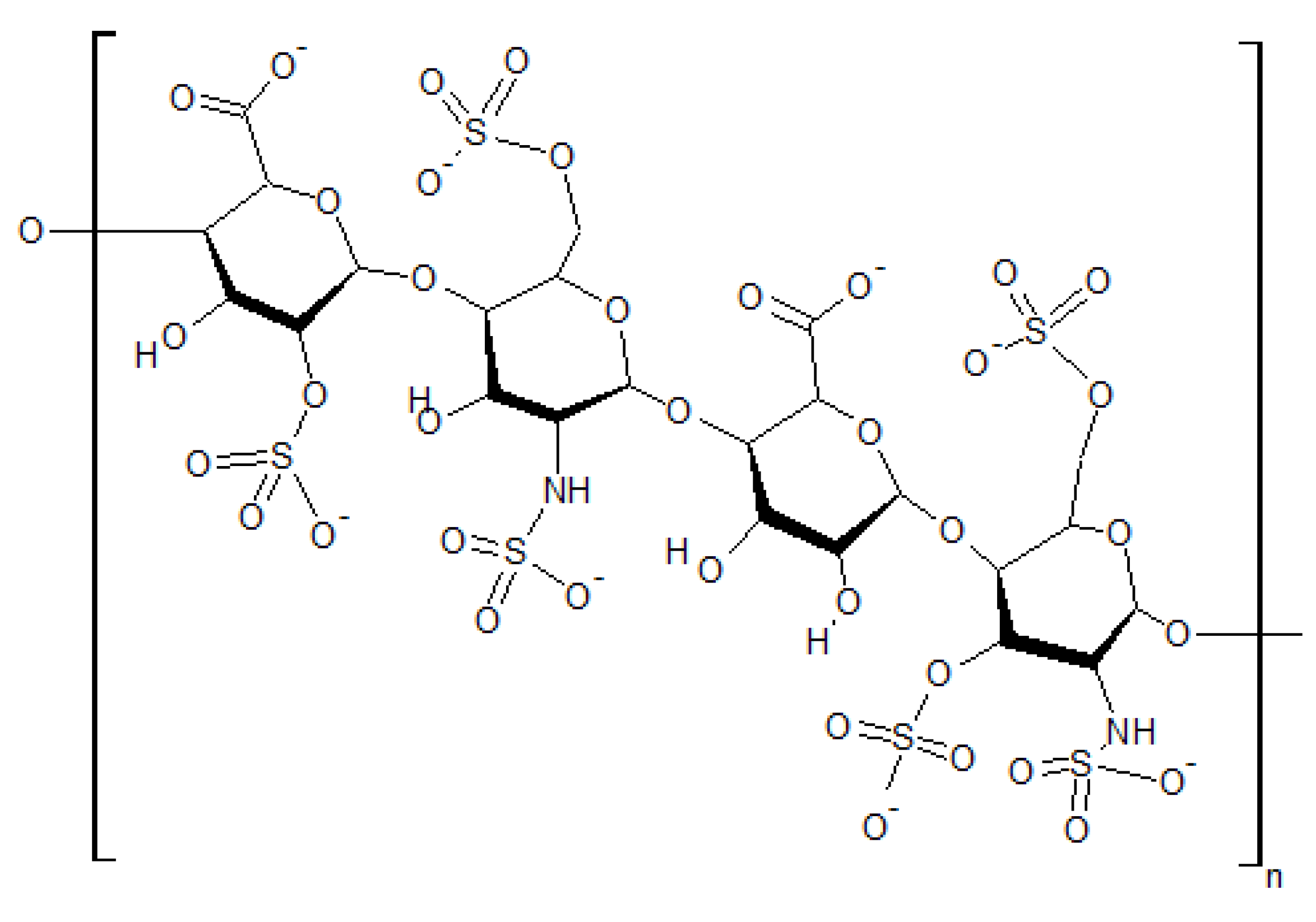
5.2.1. Displacement of Endogenous Cations

Al3+ can displace mono- and divalent cations in biological systems with deleterious consequences, as indicated by its ability to inhibit Na+/K+- ATPase, Ca2+-ATPase, and H+-ATPase [222]. Here, however, we will focus on a couple of specific situations involving substitution of Ca2+ by Al3+. Ca2+ plays an essential role in muscle contraction [223] and in a huge number of cell signaling pathways [224]. Hence, there is a large potential for Al3+, acting as a Ca2+ mimic, to become sequestered, in a structural entropy consuming process, in many of the same places that Ca2+ would otherwise sequester, with detrimental effect.
Al3+ has been shown to displace Ca2+ from heparan sulfate in rat liver [225]. By analogy, it seems likely that Al3+ could also displace Ca2+ from HSPGs throughout the human body, including those found in the stomach, small intestine, pancreas, muscle [226], as well as in lysosomes [227], the Golgi apparatus, and the glycocalyces of plasmalemmal membranes, including the mitochondrial and neuronal membranes of the human brain.
Environmental exposures to metals are well known to act as sensitizers for thrombohemorrhagic phenomena and calciphylaxis [228,229]. We hypothesize, therefore, that Al3+ , possibly in conjunction with various other exogenous metal cations, sensitizes for metastatic calcification and calciphylaxis. Calciphylaxis is an under-diagnosed condition of induced systemic hypersensitivity in which tissues respond to appropriate challenging agents with a sudden, but sometimes evanescent local calcification [228,230]. Metastatic calcification is a condition in which various calcium phosphate deposits accumulate in otherwise-normal tissues [231]. It usually sets in when the product of the serum calcium and phosphate levels in mg/dl exceeds 70, but has been reported to occur sometimes when this product is below this threshold [232]. Based on these observations and the results of earlier ion hydration studies by Guo and Tielrooij, we suggest that in the process of transformation, prior to metastatic calcification, various aluminum/phosphate and aluminum/sulfate ion pairs exceed a threshold incremental surface tension which disrupts biosignaling, in vivo [233,234], most likely by creating relative dehydration and unwetting of the type mentioned earlier [142]. This proposed sequence of events would be part of the general phenomenon we describe as EIWS. Recent empirical data of Marcus, indicating that Al3+ has an endergonic effect on interfacial water tension [200,235], provides substantial support for the EIWS hypothesis [2].

5.2.2. Reduction of Sulfur Bioavailability

Cationic Al binds strongly to cysteines in serum albumin in the blood stream. The absorption of Al cation onto serum albumin has a profound effect on zeta potential [236], driving it even to positive values at physiologic pH with sufficient concentrations of aluminum hydroxide. Also of note, Li (1992) showed that the charge of bubbles exhibits “unusual positive surface charge characteristics” in solutions of trivalent Al cations [237]. Analysis of their results indicated that the reversal of bubble charge can be attributed to specific adsorption of Al3+ and its hydroxo complexes at the gas-liquid interface in the low pH range and to precipitation of aluminum hydroxide in the intermediate pH range. Sulfate is responsible for binding to cationic metalloneurotoxins [238] like mercury and Al and expelling them through the kidneys [239]. Such action would however also lead to a further reduction in the bioavailability of sulfate.

5.2.3. Coagulant Action: Relevant Observations from Water Purification Chemistry

From water purification and wastewater treatment technology, it is well known that the zeta potential (ZP) decreases (becomes less negative) with the concentration of alum while the coagulation of colloidal particles bears a biphasic relation to the concentration of alum [240,241]. Double layer repression can be achieved by increasing the ionic strength of the solution by adding additional ionic species, preferably high valence ions. For this reason, the typical chemicals used in double layer repression are those that produce cations with a large charge such as Al3+ and Fe3+. Thus, chemicals such as Al2(SO4)·14H2O (alum) and FeCl3 are often used as coagulants. These salts also produce coagulation because of their charge suppression and bridging capability. AlCl3 and Al2(SO4)3 are commonly used coagulants against negative colloids with a relative power of coagulation of 1000× and > 1000×, respectively, compared to the coagulating power of NaCl and Na2SO4 [241].
The most effective Al salts as coagulants are Al2(SO4)3·14H2O or Al2(SO4)3·18H2O (alum). According to Droste [242], when added in significant amounts, the ions from these salts react with the OH¯ or bicarbonate and carbonate ions in solution to produce the corresponding insoluble hydroxides (Al(OH)3 or Fe(OH)3). When precipitation of the hydroxides occurs, coagulation of colloids is observed. The solubility of Al(OH)3 is a function of the pH, and thus the pH of coagulation is critical [241,242]. The precipitation of Al hydroxides proceeds through the formation of polymeric hydrocomplexes, which are positively charged if the pH is below their isoelectric point. They are thus adsorbed on the surface of the colloids producing charge suppression and coagulation. Insufficient alkalinity allows the pH to drop to a point where the aluminum ion becomes highly soluble [241].
Our biomembranes are predominantly negatively-charged, with net negative charge densities, by means of the hyaluronate, phosphate, and biosulfate moieties at the aqueous interphase. Moreover, the pH is thought to be acidic adjacent to the aqueous interphase [7] where the protomerism of nanomolecular ensembles of CD water produces long-range, dynamic, charge-separation by means of the Grotthuss phenomenon [103,104,105,106,108], The in vivo toxicity of hydrated aluminum sulfate to our bodies, whether orally, parenterally, or topically, may prove to be both autocatalytic and systemic, by virtue of interference with the Grotthuss phenomenon and the Josephson phenomenon [243,244]. To wit, insoluble aluminum hydroxide might readily form a floc precipitate responsible for colloid removal in vivo, via the following reaction [241,242]:
Al2(SO4)3·18H2O + 6H2O ⇔ 2Al(OH)3 + 6H+ + 3SO42− + 18 H2O
Multivalent metal ions such as Al ions form very sparingly soluble precipitates in the presence of phosphate ions. The reaction involved in phosphate precipitation is [241]:
Al2(SO4)3·14H2O (alum) + 2PO4−3 ⇔ 2AlPO4 + 3SO4−2 + 14H2O
Based on the aforementioned wastewater treatment data [241,242], it is quite striking to us that, of all the metal sulfates analyzed by Pogue, et al., aluminum sulfate showed by far the greatest ability to induce intracellular reactive oxygen species (ROS) [245], and potentially, therefore, the greatest potential epigenetic contribution to ROS-generation and ROS-mediated neurological dysfunction [245].

5.2.4. Induction of Oxidative, Genotoxic, and Protein Conformational Stress

Al induces an oxidative burst, cell wall NADH peroxidase activity, and DNA damage in plants [246,247,248]. A number of the currently marketed vaccines contain Al salts as adjuvants in nearly milligram quantities [249], and they have been widely used as adjuvants for the last seven decades. Exley’s review (2012) discusses Al DNA complexes and presents data supportive of cationic Al inducing oxidative stress as a potent pro-oxidant [250,251], in vivo [252], including the possible formation of Al superoxide semi-reduced radical cation complexes, in vivo. Exley’s work also suggests the possible formation of Al peroxynitrite semi-reduced cation complexes, in vivo. To summarize, there is today a large and growing body of data suggesting that cationic Al produces oxidative stress [251,252], genotoxic stress [245,253], and IWS [2].
There is ample literature to support the conclusion that Al raises blood surface tension, leading to an increase in surface tension of intracellular, extracellular, and interstitial water, resulting in IWS [2]. Al significantly affects intracellular protein turnover, most likely triggering catastrophic events for cellular life [254]. By binding with nucleic acids, Al interferes with intracellular protein metabolism [236,237,238]. Interestingly, Sin Hang Lee has identified naked non-proliferating HPV-16 L1 gene DNA fragments in non-B-conformation, in the macrophages of postmortem blood and spleen, apparently protected from degradation by binding firmly to the particulate aluminum adjuvant used in vaccine formulation [146].
Al has been demonstrated in multiple studies to inhibit hexokinase function, the first step in glycolysis. In 1979, Womack and Colowick demonstrated proton-dependent inhibition of yeast and brain hexokinases by Al in ATP preparations [255]. In 1984, Lai and Blass demonstrated inhibition of brain glycolysis by Al in rat brain with IC50 values between 4 and 9 microM for cytosolic and mitochondrial hexokinase inhibition [256]. In 1994, Exley et al. [257] demonstrated that Al inhibits hexokinase activity in vitro.
Al appears to synergistically enhance the neurotoxic hazards caused by fluoride [258,259,260,261]. In studies on Methanosarcina thermophila by Miles et al, aluminum fluoride in the form of AlF3 or AlF4 was proposed to mimic the phosphoryl group in the catalytic transition state of acetate kinase [262,263,264]. See also Figure 9 (below).
Figure 9. Fluoroaluminum complexes as transition state analogues for kinases, phosphatases, sulfatases, and sulfotransferases. Reproduced here from Wittinghofer (1997) [262] with permission of Elsevier.
Entropy 15 03822 g009
In 1993, Maruta et al. analyzed stable myosin-ADP-aluminum fluoride complexes using 19F NMR. They showed that, while the complexes’ binding to actin was weak, a distinct conformational change was induced, suggesting that aluminum fluoride plays a role as a phosphate analog [265], as further corroborated by [266]. In 1995, Ponomarev et al. demonstrated that aluminum fluoride forms complexes with ADP which act as transition-state analogs of myosin ATPase, inducing conformational changes and inhibition of ATPase [267]. Yuan et al. have recently shown that Al overload increases oxidative stress (H2O2) in the hippocampus, diencephalon, cerebellum, and brain stem in neonatal rats [268]. There is also a substantial body of data to suggest that there is no tolerable lower dose range below which freedom from IWS can be safely assumed. Haley demonstrated that the presence of Al dramatically increased the rate of neuronal death caused by thimerosal, the mercury-containing preservative still being used in many multidose vaccines [269]. Given such empirical findings and sound theoretical reasoning, we have argued that fluoride, Al, and mercury should be eliminated from the food supply because of the high-likelihood of epigenetic, supramolecular, toxic synergy, associated with the combined effect of these ions on IWS.
The Al3+ cation, despite being a non-redox-active metal, is a pro-oxidant both in in vitro preparations and in vivo, facilitating both superoxide- and iron-driven biological oxidation [250]. In 1992, Fridovich et al. suggested that the facilitation of superoxide-driven biological oxidation by Al was due to an interaction between the metal and the superoxide radical anion [270]. In 2009, Pogue et al. presented data which underscores the potential of nanomolar Al to drive genotoxic mechanisms characteristic of neurodegenerative disease processes [253]. In 2012, Exley reviewed the coordination chemistry of Al, including the role of Al as a pro-oxidant, Al as an excitotoxin, and Al-DNA binding [252].

5.2.5. Induction of Interfacial Water Stress

Although the mechanisms of Al toxicity have not yet been completely elucidated, a number of persuasive studies have been published [252,271,272,273]. Burrell and Exley [272] reported clear links between toxicity in infants and parenteral exposure to Al. Al overload has been associated with anemia. Inhibition of erythroid progenitor cells by Al has been demonstrated by both in vitro and in vivo assays. Severe morphological changes of erythrocytes were induced by Al, and traces of the metal were detected inside cells with abnormal shape [274] or attached to the erythrocyte membrane [275]. Al toxicity continues to be a problem for chronic hemodialysis patients. Vittori et al. reported the appearance of erythrocytes with abnormal shapes wherein high amounts of Al were found to be attached to the cell membrane [271]. Long-term incubation of human erythrocytes with Al induced signs of eryptosis―phosphatidylserine externalization, increased intracellular calcium, and band 3 degeneration.
As mentioned earlier, Al induces an oxidative burst, NADH peroxidase activity, and DNA damage in plants [246,247,248]. Data have been presented suggesting that Al uncouples erythrocytic NOS, interfering with sulfate synthesis, possibly by displacing zinc in the cavity formed between the two monomers of the molecule [2,276]. This disruption results in peroxynitrite production through the reaction of superoxide with nitric oxide. In 2011, Kawahara and Kato-Negishi provided a thoroughly referenced table summarizing the effects of Al on the central nervous system which include adverse effects upon the nucleus and gene expression, cellular function, membrane lipids, and higher functions [222]. If Al-superoxide semi-reduced radical cation (and/or Al-peroxynitrite complexes) suggested by Exley (2012) are formed, a mechanism for both oxidative and genotoxic stress may be adverse sequelae [252].

6. Application of EIWS to Specific Pathologies

With all the foregoing in mind, the hypotheses and evidence from Section 4 and Section 5, concerning life-enabling structured water at biological interfaces and exogenous interfacial water stress, can be applied to the etiology of several specific disease states, including breast cancer, neurological disease, and infectious disease. The purpose is to demonstrate, with these specific examples, how our central hypothesis of EIWS as the primary driver of disease development is upheld by abundant evidence from the literature.

6.1. Breast Cancer

Based on the foregoing, it appears that the pathological cell division characteristic of cancer is precipitated by change in interfacial water structure and tension. In 2011, Abramczyk et al. presented the first Raman “optical biopsy” images of the non-cancerous and cancerous (infiltrating ductal cancer) human breast tissue [277]. A marked red-shift of the maximum peak position of the OH stretching mode was observed confirming that the vibrational properties of the interfacial water observed in restricted biological environments differs drastically from those in bulk water.
As mentioned in Section 2 above, results of MRI studies point to substantial differences in water structure in normal and cancerous cells, with water appearing to have less structure in the tumor cells [7,8,9]. Consistent with the MRI findings, atomic force microscopy (AFM) studies indicate that breast, ovary, lung, and pancreas cancer cells are “softer” (more “compliant”) than corresponding normal cells [278,279,280]. Results of recent microfluidics studies reveal a strong correlation between malignancy (metastasizing ability) and flexibility of breast cancer cells; the “tumor-initiating cells” are described as showing “stem-cell-like deformability” [281].
Swaminathan et al. have demonstrated that cancer cells with the greatest invasive potential are five times less stiff than cells with the lowest potential, and that pharmacological methods to decrease cell stiffness increase invasiveness [282]. A decrease in membrane stiffness in cancer cells has been demonstrated through AFM technology in association with disruption of the microtubules by exposure to nocodazole [218], which leads to disruption in lipid transport and caveolin function [100]. It can be inferred that aluminum also disrupts the microtubules, leading to the observed “soft” characteristic. Indeed, it has been demonstrated that aluminum disrupts microtubules in plants [283]. Examination of the cytoskeletal organization in in vitro cultures of breast cancer cells revealed a cancerous cell type that was characterized by a small round shape, which grew in multilayered colonies. Cells with this morphological and growth feature were severely impoverished in microtubules [284]. Studies by Beall et al. [9,284] have shown that the cancerous state is associated with an increase in motional freedom of water molecules, and that the slowest-growing cells demonstrated more restricted water mobility, along with more abundant networks of polymerized microtubules. Thus, disruption of the cytoskeleton structure appears to be a characteristic feature of the breast cancer phenotype.
While the most malignant cancer cells are “softer” than normal cells, the cancer tissue (ECM) surrounding the cells is generally stiffer than normal tissue [285]. Data from both experimental and computational studies indicate that dehydration of collagen, the principal ECM protein, increases its stiffness [286]. Karamichos (2007) demonstrated empirically that increased collagen matrix stiffness significantly delayed the onset and lowered the amount of mechanical (tension) force generated by the host cell [287], in vitro. Results of both theoretical and experimental studies support the proposal that the cell’s tension force is inversely related to its deformability as described above [288,289]. Hence, the decreased force generation reported by Karamichos and coworkers implies that higher collagen matrix stiffness pushed the cells toward a more cancer-like state. Their observation that the cells became detached and developed a round morphology upon starvation (via removal of their fetal calf serum growth medium) is consistent with this hypothesis, as increased roundness (loss of anisotropy) has also been reported for cancer cells in MRI studies (to be discussed in Section 6.2 below).
Within the last decade, a series of elegant experiments were conducted to address whether the target of carcinogens resides in the epithelial tissue or in the stroma (connective tissue) of the mammary gland [290,291,292]. It was observed experimentally that the tissue recombination of stroma exposed to a carcinogen with normal unexposed epithelial cells resulted in neoplasms, whereas the reverse combination did not, suggesting that the stroma, rather than individual cells in the epithelium, was the target of the carcinogen [290]. Subsequently, when tumor cells were inoculated into rats of different ages, it was observed that in adult rats those tumor cells generated phenotypically normal mammary ducts, thereby establishing the possibility of “normalizing” (i.e., reversing) the tumor phenotype of rat mammary gland cells [291]. As pointed out by Sonnensheim and Soto, these results fit the tissue organization field theory (TOFT) and challenge the somatic mutation theory (SMT) [293, p. 91]. Under the TOFT, which was first proposed by Sonnenschein and Soto in 1999 [294], proliferation and motility, as constitutive, dominant properties of all cells, are thought to represent the default state of all cells. Altered tissue architecture facilitates the expression of these two constitutive states that directly relate to tumor growth and metastasis. Further, TOFT posits that carcinogenesis, like histogenesis (the formation of tissue) and organogenesis (the formation of organs), is a supracellular phenomenon, meaning that it occurs at the tissue level of biological organization. Thus, under the TOFT it is logical to conclude that an important distinction between a stem cell and a cancer cell is that development has gone awry [295].
Based on these MRI and AFM studies, and the various spectroscopic investigations discussed in Section 2 that point to water structure changes driving protein and DNA structure changes, we believe that the first step toward cancer is not a genetic mutation triggered by any of a variety of environmentally or genetically based agents, but rather is caused by interfacial water stress produced by any one or any combination of various xenobiotics [2]. More specifically, we can propose that Al3+ and EIWS can cause relative dehydration and unwetting of collagen, resulting in increased collagen “stiffness”, i.e. increased ECM “stiffness”, leading to cancer. Consideration of breast cancer provides strong support for this proposed scenario.
A number of studies have linked breast cancer with aluminum cation―a “classic” exogenous interfacial water stressor as discussed in Section 5 above―although none have proposed a compelling mechanism by which aluminum could cause or contribute to such cancer. Silva et al. (2012) found that the following metals were capable of binding to cellular estrogen receptors and then mimicking the actions of physiological estrogens: “aluminium, antimony, arsenite, barium, cadmium, chromium (Cr(II)), cobalt, copper, lead, mercury, nickel, selenite, tin and vanadate” [296,297]. Al has recently been identified in breast cancer tissue [298,299], with a significantly higher concentration in the upper outer quadrant of the breast than in the inner (middle and medial) regions. In 2012, Sappino et al. [299] found that, in MCF-10A human mammary epithelial cells, a well-established normal human mammary epithelial cell model, long-term exposure to aluminum chloride (AlCl3) concentrations of 10–300 μM, i.e. up to 100,000-fold lower than those found in antiperspirants, and in the range of those recently measured in the human breast, results in loss of contact inhibition and anchorage-independent growth. This finding should serve as a sentinel warning that environmental Al exposures [252] from dietary [300], parenteral [301], and topical sources [302], may have oncogenic [299,303] and epigenetic [245,253] consequences.
In addition to surveying the pathological effects of aluminum cation as a quintessential exogenous interfacial water stressor in Section 5 above, we also alluded to an additional route by which EIWS could give rise to uncontrolled cell division. Experiments reported in 1985, involving detergent treatment of rat embryo fibroblast cells, resulted in disruption of the cell membrane HSPGs, thereby implicating these biosulfates as an essential link between the extracellular matrix (ECM) and intracellular cytoskeleton [221]. As noted above, the resulting disconnection of the cytoskeleton from the cell membrane could redirect the cytoskeleton toward the cell nucleus and lead to cancerous cell division. Support for this hypothesis is provided by the studies of Kanthou and Tozer [304], who reported that human endothelial cells exposed to an exogenous interfacial water stressor, combretastatin A-4-phosphate, formed blebs (local bulges in the plasma membrane resulting from decoupling of the cytoskeleton), followed by disruptions in the microtubules and cytoskeleton that ultimately had highly adverse effects on cell viability. In addition, microwave dielectric spectroscopy, pulse-field gradient spin-echo 1H-NMR, and fluorescence spectroscopy studies have revealed increased water mobility—implying reduced water structure—around the F-actin component of the cytoskeleton [305,306].
According to Pollack, when mitosis and cytokinesis go rampantly out of control, the result, unfortunately, is cancer. Relevant to developing our central hypothesis, he goes on to say at page 221, “a disordered aqueous environment may thus facilitate mitosis―the cell will be biased toward replication” [7]. He predicts a therapeutic course that could prove effective, which involves water, when he states that agents which promote water ordering are predicted to inhibit tumor proliferation. The findings reported in this paper support Pollack’s conclusions. The therapeutic approach he recommends is consistent with the hope of lessening EIWS by restoring and preserving structural entropy consumption [139,140,141,142,143].

6.2. Neurological Disease

Diffusion tensor imaging (DTI) is a recently developed MRI technique that can measure macroscopic, microscopic (cellular), and molecular level physical properties of interfacial water, in vivo, noninvasively. DTI estimation provides scalar information (fractional anisotropy, mean diffusivity) and vector maps that can provide additional contrast mechanisms to those of conventional MRI [307]. Of particular interest is a scalar quantity called fractional anisotropy (FA), which refers to the extent of directional restriction of diffusion: an FA value of 1 corresponds to diffusion being restricted to only one direction, while an FA value of zero denotes a diffusion process which is equally restricted or unrestricted in all directions. Reviews of the physical basis and burgeoning literature on preclinical and clinical applications of DTI of various organs, including the brain and spinal cord, have been written [307,308,309,310,311,312], and it is logical to infer, based on findings reported here, that the fractional anisotropy (FA) noted on DTI images in differing states of health may well be accounted for by the differences in behavior of water molecules near hydrophilic and hydrophobic surfaces, as noted by Despa [60]. More particularly, differences in FA seen in DTI studies of neurologic and oncologic disease compared to healthy controls, may be ascribed directly to the dissimilarity in the behavior of water molecules at hydrophilic sites as contrasted with hydrophobic sites, the net result probably being owed to destructuring of interfacial water at the interphase of the tissue being studied.
The potential of DTI for basic neuroscience research is considerable and still evolving, but DTI studies of brain tissue in patients with a variety of neurological disorders have already yielded remarkable results. In 2004, Barnea-Goraly et al. observed reduced FA values in a preliminary study of white matter structure in individuals with autism [313]. A subsequent DTI study involving school-aged autistic children revealed significant reduction of FA and impairment of white matter microstructure, possibly associated with reduced connectivity in corpus callosum, internal capsule, and superior and middle cerebellar peduncles [314]. In 2006, DTI was employed to show that decreases in anisotropy were most prominent in the frontal and callosal areas, and particularly widespread in the frontal white matter regions in schizophrenia patients [315,316]. In 2010, Friese et al. provided multivariate analyses to show that deformation-based morphometry (DBM) and DTI data can be used to discriminate between healthy participants and patients with Alzheimer’s disease with comparable accuracy [315,317,318].
In 2010, Inglese and Bester employed diffusion-weighted MRI to study multiple sclerosis (MS) patients [319]. In 2007, Agosta et al. used DTI to obtain mean diffusivity (MD) and FA information in assessing cervical cord damage in MS patients [320]. In 2002, Cercignani et al. utilized DTI measurement of MD, FA, and inter-voxel coherence in MS lesions and normal-appearing white matter (NAWM) [321]. Interestingly, in 2004, Law et al. demonstrated that the NAWM of patients with relapsing remitting MS (RRMS), shows decreased perfusion compared with that of controls [322] by using dynamic susceptibility contrast material-enhanced perfusion magnetic resonance (MR) imaging. In 2012, Hasan et al. employed quantitative DTI in the study of brain tissue neurodegeneration in MS patients [323] with lesion-driven injury and neurodegeneration in relapsing remitting MS (RRMS). Widespread cerebral pathology and a neurodegenerative injury component that was independent from lesions in RRMS were demonstrated.
Since diffusion anisotropy provides microscopic (cellular level) anatomical and molecular information concerning interfacial water properties in tissues, the above-mentioned DTI studies of autism, schizophrenia, Alzheimer’s disease, and multiple sclerosis all suggest that interfacial water is becoming relatively destructured early in the neurodegenerative or neuroimmune disease process. It should be underscored that the biophysical properties of water in tissues are directly accountable for the measured DTI parameters. The biophysical state of interfacial water in tissues is what is actually being measured. Importantly, the observation that anisotropy precedes myelination [307] supports the view that interfacial water structure is the predominant factor affecting cellular membrane permeability, myelination, axonal integrity, and compartmentalization. Based on that central thesis, it must be predicted that, in many neurodegenerative and neuroimmune diseases, loss of anisotropy, loss of curvature, increase in diffusion magnitude, and loss of stiffness (softening), are directly attributed to destructuring of interfacial water, which precedes overt signs and symptoms of oncologic, neurologic, and infectious disease.

6.3. Infectious Disease

There is a growing body of evidence in the literature showing that exogenous interfacial water stress (EIWS) promotes infectious diseases. Infectious agents are, evidently, only taking advantage of the damage to the biological milieu wrought by EIWS. This EIWS-induced damage can include protein misfolding and/or aggregation such as that manifested in prion diseases [324,325], as well as facilitation of the membrane fusion process by which pathogenic bacteria, viruses, or even prions (pathologically misfolded proteins) invade host cells. Both of these types of pathological changes that decrease resistance to infection are considered in more detail below.
The three-dimensional structure adopted by peptides and proteins depends not only on the primary sequence, but also on conditions such as solvent polarity. The dynamic hydrating solvent, i.e., the composite nanoclusters of magnetized water, largely determine peptide and protein structure. In 1996, Kuntz et al. hypothesized that the structural states of peptides are a monomeric alpha helix and an aggregated antiparallel beta sheet. Conditions encouraging aggregation tend to favor the sheet; conditions discouraging aggregation tend to favor the helix. They suggested that consideration of solution-dependent conformational changes may have a bearing on certain biological processes [326]. Their theory is consistent with findings and theory reported here. As discussed in Section 4 and Section 5 above, the evidence points to water-driven hydrophobic effects playing the main role in determining protein conformation and protein-protein association [58,59,60,212]. An alternative hypothesis involving “partially hindered polar hydration” of the protein backbone has been proposed by Fernandez [205,206,327,328]. Although Fernandez rationalized the transformation from cellular (healthy) to scrapie-like (pathological, prion-type) conformation by analyzing the pattern of under-desolvated hydrogen bonds (UDHBs) [327,328], both the hydrophobic effect interpretation and the “hindered polar hydration” hypothesis assign a dominant role to hydration in driving protein structure.
In contrast with the Fernandez proposal [206], EIWS of the type provided by cationic surfactants must be about an equal if not a greater factor, in generating biological interfacial tension that can drive abnormal cell aggregation [2]. It has long been known that alum, an exogenous interfacial water stressor, agglutinates red blood cells and makes them susceptible to phagocytosis [329,330]. Experimental comparisons were made between the direct action of various metals on red cells and their ability to support or potentiate the action of complement [331,332]. Multivalent metals which were found to form metallo-protein complexes on the red-cell membrane demonstrated a general property of altering the hydration and reactivity of cell membranes and of protein structures. Al3+, Th4+, Fe3+, Cr3+, Ce3+, and Pb2+ (particularly Fe3+ and Cr3+) have been shown to render pneumococci and typhoid bacilli susceptible to phagocytosis [329,330]. A correlation was observed between the capacity of these metals and of vegetable tannins to agglutinate bacteria and to make them susceptible to phagocytosis [331].
In addition to the protein misfolding/aggregation associated specifically with prion diseases, fusion of host cell membranes with those of infectious bacterial cells or viruses is an essential step in the general infection process and immune response. Recent reviews described the complex protein/lipid interactions that take place in membrane fusion events and the role of membrane curvature in influencing fusogenicity [333,334]. Direct force measurements involving surfactant/lipid bilayers support the inference that the fusion process is driven by the same type of hydrophobic effect that determines protein structure [335]. Based on the discussion in Section 4 and Section 5 above, it appears that host cell susceptibility to membrane fusion is controlled by interphase water structure near the outer cell membrane, with this water structure, in turn, being influenced by exogenous interfacial water stressors and the degree of sulfation of the HSPGs (heparan sulfate proteoglycans) on the membrane’s outer surface.
Small molecules and ions, most notably cations, can induce membrane fusion [336], which is subsequently mediated by so-called fusion peptides such as the envelope glycoproteins of influenza virus (hemagluttinin, HA0) and human immunodeficiency virus (HIV-1; gp160) [337]. Both glycoprotein-41 (gp41) and hemagluttinin-2 (HA2) undergo a conformational change to a state that can catalyze fusion of the viral envelope with a cell membrane. It may be inferred that EIWS induces an unfolded protein response (UPR) in HA and gp41, exposing localized hydrophobic regions, the so-called “fusion peptides”, consisting of exposed N-terminal amphipathic, glycine-rich regions [337] which, subsequent to the EIWS and UPR, excludes water via the hydrophobic effect [59,60]. The temporal sequence is material because the EIWS, associated most prominently with cationic small molecules and cations, evidentally precedes the earliest steps in infection and infectivity. Evidence suggests that EIWS predisposes to infection and facilitates infectivity.
In Section 4 above, we discussed studies performed with sulfonated fluoropolymers and carbon nanotube sheets that revealed significant structuring of water near these types of surfaces [72,73,74,112]. Based on those results, and the recent report of increased susceptibility to attack from herpes viruses in cells with a low degree of N-sulfation in the HSPG layer of their outer membrane surface [338] it is inferred that highly-sulfated HSPGs on the outer surface of cell membranes play an important role in maintaining life-enabling extracellular water structure and resistance to infectious invasion.
Viewing EIWS as the basis for susceptibility to infectious disease changes the steps to take toward prevention and treatment of such pathologies. Specifically, replenishment of biosulfate levels (to lower interfacial water stress), silver hydrosols (to increase the negativity of zeta potential), adequate sunlight, drinking magnetized water, grounding, electromagnetic therapy, and certain anionic amphiphilic surfactants (e.g., pantothenate, ascorbate, resveratrol, curcumin, capric acid, lauric acid, alpha lipoic acid), might help prevent “infection”. Also, avoidance of microwave irradiated food and water is prudent. Thus, according to the central thesis of this paper, the possibility of “electromagnetic vaccination” of the type hypothesized by Liboff [339], and preventative and therapeutic strategies which aim to lower IWS and raise ZP, make much more sense than a “lock-key” molecular biology, gene therapy mentality to human disease prevention. Liboff seems to suggest that electromagnetic therapy (EMT) needs to be microbe-specific. However, while this may prove to be true, it is conceivable that EMT could simply be employed to “energy-load” our EZs and CDs of structured water. If a non-invasive, safe method could be developed whereby EMT thickened EZ water by generating and repleting polymolecular gyroscopic nanoclusters of magnetized water, such a method might cut off the initial common pathway to inflammation, disease and death [2]. If so, the initial pathway of Ebola virus [340], or alternatively a snake-bite toxin, to take just two comprehensible examples, could be blocked early, before the descent into damage and disorder becomes irreversible.
Exposure to cationic EIWS from aluminum, mercury, and so forth, is thought to be a sensitizer for thrombohemorrhagic phenomena [228]. In the setting of sensitizing IWS, a microbe can be the provocation to a generalized thrombohemorrhagic phenomenon (THP-G) or, in some circumstances, to a generalized Sanarelli-Shwartzman phenomenon (SSP-G). The commensal bacteria that colonize the body, according to the research and theory discussed in this paper, are opportunists, as is any highly infective virus. Blocking the portal of entry by lowering IWS and raising the ZP, in the light of relevant current research and theory, makes sense. By contrast, seeding the atmosphere with aluminum nanoparticulates, putting toxins such as fluoride in drinking water, salting injections for humans (not to mention animals) with aluminum salts and ethyl mercury, exposing younger and younger infants to more and more known pathogens, and continuing to multiply toxic exposures and their interactions through pesticides, preservatives, and downstream effects of all the foregoing is inconsistent with the best of current theory and research.

6.4. EIWS and Disease

NMR, DTI, and AFM data show that cancer cells are generally softer, rounder, and more compliant than healthy tissue. NMR, DTI, AFM, FTIR, X-ray diffraction, neutron diffraction, KITA, and dielectric spectroscopic studies show that structured nanoclusters of water become destructured, initially in the extracellular matrix (ECM) and subsequently in the cytoplasm, when the conducting pathways of the microtubules and cytoskeleton are disrupted by EIWS―precipitated by cationic surfactants or other known stressors [258,228]. The central thesis of this paper, however, is not at odds with the likelihood that coherence [341] between structured water in the ECM and structured water in the cytoplasm may result in concomitant destructuring of ECM water and cytoplasmic water by EIWS [342]. Nuclear magnetic resonance signal widths are much broader inside normal cells, showing that intracellular water is far more structured than extracellular or pure water [343]. The relationship of intracellular water and the cytoskeleton, however, has been reviewed [344]. The amount of “free” versus “bound” water [345], K+ ions and the cytoskeleton are all involved in the differences between normal and cancer cells [346]. It can be inferred therefore that the critical distinction between normal and cancer cells lies in a subtle imbalance in intracellular and extracellular interfacial water tension. In conjunction with Gilbert Ling’s Association-Induction hypothesis [347] and Matveev’s native aggregation hypothesis [348], it is inferred here that water structure is altered by biomolecules as well as by disease-enabling entities such as certain solvated ions, and in turn water dynamics and structure affect the function of biomolecular interactions. Although the structural and dynamical alterations may be subtle and though they may require highly specialized measurement tools, they perturb a balanced system sufficiently to facilitate disease.
Based on review of the literature and the empirically based inferences we have proposed here, we assert that dynamically-structured nanoclusters of magnetized water provide essential building blocks and functional elements in living systems, and that disruption of them causes disease. Our thesis differs substantively from that of Gryder, Nelson, and Shepard [349], in positing that structured interfacial water orchestrates all of the highly-stereotyped biophysical processes underlying life. Whereas Gryder et al. applied the central dogma, the SMT, to Oller’s biosemiotic entropy hypothesis [1], our EIWS thesis is supportive and furthers the TOFT, which, as expressed by Sonnenschein and Soto, removes the gene from the driver’s seat (genetic determinism) and introduces the organism and its ability to self-organize as the conceptual focus (organicism) of the biology of cancer [293,295,350]. In 2004, Jones et al. noted that the interaction of proteins with a large array of polyanions is characterized by a lower degree of specificity than seen with most commonly recognized macromolecular interactions [351]. In support of the central thesis of our paper, we have detailed how the HSPGs (ubiquitous endogenous polyanions), and biointerfacial water dynamics, upon disruption by EIWS, in a highly-stereotyped, pluricausal process, might lead to cancer, and many other idiopathic diseases of today.
Our central thesis is that anything contributing to EIWS, anything that destructures nanoclusters of magnetized water, must result in an increase in entropy, “short circuiting”, energy-unloading of membrane potentials, promotion of the unfolded protein response (UPR), the unfolded DNA response (UDR), and apoptosis. Indeed, we propose that anything that destructures interfacial water, sensitizes and often provokes a branching cascade of chain reactions leading to inflammation, disease, and death [2]. A profound message of hope now exists that the neoplastic phenotype might be normalized. Evidence for this comes from studies showing that screen-detected invasive breast cancers may sometimes regress naturally without treatment [352]. This message is supported by a body of literature over the last four decades which has now been confirmed and strengthened via utilization of tools that permit researchers to unequivocally identify normal cells that once were cancer cells [292]. We suggest that cancer prevention, if not cures, are now foreseeable. Moreover, under the TOFT and EIWS thesis, many diseases, including infectious and neurologic, will be preventable, if not curable.

7. Conclusions

We have presented a new conceptual framework in which pathology can be traced back to initial disruption of the coherent structure of water by very subtle stimuli. Research evidence supports the view that exogenous interfacial water stress―an excessive increase in interfacial water tension at biological surfaces caused by chemical and biologic intoxicants such as, for example, the metalloneurotoxin cationic Al―is the primary means and locus of pathological extracellular and intracellular changes leading to cancer, neurologic disease, and infectious disease. Our view is thus primarily a supramolecular/biophysical view of the etiology of cancer and other diseases. Both gene structure and protein structure, according to our thesis, are slaved to the biophysical status of interfacial water; hence, biomacromolecular structures react to supramolecular events. The proposed function of nanomolecular clusters of coherent water in water CDs is discussed. The hypothesis is presented that cationic Al, for example, effectively “short-circuits” the coherent nano-engines of our biomembranes, dramatically disrupting the delicately-balanced structural entropy consumption, necessary for charge separation, and transmission of both energy and information throughout the body. Concomitant increase in interfacial water stress and softening of tissues, with associated disruption of the cytoskeleton, has now been documented by multiple spectroscopic modalities. We further argue that biosulfates may play an important role in maintaining the water-based protomeric domains that sustain the healthy functioning of organelles. This model is thus a supramolecular, mesoscopic, and potentially epigenetic model of cancer induction which may serve as a useful model for better understanding many idiopathic and probably pluricausal diseases of today, including neurologic and infectious diseases. We anticipate and predict that the hypotheses presented herein are provable, both theoretically and empirically.

Acknowledgments

The authors gratefully acknowledge discussions with George Czerlinski, Martin Gruebele, Martina Havenith, and Nada Verdel. The authors are also grateful to the five anonymous reviewers, whose thoughtful analyses have contributed significantly to the improvement of this paper. This work was funded in part by Quanta Computers, Taipei, Taiwan, under the auspices of the Qmulus Project.

Conflicts of interest

The authors declare no conflict of interest.

References

  1. Oller, J.W. The antithesis of entropy: Biosemiotic communication from genetics to human language with special emphasis on the immune systems. Entropy 2010, 12, 631–705. [Google Scholar] [CrossRef]
  2. Davidson, R.M.; Seneff, S. The initial common pathway of inflammation, disease, and sudden death. Entropy 2012, 14, 1399–1442. [Google Scholar] [CrossRef] [Green Version]
  3. Oschman, J.L. Energy Medicine: The Scientific Basis; Churchill Livingston: New York, NY, USA, 2000. [Google Scholar]
  4. Sternickel, K.; Braginski, A.I. Biomagnetism using SQUIDs: status and perspectives. Supercond. Sci. Technol. 2006, S160–S171. [Google Scholar] [CrossRef]
  5. Wiltschko, W.; Wiltschko, R. Magnetic orientation and magnetoreception in birds and other animals. J. Comp. Physiol. A 2005, 191, 675–693. [Google Scholar] [CrossRef] [PubMed]
  6. Damadian, R.V. Apparatus and method for detecting cancer in tissue. U.S. Patent Number 3,789,832, 4 February 1974. [Google Scholar]
  7. Pollack, G. Cells, Gels and the Engines of Life: A New, Unifying Approach to Cell Function; Ebner & Sons: Seattle, WA, USA, 2001. [Google Scholar]
  8. Damadian, R. Tumor detection by nuclear magnetic resonance. Science 1971, 171, 1151–1153. [Google Scholar] [CrossRef] [PubMed]
  9. Beall, P.T.; Brinkley, B.R.; Chang, D.C.; Hazlewood, C.F. Microtubule complexes correlated with growth rate and water proton relaxation times in human breast cancer cells. Cancer Res. 1982, 42, 4124–4130. [Google Scholar] [CrossRef]
  10. Cameron, I.L.; Ord, V.A.; Fullerton, G.D. Characterization of proton NMR relaxation times in normal and pathological tissues by correlation with other tissue parameters. Magn. Reson. Imaging 1984, 2, 97–106. [Google Scholar] [CrossRef]
  11. Cameron, I.L.; Hunter, K.E.; Ord, V.A.; Fullerton, G.D. Relationships between ice crystal size, water content and proton NMR relaxation times in cells. Physiol. Chem. Phys. Med. NMR 1985, 17, 371–386. [Google Scholar] [PubMed]
  12. Cameron, I.L.; Cook, K.R.; Edwards, D.; Fullerton, G.D.; Schatten, G.; Schatten, H.; Zimmerman, A.M.; Zimmerman, S. Cell cycle changes in water properties in sea urchin eggs. J. Cell. Physiol. 1987, 133, 14–24. [Google Scholar] [CrossRef] [PubMed]
  13. Ford, R.C.; Ruffle, S.V.; Ramirez-Cuesta, A.J.; Michararias, I.; Beta, I.; Miller, A.; Li, J. Inelastic incoherent neutron scattering measurements of intact cells and tissues and detection of interfacial water. J. Am. Chem. Soc. 2004, 126, 4682–4688. [Google Scholar] [CrossRef] [PubMed]
  14. Mamontov, E.; Chu, X.-Q. Water-protein dynamic coupling and new opportunities for probing it at low to physiological temperatures in aqueous solutions. Phys. Chem. Chem. Phys. 2012, 14, 11573–11588. [Google Scholar] [CrossRef] [PubMed]
  15. Fu, L.; Wang, Z.; Yan, E.C.Y. Chiral Vibrational Structures of Proteins at Interfaces Probed by Sum Frequency Generation Spectroscopy. Int. J. Mol. Sci. 2011, 12, 9404–9425. [Google Scholar] [CrossRef] [PubMed]
  16. O’Brien, J.T.; Prell, J.S.; Bush, M.F.; Williams, E.R. Sulfate ion patterns water at long distance. J. Am. Chem. Soc. 2010, 132, 8248–8249. [Google Scholar] [CrossRef] [PubMed]
  17. Frauenfelder, H.; Chen, G.; Berendzen, J.; Fenimore, P.W.; Jansson, H.; McMahon, B.H.; Stroe, I.R.; Swenson, J.; Young, R.D. A unified model of protein dynamics. Proc. Natl. Acad. Sci. USA 2009, 106, 5129–5134. [Google Scholar] [CrossRef] [PubMed]
  18. Kim, S.J.; Born, B.; Havenith, M.; Gruebele, M. Real-time detection of protein-water dynamics upon protein folding by terahertz absorption spectroscopy. Angew. Chem. Int. Ed. 2008, 47, 6486–6489. [Google Scholar] [CrossRef] [PubMed]
  19. Heyden, M.; Ebbinghaus, S.; Havenith, M. Terahertz spectroscopy as a tool to study hydration dynamics. In Encyclopedia of Analytical Chemistry; John Wiley & Sons, Ltd.: New York, NY, USA, 2006; pp. 1–19. [Google Scholar]
  20. Heyden, M.; Sun, J.; Funkner, S.; Mathias, G.; Forbert, H.; Havenith, M.; Marx, D. Dissecting the THz spectrum of liquid water from first principles via correlations in time and space. Proc. Natl. Acad. Sci. USA 2010, 107, 12068–12073. [Google Scholar] [CrossRef] [PubMed]
  21. Ebbinghaus, S.; Meister, K.; Born, B.; DeVries, A.L.; Gruebele, M.; Havenith, M. Antifreeze glycoprotein activity correlates with long-range protein-water dynamics. J. Am. Chem. Soc. 2010, 132, 12210–12211. [Google Scholar] [CrossRef] [PubMed]
  22. Luong, T.Q.; Verma, P.K.; Mitra, R.K.; Havenith, M. Do hydration dynamics follow the structural perturbation during thermal denaturation of a protein: A terahertz absorption study. Biophys. J. 2011, 101, 925–933. [Google Scholar] [CrossRef] [PubMed]
  23. Grossman, M.; Born, B.; Heyden, M.; Tworowski, D.; Fields, G.B.; Sagi, I.; Havenith, M. Correlated structural kinetics and retarded solvent dynamics at the metalloprotease active site. Nat. Struct. Mol. Biol. 2011, 18, 1102–1108. [Google Scholar] [CrossRef] [PubMed]
  24. Heugen, U.; Schwaab, G.; Brundermann, E.; Heyden, M.; Yu, X.; Leitner, D.M.; Havenith, M. Solute-induced retardation of water dynamics probed directly by terahertz spectroscopy. Proc. Natl. Acad. Sci. USA 2006, 103, 12301–12306. [Google Scholar] [CrossRef] [PubMed]
  25. Havenith, M. Watching the dance of water in the hydration shell of ions and biomolecules in the THz frequency range. Keynote Lecture. In Proceedings of 86th Am. Chem. Soc. Colloid & Surface Science Symposium, Johns-Hopkins University, Baltimore, MD, USA, 11 June 2012.
  26. Jansson, H.N.; Bergman, R.; Swenson, J. Role of solvent for the dynamics and the glass transition of proteins. J. Phys. Chem. B 2011, 115, 4099–4109. [Google Scholar] [CrossRef] [PubMed]
  27. Sterpone, F.; Stirnemann, G.; Laage, D. Magnitude and molecular origin of water slowdown next to a protein. J. Am. Chem. Soc. 2012, 134, 4116–4119. [Google Scholar] [CrossRef] [PubMed]
  28. Feig, M.; Sugita, Y. Variable interactions between protein crowders and biomolecular solutes are important in understanding cellular crowding. J. Phys. Chem. B 2011, 116, 599–605. [Google Scholar] [CrossRef] [PubMed]
  29. Harada, R.; Sugita, Y.; Feig, M. Protein crowding affects hydration structure and dynamics. J. Am. Chem. Soc. 2012, 134, 4842–4849. [Google Scholar] [CrossRef] [PubMed]
  30. Miklos, A.C.; Sarkar, M.; Wang, Y.; Pielak, G.J. Protein crowding tunes protein stability. J. Am. Chem. Soc. 2011, 133, 7116–7120. [Google Scholar] [CrossRef] [PubMed]
  31. Schlesinger, A.P.; Wang, Y.; Tadeo, X.; Millet, O.; Pielak, G.J. Macromolecular crowding fails to fold a globular protein in cells. J. Am. Chem. Soc. 2011, 133, 8082–8085. [Google Scholar] [CrossRef] [PubMed]
  32. Combet, S.; Zanotti, J.-M. Further evidence that interfacial water is the main “driving force” of protein dynamics: A neutron scattering study on perdeuterated C-phycocyanin. Phys. Chem. Chem. Phys. 2012, 14, 4927–4934. [Google Scholar] [CrossRef] [PubMed]
  33. Pagnotta, S.E.; Cerveny, S.; Alegria, A.; Colmenero, J. The dynamical behavior of hydrated glutathione: a model for protein-water interactions. Phys. Chem. Chem. Phys. 2010, 12, 10512–10517. [Google Scholar] [CrossRef] [PubMed]
  34. Rodriguez-Arteche, I.; Cerveny, S.; Alegria, A.; Colmenero, J. Dielectric spectroscopy in the GHz region on fully hydrated zwitterionic amino acids. Phys. Chem. Chem. Phys. 2012, 14, 11352–11362. [Google Scholar] [CrossRef] [PubMed]
  35. Gruebele, M. Biological water: A flexible designer fluid? Water Conditioning & Purification 2009, 51, 2. http://www.wcponline.com/TOC.cfm?ISN=140 (accessed on Sep. 9, 2013). [Google Scholar]
  36. Chen, S.-H.; Lagii, M.; Chu, X.-Q.; Zhang, Y.; Kim, C.; Faraone, A.; Fratini, E.; Baglioni, S. Dynamics of a globular protein and its hydration water studied by neutron scattering and MD simulations. Spectroscopy 2010, 1–24. [Google Scholar] [CrossRef]
  37. Khodadadi, S.; Roh, J.H.; Kisluik, A.; Mamontov, E.; Tyagi, M.; Woodson, S.A.; Briber, R.M.; Solokov, A.P. Dynamics of biological macromolecules: Not a simple slaving by hydration water. Biophys. J. 2010, 98, 1321–1326. [Google Scholar] [CrossRef] [PubMed]
  38. Fuxreiter, M.; Mezei, M.; Simon, I.; Osmany, R. Interfacial water as a “hydration fingerprint” in the noncognate complex of BamHI. Biophys. J. 2005, 89, 903–911. [Google Scholar] [CrossRef] [PubMed]
  39. Geckeler, K.E.; Rupp, F.; Geis-Gerstorfer, J. Interfaces and interphases of (bio)materials: Definitions, structures, and dynamics. Adv. Mater. 1997, 9, 513–518. [Google Scholar] [CrossRef]
  40. Hofmeister, F. Naunyn-Schmiedebergs Zur Lehre von der Wirkung der Salze (Article in German). Arch. Pharmacol. 1888, 24, 247–260. [Google Scholar] [CrossRef]
  41. Collins, K.D. Charge density-dependent strength of hydration and biological structure. Biophys. J. 1997, 72, 65–76. [Google Scholar] [CrossRef] [PubMed]
  42. Zhang, Y.; Cremer, P.S. Interactions between macromolecules and ions: The Hofmeister series. Curr. Opin. Chem. Biol. 2006, 10, 658–663. [Google Scholar] [CrossRef] [PubMed]
  43. Collins, K.D.; Neilson, G.W.; Enderby, J.E. Ions in water: Characterizing forces that control chemical processes and physical structure. Biophys. J. 2007, 128, 95–104. [Google Scholar]
  44. Lo Nostro, P.; Ninham, B.W. Hofmeister phenomena: An update on ion specificity in biology. Chem. Rev. 2012, 112, 2286–2322. [Google Scholar] [PubMed]
  45. Barnes, F.S. Interaction of direct current and extremely low-frequency electric fields with biological materials and systems. In Bioengineering and Biophysical Aspects of Electromagnetic Fields; Barnes, F., Greenebaum, B., Eds.; CRC Press: Boca Raton, FL, USA, 2006; p. 120. [Google Scholar]
  46. Sietze Reitsma, S.; Slaaf, D.W.; Vink, H.; van Zandvoort, M.A.M.J.; oude Egbrink, M.G.A. The endothelial glycocalyx: Composition, functions, and visualization. Pflugers. Arch. 2007, 454, 345–359. [Google Scholar] [CrossRef] [PubMed]
  47. Lum, K.; Chandler, D.; Weeks, J.D. Hydrophobicity at small and large length scales. J. Phys. Chem. 1999, 103, 4570–4577. [Google Scholar] [CrossRef]
  48. Zangi, R. Driving force for hydrophobic interaction at different length scales. J. Phys. Chem. B 2011, 115, 2303–2311. [Google Scholar] [CrossRef] [PubMed]
  49. Goertz, M.P.; Houston, J.E.; Zhu, X.-Y. Hydrophilicity and the viscosity of interfacial water. Langmuir 2007, 23, 5491–5497. [Google Scholar] [CrossRef] [PubMed]
  50. Sendner, C.; Horinek, D.; Bocquet, L.; Netz, R.R. Interfacial water at hydrophobic and hydrophilic surfaces: Slip, viscosity, and diffusion. Langmuir 2009, 25, 10768–10781. [Google Scholar] [CrossRef] [PubMed]
  51. Sommer, A.P.; Hodeck, K.F.; Zhu, D.; Kothe, A.; Lange, K.M.; Fecht, H.-J.; Aziz, E.F. Breathing volume into interfacial water with laser light. J. Phys. Chem. Lett. 2011, 2, 562–565. [Google Scholar] [CrossRef]
  52. Huang, D.M.; Chandler, D. Temperature and length scale dependence of hydrophobic effects and their possible implications for protein folding. Proc. Natl. Acad. Sci. USA 2000, 97, 8324–8327. [Google Scholar] [CrossRef] [PubMed]
  53. Patel, A.J.; Varilly, P.; Jamadagni, S.N.; Hagan, M.F.; Chandler, D.; Garde, S. Sitting at the edge: How biomolecules use hydrophobicity to tune their interactions and function. J. Phys. Chem. B 2012, 116, 2498–2503. [Google Scholar] [CrossRef] [PubMed]
  54. Patel, A.J.; Varilly, P.; Jamadagni, S.N.; Acharya, H.; Garde, S.; Chandler, D. Extended surfaces modulate hydrophobic interactions of neighboring solutes. Proc. Natl. Acad. Sci. USA 2011, 108, 17678–17683. [Google Scholar] [CrossRef] [PubMed]
  55. Sarupria, S.; Garde, S. Quantifying water density fluctuations and compressibility of hydration shells of hydrophobic solutes and proteins. Phys. Rev. Lett. 2009, 103, 037803. [Google Scholar] [CrossRef] [PubMed]
  56. Acharya, H.; Vembanur, S.; Jamadagni, S.N.; Garde, S. Mapping hydrophobicity at the nanoscale: Applications to heterogeneous surfaces and proteins. Faraday Discuss. 2010, 146, 353–365. [Google Scholar] [CrossRef] [PubMed]
  57. Garde, S.; Patel, A.J. Unraveling the hydrophobic effect, one molecule at a time. Proc. Natl. Acad. Sci. USA 2011, 108, 16491–16492. [Google Scholar] [CrossRef] [PubMed]
  58. Rezus, Y.L.A.; Bakker, H.J. Observation of immobilized water molecules around hydrophobic groups. Phys. Rev. Lett. 2007, 99, 148301. [Google Scholar] [CrossRef] [PubMed]
  59. Despa, F.; Fernández, A.; Berry, R.S. Dielectric Modulation of Biological. Water. Phys. Rev. Lett. 2004, 93, 228104. [Google Scholar] [CrossRef] [PubMed]
  60. Despa, F. Biological water: Its vital role in macromolecular structure and function. Ann. NY Acad. Sci. 2005, 1066, 1–11. [Google Scholar] [CrossRef] [PubMed]
  61. Ashbaugh, H.S.; Asthagiri, D.; Pratt, L.R.; Rempe, S.B. Hydration of krypton and consideration of clathrate models of hydrophobic effects from the perspective of quasi-chemical theory. Biophys. Chem. 2003, 105, 323–338. [Google Scholar] [CrossRef]
  62. Liang, S.; Kusalik, P.G. Nucleation of gas hydrates within constant energy systems. J. Phys. Chem. B 2013, in press. [Google Scholar] [CrossRef] [PubMed]
  63. Blokzijl, W.; Engberts, J.B.F.N. Hydrophobic Effects. Opinions and Facts. Angewandte. Chemie. International Edition in English 1993, 32, 1545–1579. [Google Scholar] [CrossRef]
  64. Dixit, S.; Crain, J.; Poon, W.C.K.; Finney, J.L.; Soper, A.K. Molecular segregation observed in a concentrated alcohol-water solution. Nature 2002, 416, 829–832. [Google Scholar] [CrossRef] [PubMed]
  65. Ball, P. Water as an Active Constituent in Cell Biology. Chemical Reviews 2007, 108, 74–108. [Google Scholar] [CrossRef] [PubMed]
  66. Keutsch, F.N.; Saykally, R.J. Water clusters: Untangling the mysteries of the liquid, one molecule at a time. Proc. Natl. Am. Sci. USA 2011, 98, 10533–10540. [Google Scholar] [CrossRef] [PubMed]
  67. Csajka, F.S.; Chandler, D. Transition pathways in a many-body system: Application to hydrogen-bond breaking in water. J. Chem. Phys. 1998, 109, 1125–1133. [Google Scholar] [CrossRef]
  68. Xantheas, S.S. Ab initio studies of cyclic water clusters (H2O)n, n = 1–6. III. Comparison of density functional with MP2 results. J. Chem. Phys. 1995, 102, 4505–4517. [Google Scholar] [CrossRef]
  69. Nauta, K.; Miller, R.E. Formation of cyclic water hexamer in liquid helium: The smallest piece of ice. Science 2000, 287, 293–295. [Google Scholar] [CrossRef] [PubMed]
  70. Pérez, C.; Muckle, M.T.; Zaleski, D.P.; Seifert, N.A.; Temelso, B.; Shields, G.C.; Kisiel, Z.; Pate, B.H. Structures of cage, prism, and book isomers of water hexamer from broadband rotational spectroscopy. Science 2012, 336, 897–901. [Google Scholar] [CrossRef] [PubMed]
  71. Piatkowski, L.; de Heij, J.; Bakker, H.J. Probing the distribution of water molecules hydrating lipid membranes with ultrafast forster vibrational energy transfer. J. Phys. Chem. B 2013, in press. [Google Scholar] [CrossRef] [PubMed]
  72. Pries, A.R.; Secomb, T.W.; Gaehtgens, P. The endothelial surface layer. Pflugers. Arch. EJP 2000, 440, 653–666. [Google Scholar] [CrossRef]
  73. Tabuchi, A.; Mertens, M.; Kuppe, H.; Pries, A.R.; Kuebler, W.M. Intravital microscopy of the murine pulmonary microcirculation. J. Appl. Physiol. 2008, 104, 338–346. [Google Scholar] [CrossRef] [PubMed]
  74. Chai, B.-H.; Zheng, J.-M.; Zhao, Q.; Pollack, G.H. Spectroscopic studies of solutes in aqueous solution. J. Phys. Chem. A 2008, 112, 2242–2247. [Google Scholar] [CrossRef] [PubMed]
  75. Pollack, G.H.; Figueroa, X.; Zhao, Q. Review: Molecules, water, and radiant energy: New clues for the origin of life. Int. J. Mol. Sci. 2009, 10, 1419–1429. [Google Scholar] [CrossRef] [PubMed]
  76. Chai, B.; Yoo, H.; Pollack, G.H. Effect of Radiant Energy on Near-Surface Water. J. Phys. Chem. B 2009, 113, 13953–13958. [Google Scholar] [CrossRef] [PubMed]
  77. Zheng, J.M.; Chin, W.C.; Khijniak, E.; Khijniak, E., Jr.; Pollack, G.H. Surfaces and interfacial water: Evidence that hydrophilic surfaces have long-range impact. Adv. Colloid Interface Sci. 2006, 127, 19–27. [Google Scholar] [CrossRef] [PubMed]
  78. Del Giudice, E.; Spinetti, P.R.; Tedeschi, A. Water dynamics at the root of metamorphosis in living organisms. Water 2010, 2, 566–586. [Google Scholar] [CrossRef]
  79. Lo, S.Y.; Geng, X.; Gann, D. Evidence for the existence of stable-water-clusters at room temperature and normal pressure. Phys. Lett. A 2009, 373, 3872–3876. [Google Scholar] [CrossRef]
  80. Rai, D.; Kulkarni, A.D.; Gejji, S.P.; Pathak, R.K. Water clusters (H2O)n, n=6–8, in external electric fields. J. Chem. Phys. 2008, 128, 034310. [Google Scholar] [CrossRef] [PubMed]
  81. Acosta-Gutiérrez, S.; Hernández-Rojas, J.; Bretón, J.; Llorente, J.M.; Wales, D.J. Physical properties of small water clusters in low and moderate electric fields. J. Chem. Phys. 2011, 135, 124303. [Google Scholar] [CrossRef] [PubMed]
  82. Chang, K.-T.; Weng, C.-I. The effect of an external magnetic field on the structure of liquid water using molecular dynamics simulation. J. App. Phys. 2006, 100, 043917. [Google Scholar] [CrossRef]
  83. Pang, X.; Deng, B. Investigation of changes in properties of water under the action of a magnetic field. Sci. China Ser. G-Phys. Mech. Astron. 2008, 51, 1621–1632. [Google Scholar] [CrossRef]
  84. Pang, X.-F.; Deng, B. The changes of macroscopic features and microscopic structures of water under influence of magnetic field. Physica. B 2008, 403, 3571–3577. [Google Scholar] [CrossRef]
  85. Pang, X.-F.; Deng, B. Infrared absorption spectra of pure and magnetized water at elevated temperatures. EPL 2010, 92, 65001. [Google Scholar] [CrossRef]
  86. Pang, X.-F.; Deng, B.; Tang, B. Influences of magnetic field on macroscopic properties of water. Mod. Phys. Lett. B 2012, 26, 1250069. [Google Scholar] [CrossRef]
  87. Pang, X.F. The conductivity properties of protons in ice and mechanism of magnetization of liquid water. Eur. Phys. J. B 2006, 49, 5–23. [Google Scholar] [CrossRef]
  88. Mohri, K.; Fukushima, M. Milligauss magnetic field triggering reliable self-organization of water with long-range ordered proton transport through cyclotron resonance. IEEE Trans. Magn. 2003, 39, 3328–3330. [Google Scholar] [CrossRef]
  89. Mohri, K.; Uchiyama, T. Detection of human microvibration transmitted along solid using pico-tesla magneto-impedance sensor. IEEJ TEEE 2010, 5, 378–379. [Google Scholar] [CrossRef]
  90. Fukushima, M.; Mohri, K.; Kataoka, T.; Matsumoto, M. Milli gauss pulsed magnetic field applied phosphate buffeted saline solution elevates intracellular Ca2+ level and stimulates phagocytic activity of human neutrophils. Trans. Magn. Soc. Japan 2002, 2, 15–18. [Google Scholar] [CrossRef]
  91. Fukushima, M.; Kataoka, T.; Sugiyama, N.; Mohri, K. Milligauss magnetic field applied pure water exert firefly luciferin-luciferase luminescence and induce intracellular calcium elevation of CHO cells without ATP. IEEE Trans. Magn. 2005, 41, 4188–4190. [Google Scholar] [CrossRef]
  92. Johansson, B.; Sukhotskaya, S. Allometric scaling behavior—A quantum dissipative state implies a reduction in thermal infrared emission and fractal ordering in distilled coherent water. Water 2012, 3, 100–121. [Google Scholar]
  93. Del Giudice, E.G.; Tedeschi, A.; Vitiello, G.; Voeikov, V. Coherent structures in liquid water close to hydrophilic surfaces. J. Phys. Conf. Ser. 2013, 442, 012028. [Google Scholar] [CrossRef]
  94. Jung, Y.; Marcus, R.A. On the nature of organic catalysis “on water”. J. Am. Chem. Soc. 2007, 129, 5492–5502. [Google Scholar] [CrossRef] [PubMed]
  95. Sengupta, S.; Dey, K.K.; Muddana, H.S.; Tabouillot, T.; Ibele, M.E.; Butler, P.J.; Sen, A. Enzyme molecules as nanomotors. J. Am. Chem. Soc. 2013, 135, 1406–1414. [Google Scholar] [CrossRef] [PubMed]
  96. Yu, H.; Jo, K.; Kounovsky, K.L.; Pablo, J.J.d.; Schwartz, D.C. Molecular propulsion: Chemical sensing and chemotaxis of DNA driven by RNA polymerase. J. Am. Chem. Soc. 2009, 131, 5722–5723. [Google Scholar] [CrossRef] [PubMed]
  97. Baraban, L.; Harazim, S.M.; Sanchez, S.; Schmidt, O.G. Chemotactic behavior of catalytic motors in microfluidic channels. Angewandte Chemie 2013, 125, 5662–5666. [Google Scholar] [CrossRef]
  98. Gartzke, J.; Lange, K. Cellular target of weak magnetic fields: Ionic conduction along actin filaments. American journal of physiology. Cell. Physiol. 2002, 283, C1333–C1346. [Google Scholar] [CrossRef] [PubMed]
  99. Lange, K. Fundamental role of microvilli in the main functions of differentiated cells. J. Cell Physiol. 2011, 226, 896–927. [Google Scholar] [CrossRef] [PubMed]
  100. Mundy, D.I.; Machleidt, T.; Ying, Y.-S.; Anderson, R.G.W.; Bloom, G.S. Dual control of caveolar membrane traffic by microtubules and the actin cytoskeleton. J. Cell Sci. 2002, 115, 4327–4339. [Google Scholar] [CrossRef] [PubMed]
  101. Collot, M.; Louvard, D.; Singer, S.J. Lysosomes are associated with microtubules and not with intermediate filaments in cultured fibroblasts. Proc. Nat. Acad. Sci. USA 1984, 81, 788–792. [Google Scholar] [CrossRef] [PubMed]
  102. Boldogh, I.R.; Pon, L.A. Interactions of mitochondria with the actin cytoskeleton. Biochim. Biophys. Acta. 2006, 1763, 450–462. [Google Scholar] [CrossRef] [PubMed]
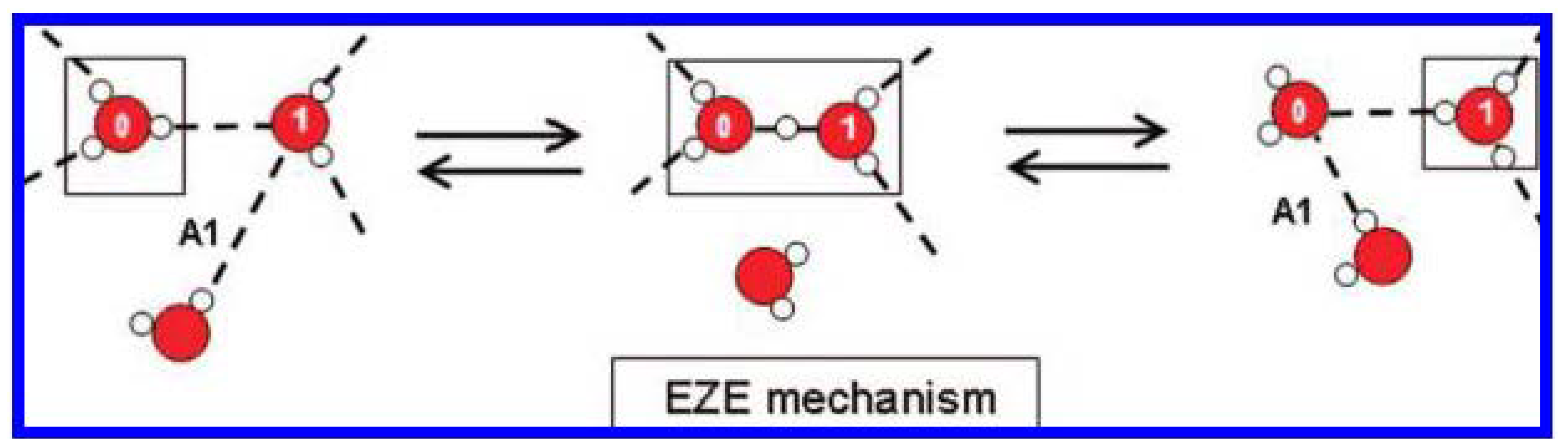
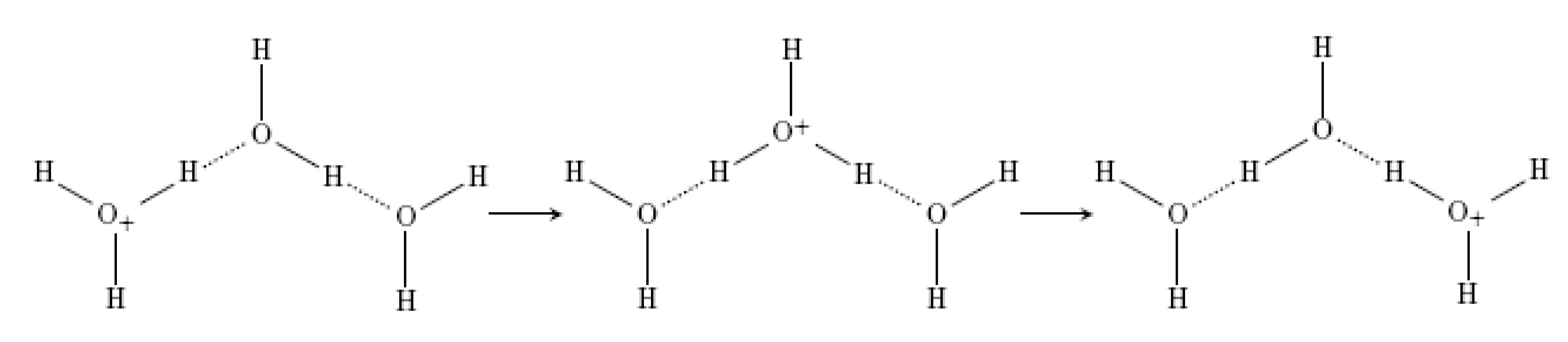
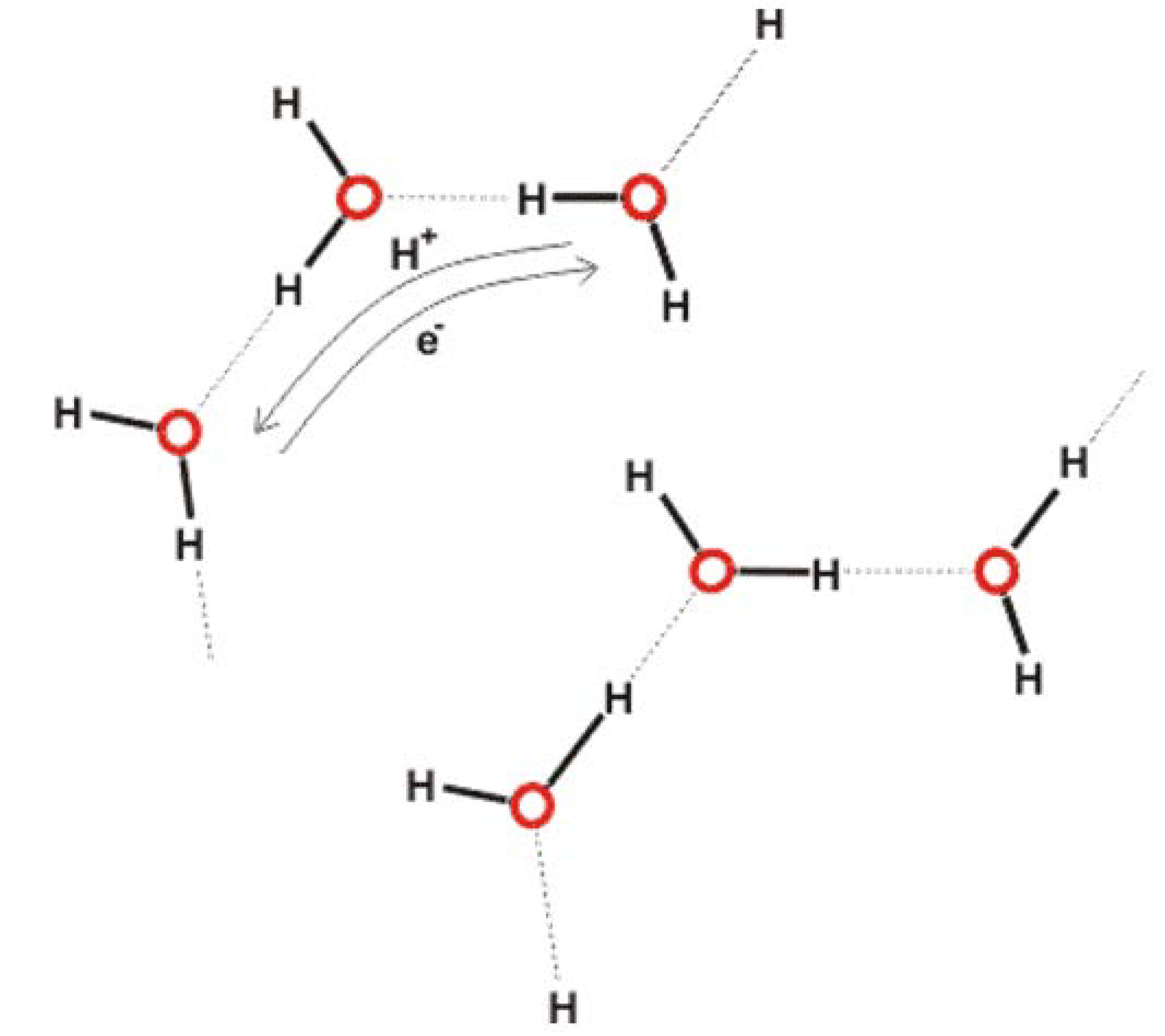
  103. Agmon, N. The Grotthuss mechanism. Chem. Phys. Lett. 1995, 244, 456–462. [Google Scholar] [CrossRef]
  104. Agmon, N. Salt effect on transient proton transfer to solvent and microscopic proton mobility. Chem. Phys. Lett. 1995, 64, 161–195. [Google Scholar] [CrossRef]
  105. Markovitch, O.; Chen, H.; Izvekov, S.; Paesani, F.; Voth, G.A.; Agmon, N. Special pair dance and partner selection: Elementary steps in proton transport in liquid water. J. Phys. Chem. B 2008, 112, 9456–9466. [Google Scholar] [CrossRef] [PubMed]
  106. Verdel, N.; Jerman, I.; Bukovec, P. The “Autothixotropic” phenomenon of water and its role in proton transfer. Int. J. Mol. Sci. 2011, 12, 7481–7494. [Google Scholar] [CrossRef] [PubMed]
  107. Verdel, N.; Jerman, I.; Krasovec, R.; Bukovec, P.; Zupancic, M. Possible time-dependent effect of ions and hydrophilic surfaces on the electrical conductivity of aqueous solutions. Int. J. Mol. Sci. 2012, 13, 4048–4068. [Google Scholar] [PubMed]
  108. Feng, S.; Voth, G.A. Proton solvation and transport in hydrated nafion. J. Phys. Chem. B 2011, 115, 5903–5912. [Google Scholar] [CrossRef] [PubMed]
  109. Jorn, R.; Voth, G.A. Mesoscale simulation of proton transport in proton exchange membranes. J. Phys. Chem. C 2012, 116, 10476–10489. [Google Scholar] [CrossRef]
  110. Zundel, G.; Metzer, H. Energiebänder der tunnelnden Überschuβ-Protonen in flüssigen Säuren. Eine IR-spektroskopische Untersuchung der Natur der Gruppierungen H5O2+. Z. Phys. Chem. 1968, 58, 225–245. (in German). [Google Scholar] [CrossRef]
  111. Knight, C.; Voth, G.A. The curious case of the hydrated proton. Acc. Chem. Res. 2012, 45, 101–109. [Google Scholar] [CrossRef]
  112. Habenicht, B.F.; Paddison, S.J. Ab initio simulations of the effects of nanoscale confinement on proton transfer in hydrophobic environments. J. Phys. Chem. B 2011, 115, 10826–10835. [Google Scholar]
  113. Martin Chaplin talks about the importance of water, in advance of the Colours of Water festival, March, 2013. http://www.i-sis.org.uk/coloursofwater/.
  114. Chaplin, M. What is Liquid Water? ISIS Report. Available online: http://www.i-sis.org.uk/What_is_Liquid_Water.php/ (accessed on 20 April 2013).
  115. Czerlinski, G.; Ypma, T. Stabilization of aqueous electromeric nano-domains. J. Comput. Theor. Nanos. 2011, 8, 1400–1408. [Google Scholar] [CrossRef]
  116. Czerlinski, G.; Ypma, T. Homeopathic potentization based on nanoscale domains. J. Altern. Complem. Med. 2011, 17, 1165–1173. [Google Scholar] [CrossRef] [PubMed]
  117. Mizuse, K.; Kuo, J.-L.; Fujii, A. Structural trends of ionized water networks: Infrared spectroscopy of water cluster radical cations (H2O)n+ (n = 3–11). Chem. Sci. 2011, 2, 868–876. [Google Scholar] [CrossRef]
  118. Fröhlich, H. Bose condensation of strongly excited longitudinal electric modes. Phys. Lett. A 1968, 26, 402–403. [Google Scholar] [CrossRef]
  119. Fröhlich, H. Long-range coherence and energy storage in biological systems. Int. J. Quantum Chem. 1968, 2, 641–649. [Google Scholar] [CrossRef]
  120. Pokorný, J.; Foletti, A.; Kobilková, J.; Jandová, A.; Vrba, J.; Vrba, J., Jr.; Nedbalová, M.; Čoček, A.; Danani, A.; Tuszyński, J.A. Biophysical insights into cancer transformation and treatment. Sci. World J. 2013. Article ID 195028. [Google Scholar] [CrossRef]
  121. Pohl, H.A. Oscillating fields about growing cells. Int. J. Quant. Chem. 1980, 7, 411–431. [Google Scholar] [CrossRef]
  122. IUPAC. Compendium of Chemical Terminology, 2nd ed.; McNaught, A.D., Wilkinson, A., Eds.; Blackwell Scientific Publications: Oxford, UK, 1997. [Google Scholar]
  123. Mitsui, T.; Rose, M.K.; Fomin, E.; Ogletree, D.F.; Salmeron, M. Water Diffusion and Clustering on Pd(111). Science 2002, 297, 1850–1852. [Google Scholar] [CrossRef] [PubMed]
  124. Smith, C.W. Fröhlich’s interpretation of biology through theoretical physics Chapter 7. In Herbert Fröhlich FRS: A physicist ahead of his time; Hyland, G.J., Rowlands, P., Eds.; The University of Liverpool: Liverpool, UK, 2006; pp. 91–138, (2nd edition 2008, pp.107–154). [Google Scholar]
  125. Ahmed, N.A.G.; Calderwood, J.H.; Fröhlich, H.; Smith, C.W. Evidence for collective magnetic effects in an enzyme: Likelihood of room temperature superconductive regions. Phys. Lett. 1975, 53A, 129–130. [Google Scholar] [CrossRef]
  126. Milani, M.; Del Giudice, E.; Doglia, S.; Vitiello, G.; Smith, C.W. Superconductive and Josephson-like behaviour of cells. La Radiologica. Medica.Radiol. Med. 1991, 81, 51–55. [Google Scholar]
  127. Stahler, J.; Bovensiepen, U.; Meyer, M.; Wolf, M. A surface science approach to ultrafast electron transfer and solvation dynamics at interfaces. Chem. Soc. Rev. 2008, 37, 2180–2190. [Google Scholar] [CrossRef] [PubMed]
  128. Liang, H.-W.; Guan, Q.-F.; Zhu, Z.; Song, L.-T.; Yao, H.-B.; Lei, X.; Yu, S.-H. Highly conductive and stretchable conductors fabricated from bacterial cellulose. NPG Asia Mater. 2012, 4, e19. [Google Scholar] [CrossRef]
  129. Gascoyne, P.R.; Pethig, R.; Szent-Gyorgyi, A. Water structure-dependent charge transport in proteins. Proc. Natl. Acad. Sci. USA 1981, 78, 261–265. [Google Scholar] [CrossRef] [PubMed]
  130. Careri, G.; Giansanti, A.; Rupley, J.A. Critical exponents of protonic percolation in hydrated lysozyme powders. Phys. Rev. A 1988, 37, 2703–2705. [Google Scholar] [CrossRef] [PubMed]
  131. Craddock, T.J.A.; Tuszynski, J.A.; Priel, A.; Freedman, H. Microtubule ionic conduction and its implications for higher cognitive functions. J. Integr. Neurosci. 2010, 9, 103–122. [Google Scholar] [CrossRef] [PubMed]
  132. Polcari, A.; Romano, P.; Sabatino, L.; Del Vecchio, E.; Consales, M.; Cusano, A.; Cutolo, A.; Colantuoni, V. Electrical and optical characterization of DNA molecules as a function of concentration in aqueous solution. J. Appl. Phys. 2011, 109, 074703. [Google Scholar] [CrossRef]
  133. Sontz, P.A.; Muren, N.B.; Barton, J.K. DNA charge transport for sensing and signaling. Accounts Chem. Res. 2012, 45, 1792–1800. [Google Scholar] [CrossRef] [PubMed]
  134. Careri, G.; Giansanti, A.; Rupley, J.A. Proton percolation on hydrated lysozyme powders. Proc. Natl. Acad. Sci. USA 1986, 83, 6810–6814. [Google Scholar] [CrossRef] [PubMed]
  135. Careri, G.; Milotti, E. Simulation of protonic fluctuations in hydrated protein powders. Phys. Rev. E 2003, 67, 051923. [Google Scholar] [CrossRef]
  136. Brovchenko, I.; Krukau, A.; Oleinikova, A.; Mazur, A.N. Water percolation governs polymorphic transitions and conductivity of DNA. Phys. Rev. Lett. 2006, 97, 137801. [Google Scholar] [CrossRef] [PubMed]
  137. Czerlinski, G.; Ypma, T. Domains of water molecules provide mechanisms of potentization in homeopathy. Water 2010, 2, 1–14. [Google Scholar]
  138. Czerlinski, G.; Ypma, T. The targets of information-carrying nanodomains. J. NanoSci. Nanotech. 2012, 12, 2239–2247. [Google Scholar] [CrossRef]
  139. Wang, G.M.; Sevick, E.M.; Mittag, E.; Searles, D.J.; Evans, D.J. Experimental demonstration of violations of the second law of thermodynamics for small systems and short time scales. Phys. Rev. Lett. 2002, 89, 050601. [Google Scholar] [CrossRef] [PubMed]
  140. Cox, R.A. A greatly under-appreciated fundamental principle of physical organic chemistry. Intl. J. Mol. Sci. 2011, 12, 8316–8332. [Google Scholar] [CrossRef] [PubMed]
  141. Tanvir, S.; Qiao, L. Surface tension of nanofluid-type fuels containing suspended nanomaterials. Nanoscale Res. Lett. 2012, 7, 226. [Google Scholar] [CrossRef] [PubMed]
  142. Sharma, S.; Debenedetti, P.G. Evaporation rate of water in hydrophobic confinement. Proc. Natl. Acad. Sci. 2012, 109, 4365–4370. [Google Scholar] [CrossRef] [PubMed]
  143. Mori, A.; Suzuki, Y. Grand potential formalism of interfacial thermodynamics for critical nucleus. Nat. Sci. 2013, 5, 631–639. [Google Scholar] [CrossRef]
  144. Belloni, L.; Allweiss, L.; Guerrieri, F.; Pediconi, N.; Volz, T.; Pollicino, T.; Petersen, J.; Raimondo, G.; Dandri, M.; Levrero, M. IFN-α inhibits HBV transcription and replication in cell culture and in humanized mice by targeting the epigenetic regulation of the nuclear cccDNA minichromosome. J. Clin. Invest. 2012, 122, 529–537. [Google Scholar] [CrossRef] [PubMed]
  145. Shibata, Y.; Kumar, P.; Layer, R.; Willcox, S.; Gagan, J.R.; Griffith, J.D.; Dutta, A. Extrachromosomal microDNAs and chromosomal microdeletions in normal tissues. Science 2012, 336, 82–86. [Google Scholar] [CrossRef] [PubMed]
  146. Lee, S. Detection of human papillomavirus L1 gene DNA fragments in postmortem blood and spleen after Gardasil® vaccination—A case report. Adv. Biosci. Biotechnol. 2012, 3, 1214–1224. [Google Scholar] [CrossRef]
  147. Rich, A.; Zhang, S. Z-DNA: The long road to biological function. Nature 2003, 4, 568–572. [Google Scholar] [CrossRef] [PubMed]
  148. Montagnier, L.; Aissa, J.; Ferris, S.; Montagnier, J.L.; Lavallee, C. Electromagnetic signals are produced by aqueous nanostructures derived from bacterial DNA sequences. Interdiscipl. Sci. Comp. Life Sci. 2009, 1, 81–90. [Google Scholar] [CrossRef] [PubMed]
  149. Voeikov, V.L.; Naletov, V.I. Weak photon emission of non-linear chemical reactions of amino acids and sugars in aqueous solutions. In Biophotons; Chang, J.-J., Ed.; Kluwer Academic Publishers: Dordrecht, The Netherlands, 1998; pp. 93–108. [Google Scholar]
  150. Montagnier, L.; Aissa, J.; Del Giudice, E.; Lavallee, C.; Tedeschi, A.; Vitiello, G. DNA waves and water. J. Phys. Conf. Ser. 2011, 306, 012007. [Google Scholar] [CrossRef]
  151. Voeikov, V.L.; Del Giudice, E. Water respiration—The basis of the living state. Water 2009, 1, 52–75. [Google Scholar]
  152. Preparata, G. QED Coherence and Matter; World Scientific: City, Singapore, 1995. [Google Scholar]
  153. Arani, R.; Bono, I.; Del Giudice, E.; Preparata, G. QED coherence and the thermodynamics of water. Int. J. Mod. Phys. B 1995, 9, 1813–1841. [Google Scholar] [CrossRef]
  154. Del Giudice, E.; Fuchs, E.; Vitiello, G. Collective molecular dynamics of a floating water bridge. Water 2010, 2, 69–82. [Google Scholar]
  155. Marchettini, N.; Del Giudice, E.; Voeikov, V.; Tiezzi, E. Water: A medium where dissipative structures are produced by a coherent dynamics. J. Theor. Biol. 2010, 265, 511–516. [Google Scholar] [CrossRef] [PubMed]
  156. Kobayashi, M.; Takeda, M.; Sato, T.; Yamazaki, Y.; Kaneko, K.; Ito, K.; Kato, H.; Inaba, H. In vivo imaging of spontaneous ultraweak photon emission from a rat’s brain correlated with cerebral energy metabolism and oxidative stress. Neurosci. Res. 1999, 34, 103–113. [Google Scholar] [CrossRef]
  157. Curtis, B.D.; Hurtak, J.J. Consciousness and quantum information processing: uncovering the foundation for a medicine of light. J. Altern. Complement. Med. 2004, 10, 27–39. [Google Scholar] [CrossRef] [PubMed]
  158. Kim, H.W.; Sim, S.B.; Kim, C.K.; Kim, J.; Choi, C.; You, H.; Soh, K.S. Spontaneous photon emission and delayed luminescence of two types of human lung cancer tissues: adenocarcinoma and squamous cell carcinoma. Cancer Lett. 2005, 229, 283–289. [Google Scholar] [CrossRef] [PubMed]
  159. Kim, J.; Choi, C.; Lim, J.; You, H.; Sim, S.B.; Yom, Y.K.; Kim, E.H.; Soh, K.S. Measurements of spontaneous ultraweak photon emission and delayed luminescence from human cancer tissues. J. Altern. Complement. Med. 2005, 11, 879–884. [Google Scholar] [CrossRef] [PubMed]
  160. Whissell, P.D.; Persinger, M.A. Developmental effects of perinatal exposure to extremely weak 7 Hz magnetic fields and nitric oxide modulation in the Wistar albino rat. Int. J. Dev. Neurosci. 2007, 25, 433–439. [Google Scholar] [CrossRef] [PubMed]
  161. Del Giudice, E.; Fleischmann, M.; Preparata, G.; Talpo, G. On the “unreasonable” effects of ELF magnetic fields upon a system of ions. Bioelectromagnetics 2002, 23, 522–530. [Google Scholar] [CrossRef] [PubMed]
  162. Tafur, J.; Van Wijk, E.P.; Van Wijk, R.; Mills, P.J. Biophoton detection and low-intensity light therapy: A potential clinical partnership. Photomed. Laser Surg. 2010, 28, 23–30. [Google Scholar] [CrossRef] [PubMed]
  163. Ho, M.-W. Quantum Coherent Water, Non-thermal EMF Effects, and Homeopathy. ISIS Report. Available online: http://www.i-sis.org.uk/Quantum_Coherent_Water_Homeopathy.php/ (accessed on 31 January 2013).
  164. Binhi, V.N.; Savin, A.V. Molecular gyroscopes and biological effects of weak extremely low-frequency. Phys. Rev. E Stat. Nonlin. Soft Matter Phys. 2002, 65, 051912. [Google Scholar] [CrossRef] [PubMed]
  165. Higgins, M.J.; Polcik, M.; Fukuma, T.; Sader, J.E.; Nakayama, Y.; Jarvis, S.P. Structured water layers adjacent to biological membranes. Biophys. J. 2006, 91, 2532–2542. [Google Scholar] [CrossRef] [PubMed]
  166. Binhi, V.N. A few remarks on combined action of DC and AC magnetic fields on ion motion in a macromolecule. Bioelectromagnetics 2007, 28, 409–412. [Google Scholar] [CrossRef] [PubMed]
  167. Giuliani, L.; D’Emilia, E.; Grimaldi, S.; Lisi, A.; Bobkova, N.; Zhadin, M.N. Investigating the ICR effect in a Zhadin’s cell. Intl. J. Biomed. Sci. 2009, 5, 181–186. [Google Scholar]
  168. Zhadin, M. Quantum electrodynamical mechanisms of resonant effects development inside coherence domains at combined magnetic fields. In Proceedings of Action Progress in Electromagnetics, Research Symposium Abstracts, Moscow, Russia, 18–21 August 2009; p. 416.
  169. Liboff, A.R. The charge-to-mass ICR signature in weak ELF bioelectromagnetic effects. In Advances in Electromagnetic Fields in Living Systems, Chapter 6; Lin, J.C., Ed.; Springer Science+Business Media: New York, NY, USA, 2005; Volume 4. [Google Scholar]
  170. Vazquez, A. Optimal cytoplasmatic density and flux balance model under macromolecular crowding effects. J. Theor. Biol. 2010, 264, 356–359. [Google Scholar] [CrossRef] [PubMed]
  171. Silverstein, K.A.T.; Haymet, A.D.J.; Dill, K.A. A simple model of water and the hydrophobic effect. J. Am. Chem. Soc. 1998, 120, 3166–3175. [Google Scholar] [CrossRef]
  172. Latham, M.P.; Kay, L.E. Is buffer a good proxy for a crowded cell-like environment? A comparative NMR study of calmodulin side-chain dynamics in buffer and E. coli lysate. PloS One 2012, 7, e48226. [Google Scholar] [CrossRef] [PubMed]
  173. Giuliani, L.; Grimaldi, S.; Lisi, A.; D’Emilia, E.; Bobkova, N.; Zhadin, M. Action of combined magnetic fields on aqueous solution of glutamic acid: the further development of investigations. Biomagn. Res. Technol. 2008, 6, 1. [Google Scholar] [CrossRef] [PubMed]
  174. Zhadin, M.; Giuliani, L. Some problems in modern bioelectromagnetics. Electromagn. Biol. Med. 2006, 25, 227–243. [Google Scholar] [CrossRef] [PubMed]
  175. Liboff, A.R. The Ion Cyclotron Resonance Hypothesis. In Bioengineering and Biophysical Aspects of Electromagnetic Fields; Barnes, F., Greenebaum, B., Eds.; CRC Press, Taylor & Francis Group: Boca Raton, FL, USA, 2006; pp. 261–292. [Google Scholar]
  176. Zhadin, M.N.; Novikov, V.V.; Barnes, F.S.; Pergola, N.F. Combined action of static and alternating magnetic fields on ionic current in aqueous glutamic acid solution. Bioelectromagnetics 1998, 19, 41–45. [Google Scholar] [CrossRef]
  177. Novikov, V.V.; Zhadin, M.N. Combined action of weak constant and variable low-frequency magnetic fields on ionic currents in aqueous solutions of amino acid. Biophysics 1994, 994, 41–45. [Google Scholar]
  178. Luby-Phelps, K. Cytoarchitecture and physical properties of cytoplasm: Volume, viscosity, diffusion, intracellular surface area. Int. Rev. Cytol. 2000, 192, 189–221. [Google Scholar] [PubMed]
  179. Aggeli, A.; Nyrkova, I.A.; Bell, M.; Harding, R.; Carrick, L.; McLeish, T.C.; Semenov, A.N.; Boden, N. Hierarchical self-assembly of chiral rod-like molecules as a model for peptide beta-sheet tapes, ribbons, fibrils, and fibers. Proc. Natl. Acad. Sci. USA 2001, 98, 11857–11862. [Google Scholar] [CrossRef] [PubMed]
  180. Marchesan, S.; Waddington, L.; Easton, C.D.; Winkler, D.A.; Goodall, L.; Forsythe, J.; Hartley, P.G. Unzipping the role of chirality in nanoscale self-assembly of tripeptide hydrogels. Nanoscale 2012, 4, 6752–6760. [Google Scholar] [CrossRef] [PubMed]
  181. Zhao, K.; Bruinsma, R.; Mason, T.G. Local chiral symmetry breaking in triatic liquid crystals. Nat. Comm. 2012, 3, 801. [Google Scholar] [CrossRef] [PubMed]
  182. Boncheva, M.; Andreev, S.A.; Mahadevan, L.; Winkleman, A.; Reichman, D.R.; Prentiss, M.G.; Whitesides, S.; Whitesides, G.M. Magnetic self-assembly of three-dimensional surfaces from planar sheets. Proc. Natl. Am. Sci. USA 2005, 102, 3924–3929. [Google Scholar] [CrossRef] [PubMed]
  183. Mirica, K.A.; Ilievski, F.; Ellerbee, A.K.; Shevkoplyas, S.S.; Whitesides, G.M. Using magnetic levitation for three dimensional self-assembly. Adv. Mater. 2011, 23, 4134–4140. [Google Scholar] [CrossRef] [PubMed]
  184. Brown, P.; Butts, C.P.; Cheng, J.; Eastoe, J.; Russell, C.A.; Smith, G.N. Magnetic emulsions with responsive surfactants. Soft Matter 2012, 8, 7545–7546. [Google Scholar] [CrossRef]
  185. Brown, P.; Khan, A.M.; Armstrong, J.P.K.; Perriman, A.W.; Butts, C.P.; Eastoe, J. Magnetizing DNA and proteins using responsive surfactants. Adv. Matr. 2012, 24, 6244–6247. [Google Scholar] [CrossRef] [PubMed]
  186. Brown, P.; Butts, C.P.; Eastoe, J.; Glatzel, S.; Grillo, I.; Hall, S.H.; Rogers, S.; Trickett, K. Microemulsions as tunable nanomagnets. Soft Matter 2012, 8, 11609–11612. [Google Scholar] [CrossRef]
  187. Banquy, X.; Kristiansen, K.; Lee, D.W.; Israelachvili, J.N. Adhesion and hemifusion of cytoplasmic myelin lipid membranes are highly dependent on the lipid composition. Biochim. Biophys. Acta. 2012, 1818, 402–410. [Google Scholar] [CrossRef]
  188. Sherman, I.A. Interfacial tension effects in the microvasculature. Microvasc. Res. 1981, 22, 296–307. [Google Scholar] [CrossRef]
  189. Rowlinson, J.S.; Widom, B. Molecular Theory of Capillarity; Dover Press: Mineola, NY, USA, 1982. [Google Scholar]
  190. Kashiwagi, T.; Kunishima, N.; Suzuki, C.; Tsuchiya, F.; Nikkuni, S.; Arata, Y.; Morikawa, K. The novel acidophilic structure of the killer toxin from halotolerant yeast demonstrates remarkable folding similarity with a fungal killer toxin. Structure 1997, 5, 81–94. [Google Scholar] [CrossRef]
  191. Mennerick, S.; Lamberta, M.; Shu, H.-J.; Hogins, J.; Wang, C.; Covey, D.F.; Eisenman, L.N.; Zorumski, C.F. Effects on membrane capacitance of steroids with antagonist properties at GABAA receptors. Biophys. J. 2008, 95, 176–185. [Google Scholar] [CrossRef] [PubMed]
  192. Chisari, M.; Wu, K.; Zorumski, C.F.; Mennerick, S. Hydrophobic anions potently and uncompetitively antagonize GABA(A) receptor function in the absence of a conventional binding site. Br. J. Pharmacol. 2011, 164, 667–680. [Google Scholar] [CrossRef] [PubMed]
  193. Walle, T.; Hsieh, F.; DeLegge, M.H.; Oatis, J.E., Jr.; Walle, U.K. High absorption but very low bioavailability of oral resveratrol in humans. Drug Metab. Dispos. 2004, 32, 1377–1382. [Google Scholar] [CrossRef] [PubMed]
  194. McCully, K.S. Chemical pathology of homocysteine. V. Thioretinamide, thioretinaco, and cystathionine synthase function in degenerative diseases. Ann. Clin. Lab. Sci. 2011, 41, 300–313. [Google Scholar]
  195. Shwartz, E.R.; Adamy, L. Effect of ascorbic acid on arylsulfatase activities and sulfated proteoglycan metabolism in chondrocyte cultures. J. Clin. Invest. 1977, 60, 96–106. [Google Scholar] [CrossRef] [PubMed]
  196. Boskey, A.L.; Blank, R.D.; Doty, S.B. Vitamin C-sulfate inhibits mineralization in chondrocyte cultures: A caveat. Matrix Biol. 2001, 20, 99–106. [Google Scholar] [CrossRef]
  197. Verlangieri, J.; Mumma, R.O. In vivo sulfation of cholesterol by ascorbic acid 2-sulfate. Atherosclerosis 1973, 17, 37–48. [Google Scholar] [CrossRef]
  198. Chen, X.; Resh, M.D. Cholesterol depletion from the plasma membrane triggers ligand-independent activation of the epidermal growth factor receptor. J. Biol. Chem. 2002, 277, 49631–49637. [Google Scholar] [CrossRef] [PubMed]
  199. Jiang, B.-H.; Liu, L.-Z. PI3K/PTEN signaling in angiogenesis and tumorigenesis. Adv. Cancer Res. 2009, 102, 19–65. [Google Scholar] [CrossRef] [PubMed]
  200. Marcus, Y. Individual ionic surface tension increments in aqueous solutions. Langmuir 2013, 29, 2881–2888. [Google Scholar] [CrossRef] [PubMed]
  201. Anand, S.; Paxson, A.T.; Dhiman, R.; Smith, J.D.; Varanasi, K.K. Enhanced condensation on lubricant-impregnated nanotextured surfaces. ACS Nano. 2012, 6, 10122–10129. [Google Scholar] [CrossRef] [PubMed]
  202. Quéré, D. Wetting and roughness. Annu. Rev. Mater. Res. 2008, 38, 71–99. [Google Scholar] [CrossRef]
  203. Ellinas, K.; Tserepi, A.; Gogolides, E. From superamphiphobic to amphiphilic polymeric surfaces with ordered hierarchical roughness fabricated with colloidal lithography and plasma nanotexturing. Langmuir 2011, 27, 3960–3969. [Google Scholar] [CrossRef] [PubMed]
  204. Campos, R.; Kataky, R. Electron transport in supported and tethered lipid bilayers modified with bioelectroactive molecules. J. Phys. Chem. B 2012, 116, 3909–3917. [Google Scholar] [CrossRef] [PubMed]
  205. Fernández, A. Nanoscale thermodynamics of biological interfacial tension. Proc. R. Soc. A 2011, 467, 559–568. [Google Scholar] [CrossRef]
  206. Fernandez, A. Epistructural tension promotes protein associations. Phys. Rev. Lett. 2012, 108, 188102. [Google Scholar] [CrossRef] [PubMed]
  207. Brecher, G.; Bessis, M. Present status of spiculed red cells and their relationship to the discocyte-echinocyte transformation: A critical review. Blood 1972, 40, 333–344. [Google Scholar] [PubMed]
  208. Absolom, D.R. Measurement of surface properties of phagocytes, bacteria, and other particles. Methods Enzymol. 1986, 132, 16–95. [Google Scholar] [PubMed]
  209. Gallez, D.; Coakley, W.T. Interfacial instability at cell membranes. Prog. Biophys. Mol. Biol. 1986, 48, 155–199. [Google Scholar] [CrossRef]
  210. Berthelot, M. Sur quelques phénomènes de dilatation forcée des liquids. Annales. de Chimie. et de Physique. 1850, 30, 232–237, (English translation). [Google Scholar]
  211. Larios, E.; Gruebele, M. Protein stability at negative pressure. Methods 2010, 52, 51–56. [Google Scholar] [CrossRef] [PubMed]
  212. Grigera, J.R.; Andres, N.; McCarthy, A.N. The behavior of the hydrophobic effect under pressure and protein denaturation. Biophys. J. 2010, 98, 1626–1631. [Google Scholar] [CrossRef] [PubMed]
  213. Defay, R.; Prigogine, I. Surface Tension and Adsorption; Wiley: Hoboken, NJ, USA, 1966. [Google Scholar]
  214. Janmey, P.A. Gel-sol transition of the cytoplasm and its regulation. AIP Conf. Proc. 1980, 226, 304–325. [Google Scholar]
  215. Debenedetti, P.G.; Stillinger, F.H. Supercooled liquids and the glass transition. Nature 2001, 410, 259–267. [Google Scholar] [CrossRef] [PubMed]
  216. Torre, R.; Bartolini, P.; Righini, R. Structural relaxation in supercooled water by time-resolved spectroscopy. Nature 2004, 428, 296–299. [Google Scholar] [CrossRef] [PubMed]
  217. Kim, C.U.; Barstow, B.; Tate, M.W.; Gruner, S.M. Evidence for liquid water during the high-density to low-density amorphous ice transition. Proc. Natl. Am. Sci. USA 2009, 106, 4596–4600. [Google Scholar] [CrossRef] [PubMed]
  218. Pelling, A.E.; Dawson, D.W.; Carreon, D.M.; Christiansen, J.J.; Shen, R.R.; Teitell, M.A.; Gimzewski, J.K. Distinct contributions of microtubule subtypes to cell membrane shape and stability. Nanomedicine 2007, 3, 43–52. [Google Scholar] [CrossRef] [PubMed]
  219. Zhou, E.H.; Trepat, X.; Park, C.Y.; Lenormand, G.; Oliver, M.N.; Mijailovich, S.M.; Hardin, C.; Weitz, C.A.; Butler, J.P.; Fredberg, J.J. Universal behavior of the osmotically compressed cell and its analogy to the colloidal glass transition. Proc. Natl. Am. Sci. USA 2009, 106, 10632–10637. [Google Scholar]
  220. Huang, D.M.; Chandler, D. The hydrophobic effect and the influence of solute-solvent attractions. J. Phys. Chem. B 2002, 106, 2047–2053. [Google Scholar] [CrossRef]
  221. Woods, A.; Couchman, J.R.; Höök, M. Heparan sulfate proteoglycans of rat embryo fibroblasts. A hydrophobic form may link cytoskeleton and matrix components. J. Biol. Chem. 1985, 260, 10872–10879. [Google Scholar] [PubMed]
  222. Kawahara, M.; Kato-Negishi, M. Link between aluminum and the pathogenesis of Alzheimer’s disease: The integration of the aluminum and amyloid cascade hypotheses. Int. J. Alzheimers. Dis. 2011, 276393. [Google Scholar] [CrossRef] [PubMed]
  223. Szent-Györgyi, A.G. Calcium regulation of muscle contraction. Biophys. J. 1975, 15, 707–723. [Google Scholar] [CrossRef]
  224. Berridge, M.J.; Lipp, P.; Bootman, M.D. The versatility and universality of calcium signaling. Nat. Rev. Mol. Cell Biol. 2000, 1, 11–21. [Google Scholar] [CrossRef] [PubMed]
  225. Kojima, S.; Hama, Y.; Sasaki, T.; Kubodera, A. Elevated uptake of 67Ga and increased heparan sulfate content in liver-damaged rats. Eur. J. Nucl. Med. 1983, 8, 52–59. [Google Scholar] [CrossRef] [PubMed]
  226. Ando, A.; Ando, I.; Hiraki, T.; Hisada, K. 67Ga-binding substances in stomach, small intestine, pancreas, and muscle. Eur. J. Nucl. Med. 1985, 11, 235–239. [Google Scholar] [CrossRef] [PubMed]
  227. Hama, Y.; Sasaki, T.; Kojima, S.; Kubodera, A. 67Ga accumulation and heparan sulfate metabolism in lysosomes. Eur. J. Nucl. Med. 1984, 9, 51–56. [Google Scholar] [CrossRef] [PubMed]
  228. Selye, H. Thrombohemorrhagic Phenomena; Charles, C., Ed.; Thomas: Springfield, IL, USA, 1966. [Google Scholar]
  229. Selye, H. In Vivo: The Case for Supramolecular Biology; Liveright Publishing Corporation: New York, NY, USA, 1967. [Google Scholar]
  230. Mastruserio, D.N.; Nguyen, E.Q.; Nielsen, T.; Hessel, A.; Pellegrini, A.E. Calciphylaxis associated with metastatic breast carcinoma. J. Am. Acad. Dermatol. 1999, 41, 295–298. [Google Scholar] [CrossRef] [PubMed]
  231. Goel, S.K.; Bellovich, K.; McCullough, P.A. Treatment of severe metastatic calcification and calciphylaxis in dialysis patients. Int. J. Nephrol. 2011, 2011. Article ID 701603. [Google Scholar] [CrossRef] [PubMed]
  232. Block, G.A.; Hulbert-Shearon, T.E.; Levin, N.W.; Port, F.K. Association of serum phosphorus and calcium x phosphate product with mortality risk in chronic hemodialysis patients: a national study. Am. J. Kidney Dis. 1998, 31, 607–617. [Google Scholar] [CrossRef] [PubMed]
  233. Guo, F.; Friedman, J.M. Charge density-dependent modifications of hydration shell waters by Hofmeister ions. J. Am. Chem. Soc. 2009, 131, 11010–11018. [Google Scholar] [CrossRef] [PubMed]
  234. Tielrooij, K.J.; Garcia-Araez, N.; Bonn, M.; Bakker, H.J. Cooperativity in ion hydration. Science 2010, 328, 1006–1009. [Google Scholar] [CrossRef] [PubMed]
  235. Marcus, Y. Volumetric properties of molten salt hydrates. J. Chem. Eng. Data 2013, 58, 488–491. [Google Scholar] [CrossRef]
  236. Rezwan, K.; Meier, L.P.; Rezwan, M.; Vöörös, J.; Textor, M.; Gauckler, L.J. Bovine serum albumin adsorption onto colloidal Al2O3 particles: a new model based on zeta potential and UV-vis measurements. Langmuir 2004, 20, 10055–10061. [Google Scholar] [CrossRef] [PubMed]
  237. Li, C.; Somasundaran, P. Reversal of bubble charge in multivalent inorganic salt solutions: Effect of aluminum. J. Colloid. Interface Sci. 1992, 148, 587–591. [Google Scholar] [CrossRef]
  238. Nday, C.; Salifoglou, A. The influence of the environmental metallotoxin Al(III) on neuronal cell structures linked to neurodegeneration. J. Agroaliment. Proc. Tech. 2012, 18, 208–211. [Google Scholar]
  239. Lipinski, B.; Jeljaszewicz, J. A hypothesis for the pathogenesis of the generalized Shwartzman reaction. J. Infect. Dis. 1969, 120, 160–168. [Google Scholar] [CrossRef] [PubMed]
  240. Riddick, T. Control of Colloid Stability through Zeta Potential (with a Closing Chapter on its Relationship to Cardiovascular Disease); Livingston Pub. Co.: Wynnewood, PA, USA, 1968. [Google Scholar]
  241. Armenante, P.M. Coagulation & Flocculation. Available online: http://cpe.njit.edu/dlnotes/che685/cls07–1.pdf (accessed on 23 January 2013).
  242. Droste, R.L. Theory and Practice of Water and Wastewater Treatment; John Wiley & Sons: New York, NY, USA, 1997; pp. 384–415. [Google Scholar]
  243. Del Giudice, E.; Doglia, S.; Milani, M.; Smith, C.W.; Vitiello, G. Magnetic flux quantization and Josephson behaviour in living systems. Phys. Scr. 1989, 40, 786. [Google Scholar] [CrossRef]
  244. Hunt, R.W.; Zavalin, A.; Bhatnagar, A.; Chinnasamy, S.; Das, K.C. Electromagnetic biostimulation of living cultures for biotechnology, biofuel and bioenergy applications. Int. J. Mol. Sci. 2009, 10, 4515–4558. [Google Scholar] [CrossRef] [PubMed]
  245. Pogue, A.I.; Jones, B.M.; Bhattacharjee, S.; Percy, M.E.; Zhao, Y.; Lukiw, W.J. Metal-sulfate induced generation of ROS in human brain cells: Detection using an isomeric mixture of 5- and 6-carboxy-2,7-dichlorofluoresce in diacetate (carboxy-DCFDA) as a cell permeant tracer. Int. J. Mol. Sci. 2012, 13, 9615–9626. [Google Scholar] [CrossRef] [PubMed]
  246. Patra, J.; Baisakhi, B.; Mohapatro, M.K.; Panda, B.B. Aluminium triggers genotoxic adaptation to methyl mercuric chloride and ethyl methane sulfonate, but not to maleic hydrazide in plant cells in vivo. Mutat. Res. 2000, 465, 1–9. [Google Scholar] [CrossRef]
  247. Achary, V.M.M.; Panda, B.B. Aluminium-induced DNA damage and adaptive response to genotoxic stress in plant cells are mediated through reactive oxygen intermediates. Mutagenesis 2010, 25, 201–209. [Google Scholar] [CrossRef] [PubMed]
  248. Achary, V.M.; Parinandi, N.L.; Panda, B.B. Aluminum induces oxidative burst, cell wall NADH peroxidase activity, and DNA damage in root cells of Allium cepa L. Enviro. Mol. Mutagen. 2012, 53, 550–560. [Google Scholar] [CrossRef] [PubMed]
  249. Baylor, N.W.; Egan, W.; Richman, P. Aluminum salts in vaccines–US perspective. Vaccine 2002, 20, S18–S23. [Google Scholar] [CrossRef]
  250. Exley, C. The pro-oxidant activity of aluminum. Free Radic. Biol. Med. 2004, 36, 380–387. [Google Scholar] [CrossRef] [PubMed]
  251. Mujika, J.I.; Ruiperez, F.; Infante, I.; Ugalde, J.M.; Exley, C.; Lopez, X. Pro-oxidant activity of aluminum: stabilization of the aluminum superoxide radical ion. J. Phys. Chem. A 2011, 115, 6717–6723. [Google Scholar] [CrossRef] [PubMed]
  252. Exley, C. The coordination chemistry of aluminium in neurodegenerative disease. Coord. Chem. Rev. 2012, 256, 2142–2146. [Google Scholar] [CrossRef]
  253. Pogue, A.I.; Li, Y.Y.; Cui, J.-G.; Zhao, Y.; Kruck, T.P.A.; Percy, M.E.; Tarr, M.A.; Lukiw, W.J. Characterization of an NF-jB-regulated, miRNA-146a-mediated down-regulation of complement factor H (CFH) in metal-sulfate-stressed human brain cells. J. Inorg. Biochem. 2009, 103, 1591–1595. [Google Scholar] [CrossRef] [PubMed]
  254. Lupidi, G.; Angeletti, M.; Eleuteri, A.M.; Fioretti, E.; Marini, S.; Gioia, M.; Coletta, M. Aluminum modulation of proteolytic activities. Coord. Chem. Rev. 2002, 228, 263–269. [Google Scholar] [CrossRef] [Green Version]
  255. Womack, F.C.; Colowick, S.P. Proton-dependent inhibition of yeast and brain hexokinases by aluminum in ATP preparations. Proc. Natl. Acad. Sci. USA 1979, 76, 5080–5084. [Google Scholar] [CrossRef] [PubMed]
  256. Lai, J.C.; Blass, J.P. Inhibition of brain glycolysis by aluminum. J. Neurochem. 1984, 42, 438–446. [Google Scholar] [CrossRef] [PubMed]
  257. Exley, C.; Price, N.C.; Birchall, J.D. Aluminum inhibition of hexokinase activity in vitro: A study in biological availability. J. Inorg. Biochem. 1994, 54, 297–304. [Google Scholar] [CrossRef]
  258. Li, L. The biochemistry and physiology of metallic fluoride: Action, mechanism, and implications. Crit. Rev. Oral Biol. Med. 2003, 4, 100–114. [Google Scholar] [CrossRef]
  259. Isaacson, R.A.; Varner, J.A.; Jensen, K.F. Toxin-induced blood vessel inclusions caused by the chronic administration of aluminum and sodium fluoride and their implications for dementia. Ann. NY Acad. Sci. 1997, 825, 152–166. [Google Scholar] [CrossRef] [PubMed]
  260. Varner, J.A.; Jensen, K.F.; Horvath, W.; Isaacson, R.L. Chronic administration of aluminum-fluoride or sodium-fluoride to rats in drinking water: Alterations in neuronal and cerebrovascular integrity. Brain Res. 1998, 784, 284–298. [Google Scholar] [CrossRef]
  261. Kaur, T.; Bijarnia, R.K.; Nehru, B. Effect of concurrent chronic exposure of fluoride and aluminum on rat brain. Drug Chem. Toxicol. 2009, 32, 215–221. [Google Scholar] [CrossRef] [PubMed]
  262. Wittinghofer, A. Signaling mechanistics: Aluminum fluoride for molecule of the year. Curr. Biol. 1997, 7, R682–R685. [Google Scholar] [CrossRef]
  263. Braig, K.; Menz, R.I.; Montgomery, M.G.; Leslie, A.G.W.; Walker, J.E. Structure of bovine mitochondrial F1-ATPase inhibited by Mg2+ADP and aluminium fluoride. Structure 2000, 8, 567–573. [Google Scholar] [CrossRef]
  264. Miles, R.D.; Gorrell, A.; Ferry, J.G. Evidence for a transition state analog, MgADP-aluminum fluoride-acetate, in acetate kinase from Methanosarcina thermophila. J. Biol. Chem. 2002, 277, 22547–22552. [Google Scholar] [CrossRef] [PubMed]
  265. Maruta, S.; Henry, G.D.; Sykes, B.D.; Ikebe, M. Formation of the stable myosin-ADP-aluminum fluoride and myosin-ADP-beryllium fluoride complexes and their analysis using 19F NMR. J. Biol. Chem. 1993, 268, 7093–7100. [Google Scholar] [PubMed]
  266. Werber, M.M.; Peyser, Y.M.; Muhlrad, A. Characterization of stable beryllium fluoride, aluminum fluoride, and vanadate containing myosin subfragment 1-nucleotide complexes. Biochemistry 1992, 31, 7190–7197. [Google Scholar] [CrossRef] [PubMed]
  267. Ponomarev, M.A.; Timofeev, V.P.; Levitsky, D.I. The difference between ADP-beryllium fluoride and ADP-aluminum fluoride complexes of the spin-labeled myosin subfragment 1. FEBS Lett. 1995, 371, 261–263. [Google Scholar] [CrossRef]
  268. Yuan, C.Y.; Lee, Y.J.; Hsu, G.S. Aluminum overload increases oxidative stress in four functional brain areas of neonatal rats. J. Biomed. Sci. 2012, 19, 51. [Google Scholar] [CrossRef] [PubMed]
  269. Haley, B. Mercury toxicity: Genetic susceptibility and synergistic effects. Medical Veritas. 2005, 2, 535–542. [Google Scholar] [CrossRef]
  270. Kong, S.; Liochev, S.; Fridovich, I. Aluminum (III) facilitates the oxidation of NADH by the superoxide anion. Free Radic. Biol. Med. 1992, 13, 79–81. [Google Scholar] [CrossRef]
  271. Vota, D.M.; Crisp, R.L.; Nesse, A.B.; Vittori, D.C. Oxidative stress due to aluminum exposure induces eryptosis which is prevented by erythropoietin. J. Cell. Biochem. 2012, 113, 1581–1589. [Google Scholar] [PubMed]
  272. Burrell, S.-A.M.; Exley, C. There is (still) too much aluminium in infant formulas. BMC Pediatr. 2010, 10, 63. [Google Scholar] [CrossRef] [PubMed]
  273. de Oliveira, S.R.; Bohrer, D.; Garcia, S.C.; do Nascimento, P.C.; Noremberg, S. Aluminum content in intravenous solutions for administration to neonates: Role of product preparation and administration methods. JPEN J. Parenter. Enteral. Nutr. 2010, 34, 322–328. [Google Scholar] [CrossRef] [PubMed]
  274. Vittori, D.; Nesse, A.; Pérez, G.; Garbossa, G. Morphologic and functional alterations of erythroid cells induced by long-term ingestion of aluminium. J. Inorg. Biochem. 1999, 76, 113–120. [Google Scholar] [CrossRef]
  275. Vittori, D.; Garbossa, G.; Lafourcade, C.; Pérez, G.; Nesse, A. Human erythroid cells are affected by aluminium. Alteration of membrane band 3 protein. Biochim. Biophys. Acta 2002, 1558, 142–150. [Google Scholar] [CrossRef] [PubMed]
  276. Seneff, S.; Lauritzen, A.; Davidson, R.; Lentz-Marino, L. Is endothelial nitric oxide synthase a moonlighting protein whose day job is cholesterol sulfate synthesis? Implications for cholesterol transport, diabetes and cardiovascular disease. Entropy 2012, 14, 2492–2530. [Google Scholar] [CrossRef] [Green Version]
  277. Abramczyk, H.; Brozek-Pluska, B.; Surmacki, J.; Jablonska-Gajewicz, J.; Kordek, R. Hydrogen bonds of interfacial water in human breast cancer tissue compared to lipid and DNA interfaces. JBC 2011, 2, 158–169. [Google Scholar] [CrossRef]
  278. Oertle, P.; Hyotyla, J.T.; Aebi, U.; Bentires-Alj, M.; Lim, R.Y.H.; Schoenenberger, C.-A. The nanomechanical signature of breast cancer. Nat. Nano 2012, 7, 757–765. [Google Scholar]
  279. Xu, W.; Mezencev, R.; Kim, B.; Wang, L.; McDonald, J.; Sulchek, T. Cell stiffness is a biomarker of the metastatic potential of ovarian cancer cells. PLoS One 2012, 7, e46609. [Google Scholar] [CrossRef] [PubMed]
  280. Cross, S.E.; Jin, Y.-S.; Rao, J.; Gimzewski, J.K. Nanomechanical analysis of cells from cancer patients. Nat. Nano 2007, 2, 780–783. [Google Scholar] [CrossRef] [PubMed]
  281. Zhang, W.; Kai, K.; Choi, D.S.; Iwamoto, T.; Nguyen, Y.H.; Wong, H.; Landis, M.D.; Ueno, N.T.; Chang, J.; Qin, L. Microfluidics separation reveals stem-cell-like deformability of tumor-initiating cells. Proc. Natl. Acad. Sci. USA 2012, 109, 18707–18712. [Google Scholar] [CrossRef] [PubMed]
  282. Swaminathan, V.; Mythreye, K.; O'Brien, E.T.; Berchuck, A.; Blobe, G.C.; Superfine, R. Mechanical stiffness grades metastatic potential in patient tumor cells and in cancer cell lines. Cancer Res. 2011, 71, 5075–5080. [Google Scholar] [CrossRef] [PubMed]
  283. Sivaguru, M.; Pike, S.; Gassmann, W.; Baskin, T.I. Aluminum rapidly depolymerizes cortical microtubules and depolarizes the plasma membrane: Evidence that these responses are mediated by a glutamate receptor. Plant. Cell. Physiol. 2003, 44, 667–675. [Google Scholar] [CrossRef] [PubMed]
  284. Brinkley, B.R.; Beall, P.T.; Wible, L.J.; Mace, M.L.; Turner, D.S.; Cailleau, R.M. Variations in cell form and cytoskeleton in human breast carcinoma cells in vitro ,in vitro. Cancer Res. 1980, 40, 3118–3129. [Google Scholar] [PubMed]
  285. Schedin, P.; Keely, P.J. Mammary gland ECM remodeling, stiffness, and mechanosignaling in normal development and tumor progression. Cold Spring Harb. Perspect. Biol. 2011, 3, a003228. [Google Scholar] [CrossRef] [PubMed]
  286. Gautieri, A.; Vesentini, S.; Radaelli, A.; Buehler, M.J. Hierarchical structure and nanomechanics of collagenmMicrofibrils from the atomistic scale up. Nano Lett. 2011, 11, 757–766. [Google Scholar] [CrossRef] [PubMed]
  287. Karamichos, D.; Brown, R.A.; Mudera, V. Collagen stiffness regulates cellular contraction and matrix remodeling gene expression. J. Biomed. Mat. Res. Part A 2007, 83A, 887–894. [Google Scholar] [CrossRef] [PubMed]
  288. Butcher, D.T.; Alliston, T.; Weaver, V.M. A tense situation: Forcing tumour progression. Nat. Rev. Cancer 2009, 9, 108–122. [Google Scholar] [CrossRef] [PubMed]
  289. Wirtz, D.; Konstantopoulos, K.; Searson, P.C. The physics of cancer: The role of physical interactions and mechanical forces in metastasis. Nat. Rev. Cancer 2011, 11, 512–522. [Google Scholar] [CrossRef] [PubMed]
  290. Maffini, M.V.; Soto, A.M.; Calabro, J.M.; Ucci, A.A.; Sonnenschein, C. The stroma as a crucial target in rat mammary gland carcinogenesis. J. Cell Sci. 2004, 117, 1495–1502. [Google Scholar] [CrossRef] [PubMed]
  291. Maffini, M.V.; Calabro, J.M.; Soto, A.M.; Sonnenschein, C. Stromal Regulation of Neoplastic Development: Age-dependent normalization of neoplastic mammary cells by mammary stroma. Am. J. Pathol. 2005, 167, 1405–1410. [Google Scholar] [CrossRef]
  292. Booth, B.W.; Boulanger, C.A.; Anderson, L.H.; Smith, G.H. The normal mammary microenvironment suppresses the tumorigenic phenotype of mouse mammary tumor virus-neu-transformed mammary tumor cells. Oncogene 2011, 30, 679–689. [Google Scholar] [CrossRef] [PubMed]
  293. Sonnenschein, C.; Soto, A.M. Cancer genes: The vestigial remains of a fallen theory. In Genetic Explanations: Sense and Nonsense; Krimsky, S., Gruber, J., Eds.; Harvard University Press: Cambridge, MA, USA, 2013; pp. 81–93. [Google Scholar]
  294. Sonnenschein, C.; Soto, A.M. The Society of Cells: Cancer and Control of Cell Proliferation; Bios Scientific Publishers: Oxford, UK, 1999. [Google Scholar]
  295. Sonnenschein, C.; Soto, A.M. Theories of carcinogenesis: An emerging perspective. Seminars in Cancer Biology 2008, 18, 372–377. [Google Scholar] [CrossRef] [PubMed]
  296. Darbre, P.D. Metalloestrogens: An emerging class of inorganic xenoestrogens with potential to add to the oestrogenic burden of the human breast. J. Appl. Toxicol. 2006, 26, 191–197. [Google Scholar] [CrossRef]
  297. Silva, N.; Peiris-John, R.; Wickremasinghe, R.; Senanayake, H.; Sathiakumar, N. Cadmium a metalloestrogen: Are we convinced? JAT 2012, 32, 318–332. [Google Scholar] [CrossRef] [PubMed]
  298. Exley, C.; Charles, L.M.; Barr, L.; Martin, C.; Polwart, A.; Darbre, P.D. Aluminium in human breast tissue. J. Inorg. Biochem. 2007, 101, 1344–1346. [Google Scholar] [CrossRef] [PubMed]
  299. Sappino, A.P.; Buser, R.; Lesne, L.; Gimelli, S.; Bena, F.; Belin, D.; Mandriota, S.J. Aluminium chloride promotes anchorage-independent growth in human mammary epithelial cells. JAT 2012, 32, 233–243. [Google Scholar] [CrossRef] [PubMed]
  300. Al Zubaidy, E.A.H.; Mohammad, F.S.; Bassioni, G. Effect of pH, salinity and temperature on aluminum cookware. Int. J. Electrochem. Sci. 2011, 6, 6424–6441. [Google Scholar]
  301. Tomljenovic, L.; Shaw, C.A. Do aluminum vaccine adjuvants contribute to the rising prevalence of autism? J. Inorg. Biochem. 2011, 105, 1489–1499. [Google Scholar] [CrossRef] [PubMed]
  302. Seneff, S.; Lauritzen, A.; Davidson, R.M.; Lentz-Marino, L. Is encephalopathy a mechanism to renew sulfate in autism? Entropy 2013, 15, 372–406. [Google Scholar] [CrossRef] [Green Version]
  303. McGrath, K.G. An earlier age of breast cancer diagnosis related to more frequent use of antiperspirants/deodorants and underarm shaving. Eur. J. Cancer Prev. 2003, 12, 479–485. [Google Scholar] [CrossRef] [PubMed]
  304. Kanthou, C.; Tozer, G.M. The tumor vascular targeting agent combretastatin A-4-phosphate induces reorganization of the actin cytoskeleton and early membrane blebbing in human endothelial cells. Blood 2002, 99, 2060–2069. [Google Scholar] [CrossRef] [PubMed]
  305. Kabir, S.R.; Yokoyama, K.; Mihashi, K.; Kodama, T.; Suzuki, M. Hyper-mobile water is induced around actin filaments. Biophys. J. 2003, 85, 3154–3161. [Google Scholar] [CrossRef]
  306. Wazawa, T.; Sagawa, T.; Ogawa, T.; Morimoto, N.; Kodama, T.; Suzuki, M. Hypermobility of water around actin filaments revealed using pulse-field gradient spin-echo 1H NMR and fluorescence spectroscopy. Biochem. Biophys. Res. Commun. 2011, 404, 985–990. [Google Scholar] [CrossRef] [PubMed]
  307. Hasan, K.M.; Narayana, P.A. DTI parameter optimization at 3.0 T: Potential application in entire normal human brain mapping and multiple sclerosis research. Medicamundi 2005, 491, 30–45. [Google Scholar]
  308. Basser, P.J. Relationships between diffusion tensor and q-space MRI. Mag. Res. Med. 2002, 47, 392–397. [Google Scholar] [CrossRef] [PubMed]
  309. Beaulieu, C. The basis of anisotropic water diffusion in the nervous system—A technical review. NMR Biomed. 2002, 15, 4350–455. [Google Scholar] [CrossRef] [PubMed]
  310. Horsfield, M.A.; Jones, D.K. Applications of diffusion-weighted and diffusion tensor MRI to white matter diseases—A review. NMR Biomed. 2002, 15, 570–577. [Google Scholar] [CrossRef] [PubMed]
  311. Neil, J.; Miller, J.; Mukherjee, P.; Hüppi, P.S. Diffusion tensor imaging of normal and injured developing human brain—A technical review. NMR Biomed. 2002, 15, 543–552. [Google Scholar] [CrossRef] [PubMed]
  312. Mori, S. Introduction to Diffusion Tensor Imaging; Elsevier Science: Maryland Heights, MO, USA, 2007. [Google Scholar]
  313. Barnea-Goraly, N.; Kwon, H.; Menon, V.; Eliez, S.; Lotspeich, L.; Reiss, A.L. White matter structure in autism: preliminary evidence from diffusion tensor imaging. Biol. Psych. 2004, 55, 323–326. [Google Scholar] [CrossRef]
  314. Brito, A.R.; Vasconcelos, M.M.; Domingues, R.C.; Hygino da Cruz, L.C., Jr.; Rodrigues Lde, S.; Gasparetto, E.L.; Calcada, C.A. Diffusion tensor imaging findings in school-aged autistic children. J. Neuroimag. 2009, 19, 337–343. [Google Scholar] [CrossRef] [PubMed]
  315. Le Bihan, D.; Van Zijl, P. From the diffusion coefficient to the diffusion tensor. NMR Biomed. 2002, 15, 431–434. [Google Scholar] [CrossRef] [PubMed]
  316. Buchsbaum, M.S.; Friedman, J.; Buchsbaum, B.R.; Chu, K.W.; Hazlett, E.A.; Newmark, R.; Schneiderman, J.S.; Torosjan, Y.; Tang, C.; Hof, P.R.; et al. Diffusion tensor imaging in schizophrenia. Biol. Psch. 2006, 60, 1181–1187. [Google Scholar] [CrossRef] [PubMed]
  317. Murata, T.; Higano, S.; Tamura, H.; Mugikura, S.; Takahashi, S. Colour-coded fractional anisotropy images: differential visualisation of white-matter tracts–preliminary experience. Neuroradiology 2002, 44, 822–824. [Google Scholar] [PubMed]
  318. Friese, U.; Meindl, T.; Herpertz, S.C.; Reiser, M.F.; Hampel, H.; Teipel, S.J. Diagnostic utility of novel MRI-based biomarkers for Alzheimer’s disease. JAD 2010, 20, 477–490. [Google Scholar] [PubMed]
  319. Inglese, M.; Bester, M. Diffusion imaging in multiple sclerosis: Research and clinical implications. NMR Biomed. 2010, 23, 865–872. [Google Scholar] [CrossRef] [PubMed]
  320. Agosta, F.; Absinta, M.; Sormani, M.P.; Ghezzi, A.; Bertolotto, A.; Montanari, E.; Comi, G.; Filippi, M. In vivo assessment of cervical cord damage in MS patients: A longitudinal diffusion tensor MRI study. Brain 2007, 130, 2211–2219. [Google Scholar] [CrossRef] [PubMed]
  321. Cercignani, M.; Bozzali, M.; Iannucci, G.; Comi, G.; Filippi, M. Intra-voxel and inter-voxel coherence in patients with multiple sclerosis. J. Neurol. 2002, 249, 875–883. [Google Scholar] [CrossRef] [PubMed]
  322. Law, M.; Saindane, A.M.; Ge, Y.; Babb, J.S.; Johnson, G.; Mannon, L.J.; Herbert, J.; Grossman, R.I. Microvascular abnormality in relapsing-remitting multiple sclerosis: Perfusion MR imaging findings in normal-appearing white matter. Radiology 2004, 231, 645–652. [Google Scholar] [CrossRef] [PubMed]
  323. Hasan, K.M.; Walimuni, I.S.; Abid, H.; Wolinsky, J.S.; Narayana, P.A. Multimodal quantitative MRI investigation of brain tissue neurodegeneration. JMRI 2012, 35, 1300–1311. [Google Scholar] [CrossRef] [PubMed]
  324. Schmerr, M.J.; Jenny, A.; Cutlip, R.C. Use of capillary sodium dodecyl sulfate gel electrophoresis to detect the prion. J. Chromatogr. B. 1997, 697, 223–229. [Google Scholar] [CrossRef]
  325. Kretlow, A.; Wang, Q.; Kneipp, J.; Lasch, P.; Beekes, M.; Miller, L.; Naumann, D. FTIR-microspectroscopy of prion-infected nervous tissue. Biochim. Biophys. Acta 2006, 1758, 948–959. [Google Scholar] [CrossRef] [PubMed]
  326. Cerpa, R.; Cohen, F.E.; Kuntz, I.D. Conformational switching in designed peptides: The helix/sheet transition. Folding Design 1996, 1, 91–101. [Google Scholar] [CrossRef]
  327. Fernández, A.; Scotty, R. Dehydron: A structurally encoded signal for protein interaction. Biophys. J. 2003, 85, 1914–1928. [Google Scholar] [CrossRef]
  328. Fernández, A. Insufficient hydrogen-bond desolvation and prion-related disease. Eur. J. Biochem. 2002, 269, 4165–4168. [Google Scholar] [CrossRef] [PubMed]
  329. Neufeld, F.; Etinger-Tulczynska, R. Beitrag zur wirkungsweise der phagozytoseerre- genden immunkorpcr (in German). Zbl. Bakt. Abt. I 1929, 114, 252. [Google Scholar]
  330. Jandl, J.H.; Simmons, R.L. The agglutination and sensitization of red cells by metallic cations: Interactions between multivalent metals and the red-cell membrane. Brit. J. Haemat. 1957, 3, 19–38. [Google Scholar] [CrossRef] [PubMed]
  331. Levine, L.; Cowan, K.M.; Osler, A.G.; Mayer, M.M. Studies on the role of Ca2+ and Mg2+ in complement fixation and immune hemolysis. J. Immunol. 1953, 71, 359–367. [Google Scholar] [PubMed]
  332. Hinz, C.F., Jr.; Pillemer, L. The requirement for the properdin system in the hemolysis of human erythrocytes treated with tannic acid. J. Clin. Invest. 1955, 34, 912. [Google Scholar]
  333. Chernomordik, L.V.; Kozlov, M.M. Protein-lipid interplay in fusion and fission of biological membranes. Ann. Rev. Biochem. 2003, 72, 175–207. [Google Scholar] [CrossRef] [PubMed]
  334. Martens, S.; McMahon, H.T. Mechanisms of membrane fusion: Disparate players and common principles. Nature Rev. Mol. Cell. Biol. 2008, 9, 543–556. [Google Scholar] [CrossRef] [PubMed]
  335. Donaldson, S.H.; Lee, C.T.; Chmelka, B.F.; Israelachvili, J.N. General hydrophobic interaction potential for surfactant/lipid bilayers from direct force measurements between light-modulated bilayers. Proc. Natl. Acad. Sci. USA 2011, 108, 15699–15704. [Google Scholar] [CrossRef] [PubMed]
  336. Roy, S.M.; Sarkar, M. Membrane fusion induced by small molecules and ions. J. Lipids 2011, 2011. Article ID 528784. [Google Scholar]
  337. Haque, M.E.; Koppaka, V.; Axelsen, P.H.; Lentz, B.R. Properties and structures of the influenza and HIV fusion peptides on lipid membranes: Implications for a role in fusion. Biophys. J. 2005, 89, 3183–3194. [Google Scholar] [CrossRef] [PubMed]
  338. Shukla, D.; Spear, P.G. Herpes viruses and heparan sulfate: an intimate relationship in aid of viral entry. J. Clin. Invest. 2001, 103, 503–510. [Google Scholar] [CrossRef]
  339. Liboff, A.R. Electromagnetic vaccination. Med. Hypotheses 2012, 79, 331–333. [Google Scholar] [CrossRef] [PubMed]
  340. Twenhafel, N.A.; Mattix, M.E.; Johnson, J.C.; Robinson, C.G.; Pratt, W.D.; Cashman, K.A.; Wahl-Jensen, V.; Terry, C.; Olinger, G.G.; Hensley, L.E.; et al. Pathology of experimental aerosol Zaire ebolavirus infection in rhesus macaques. Vet. Pathol. 2012, in press. [Google Scholar] [CrossRef] [PubMed]
  341. Salari, V.; Tuszynski, J.; Rahnama, M.; Bernroider, G. Plausibility of quantum coherent states in biological systems. J. Phys.Conf. Ser. 2011, 306, 012075. [Google Scholar] [CrossRef]
  342. Sear, R.P. The cytoplasm of living cells: A functional mixture of thousands of components. J. Phys. 2005, 17, S3587. [Google Scholar] [CrossRef]
  343. Hazlewood, C.F. A role for water in the exclusion of cellular sodium—Is a sodium pump needed? Cardiovasc. Dis. Bull. Texas Heart Inst. 1975, 2, 83–104. [Google Scholar]
  344. Leterrier, J.F. Water and the cytoskeleton. Cell. Mol. Biol. (Noisy-le-grand) 2001, 47, 901–923. [Google Scholar]
  345. McIntyre, G.I. Increased cell hydration promotes both tumor growth and metastasis: A biochemical mechanism consistent with genetic signatures. Med. Hypotheses 2007, 69, 1127–1130. [Google Scholar] [CrossRef] [PubMed]
  346. Toral, C.; Mendoza-Garrido, M.E.; Azorín, E.; Hernández-Gallegos, E.; Gomora, J.C.; Delgadillo, D.M.; Solano-Agama, C.; Camacho, J. Effect of extracellular matrix on adhesion, viability, actin cytoskeleton and K+ currents of cells expressing human ether à go-go channels. Life Sci. 2007, 81, 255–265. [Google Scholar] [CrossRef]
  347. Ling, G.N. A Physical Theory of the Living State: The Association-Induction Hypothesis; Blaisdell Publishing Company: New York, NY, USA, 1962. [Google Scholar]
  348. Jaeken, L.; Matveev, V.V. Coherent behavior and the bound state of water and K+ imply another model of bioenergetics: Negative entropy instead of high-energy bonds. The Open Biochem. J. 2012, 6, 139–159. [Google Scholar] [CrossRef] [PubMed]
  349. Gryder, B.E.; Nelson, C.W.; Shepard, S.S. Biosemiotic entropy of the genome: Mutations and epigenetic imbalances resulting in cancer. Entropy 2013, 15, 234–261. [Google Scholar] [CrossRef]
  350. Soto, A.M.; Sonnenschein, C. The somatic mutation theory of cancer: Growing problems with the paradigm? BioEssays 2004, 26, 1097–1107. [Google Scholar] [CrossRef] [PubMed]
  351. Jones, L.S.; Yazzie, B.; Middaugh, C.R. Polyanions and the proteome. Mol. Cell. Proteomics 2004, 3, 746–769. [Google Scholar] [CrossRef] [PubMed]
  352. Zahl, P.-H.; Mæhlen, J.; Welch, G. The natural history of invasive breast cancers detected by screening mammography. Arch. Intern. Med. 2008, 168, 2311–2316. [Google Scholar] [CrossRef] [PubMed]

