12 pages, 942 KiB  
Article
Epidemiology of Community-Acquired Sepsis: Data from an E-Sepsis Registry of a Tertiary Care Center in South India
by Fabia Edathadathil, Soumya Alex, Preetha Prasanna, Sangita Sudhir, Sabarish Balachandran, Merlin Moni, Vidya Menon, Dipu T. Sathyapalan and Sanjeev Singh
Pathogens 2022, 11(11), 1226; https://doi.org/10.3390/pathogens11111226 - 24 Oct 2022
Cited by 4 | Viewed by 2879
Abstract
The study aims to characterize community-acquired sepsis patients admitted to our 1300-bedded tertiary care hospital in South India from the Surviving Sepsis Campaign (SSC) guideline-compliant e-sepsis registry stratified by focus of infection. The prospective observational study recruited 1009 adult sepsis patients presenting to [...] Read more.
The study aims to characterize community-acquired sepsis patients admitted to our 1300-bedded tertiary care hospital in South India from the Surviving Sepsis Campaign (SSC) guideline-compliant e-sepsis registry stratified by focus of infection. The prospective observational study recruited 1009 adult sepsis patients presenting to the emergency department at the center based on Sepsis-2 criteria for a period of three years. Of the patients, 41% were between 61 and 80 years with a mean age of 57.37 ± 13.5%. A total of 13.5% (136) was under septic shock and in-hospital mortality for the study cohort was 25%. The 3 h and 6 h bundle compliance rates observed were 37% and 49%, respectively, without significant survival benefits. Predictors of mortality among patients with bloodstream infections were septic shock (p = 0.01, OR 2.4, 95% CI 1.23–4.79) and neutrophil-to-lymphocyte ratio (p = 0.008, OR 1.01, 95% CI 1.009–1.066). The presence of Acinetobacter (p = 0.005, OR 4.07, 95% CI 1.37–12.09), Candida non-albicans (p = 0.001, OR16.02, 95% CI 3.0–84.2) and septic shock (p = 0.071, OR 2.5, 95% CI 0.97–6.6) were significant predictors of mortality in patients with community-acquired pneumonia. The registry has proven to be a key data source detailing regional microbial etiology and clinical outcomes of adult sepsis patients, enabling comprehensive evaluation of regional community-acquired sepsis to tailor institutional sepsis treatment protocols. Full article
(This article belongs to the Special Issue Viral Diseases, Bacterial Infections, and Antimicrobial Resistance)
Show Figures

Figure 1

7 pages, 1347 KiB  
Article
European Hares, Lepus europaeus, Represent a Reservoir Host for Thelazia callipaeda in Romania
by Vlad-Dan Cotuțiu, Andrei Daniel Mihalca, Katarzyna Anna Hołówka, Angela Monica Ionică, Cristina Daniela Cazan and Călin Mircea Gherman
Pathogens 2022, 11(11), 1225; https://doi.org/10.3390/pathogens11111225 - 24 Oct 2022
Cited by 8 | Viewed by 2002
Abstract
Thelaziosis caused by Thelazia callipaeda is an emerging disease in Europe. Only two reports of naturally infected lagomorphs have been published so far. The aim of this study was to evaluate the status of the Romanian populations of European brown hares, Lepus europaeus [...] Read more.
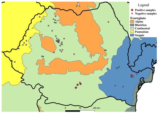
Thelaziosis caused by Thelazia callipaeda is an emerging disease in Europe. Only two reports of naturally infected lagomorphs have been published so far. The aim of this study was to evaluate the status of the Romanian populations of European brown hares, Lepus europaeus as reservoir hosts for T. callipaeda. Between November 2019 and November 2021, the eyes of 326 L. europaeus carcasses were examined for the presence of ocular parasites. Nematodes were stored in plastic vials with physiological saline, followed by morphological and molecular identification. QGis 3.20 and EpiInfoTM 7 were used for mapping and statistical analysis. Four (1.23%) hares harbored T. callipaeda infection, with a total of 84 nematodes collected (mean intensity 21 nematodes/host), with 45 males, 39 females (two sexually immature, seven with only eggs, and 30 with eggs and larvae). One specimen from each host was successfully sequenced resulting in a 100% similarity with several other sequences of T. callipaeda haplotype 1. Statistical analysis revealed no significant results. The current study represents a first report of T. callipaeda in the European brown hare in Romania, and the second in Europe, also reiterating the role of lagomorphs as reservoir hosts for this zoonotic ocular nematode. Full article
(This article belongs to the Special Issue Pets, Wildlife and Parasites)
Show Figures

Figure 1

6 pages, 369 KiB  
Communication
Serological Screening for Middle East Respiratory Syndrome Coronavirus and Hepatitis E Virus in Camels in Kazakhstan
by Kobey Karamendin, Aigerim Seidalina, Temirlan Sabyrzhan, Sardor Nuralibekov, Yermukhammet Kasymbekov, Symbat Suleimenova, Elizaveta Khan, Oralbek Alikhanov, Uldana Narsha, Kalya Erkekulova and Aidyn Kydyrmanov
Pathogens 2022, 11(11), 1224; https://doi.org/10.3390/pathogens11111224 - 24 Oct 2022
Viewed by 2022
Abstract
After the recent Middle East Respiratory Syndrome coronavirus (MERS–CoV) pandemic in 2013, more attention has been paid to the camel as an important source of zoonotic viral infections. Almost simultaneously, in 2013, new genotypes 7 and 8 of the hepatitis E virus (HEV) [...] Read more.
After the recent Middle East Respiratory Syndrome coronavirus (MERS–CoV) pandemic in 2013, more attention has been paid to the camel as an important source of zoonotic viral infections. Almost simultaneously, in 2013, new genotypes 7 and 8 of the hepatitis E virus (HEV) were discovered in dromedary and Bactrian camels, respectively. HEV 7 was further shown to be associated with chronic viral hepatitis in a transplant recipient. In this study, serological screening for antibodies to MERS-CoV and hepatitis E virus was carried out on large camel farms in the south and west of Kazakhstan. 6.42% of the tested camels were found to be positive for antibodies to the hepatitis E virus, which indicates its circulation in local camel population. For the first time, antibodies to the hepatitis E virus were found in Bactrians, which have been little studied to date. Antibodies to MERS-CoV were not found in the camel sera. Full article
(This article belongs to the Special Issue Emerging Infections in Domestic Animals)
Show Figures

Figure 1

9 pages, 1080 KiB  
Article
Occurrence and Genetic Diversity of the Zoonotic Enteric Protozoans and Enterocytozoon bieneusi in Père David’s Deer (Elaphurus davidianus) from Beijing, China
by Peiyang Zhang, Qingxun Zhang, Shuyi Han, Guohui Yuan, Jiade Bai and Hongxuan He
Pathogens 2022, 11(11), 1223; https://doi.org/10.3390/pathogens11111223 - 23 Oct 2022
Cited by 6 | Viewed by 1939
Abstract
Cryptosporidium spp., Blastocystis, Giardia duodenalis, Balantioides coli, Pentatrichomonas hominis, and Enterocytozoon bieneusi are enteric protozoan parasites and fungal species in humans and animals. Père David’s deer is an endangered species in China, but the prevalence of enteric protozoans in [...] Read more.
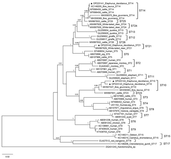
Cryptosporidium spp., Blastocystis, Giardia duodenalis, Balantioides coli, Pentatrichomonas hominis, and Enterocytozoon bieneusi are enteric protozoan parasites and fungal species in humans and animals. Père David’s deer is an endangered species in China, but the prevalence of enteric protozoans in this species still needs to be further studied. Thus, we investigated the prevalence and genetic diversity of zoonotic parasites in Père David’s deer during the period of 2018–2021. Among the 286 fecal samples collected from Père David’s deer in the Nanhaizi Nature Reserve, 83 (29.0%) were positive for Blastocystis, 70 (24.5%) were positive for E. bieneusi, while other protozoan parasites were negative. Based on a phylogenetic analysis, three Blastocystis subtypes (ST10, ST14, and ST21) and ten E. bieneusi genotypes (Genotype D, MWC_d1, HLJD-V, Peru6, BEB6, BJED-I to BJED-I V) were identified. In addition, the Blastocystis subtype ST14 and the E. bieneusi genotype D and Peru6 were first detected in Père David’s deer. Our study first reports the presence of two enteric protozoans in Père David’s deer during a 4-year active surveillance and provides more information about zoonotic subtypes/genotypes of Blastocystis and E. bieneusi in deer. Full article
(This article belongs to the Special Issue Parasitic Diseases of Domestic, Wild, and Exotic Animals (Volume II))
Show Figures

Figure 1

7 pages, 638 KiB  
Article
Procalcitonin as a Potential Biomarker in the Study of Babesiosis Caused by B. microti
by Michael Lum, Caitlin Gauvin, Sophia K. Pham, Aikaterini Papamanoli, Eric D. Spitzer, Andreas P. Kalogeropoulos and Luis A. Marcos
Pathogens 2022, 11(11), 1222; https://doi.org/10.3390/pathogens11111222 - 23 Oct 2022
Cited by 1 | Viewed by 2343
Abstract
Procalcitonin is gaining momentum in the study of protozoal sepsis, but its utility as a biomarker has yet to be fully discovered in human babesiosis. A total of 33 cases of acute babesiosis dating between 2012 and 2019 were retrospectively collected from Stony [...] Read more.
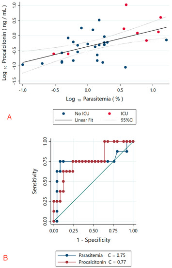
Procalcitonin is gaining momentum in the study of protozoal sepsis, but its utility as a biomarker has yet to be fully discovered in human babesiosis. A total of 33 cases of acute babesiosis dating between 2012 and 2019 were retrospectively collected from Stony Brook University Hospital (SBUH) and Stony Brook South Hampton Hospital (SHH), both of which are located on Long Island, NY. Cases were cross-referenced for the need for ICU admission, and the procalcitonin levels were measured by the use of BRAHMS Elecsys assay at SBUH and BRAHMS Architect assay at SHH. Our study demonstrated that the log-transformed procalcitonin levels had a linear correlation with log-transformed maximum parasitemia, which suggests that procalcitonin directly correlates with parasitemia values. Furthermore, when comparing values that predict ICU admission, our ROC analysis of procalcitonin demonstrated similar AUC values to the percentage of parasitemia, suggesting that procalcitonin may assist in determining the severity of disease. We demonstrate that procalcitonin may directly correlate with the parasitemia percentage and have prognostic capabilities, which suggests that procalcitonin may have biomarker potential in human babesiosis. Full article
Show Figures

Figure 1

12 pages, 2442 KiB  
Article
Loop-Mediated Isothermal Amplification (LAMP) for the Rapid and Sensitive Detection of Alternaria alternata (Fr.) Keissl in Apple Alternaria Blotch Disease with Aapg-1 Encoding the Endopolygalacturonase
by Baoyou Liu, Zhiwei Li, Jianfeng Du, Wei Zhang, Xiaozhi Che, Ziran Zhang, Ping Chen, Yingzi Wang, Yang Li, Shaoli Wang and Xinhua Ding
Pathogens 2022, 11(11), 1221; https://doi.org/10.3390/pathogens11111221 - 23 Oct 2022
Cited by 8 | Viewed by 2684
Abstract
Apple Alternaria blotch disease, caused by Alternaria alternata (Fr.) Keissl, is one of the most famous leaf diseases. When the disease is prevalent, it causes leaf abscission and influences the formation of flower buds and photosynthesis. Therefore, a simple, rapid, high-specificity and sensitivity [...] Read more.
Apple Alternaria blotch disease, caused by Alternaria alternata (Fr.) Keissl, is one of the most famous leaf diseases. When the disease is prevalent, it causes leaf abscission and influences the formation of flower buds and photosynthesis. Therefore, a simple, rapid, high-specificity and sensitivity method for monitoring infected leaves at early developmental stages is urgently needed, so that the occurrence and expansion of A. alternata can be controlled in time. In our research, a rapid, specific and efficient loop-mediated isothermal amplification (LAMP) method was developed to detect A. alternata within 60 min. Six primers of LAMP detection can only specifically amplify the aapg-1 gene in A. alternata but not in four other important fungi in apples. The aapg-1 gene encodes endopolygalacturonase in A. alternata, and there are significant differences among different species. Thus, it was applied as the target for LAMP primers. Compared to conventional PCR detection, our LAMP method had the same sensitivity as that of detecting as little as 1 fg of pure genomic DNA of A. alternata. When leaves were inoculated with A. alternata conidia, LAMP detected 1 × 10² conidia/mL as the minimum concentration. However, the traditional tissue isolation and identification method only isolated A. alternata from leaves inoculated with 1 × 10⁵ and 1 × 10⁶ conidia/mL, indicating that the LAMP method was more sensitive than the traditional tissue isolation and identification method for A. alternata before symptoms. Further tests also indicated that LAMP detection was more accurate and sensitive than the traditional tissue isolation and identification method for A. alternata in leaves with the Alternaria blotch symptom collected from the field. Our results showed that the LAMP-targeting the aapg-1 gene has the advantages of high sensitivity, specificity and simplicity and can be used for rapid detection and early monitoring of A. alternata in the field. LAMP is instructive for us to effectively prevent and control apple Alternaria blotch disease. Full article
Show Figures

Figure 1

11 pages, 653 KiB  
Article
Molecular and Serological Diagnostic Approach to Define the Microbiological Origin of Blood Culture-Negative Infective Endocarditis
by Amira H. El-Ashry, Khaled Saad, Ahmed A. Obiedallah, Amira Elhoufey, Hamad Ghaleb Dailah and Mohammed Salah A. Hussein
Pathogens 2022, 11(11), 1220; https://doi.org/10.3390/pathogens11111220 - 22 Oct 2022
Cited by 7 | Viewed by 2882
Abstract
Blood culture-negative infective endocarditis (BCNIE) poses a significant challenge in determining the best antibiotic regimen for this life-threatening infection, which should be treated with as specific and effective a regimen as feasible. The goal of this study was to determine the prevalence of [...] Read more.
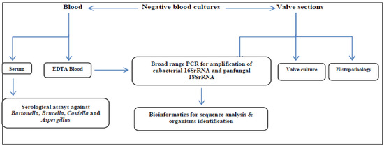
Blood culture-negative infective endocarditis (BCNIE) poses a significant challenge in determining the best antibiotic regimen for this life-threatening infection, which should be treated with as specific and effective a regimen as feasible. The goal of this study was to determine the prevalence of BCNIE among definite infective endocarditis (IE) cases and to study the impact of a molecular and serological diagnostic approach in defining the microbiological origin of BCNIE. This study included 94 definite IE cases. Serum and blood samples from BCNIE patients were tested using serological, broad-range PCR, and sequencing assays. Valve tissue sections obtained from 42 operated patients were subjected to culture and molecular studies. BCNIE accounted for 63 (67%) of the cases. Of these cases, blood PCR followed by sequencing could diagnose 11 cases. Zoonotic infective endocarditis was detected in 7 (11%) patients by serology and PCR (four Brucella, two Bartonella, and one Coxiella). Sequencing of valve PCR bands revealed 30 positive cases. Therefore, the percentage of BCNIE with unidentified etiology was reduced from 67% to 27.7% through a combination of all diagnostic procedures utilized in our study. Blood and valve PCR and sequencing assays are valuable techniques for the etiological diagnosis of BCNIE, especially in cases with previous antibiotic therapy. However, these tests should be used as part of a larger diagnostic strategy that includes serology, microscopy, and valve culture. The use of an automated blood culture system, and proper blood culture collection before ordering antibiotics, will guide IE etiological diagnosis. Full article
(This article belongs to the Special Issue Updates in Infective Endocarditis)
Show Figures

Figure 1

6 pages, 889 KiB  
Case Report
Tetratrichomoniasis in the Geese Flock—Case Report
by Piotr Falkowski, Andrzej Gaweł and Kamila Bobrek
Pathogens 2022, 11(11), 1219; https://doi.org/10.3390/pathogens11111219 - 22 Oct 2022
Cited by 3 | Viewed by 1479
Abstract
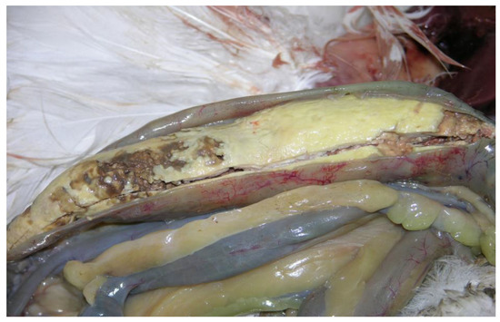
Infections caused by tetratrichomonas are commonly observed in geese. Most cases are subclinical, and the clinical form of the disease manifests itself with a greater mortality and the presence of caseous content in ceca. We describe the case of tetratrichomoniasis in a geese [...] Read more.
Infections caused by tetratrichomonas are commonly observed in geese. Most cases are subclinical, and the clinical form of the disease manifests itself with a greater mortality and the presence of caseous content in ceca. We describe the case of tetratrichomoniasis in a geese flock caused by Tetratrichomonas gallinarum, with the genetic analysis of the isolate being based on the fragments of 18SrRNA and ITS1-5.8rRNA-ITS2. Full article
(This article belongs to the Special Issue Parasitic Diseases of Domestic, Wild, and Exotic Animals (Volume II))
Show Figures

Figure 1