Pathogens 2021, 10(5), 601; https://doi.org/10.3390/pathogens10050601 - 14 May 2021
Cited by 8 | Viewed by 3066
Abstract
Previous studies on the prevalence and transmission mechanism of oxazolidinone resistance gene poxtA in CoNS are lacking, which this study addresses. By screening 763 CoNS isolates from different sources of several livestock farms in Guangdong, China, 2018–2020, we identified that the poxtA was
[...] Read more.
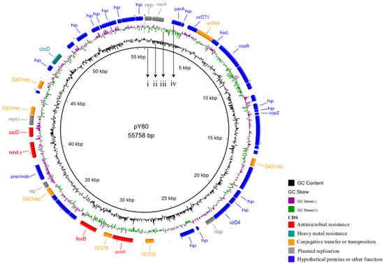
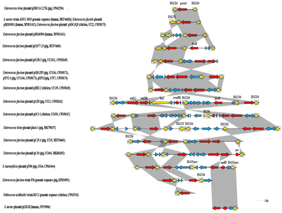
Previous studies on the prevalence and transmission mechanism of oxazolidinone resistance gene poxtA in CoNS are lacking, which this study addresses. By screening 763 CoNS isolates from different sources of several livestock farms in Guangdong, China, 2018–2020, we identified that the poxtA was present in seven CoNS isolates of pig and feed origins. Species identification and multilocus sequence typing (MLST) confirmed that seven poxtA-positive CoNS isolates were composed of five ST64-Staphylococcus haemolyticus and two Staphylococcus saprophyticus isolates. All poxtA-positive Staphylococcus haemolyticus isolates shared similar pulsed-field gel electrophoresis (PFGE) patterns. Transformation assays demonstrated all poxtA-positive isolates were able to transfer poxtA gene to Staphylococcus aureus RN4220. S1-PFGE and whole-genome sequencing (WGS) revealed the presence of poxtA-carrying plasmids in size around 54.7 kb. The plasmid pY80 was 55,758 bp in size and harbored the heavy metal resistance gene czcD and antimicrobial resistance genes, poxtA, aadD, fexB and tet(L). The regions (IS1216E-poxtA-IS1216E) in plasmid pY80 were identified in Staphylococcus spp. and Enterococcus spp. with different genetic and source backgrounds. In conclusion, this was the first report about the poxtA gene in Staphylococcus haemolyticus and Staphylococcus saprophyticus, and IS1216 may play an important role in the dissemination of poxtA among different Gram-positive bacteria.
Full article
(This article belongs to the Special Issue Staphylococcus Infections in Humans and Animals)
►
Show Figures

Figure 1


