Cells 2022, 11(2), 269; https://doi.org/10.3390/cells11020269 - 13 Jan 2022
Cited by 16 | Viewed by 6576
Abstract
The concept of allogeneic cell therapy was first presented over 60 years ago with hematopoietic stem cell transplantation. However, complications such as graft versus host disease (GVHD) and regimen-related toxicities remained as major obstacles. To maximize the effect of graft versus leukemia, while
[...] Read more.
The concept of allogeneic cell therapy was first presented over 60 years ago with hematopoietic stem cell transplantation. However, complications such as graft versus host disease (GVHD) and regimen-related toxicities remained as major obstacles. To maximize the effect of graft versus leukemia, while minimizing the effect of GVHD, donor lymphocyte infusion was utilized. This idea, which was used against viral infections, postulated that adoptive transfer of virus-specific cytotoxic T lymphocytes could reconstitute specific immunity and eliminate virus infected cells and led to the idea of banking third party cytotoxic T cells (CTLs). T cell exhaustion sometimes became a problem and difficulty arose in creating robust CTLs. However, the introduction of induced pluripotent stem cells (iPSCs) lessens such problems, and by using iPSC technology, unlimited numbers of allogeneic rejuvenated CTLs with robust and proliferative cytotoxic activity can be created. Despite this revolutionary concept, several concerns still exist, such as immunorejection by recipient cells and safety issues of gene editing. In this review, we describe approaches to a feasible “off-the-shelf” therapy that can be distributed rapidly worldwide. We also offer perspectives on the future of allogeneic cell cancer immunotherapy.
Full article
(This article belongs to the Special Issue Allogeneic Cell Cancer Immunotherapies)
►
Show Figures
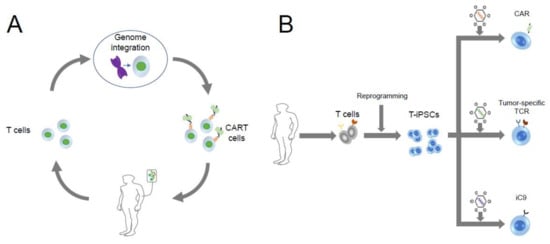
Figure 1


