by
Judith Pape 1, Auxtine Micalet 1,2, Wissal Alsheikh 1, Nadia Ezbakh 1, Rania-Iman Virjee 1 , Rawiya Al Hosni 1, Emad Moeendarbary 2 and Umber Cheema 1,*
, Rawiya Al Hosni 1, Emad Moeendarbary 2 and Umber Cheema 1,*
1
Centre for 3D Models of Health and Disease, Department of Targeted Intervention, Division of Surgery and Interventional Science, University College London, Charles Bell House, 43-45 Foley Street, London W1W 7TS, UK
2
Department of Mechanical Engineering, University College London, Gower Street, London WC1E 6BT, UK
Int. J. Mol. Sci. 2023, 24(4), 3956; https://doi.org/10.3390/ijms24043956 - 16 Feb 2023
Cited by 3 | Viewed by 3091
Abstract
Epithelial to mesenchymal transition (EMT) in cancer is the process described where cancer epithelial cells acquire mesenchymal properties which can lead to enhanced invasiveness. Three-dimensional cancer models often lack the relevant and biomimetic microenvironment parameters appropriate to the native tumour microenvironment thought to
[...] Read more.
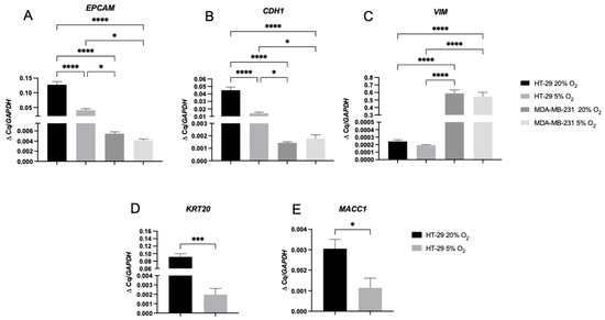
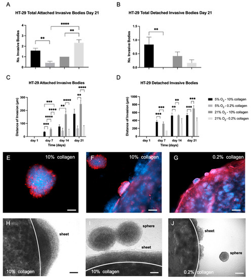
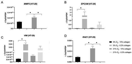
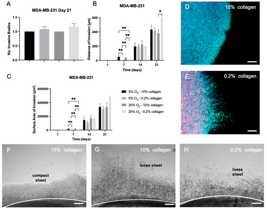
Epithelial to mesenchymal transition (EMT) in cancer is the process described where cancer epithelial cells acquire mesenchymal properties which can lead to enhanced invasiveness. Three-dimensional cancer models often lack the relevant and biomimetic microenvironment parameters appropriate to the native tumour microenvironment thought to drive EMT. In this study, HT-29 epithelial colorectal cells were cultivated in different oxygen and collagen concentrations to investigate how these biophysical parameters influenced invasion patterns and EMT. Colorectal HT-29 cells were grown in physiological hypoxia (5% O2) and normoxia (21% O2) in 2D, 3D soft (60 Pa), and 3D stiff (4 kPa) collagen matrices. Physiological hypoxia was sufficient to trigger expression of markers of EMT in the HT-29 cells in 2D by day 7. This is in contrast to a control breast cancer cell line, MDA-MB-231, which expresses a mesenchymal phenotype regardless of the oxygen concentration. In 3D, HT-29 cells invaded more extensively in a stiff matrix environment with corresponding increases in the invasive genes MMP2 and RAE1. This demonstrates that the physiological environment can directly impact HT-29 cells in terms of EMT marker expression and invasion, compared to an established cell line, MDA-MB-231, which has already undergone EMT. This study highlights the importance of the biophysical microenvironment to cancer epithelial cells and how these factors can direct cell behaviour. In particular, that stiffness of the 3D matrix drives greater invasion in HT-29 cells regardless of hypoxia. It is also pertinent that some cell lines (already having undergone EMT) are not as sensitive to the biophysical features of their microenvironment.
Full article
(This article belongs to the Special Issue State-of-the-Art Molecular Oncology in UK)
▼
Show Figures

Figure 1


