Int. J. Mol. Sci. 2022, 23(13), 7263; https://doi.org/10.3390/ijms23137263 - 30 Jun 2022
Cited by 15 | Viewed by 4139
Abstract
Mitochondrial dysfunction is a pathophysiological hallmark of most neurodegenerative diseases. Several clinical trials targeting mitochondrial dysfunction have been performed with conflicting results. Reliable biomarkers of mitochondrial dysfunction in vivo are thus needed to optimize future clinical trial designs. This narrative review highlights various
[...] Read more.
Mitochondrial dysfunction is a pathophysiological hallmark of most neurodegenerative diseases. Several clinical trials targeting mitochondrial dysfunction have been performed with conflicting results. Reliable biomarkers of mitochondrial dysfunction in vivo are thus needed to optimize future clinical trial designs. This narrative review highlights various neuroimaging methods to probe mitochondrial dysfunction. We provide a general overview of the current biological understanding of mitochondrial dysfunction in degenerative brain disorders and how distinct neuroimaging methods can be employed to map disease-related changes. The reviewed methodological spectrum includes positron emission tomography, magnetic resonance, magnetic resonance spectroscopy, and near-infrared spectroscopy imaging, and how these methods can be applied to study alterations in oxidative phosphorylation and oxidative stress. We highlight the advantages and shortcomings of the different neuroimaging methods and discuss the necessary steps to use these for future research. This review stresses the importance of neuroimaging methods to gain deepened insights into mitochondrial dysfunction in vivo, its role as a critical disease mechanism in neurodegenerative diseases, the applicability for patient stratification in interventional trials, and the quantification of individual treatment responses. The in vivo assessment of mitochondrial dysfunction is a crucial prerequisite for providing individualized treatments for neurodegenerative disorders.
Full article
(This article belongs to the Special Issue Mitochondrial Dysfunction: A Metabolic, Cardiovascular, Neurodegenerative and Neuromuscular Issue)
►
Show Figures
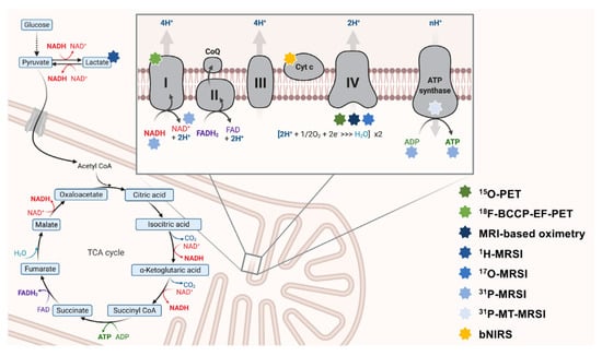
Figure 1


