by
María Teresa Ariza 1,* , Patricia Reboredo-Rodríguez 2, Luca Mazzoni 3, Tamara Yuliett Forbes-Hernández 4, Francesca Giampieri 4, Sadia Afrin 4, Massimiliano Gasparrini 4, Carmen Soria 1, Elsa Martínez-Ferri 1, Maurizio Battino 4 and Bruno Mezzetti 3,*
, Patricia Reboredo-Rodríguez 2, Luca Mazzoni 3, Tamara Yuliett Forbes-Hernández 4, Francesca Giampieri 4, Sadia Afrin 4, Massimiliano Gasparrini 4, Carmen Soria 1, Elsa Martínez-Ferri 1, Maurizio Battino 4 and Bruno Mezzetti 3,*
1
Instituto Andaluz de Investigación y Formación Agraria y Pesquera (IFAPA), Consejería de Agricultura, Pesca y Desarrollo Rural, Junta de Andalucía, IFAPA de Churriana, Cortijo de la Cruz s/n, Churriana, 29140 Málaga, Spain
2
Área de Nutrición y Bromatología, Departamento de Química Analítica y Alimentaria, Universidad de Vigo, Campus de Ourense, E-32004 Ourense, Spain
3
Dipartimento di Scienze Agrarie, Alimentari e Ambientali, Università Politecnica delle Marche, Ancona 60131, Italy
4
Department of Clinical Sciences, Faculty of Medicine, Polytechnic University of Marche, Ancona 60131, Italy
Int. J. Mol. Sci. 2016, 17(7), 1103; https://doi.org/10.3390/ijms17071103 - 11 Jul 2016
Cited by 69 | Viewed by 11034
Abstract
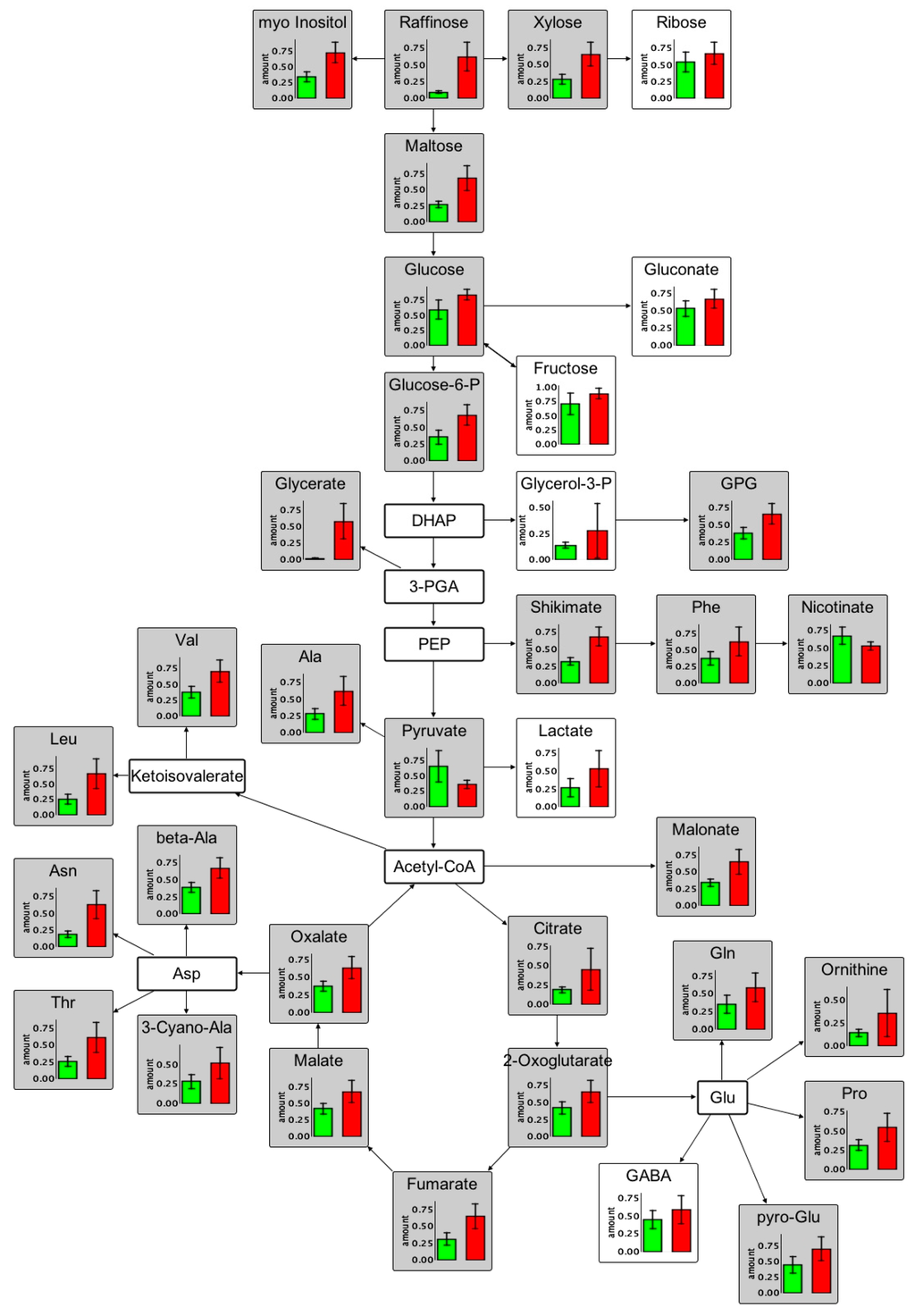
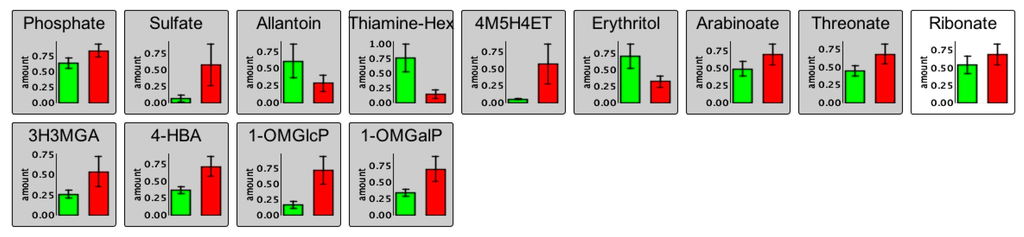
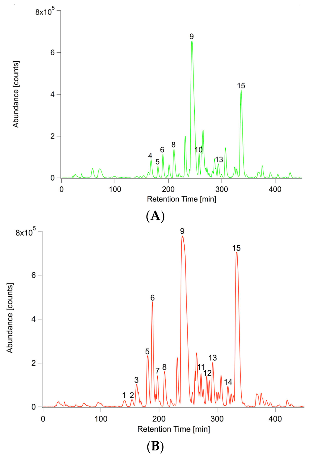
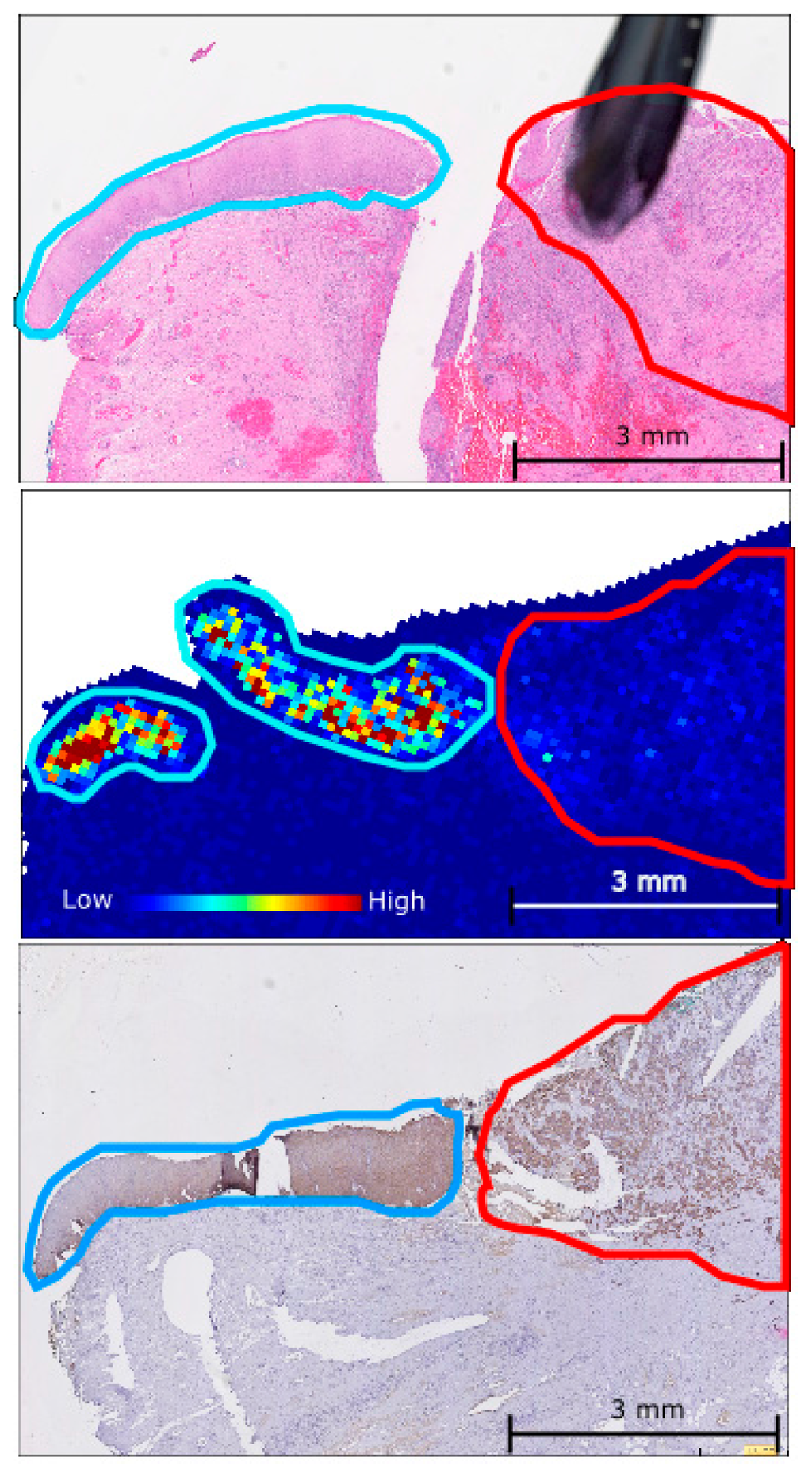
Strawberries are highly appreciated for their taste, nutritional value and antioxidant compounds, mainly phenolics. Fruit antioxidants derive from achenes and flesh, but achene contribution to the total fruit antioxidant capacity and to the bioaccessibility after intake is still unknown. In this work, the
[...] Read more.
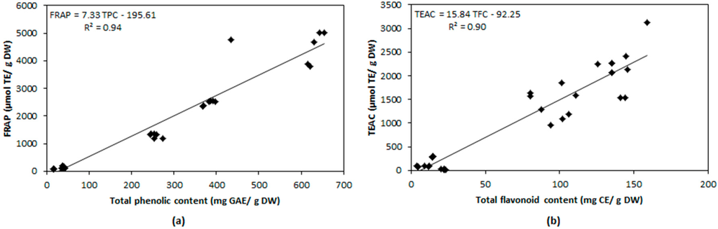
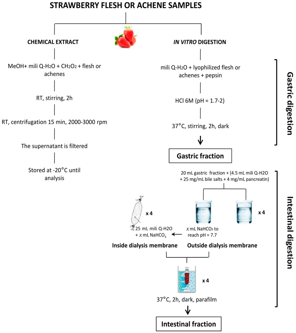
Strawberries are highly appreciated for their taste, nutritional value and antioxidant compounds, mainly phenolics. Fruit antioxidants derive from achenes and flesh, but achene contribution to the total fruit antioxidant capacity and to the bioaccessibility after intake is still unknown. In this work, the content of total phenolic compounds, flavonoids, anthocyanins and antioxidant capacity (TEAC, FRAP and DPPH) of achenes and flesh were compared in non-digested as well as in gastric and intestinal extracts after in vitro digestion. Results showed that, despite strawberry achenes represent a small fraction of the fruit, their contribution to total fruit antioxidant content was more than 41% and accounted for 81% of antioxidant capacity (TEAC). Achenes have higher quantity and different quality of antioxidants in non-digested and digested extracts. Antioxidant release was higher in the in vitro gastric digested extracts, but digestion conditions did not only affect quantity but quality, resulting in differences in antioxidant capacity and highlighting the importance of simulating physiological-like extraction conditions for assessing fruit antioxidant properties on human health. These results give new insights into the use of strawberry achenes as a source of bioactive compounds to be considered in strawberry breeding programs for improving human health.
Full article
(This article belongs to the Special Issue Macro- and Micro-nutrient Antioxidants)
▼
Show Figures
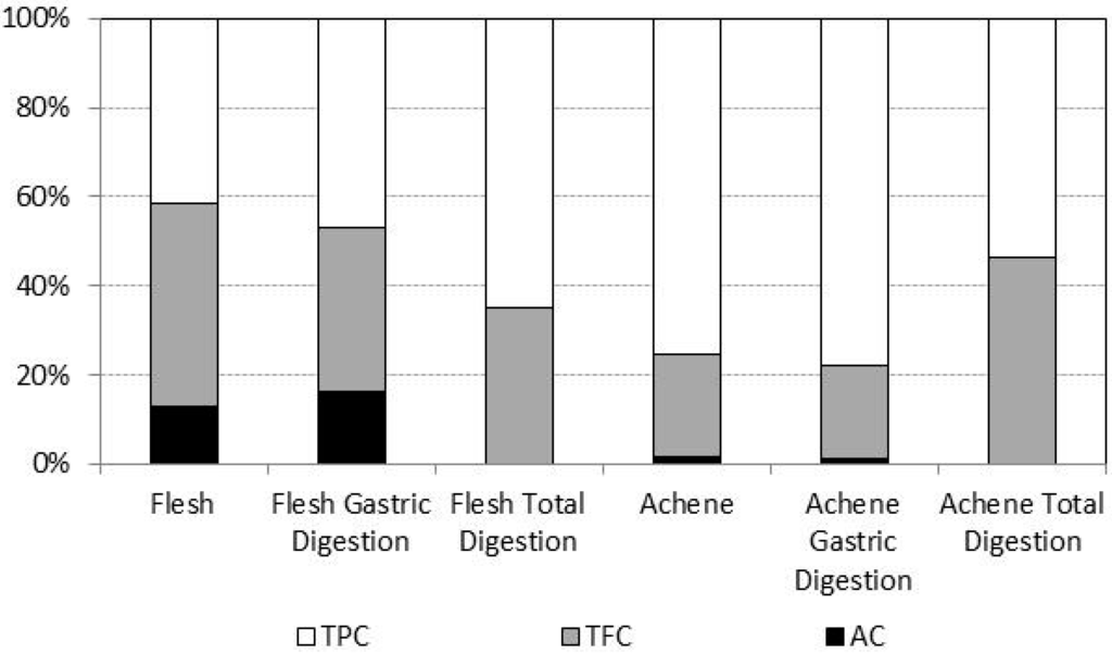
Figure 1

