16 pages, 1015 KiB  
Article
Nrf2 and Redox Status in Prediabetic and Diabetic Patients
by Angélica S. Jiménez-Osorio 1, Alejandra Picazo 2, Susana González-Reyes 1, Diana Barrera-Oviedo 2, Martha E. Rodríguez-Arellano 3 and José Pedraza-Chaverri 1,*
1 Faculty of Chemistry, Department of Biology, National Autonomous University of Mexico (UNAM), University City 04510 DF, Mexico
2 Faculty of Medicine, Department of Pharmacology, National Autonomous University of Mexico (UNAM), University City 04510 DF, Mexico
3 Research Department, Hospital Regional "Lic. Adolfo López Mateos", ISSSTE, Av. Universidad 1321, Florida 01030 DF, Mexico
Int. J. Mol. Sci. 2014, 15(11), 20290-20305; https://doi.org/10.3390/ijms151120290 - 6 Nov 2014
Cited by 89 | Viewed by 7253
Abstract
The redox status associated with nuclear factor erythroid 2-related factor-2 (Nrf2) was evaluated in prediabetic and diabetic subjects. Total antioxidant status (TAS) in plasma and erythrocytes, glutathione (GSH) and malondialdehyde (MDA) content and activity of antioxidant enzymes were measured as redox status markers [...] Read more.
The redox status associated with nuclear factor erythroid 2-related factor-2 (Nrf2) was evaluated in prediabetic and diabetic subjects. Total antioxidant status (TAS) in plasma and erythrocytes, glutathione (GSH) and malondialdehyde (MDA) content and activity of antioxidant enzymes were measured as redox status markers in 259 controls, 111 prediabetics and 186 diabetic type 2 subjects. Nrf2 was measured in nuclear extract fractions from peripheral blood mononuclear cells (PBMC). Nrf2 levels were lower in prediabetic and diabetic patients. TAS, GSH and activity of glutamate cysteine ligase were lower in diabetic subjects. An increase of MDA and superoxide dismutase activity was found in diabetic subjects. These results suggest that low levels of Nrf2 are involved in the development of oxidative stress and redox status disbalance in diabetic patients. Full article
(This article belongs to the Section Biochemistry)
Show Figures

Figure 1

24 pages, 428 KiB  
Review
Hunting the Needle in the Haystack: A Guide to Obtain Biologically Meaningful MicroRNA Targets
by Michael Karbiener, Christina Glantschnig and Marcel Scheideler *
1 RNA Biology Group, Institute of Molecular Biotechnology, Graz University of Technology, Petersgasse 14, 8010 Graz, Austria
These authors contributed equally to this work.
Int. J. Mol. Sci. 2014, 15(11), 20266-20289; https://doi.org/10.3390/ijms151120266 - 6 Nov 2014
Cited by 19 | Viewed by 7104
Abstract
MicroRNAs (miRNAs) are endogenous small non-coding RNAs of ~23 nucleotides in length that form up a novel class of regulatory determinants, with a large set of target mRNAs postulated for every single miRNA. Thousands of miRNAs have been discovered so far, with hundreds [...] Read more.
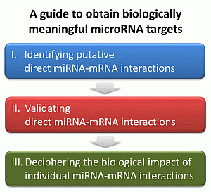
MicroRNAs (miRNAs) are endogenous small non-coding RNAs of ~23 nucleotides in length that form up a novel class of regulatory determinants, with a large set of target mRNAs postulated for every single miRNA. Thousands of miRNAs have been discovered so far, with hundreds of them shown to govern biological processes with impact on disease. However, very little is known about how they specifically interfere with biological pathways and disease mechanisms. To investigate this interaction, the hunt for direct miRNA targets that mediate the miRNA effects—the “needle in the haystack”—is an essential step. In this review we provide a comprehensive workflow of successfully applied methods starting from the identification of putative miRNA-target pairs, followed by validation of direct miRNA–mRNA interactions, and finally presenting methods that dissect the impact of particular miRNA-target pairs on a biological process or disease. This guide allows the way to be paved for obtaining biologically meaningful miRNA targets. Full article
(This article belongs to the Collection Regulation by Non-coding RNAs)
Show Figures

Graphical abstract

12 pages, 1429 KiB  
Article
Formation and Yield of Multi-Walled Carbon Nanotubes Synthesized via Chemical Vapour Deposition Routes Using Different Metal-Based Catalysts of FeCoNiAl, CoNiAl and FeNiAl-LDH
by Mohd Zobir Hussein 1,*, Adila Mohamad Jaafar 1,2,†, Asmah Hj. Yahaya 2,†, Mas Jaffri Masarudin 3,† and Zulkarnain Zainal 2,†
1 Advanced Material and Nanotechnology Laboratory, Institute of Advanced Technology (ITMA), Universiti Putra Malaysia, 43400 UPM Serdang, Selangor, Malaysia
2 Department of Chemistry, Faculty of Science, Universiti Putra Malaysia, 43400 UPM Serdang, Selangor, Malaysia
3 Department of Cell and Molecular Biology, Faculty of Biotechnology and Biomolecular Sciences, Universiti Putra Malaysia, 43400 UPM Serdang, Selangor, Malaysia
These authors contributed equally to this work.
Int. J. Mol. Sci. 2014, 15(11), 20254-20265; https://doi.org/10.3390/ijms151120254 - 5 Nov 2014
Cited by 12 | Viewed by 6836
Abstract
Multi-walled carbon nanotubes (MWCNTs) were prepared via chemical vapor deposition (CVD) using a series of different catalysts, derived from FeCoNiAl, CoNiAl and FeNiAl layered double hydroxides (LDHs). Catalyst-active particles were obtained by calcination of LDHs at 800 °C for 5 h. Nitrogen and [...] Read more.
Multi-walled carbon nanotubes (MWCNTs) were prepared via chemical vapor deposition (CVD) using a series of different catalysts, derived from FeCoNiAl, CoNiAl and FeNiAl layered double hydroxides (LDHs). Catalyst-active particles were obtained by calcination of LDHs at 800 °C for 5 h. Nitrogen and hexane were used as the carrier gas and carbon source respectively, for preparation of MWCNTs using CVD methods at 800 °C. MWCNTs were allowed to grow for 30 min on the catalyst spread on an alumina boat in a quartz tube. The materials were subsequently characterized through X-ray diffraction, Fourier transform infrared spectroscopy, surface area analysis, field emission scanning electron microscopy and transmission electron microscopy. It was determined that size and yield of MWCNTs varied depending on the type of LDH catalyst precursor that is used during synthesis. MWCNTs obtained using CoNiAl-LDH as the catalyst precursor showed smaller diameter and higher yield compared to FeCoNiAl and FeNiAl LDHs. Full article
(This article belongs to the Section Materials Science)
Show Figures

Figure 1

14 pages, 4212 KiB  
Article
Pressure Overload-Induced Cardiac Hypertrophy Response Requires Janus Kinase 2-Histone Deacetylase 2 Signaling
by Huang Ying 1,†, Mao-Chun Xu 2,†, Jing-Hua Tan 1, Jing-Hua Shen 1, Hao Wang 3,* and Dai-Fu Zhang 1,*
1 Department of Cardiology, Shanghai Pu Dong New Area People's Hospital, Shanghai 200120, China
2 Department of Cardiology, Huashan Hospital of Fudan University, Shanghai 200040, China
3 Fudan University Shanghai Medical College Centre of Medical Experiments, Shanghai 200040, China
These authors contributed equally to this work.
Int. J. Mol. Sci. 2014, 15(11), 20240-20253; https://doi.org/10.3390/ijms151120240 - 5 Nov 2014
Cited by 7 | Viewed by 6591
Abstract
Pressure overload induces cardiac hypertrophy through activation of Janus kinase 2 (Jak2), however, the underlying mechanisms remain largely unknown. In the current study, we tested whether histone deacetylase 2 (HDAC2) was involved in the process. We found that angiotensin II (Ang-II)-induced re-expression of [...] Read more.
Pressure overload induces cardiac hypertrophy through activation of Janus kinase 2 (Jak2), however, the underlying mechanisms remain largely unknown. In the current study, we tested whether histone deacetylase 2 (HDAC2) was involved in the process. We found that angiotensin II (Ang-II)-induced re-expression of fetal genes (Atrial natriuretic peptide (ANP) and brain natriuretic peptide (BNP)) in cultured cardiomyocytes was prevented by the Jak2 inhibitor AG-490 and HDAC2 inhibitor Trichostatin-A (TSA), or by Jak2/HDAC2 siRNA knockdown. On the other hand, myocardial cells with Jak2 or HDAC2 over-expression were hyper-sensitive to Ang-II. In vivo, pressure overload by transverse aorta binding (AB) induced a significant cardiac hypertrophic response as well as re-expression of ANP and BNP in mice heart, which were markedly reduced by AG-490 and TSA. Significantly, AG-490, the Jak2 inhibitor, largely suppressed pressure overload-/Ang-II-induced HDAC2 nuclear exportation in vivo and in vitro. Meanwhile, TSA or HDAC2 siRNA knockdown reduced Ang-II-induced ANP/BNP expression in Jak2 over-expressed H9c2 cardiomyocytes. Together, these results suggest that HDAC2 might be a downstream effector of Jak2 to mediate cardiac hypertrophic response by pressure overload or Ang-II. Full article
(This article belongs to the Section Biochemistry)
Show Figures

Figure 1

31 pages, 6701 KiB  
Review
Microenvironment of Tumor-Draining Lymph Nodes: Opportunities for Liposome-Based Targeted Therapy
by Siddarth Chandrasekaran and Michael R. King *
Department of Biomedical Engineering, Cornell University, Ithaca, NY 14853, USA
Int. J. Mol. Sci. 2014, 15(11), 20209-20239; https://doi.org/10.3390/ijms151120209 - 5 Nov 2014
Cited by 67 | Viewed by 17967
Abstract
The World Health Organization (WHO) recently reported that the total number of global cancer cases in 2013 reached 14 million, a 10% rise since 2008, while the total number of cancer deaths reached 8.2 million, a 5.2% increase since 2008. Metastasis is the [...] Read more.
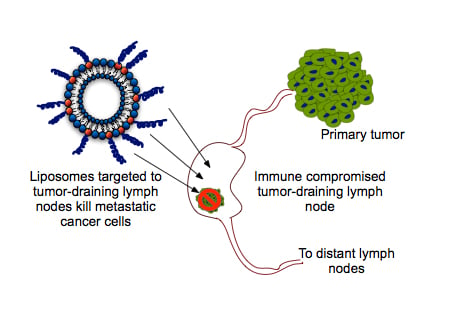
The World Health Organization (WHO) recently reported that the total number of global cancer cases in 2013 reached 14 million, a 10% rise since 2008, while the total number of cancer deaths reached 8.2 million, a 5.2% increase since 2008. Metastasis is the major cause of death from cancer, accounting for 90% of all cancer related deaths. Tumor-draining lymph nodes (TDLN), the sentinel nodes, are the first organs of metastasis in several types of cancers. The extent of metastasis in the TDLN is often used in disease staging and prognosis evaluation in cancer patients. Here, we describe the microenvironment of the TDLN and review the recent literature on liposome-based therapies directed to immune cells within the TDLN with the intent to target cancer cells. Full article
Show Figures

Graphical abstract

40 pages, 820 KiB  
Review
Current Experience in Testing Mitochondrial Nutrients in Disorders Featuring Oxidative Stress and Mitochondrial Dysfunction: Rational Design of Chemoprevention Trials
by Giovanni Pagano 1,*, Annarita Aiello Talamanca 1, Giuseppe Castello 1, Mario D. Cordero 2, Marco D'Ischia 3, Maria Nicola Gadaleta 4, Federico V. Pallardó 5, Sandra Petrović 6, Luca Tiano 7 and Adriana Zatterale 8
1 Istituto Nazionale Tumori Fondazione G. Pascale—Cancer Research Center at Mercogliano (CROM)—IRCCS, Naples I-80131, Italy
2 Research Laboratory, Dental School, Universidad de Sevilla, Sevilla 41009, Spain
3 Department of Chemical Sciences, University of Naples "Federico II", Naples I-80126, Italy
4 National Research Council, Institute of Biomembranes and Bioenergetics, Bari I-70126, Italy
5 CIBERER (Centro de Investigación Biomédica en Red de Enfermedades Raras), University of Valencia—INCLIVA, Valencia 46010, Spain
6 Vinca" Institute of Nuclear Sciences, University of Belgrade, Belgrade 11001, Serbia
7 Biochemistry Unit, Department of Clinical and Dental Sciences, Polytechnical University of Marche, Ancona I-60131, Italy
8 Genetics Unit, Azienda Sanitaria Locale (ASL) Napoli 1 Centro, Naples I-80136, Italy
Int. J. Mol. Sci. 2014, 15(11), 20169-20208; https://doi.org/10.3390/ijms151120169 - 5 Nov 2014
Cited by 25 | Viewed by 9563
Abstract
An extensive number of pathologies are associated with mitochondrial dysfunction (MDF) and oxidative stress (OS). Thus, mitochondrial cofactors termed “mitochondrial nutrients” (MN), such as α-lipoic acid (ALA), Coenzyme Q10 (CoQ10), and l-carnitine (CARN) (or its derivatives) have been tested in a number of [...] Read more.
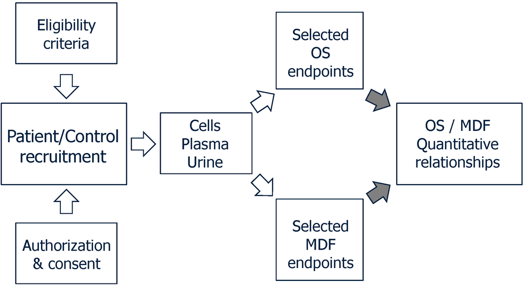
An extensive number of pathologies are associated with mitochondrial dysfunction (MDF) and oxidative stress (OS). Thus, mitochondrial cofactors termed “mitochondrial nutrients” (MN), such as α-lipoic acid (ALA), Coenzyme Q10 (CoQ10), and l-carnitine (CARN) (or its derivatives) have been tested in a number of clinical trials, and this review is focused on the use of MN-based clinical trials. The papers reporting on MN-based clinical trials were retrieved in MedLine up to July 2014, and evaluated for the following endpoints: (a) treated diseases; (b) dosages, number of enrolled patients and duration of treatment; (c) trial success for each MN or MN combinations as reported by authors. The reports satisfying the above endpoints included total numbers of trials and frequencies of randomized, controlled studies, i.e., 81 trials testing ALA, 107 reports testing CoQ10, and 74 reports testing CARN, while only 7 reports were retrieved testing double MN associations, while no report was found testing a triple MN combination. A total of 28 reports tested MN associations with “classical” antioxidants, such as antioxidant nutrients or drugs. Combinations of MN showed better outcomes than individual MN, suggesting forthcoming clinical studies. The criteria in study design and monitoring MN-based clinical trials are discussed. Full article
(This article belongs to the Special Issue Mitochondrial Dysfunction in Ageing and Diseases)
Show Figures

Figure 1

11 pages, 2080 KiB  
Article
A Microdeletion of Chromosome 9q33.3 Encompasses the Entire LMX1B Gene in a Chinese Family with Nail Patella Syndrome
by Shujuan Jiang 1, Jiubin Zhang 2, Dan Huang 1, Yuanyuan Zhang 1, Xiaoliang Liu 1, Yinzhao Wang 3, Rong He 1,* and Yanyan Zhao 1,*
1 Clinical Genetics, the Affiliated Shengjing Hospital, China Medical University, Shenyang 110004, Liaoning, China
2 Orthopedics Department, the First Affiliated Hospital, China Medical University, Shenyang 110001, Liaoning, China
3 NO. 31 Middle School in Shenyang of Liaoning Province, Shenyang 110021, Liaoning, China
Int. J. Mol. Sci. 2014, 15(11), 20158-20168; https://doi.org/10.3390/ijms151120158 - 5 Nov 2014
Cited by 5 | Viewed by 7139
Abstract
Nail patella syndrome (NPS) is an autosomal dominant disorder characterized by nail malformations, patellar apoplasia, or patellar hypoplasia. Mutations within the LMX1B gene are found in 85% of families with NPS; thus, this gene has been characterized as the causative gene of NPS. [...] Read more.
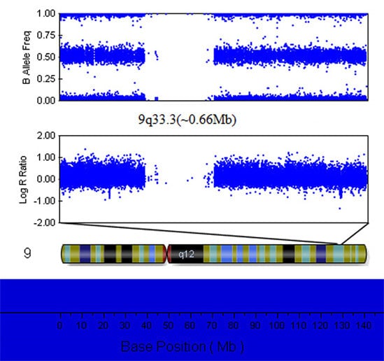
Nail patella syndrome (NPS) is an autosomal dominant disorder characterized by nail malformations, patellar apoplasia, or patellar hypoplasia. Mutations within the LMX1B gene are found in 85% of families with NPS; thus, this gene has been characterized as the causative gene of NPS. In this study, we identified a heterozygous microdeletion of the entire LMX1B gene using multiplex ligation-dependent probe amplification (MLPA) in a Chinese family with NPS. The determination of the deletion breakpoints by Illumina genome-wide DNA analysis beadchip showed that the deletion was located in chromosome 9q33.3 and spanned about 0.66 Mb in size. This heterozygous deletion provides strong evidence for haploinsufficiency as the pathogenic mechanism of NPS. Full article
(This article belongs to the Section Biochemistry)
Show Figures

Graphical abstract

24 pages, 816 KiB  
Article
MicroRNAs Associated with the Efficacy of Photodynamic Therapy in Biliary Tract Cancer Cell Lines
by Andrej Wagner 1,*, Christian Mayr 1, Doris Bach 1, Romana Illig 2, Kristjan Plaetzer 3, Frieder Berr 1, Martin Pichler 4,5, Daniel Neureiter 2 and Tobias Kiesslich 1,6
1 Department of Internal Medicine I, Paracelsus Medical University/Salzburger Landeskliniken (SALK), Muellner Hauptstrasse 48, Salzburg 5020, Austria
2 Institute of Pathology, Paracelsus Medical University/Salzburger Landeskliniken (SALK), Salzburg 5020, Austria
3 Laboratory of Photodynamic Inactivation of Microorganisms, Department of Materials Science and Physics, University of Salzburg, Salzburg 5020, Austria
4 Division of Oncology, Medical University Graz, Graz 8036, Austria
5 Department of Experimental Therapeutics, the University of Texas MD Anderson Cancer Center, Houston, TX 77054, USA
6 Institute of Physiology and Pathophysiology, Paracelsus Medical University, Salzburg 5020, Austria
Int. J. Mol. Sci. 2014, 15(11), 20134-20157; https://doi.org/10.3390/ijms151120134 - 5 Nov 2014
Cited by 19 | Viewed by 7878
Abstract
Photodynamic therapy (PDT) is a palliative treatment option for unresectable hilar biliary tract cancer (BTC) showing a considerable benefit for survival and quality of life with few side effects. Currently, factors determining the cellular response of BTC cells towards PDT are unknown. Due [...] Read more.
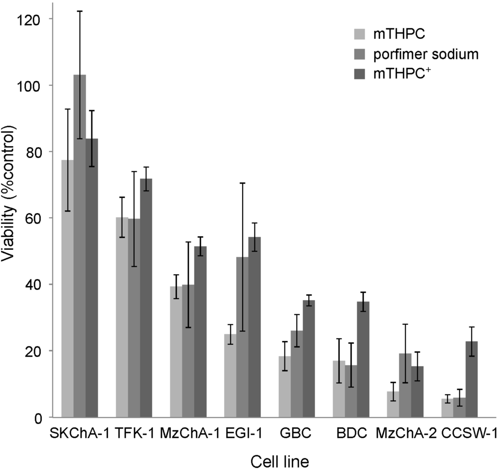
Photodynamic therapy (PDT) is a palliative treatment option for unresectable hilar biliary tract cancer (BTC) showing a considerable benefit for survival and quality of life with few side effects. Currently, factors determining the cellular response of BTC cells towards PDT are unknown. Due to their multifaceted nature, microRNAs (miRs) are a promising analyte to investigate the cellular mechanisms following PDT. For two photosensitizers, Photofrin® and Foscan®, the phototoxicity was investigated in eight BTC cell lines. Each cell line (untreated) was profiled for expression of n = 754 miRs using TaqMan® Array Human MicroRNA Cards. Statistical analysis and bioinformatic tools were used to identify miRs associated with PDT efficiency and their putative targets, respectively. Twenty miRs correlated significantly with either high or low PDT efficiency. PDT was particularly effective in cells with high levels of clustered miRs 25-93*-106b and (in case of miR-106b) a phenotype characterized by high expression of the mesenchymal marker vimentin and high proliferation (cyclinD1 and Ki67 expression). Insensitivity towards PDT was associated with high miR-200 family expression and (for miR-cluster 200a/b-429) expression of differentiation markers Ck19 and Ck8/18. Predicted and validated downstream targets indicate plausible involvement of miRs 20a*, 25, 93*, 130a, 141, 200a, 200c and 203 in response mechanisms to PDT, suggesting that targeting these miRs could improve susceptibility to PDT in insensitive cell lines. Taken together, the miRNome pattern may provide a novel tool for predicting the efficiency of PDT and—following appropriate functional verification—may subsequently allow for optimization of the PDT protocol. Full article
(This article belongs to the Collection Regulation by Non-coding RNAs)
Show Figures

Figure 1

17 pages, 9335 KiB  
Article
Cartilage Oligomeric Matrix Protein Gene Multilayers Inhibit Osteogenic Differentiation and Promote Chondrogenic Differentiation of Mesenchymal Stem Cells
by Peng Guo, Zhong-Li Shi, An Liu, Tiao Lin, Fang-Gang Bi, Ming-Min Shi and Shi-Gui Yan *
1 Department of Orthopaedic Surgery, the Second Affiliated Hospital, Medical College of Zhejiang University, NO. 88 Jiefang Road, Hangzhou 310009, China
These authors contributed equally to this work.
Int. J. Mol. Sci. 2014, 15(11), 20117-20133; https://doi.org/10.3390/ijms151120117 - 5 Nov 2014
Cited by 8 | Viewed by 7581
Abstract
There are still many challenges to acquire the optimal integration of biomedical materials with the surrounding tissues. Gene coatings on the surface of biomaterials may offer an effective approach to solve the problem. In order to investigate the gene multilayers mediated differentiation of [...] Read more.
There are still many challenges to acquire the optimal integration of biomedical materials with the surrounding tissues. Gene coatings on the surface of biomaterials may offer an effective approach to solve the problem. In order to investigate the gene multilayers mediated differentiation of mesenchymal stem cells (MSCs), gene functionalized films of hyaluronic acid (HA) and lipid-DNA complex (LDc) encoding cartilage oligomeric matrix protein (COMP) were constructed in this study via the layer-by-layer self-assembly technique. Characterizations of the HA/DNA multilayered films indicated the successful build-up process. Cells could be directly transfected by gene films and a higher expression could be obtained with the increasing bilayer number. The multilayered films were stable for a long period and DNA could be easily released in an enzymatic condition. Real-time polymerase chain reaction (RT-PCR) assay presented significantly higher (p < 0.01) COMP expression of MSCs cultured with HA/COMP multilayered films. Compared with control groups, the osteogenic gene expression levels of MSCs with HA/COMP multilayered films were down-regulated while the chondrogenic gene expression levels were up-regulated. Similarly, the alkaline phosphatase (ALP) staining and Alizarin red S staining of MSCs with HA/COMP films were weakened while the alcian blue staining was enhanced. These results demonstrated that HA/COMP multilayered films could inhibit osteogenic differentiation and promote chondrogenic differentiation of MSCs, which might provide new insight for physiological ligament-bone healing. Full article
(This article belongs to the Special Issue Biomimetic and Functional Materials)
Show Figures

Graphical abstract

16 pages, 3496 KiB  
Article
Overexpression of the CaTIP1-1 Pepper Gene in Tobacco Enhances Resistance to Osmotic Stresses
by Yan-Xu Yin 1,†, Shu-Bin Wang 2,†, Huai-Juan Xiao 1, Huai-Xia Zhang 1, Zhen Zhang 1, Hua Jing 1, Ying-Li Zhang 1,3, Ru-Gang Chen 1 and Zhen-Hui Gong 1,*
1 College of Horticulture, Northwest A&F University, Yangling, Shaanxi 712100, China
2 Institute of Vegetable Crops, Jiangsu Academy of Agricultural Sciences, Nanjing, Jiangsu 210014, China
3 College of Food and Biological Engineering, Xuchang University, Xuchang, Henan 461000, China
These authors contributed equally to this work.
Int. J. Mol. Sci. 2014, 15(11), 20101-20116; https://doi.org/10.3390/ijms151120101 - 4 Nov 2014
Cited by 17 | Viewed by 6994
Abstract
Both the gene expression and activity of water channel protein can control transmembrane water movement. We have reported the overexpression of CaTIP1-1, which caused a decrease in chilling tolerance in transgenic plants by increasing the size of the stomatal pore. CaTIP1-1 expression [...] Read more.
Both the gene expression and activity of water channel protein can control transmembrane water movement. We have reported the overexpression of CaTIP1-1, which caused a decrease in chilling tolerance in transgenic plants by increasing the size of the stomatal pore. CaTIP1-1 expression was strongly induced by salt and mannitol stresses in pepper (Capsicum annuum). However, its biochemical and physiological functions are still unknown in transgenic tobacco. In this study, transient expression of CaTIP1-1-GFP in tobacco suspension cells revealed that the protein was localized in the tonoplast. CaTIP1-1 overexpressed in radicle exhibited vigorous growth under high salt and mannitol treatments more than wild-type plants. The overexpression of CaTIP1-1 pepper gene in tobacco enhanced the antioxidant enzyme activities and increased transcription levels of reactive oxygen species-related gene expression under osmotic stresses. Moreover, the viability of transgenic tobacco cells was higher than the wild-type after exposure to stress. The pepper plants with silenced CaTIP1-1 in P70 decreased tolerance to salt and osmotic stresses using the detached leaf method. We concluded that the CaTIP1-1 gene plays an important role in response to osmotic stresses in tobacco. Full article
(This article belongs to the Section Biochemistry)
Show Figures

Figure 1

22 pages, 1170 KiB  
Article
Role of NADPH Oxidase and Xanthine Oxidase in Mediating Inducible VT/VF and Triggered Activity in a Canine Model of Myocardial Ischemia
by James B. Martins *, Ashok K. Chaudhary, Shuxia Jiang, Michael Kwofie, Prescott Mackie and Francis J. Miller
Division of Cardiovascular Diseases, Departments of Internal Medicine, University of Iowa and Veterans Affairs Medical Center, Iowa City, Iowa 52242, IA, USA
Int. J. Mol. Sci. 2014, 15(11), 20079-20100; https://doi.org/10.3390/ijms151120079 - 4 Nov 2014
Cited by 6 | Viewed by 5462
Abstract
Background: Ventricular tachycardia or fibrillation (VT/VF) of focal origin due to triggered activity (TA) from delayed afterdepolarizations (DADs) is reproducibly inducible after anterior coronary artery occlusion. Both VT/VF and TA can be blocked by reducing reactive oxygen species (ROS). We tested the hypothesis [...] Read more.
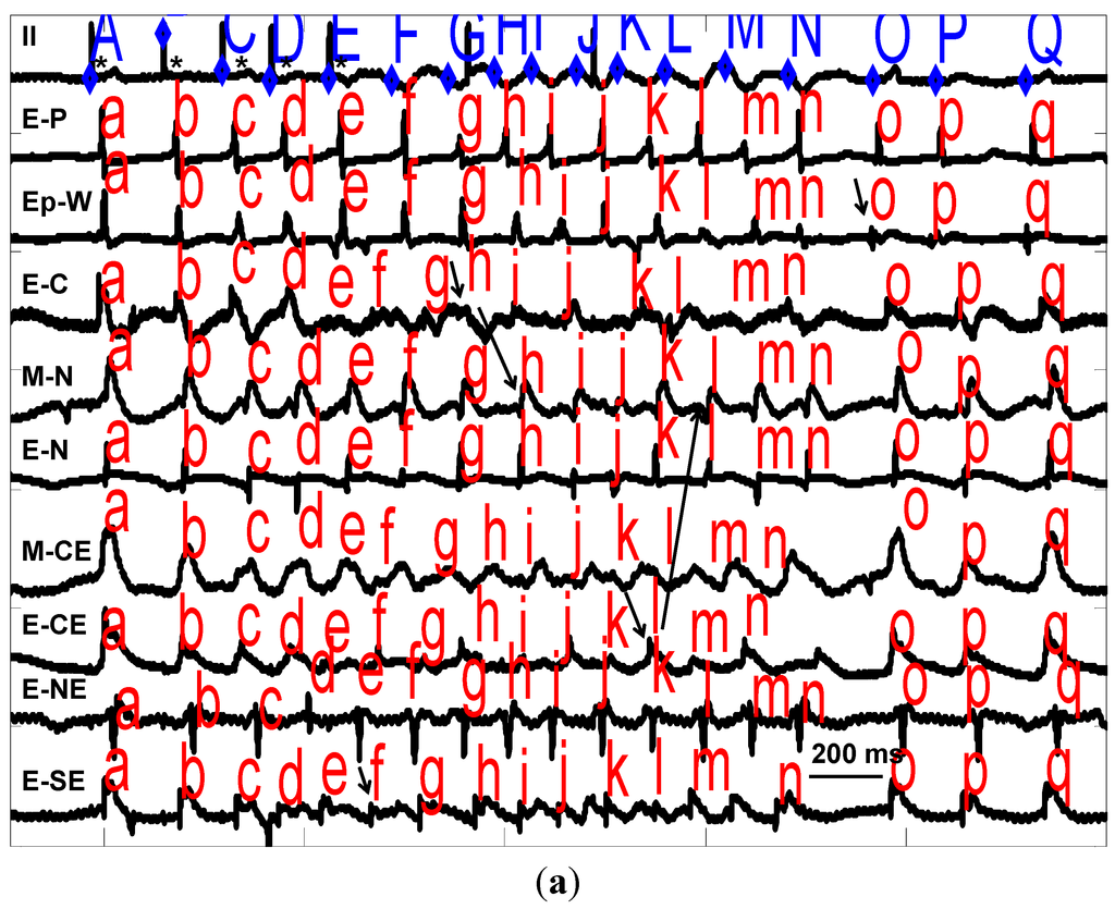
Background: Ventricular tachycardia or fibrillation (VT/VF) of focal origin due to triggered activity (TA) from delayed afterdepolarizations (DADs) is reproducibly inducible after anterior coronary artery occlusion. Both VT/VF and TA can be blocked by reducing reactive oxygen species (ROS). We tested the hypothesis that inhibition of NADPH oxidase and xanthine oxidase would block VT/VF. Methods: 69 dogs received apocynin (APO), 4 mg/kg intraveneously (IV), oxypurinol (OXY), 4 mg/kg IV, or both APO and OXY (BOTH) agents, or saline 3 h after coronary occlusion. Endocardium from ischemic sites (3-D mapping) was sampled for Rac1 (GTP-binding protein in membrane NADPH oxidase) activation or standard microelectrode techniques. Results (mean ± SE, * p < 0.05): VT/VF originating from ischemic zones was blocked by APO in 6/10 *, OXY in 4/9 *, BOTH in 5/8 * or saline in 1/27; 11/16 VT/VFs blocked were focal. In isolated myocardium, TA was blocked by APO (10−6 M) or OXY (10−8 M). Rac1 levels in ischemic endocardium were decreased by APO or OXY. Conclusion: APO and OXY suppressed focal VT/VF due to DADs, but the combination of the drugs was not more effective than either alone. Both drugs inhibited ischemic Rac1 with inhibition by OXY suggesting ROS-induced ROS. The inability to totally prevent VT/VF suggests that other mechanisms also contribute to ischemic VT. Full article
(This article belongs to the Special Issue Oxidative Stress in Cardiovascular Disease 2015)
Show Figures

Figure 1

7 pages, 688 KiB  
Editorial
Molecular Science for Drug Development and Biomedicine
by Wei-Zhu Zhong 1,* and Shu-Feng Zhou 2
1 Gordon Life Science Institute, Belmont, MA 02478, USA
2 Department of Pharmaceutical Sciences, College of Pharmacy, University of South Florida, Tampa, FL 33620, USA
Int. J. Mol. Sci. 2014, 15(11), 20072-20078; https://doi.org/10.3390/ijms151120072 - 4 Nov 2014
Cited by 86 | Viewed by 8253
Abstract
With the avalanche of biological sequences generated in the postgenomic age, molecular science is facing an unprecedented challenge, i.e., how to timely utilize the huge amount of data to benefit human beings. Stimulated by such a challenge, a rapid development has taken [...] Read more.
With the avalanche of biological sequences generated in the postgenomic age, molecular science is facing an unprecedented challenge, i.e., how to timely utilize the huge amount of data to benefit human beings. Stimulated by such a challenge, a rapid development has taken place in molecular science, particularly in the areas associated with drug development and biomedicine, both experimental and theoretical. The current thematic issue was launched with the focus on the topic of “Molecular Science for Drug Development and Biomedicine”, in hopes to further stimulate more useful techniques and findings from various approaches of molecular science for drug development and biomedicine.[...] Full article
(This article belongs to the Special Issue Molecular Science for Drug Development and Biomedicine)
0 pages, 2360 KiB  
Article
RETRACTED: Dinitrosopiperazine-Mediated Phosphorylated-Proteins Are Involved in Nasopharyngeal Carcinoma Metastasis
by Gongjun Tan 1,2,†, Xiaowei Tang 3,†, Damao Huang 2,†, Yuejin Li 1,†, Na Liu 2, Zhengke Peng 1, Zhenlin Zhang 1, Chaojun Duan 2, Jinping Lu 1, Guangrong Yan 4 and Faqing Tang 1,*
1 Medical Research Center and Clinical Laboratory, Zhuhai Hospital of Jinan University, 79 Kangning Road, Zhuhai 519000, China
2 Clinical Laboratory and Medical Research Center, Xiangya Hospital, Central South University, 87 Xiangya Road, Changsha 410008, China
3 Metallurgical Science and Engineering, Central South University, 21 Lushan South Road, Changsha 410083, China
4 Institute of Life and Health Engineering, National Engineering and Research Center for Genetic Medicine, Jinan University, 601 Huangpu Road West, Guangzhou 510632, China
These authors contributed equally to this work.
Int. J. Mol. Sci. 2014, 15(11), 20054-20071; https://doi.org/10.3390/ijms151120054 - 4 Nov 2014
Cited by 7 | Viewed by 6196 | Retraction
Abstract
N,N'-dinitrosopiperazine (DNP) with organ specificity for nasopharyngeal epithelium, is involved in nasopharyngeal carcinoma (NPC) metastasis, though its mechanism is unclear. To reveal the pathogenesis of DNP-induced metastasis, immunoprecipitation was used to identify DNP-mediated phosphoproteins. DNP-mediated NPC cell line [...] Read more.
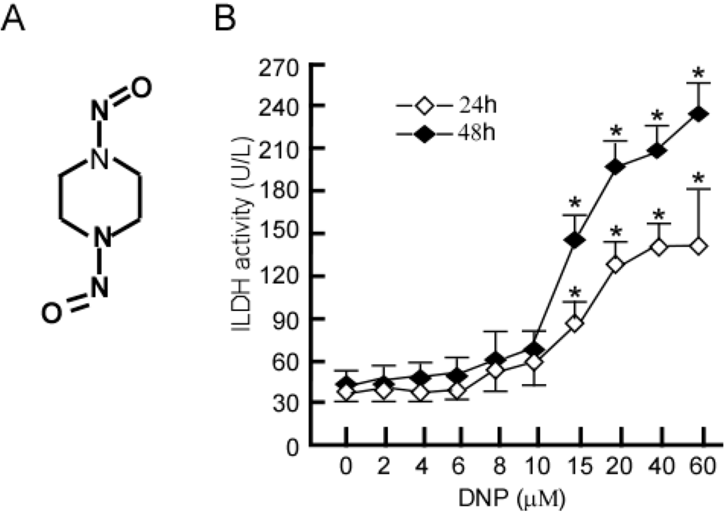
N,N'-dinitrosopiperazine (DNP) with organ specificity for nasopharyngeal epithelium, is involved in nasopharyngeal carcinoma (NPC) metastasis, though its mechanism is unclear. To reveal the pathogenesis of DNP-induced metastasis, immunoprecipitation was used to identify DNP-mediated phosphoproteins. DNP-mediated NPC cell line (6-10B) motility and invasion was confirmed. Twenty-six phosphoproteins were increased at least 1.5-fold following DNP exposure. Changes in the expression levels of selected phosphoproteins were verified by Western-blotting analysis. DNP treatment altered the phosphorylation of ezrin (threonine 567), vimentin (serine 55), stathmin (serine 25) and STAT3 (serine 727). Furthermore, it was shown that DNP-dependent metastasis is mediated in part through ezrin at threonine 567, as DNP-mediated metastasis was decreased when threonine 567 of ezrin was mutated. Strikingly, NPC metastatic tumors exhibited a higher expression of phosphorylated-ezrin at threonine 567 than the primary tumors. These findings provide novel insight into DNP-induced NPC metastasis and may contribute to a better understanding of the metastatic mechanisms of NPC tumors. Full article
(This article belongs to the Special Issue Signalling Molecules and Signal Transduction in Cells 2014)
Show Figures

Figure 1

9 pages, 790 KiB  
Article
Cytotoxic Sesterterpenoids Isolated from the Marine Sponge Scalarispongia sp.
by Yeon-Ju Lee 1,*, Jeong-Woo Lee 1, Dong-Geun Lee 1, Hyi-Seung Lee 1, Jong Soon Kang 2 and Jieun Yun 2
1 Marine Natural Product Chemistry Laboratory, Korea Institute of Ocean Science and Technology, Ansan 426-744, Korea
2 Bio-Evaluation Center, Korea Research Institute of Bioscience and Biotechnology, Cheongwon 323-833, Korea
Int. J. Mol. Sci. 2014, 15(11), 20045-20053; https://doi.org/10.3390/ijms151120045 - 4 Nov 2014
Cited by 10 | Viewed by 5549
Abstract
Eight scalarane sesterterpenoids, including four new compounds, were isolated from the marine sponge Scalarispongia sp. The structures of the new compounds were elucidated by 2D-NMR and HRMS analyses. All of the isolated compounds, with the exception of 16-O-deacetyl-12,16-epi-scalarolbutanolide, showed [...] Read more.
Eight scalarane sesterterpenoids, including four new compounds, were isolated from the marine sponge Scalarispongia sp. The structures of the new compounds were elucidated by 2D-NMR and HRMS analyses. All of the isolated compounds, with the exception of 16-O-deacetyl-12,16-epi-scalarolbutanolide, showed significant in vitro cytotoxicity (GI50 values down to 5.2 μM) against six human cancer cell lines. Full article
(This article belongs to the Section Biochemistry)
Show Figures

Figure 1

23 pages, 5691 KiB  
Article
Effects of Bofu-Tsusho-San on Diabetes and Hyperlipidemia Associated with AMP-Activated Protein Kinase and Glucose Transporter 4 in High-Fat-Fed Mice
by Cheng-Hsiu Lin 1, Yueh-Hsiung Kuo 2,3 and Chun-Ching Shih 4,*
1 Department of Internal Medicine, Feng-Yuan Hospital, Ministry of Health and Welfare, Fengyuan District, Taichung City 42055, Taiwan
2 Department of Chinese Pharmaceutical Sciences and Chinese Medicine Resources, China Medical University, Taichung City 40402, Taiwan
3 Department of Biotechnology, Asia University, Taichung City 41354, Taiwan
4 Graduate Institute of Pharmaceutical Science and Technology, College of Health Science, Central Taiwan University of Science and Technology, No. 666, Buzih Road, Beitun District, Taichung City 40601, Taiwan
Int. J. Mol. Sci. 2014, 15(11), 20022-20044; https://doi.org/10.3390/ijms151120022 - 4 Nov 2014
Cited by 25 | Viewed by 7465
Abstract
This study was undertaken to examine the effect and mechanism of Bofu-tsusho-san formula (BO) on hyperglycemia and hyperlipidemia and in mice fed with a high-fat (HF) diet. The C57BL/6J mice were received control/HF diet for 12 weeks, and oral administration of BO (at [...] Read more.
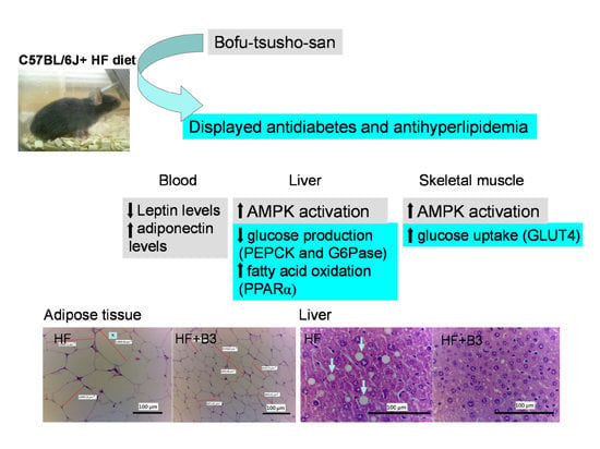
This study was undertaken to examine the effect and mechanism of Bofu-tsusho-san formula (BO) on hyperglycemia and hyperlipidemia and in mice fed with a high-fat (HF) diet. The C57BL/6J mice were received control/HF diet for 12 weeks, and oral administration of BO (at three doses) or rosiglitazone (Rosi) or vehicle for the last 4 weeks. Blood, skeletal muscle and tissues were examined by means of measuring glycaemia and dyslipidaemia-associated events. BO treatment effectively prevented HF diet-induced increases in the levels of triglyceride (TG), free fatty acid (FFA) and leptin (p < 0.01, p < 0.01, p < 0.01, respectively). BO treatment exhibited reduced both visceral fat mass and hepatic triacylglycerol content; moreover, BO treatment displayed significantly decreased both the average area of the cut of adipocytes and ballooning of hepatocytes. BO treatment exerted increased the protein contents of glucose transporter 4 (GLUT4) in skeletal muscle, and caused lowered blood glucose levels. BO treatment displayed increased levels of phosphorylated AMP-activated protein kinase (AMPK) in both skeletal muscle and liver tissue. Furthermore, BO reduced the hepatic expression of glucose-6-phosphatase (G6Pase) and phosphenolpyruvate carboxykinase (PEPCK) and glucose production. Therefore, it is possible that the activation of AMPK by BO leads to diminished gluconeogenesis in liver tissue. BO increased hepatic expressions of peroxisome proliferator-activated receptor α (PPARα), whereas down-regulating decreasing expressions of fatty acid synthesis, including sterol regulatory element binding protein 1c (SREBP1c) and fatty acid synthase (FAS), resulting in a decrease in circulating triglycerides. This study originally provides the evidence that amelioration of dyslipidemic and diabetic state by BO in HF-fed mice occurred by regulation of GLUT4, SREBP1c, FAS, PPARα, adiponectin and AMPK phosphorylation. Full article
(This article belongs to the Section Biochemistry)
Show Figures

Graphical abstract