Applicability of Supraclavicular Oxygenated and Total Hemoglobin Evaluated by Near-Infrared Time-Resolved Spectroscopy as Indicators of Brown Adipose Tissue Density in Humans

 ,
,
Abstract
:1. Introduction
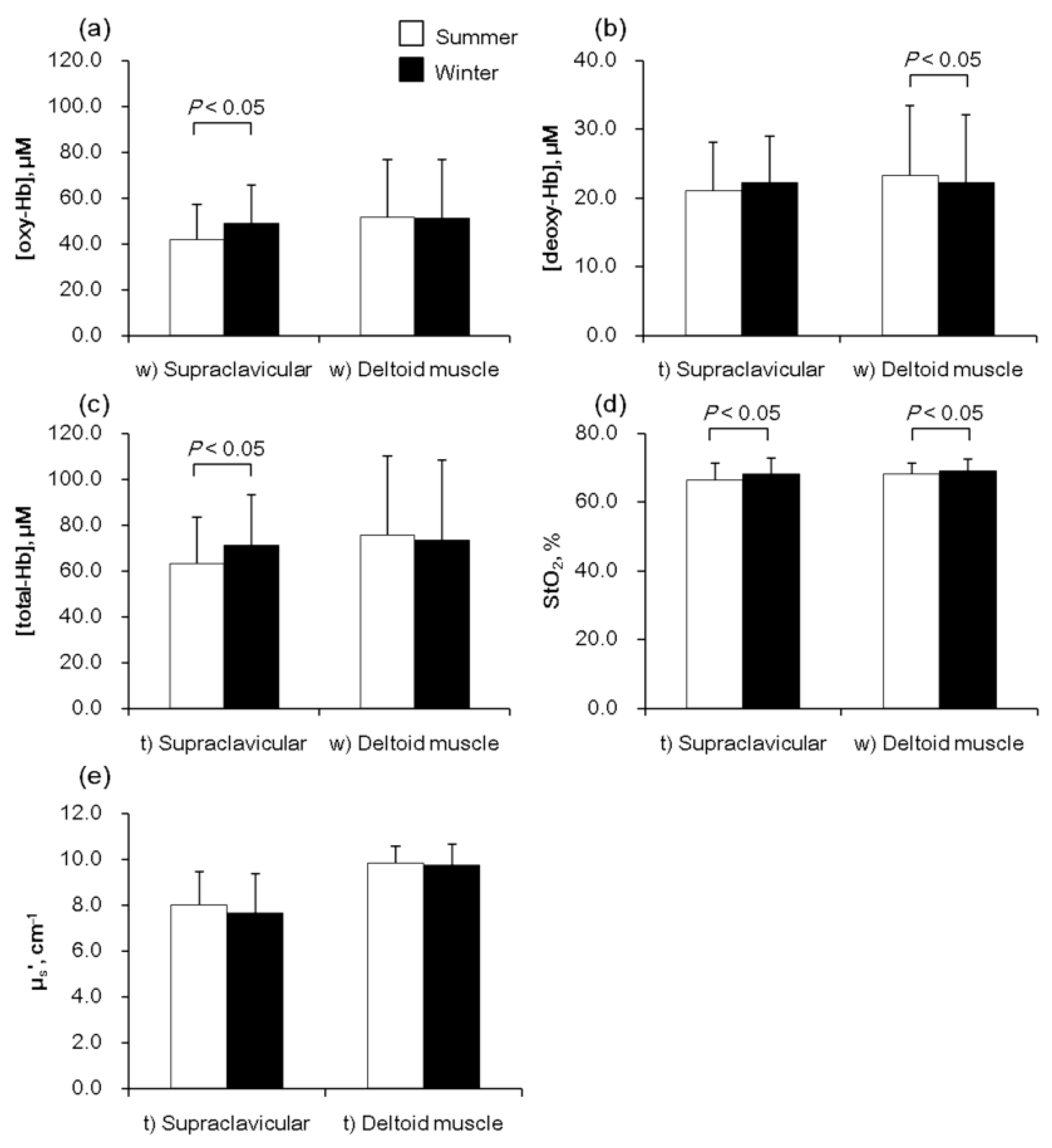
2. Results
3. Discussion
4. Materials and Methods
4.1. Subjects and Study Design
4.2. NIRTRS Parameters
4.3. Cold Induced Thermogenesis (CIT)
4.4. Statistical Analyses
5. Conclusions
Author Contributions
Funding
Conflicts of Interest
Abbreviations
| adjStO2 | adjusted supraclavicular hemoglobin oxygen saturation |
| BAT | brown adipose tissue |
| BAT-d | vascular or mitochondrial density in brown adipose tissue |
| CIT | cold-induced thermogenesis |
| CT | computed tomography |
| deoxy-Hb | deoxygenated hemoglobin |
| FDG | 18F-fluorodeoxyglucose |
| Hb | hemoglobin |
| NIRS | near-infrared spectroscopy |
| NIRCWS | near-infrared continuous-wave spectroscopy |
| NIRTRS | near-infrared time-resolved spectroscopy |
| oxy-Hb | oxygenated hemoglobin |
| PET | positron emission tomography |
| StO2 | hemoglobin oxygen saturation |
| SUVmean | mean standardized uptake value |
| total-Hb | total hemoglobin |
| µa | absorption coefficient |
| µs’ | scattering coefficient |
References
- Yoneshiro, T.; Aita, S.; Matsushita, M.; Kayahara, T.; Kameya, T.; Kawai, Y.; Iwanaga, T.; Saito, M. Recruited brown adipose tissue as an antiobesity agent in humans. J. Clin. Investig. 2013, 123, 3404–3408. [Google Scholar] [CrossRef] [PubMed] [Green Version]
- Saito, M.; Okamatsu-Ogura, Y.; Matsushita, M.; Watanabe, K.; Yoneshiro, T.; Nio-Kobayashi, J.; Iwanaga, T.; Miyagawa, M.; Kameya, T.; Nakada, K.; et al. High incidence of metabolically active brown adipose tissue in healthy adult humans: Effects of cold exposure and adiposity. Diabetes 2009, 58, 1526–1531. [Google Scholar] [CrossRef]
- Matsushita, M.; Yoneshiro, T.; Aita, S.; Kameya, T.; Sugie, H.; Saito, M. Impact of brown adipose tissue on body fatness and glucose metabolism in healthy humans. Int. J. Obes. (Lond.) 2014, 38, 812–817. [Google Scholar] [CrossRef] [PubMed]
- Blondin, D.P.; Labbe, S.M.; Tingelstad, H.C.; Noll, C.; Kunach, M.; Phoenix, S.; Guerin, B.; Turcotte, E.E.; Carpentier, A.C.; Richard, D.; et al. Increased brown adipose tissue oxidative capacity in cold-acclimated humans. J. Clin. Endocrinol. Metab. 2014, 99, E438–E446. [Google Scholar] [CrossRef] [PubMed]
- van der Lans, A.A.; Hoeks, J.; Brans, B.; Vijgen, G.H.; Visser, M.G.; Vosselman, M.J.; Hansen, J.; Jorgensen, J.A.; Wu, J.; Mottaghy, F.M.; et al. Cold acclimation recruits human brown fat and increases nonshivering thermogenesis. J. Clin. Investig. 2013, 123, 3395–3403. [Google Scholar] [CrossRef] [Green Version]
- Hanssen, M.J.; van der Lans, A.A.; Brans, B.; Hoeks, J.; Jardon, K.M.; Schaart, G.; Mottaghy, F.M.; Schrauwen, P.; van Marken Lichtenbelt, W.D. Short-term Cold Acclimation Recruits Brown Adipose Tissue in Obese Humans. Diabetes 2016, 65, 1179–1189. [Google Scholar] [CrossRef] [PubMed]
- Hanssen, M.J.; Hoeks, J.; Brans, B.; van der Lans, A.A.; Schaart, G.; van den Driessche, J.J.; Jorgensen, J.A.; Boekschoten, M.V.; Hesselink, M.K.; Havekes, B.; et al. Short-term cold acclimation improves insulin sensitivity in patients with type 2 diabetes mellitus. Nat. Med. 2015, 21, 863–865. [Google Scholar] [CrossRef] [PubMed]
- Borga, M.; Virtanen, K.A.; Romu, T.; Leinhard, O.D.; Persson, A.; Nuutila, P.; Enerback, S. Brown adipose tissue in humans: Detection and functional analysis using PET (positron emission tomography), MRI (magnetic resonance imaging), and DECT (dual energy computed tomography). Methods Enzym. 2014, 537, 141–159. [Google Scholar] [CrossRef]
- Beauvoit, B.; Chance, B. Time-resolved spectroscopy of mitochondria, cells and tissues under normal and pathological conditions. Mol. Cell Biochem. 1998, 184, 445–455. [Google Scholar] [CrossRef]
- Nirengi, S.; Yoneshiro, T.; Sugie, H.; Saito, M.; Hamaoka, T. Human brown adipose tissue assessed by simple, noninvasive near-infrared time-resolved spectroscopy. Obesity (Silver Spring) 2015, 23, 973–980. [Google Scholar] [CrossRef]
- Nirengi, S.; Homma, T.; Inoue, N.; Sato, H.; Yoneshiro, T.; Matsushita, M.; Kameya, T.; Sugie, H.; Tsuzaki, K.; Saito, M.; et al. Assessment of human brown adipose tissue density during daily ingestion of thermogenic capsinoids using near-infrared time-resolved spectroscopy. J. Biomed. Opt. 2016, 21, 091305. [Google Scholar] [CrossRef] [PubMed] [Green Version]
- Acosta, F.M.; Berchem, J.; Martinez-Tellez, B.; Sanchez-Delgado, G.; Alcantara, J.M.A.; Ortiz-Alvarez, L.; Hamaoka, T.; Ruiz, J.R. Near-Infrared Spatially Resolved Spectroscopy as an Indirect Technique to Assess Brown Adipose Tissue in Young Women. Mol. Imaging Biol. 2018. [Google Scholar] [CrossRef] [PubMed]
- Muzik, O.; Mangner, T.J.; Leonard, W.R.; Kumar, A.; Janisse, J.; Granneman, J.G. 15O PET measurement of blood flow and oxygen consumption in cold-activated human brown fat. J. Nucl. Med. 2013, 54, 523–531. [Google Scholar] [CrossRef]
- Chance, B.; Dait, M.T.; Zhang, C.; Hamaoka, T.; Hagerman, F. Recovery from exercise-induced desaturation in the quadriceps muscles of elite competitive rowers. Am. J. Physiol. 1992, 262, C766–C775. [Google Scholar] [CrossRef]
- Ferrari, M.; Mottola, L.; Quaresima, V. Principles, techniques, and limitations of near infrared spectroscopy. Can. J. Appl. Physiol. 2004, 29, 463–487. [Google Scholar] [CrossRef]
- Chance, B.; Nioka, S.; Kent, J.; McCully, K.; Fountain, M.; Greenfeld, R.; Holtom, G. Time-resolved spectroscopy of hemoglobin and myoglobin in resting and ischemic muscle. Anal. Biochem. 1988, 174, 698–707. [Google Scholar] [CrossRef]
- Hamaoka, T.; McCully, K.K.; Quaresima, V.; Yamamoto, K.; Chance, B. Near-infrared spectroscopy/imaging for monitoring muscle oxygenation and oxidative metabolism in healthy and diseased humans. J. Biomed. Opt. 2007, 12, 062105. [Google Scholar] [CrossRef]
- Hamaoka, T.; Katsumura, T.; Murase, N.; Nishio, S.; Osada, T.; Sako, T.; Higuchi, H.; Kurosawa, Y.; Shimomitsu, T.; Miwa, M.; et al. Quantification of ischemic muscle deoxygenation by near infrared time-resolved spectroscopy. J. Biomed. Opt. 2000, 5, 102–105. [Google Scholar] [CrossRef]
- Gunadi, S.; Leung, T.S.; Elwell, C.E.; Tachtsidis, I. Spatial sensitivity and penetration depth of three cerebral oxygenation monitors. Biomed. Opt. Express 2014, 5, 2896–2912. [Google Scholar] [CrossRef]
- Cohade, C.; Mourtzikos, K.A.; Wahl, R.L. “USA-Fat”: Prevalence is related to ambient outdoor temperature-evaluation with 18F-FDG PET/CT. J. Nucl. Med. 2003, 44, 1267–1270. [Google Scholar]
- Au-Yong, I.T.; Thorn, N.; Ganatra, R.; Perkins, A.C.; Symonds, M.E. Brown adipose tissue and seasonal variation in humans. Diabetes 2009, 58, 2583–2587. [Google Scholar] [CrossRef]
- Kim, S.; Krynyckyi, B.R.; Machac, J.; Kim, C.K. Temporal relation between temperature change and FDG uptake in brown adipose tissue. Eur. J. Nucl. Med. Mol. Imaging 2008, 35, 984–989. [Google Scholar] [CrossRef]
- Nirengi, S.; Sakane, N.; Amagasa, S.; Wakui, S.; Homma, T.; Kurosawa, Y.; Hamaoka, T. Seasonal differences in brown adipose tissue density and pulse rate variability in a thermoneutral environment. J. Physiol. Anthropol. 2018, 37, 6. [Google Scholar] [CrossRef]
- Yoneshiro, T.; Aita, S.; Matsushita, M.; Kameya, T.; Nakada, K.; Kawai, Y.; Saito, M. Brown adipose tissue, whole-body energy expenditure, and thermogenesis in healthy adult men. Obesity (Silver Spring) 2011, 19, 13–16. [Google Scholar] [CrossRef]
- Bruns, O.T.; Bischof, T.S.; Harris, D.K.; Franke, D.; Shi, Y.; Riedemann, L.; Bartelt, A.; Jaworski, F.B.; Carr, J.A.; Rowlands, C.J.; et al. Next-generation in vivo optical imaging with short-wave infrared quantum dots. Nat. Biomed. Eng. 2017, 1, pii: 0056. [Google Scholar] [CrossRef]
- Watanabe, M.; Yamamoto, T.; Kakuhata, R.; Okada, N.; Kajimoto, K.; Yamazaki, N.; Kataoka, M.; Baba, Y.; Tamaki, T.; Shinohara, Y. Synchronized changes in transcript levels of genes activating cold exposure-induced thermogenesis in brown adipose tissue of experimental animals. Biochim. Biophys. Acta 2008, 1777, 104–112. [Google Scholar] [CrossRef] [PubMed] [Green Version]
- Boushel, R.; Saltin, B. Ex vivo measures of muscle mitochondrial capacity reveal quantitative limits of oxygen delivery by the circulation during exercise. Int. J. Biochem. Cell Biol. 2013, 45, 68–75. [Google Scholar] [CrossRef] [PubMed]
- Delpy, D.T.; Cope, M. Quantification in tissue near-infrared spectroscopy. Philos. Trans. R. Soc. Lond. B 1997, 352, 649–659. [Google Scholar] [CrossRef] [Green Version]


| Supraclavicular Region | Deltoid Muscle Region | ||
|---|---|---|---|
| [total-Hb] | 27 °C | 0.64 ※ | 0.24 |
| 19 °C | 0.48 ※ | 0.21 | |
| [oxy-Hb] | 27 °C | 0.62 ※ | 0.25 |
| 19 °C | 0.49 ※ | 0.15 | |
| [deoxy-Hb] | 27 °C | 0.70 ※ | 0.20 |
| 19 °C | 0.40 | 0.30 | |
| StO2 | 27 °C | −0.38 | 0.20 |
| 19 °C | −0.11 | −0.24 | |
| μs’ | 27 °C | −0.02 | 0.30 |
| 19 °C | 0.08 | 0.39 |
© 2019 by the authors. Licensee MDPI, Basel, Switzerland. This article is an open access article distributed under the terms and conditions of the Creative Commons Attribution (CC BY) license (http://creativecommons.org/licenses/by/4.0/).
Share and Cite
Nirengi, S.; Fuse, S.; Amagasa, S.; Homma, T.; Kime, R.; Kuroiwa, M.; Endo, T.; Sakane, N.; Matsushita, M.; Saito, M.; et al. Applicability of Supraclavicular Oxygenated and Total Hemoglobin Evaluated by Near-Infrared Time-Resolved Spectroscopy as Indicators of Brown Adipose Tissue Density in Humans. Int. J. Mol. Sci. 2019, 20, 2214. https://doi.org/10.3390/ijms20092214
Nirengi S, Fuse S, Amagasa S, Homma T, Kime R, Kuroiwa M, Endo T, Sakane N, Matsushita M, Saito M, et al. Applicability of Supraclavicular Oxygenated and Total Hemoglobin Evaluated by Near-Infrared Time-Resolved Spectroscopy as Indicators of Brown Adipose Tissue Density in Humans. International Journal of Molecular Sciences. 2019; 20(9):2214. https://doi.org/10.3390/ijms20092214
Chicago/Turabian StyleNirengi, Shinsuke, Sayuri Fuse, Shiho Amagasa, Toshiyuki Homma, Ryotaro Kime, Miyuki Kuroiwa, Tasuki Endo, Naoki Sakane, Mami Matsushita, Masayuki Saito, and et al. 2019. "Applicability of Supraclavicular Oxygenated and Total Hemoglobin Evaluated by Near-Infrared Time-Resolved Spectroscopy as Indicators of Brown Adipose Tissue Density in Humans" International Journal of Molecular Sciences 20, no. 9: 2214. https://doi.org/10.3390/ijms20092214