Article

Fungal Extracellular Lipases from Coffee Plantation Environments for the Sustainable Management of Agro-Industrial Coffee Biomass

by
Randall Syedd-León
1,2,
Frank Solano-Campos
3,
Jorge Campos-Rodríguez
1,
Daniela Pereira-Arce
1,
Luis Roberto Villegas-Peñaranda
1,4 and
Manuel Sandoval-Barrantes
1,*
1
Biorefinery Research Laboratory (LIB), Chemistry School, Universidad Nacional, Heredia 86-3000, Costa Rica
2
Natural and Exact Sciences School, Universidad Estatal a Distancia, Mercedes de Montes de Oca, Mercedes 11503, Costa Rica
3
Plant Biotechnology Laboratory, Biological Sciences School, Universidad Nacional, Heredia 86-3000, Costa Rica
4
Teaching, Research and Production Support Laboratory (LADIP), Chemistry School, Universidad Nacional, Heredia 86-3000, Costa Rica
*
Author to whom correspondence should be addressed.
Biomass 2022, 2(2), 62-79; https://doi.org/10.3390/biomass2020005
Submission received: 18 December 2021 / Revised: 28 February 2022 / Accepted: 7 March 2022 / Published: 22 March 2022

Abstract

:
Coffee wastes have large amounts of by-products rich in phenolic compounds such as chlorogenic and caffeic acid, with potential applications for developing fine chemicals such as caffeic acid phenethyl ester (CAPE). A screening for microorganisms was undertaken in a coffee plantation environment to isolate native tropical species able to modify secondary metabolites present in this kind of biomass enzymatically. From the screening, 130 fungal strains could grow in lipase inducer media. Fungal strains were identified via ITS-based sequencing. Classification based on BLAST assigned 51 isolates to 12 different genera, including Absidia, Aspergillus, Cunninghamella, Fusarium, Metarhizium, Meyerozyma, Mucor, Neocosmospora, Papiliotrema, Penicillium, Rhizopus, and Trichoderma. DNA sequencing identified 14 putative extracellular lipases. According to the extracellular lipase activity, the most promising strain was identified as Fusarium sp. by DNA barcoding. Extracellular lipases from this strain exhibited maximal hydrolytic activity at a temperature of 45 °C, a pH of 7.00, and 200 ppm of NaCl, with an affinity towards substrates having carbon chain lengths of 8 or longer. Under these conditions, lipase instead of esterase activity is the main feature. The Km and Vmax values calculated using p-nitrophenyl palmitate (pNPP) as hydrolysis substrate were 0.003 mM and 299.8 μmol min−1 mg−1, respectively. Fusarium sp. lipases presented high stability during freeze–thawing, allowing the storage of enzyme solutions at −20 °C, but not as a lyophilized powder. According to our kinetic study, these lipases catalyzed CAPE hydrolysis, showing a progressive decrease in the concentration of the CAPE and a correspondent increase in the caffeic acid concentration as a product of this hydrolysis. Being able to carry out this type of reaction under mild conditions shows that Fusarium sp. lipases recognize CAPE as substrate and suggest CAPE synthesis (reverse reaction) and transformation can be engineered, using caffeic acid from coffee biomass, as a potential industrial application for these lipases.

Graphical Abstract

1. Introduction

Coffee is one of the most traded crops in the world. For the crop year commencing in 2020, more than 175 million 60 kg bags were produced [1]. The coffee fruit is considered a berry or cherry from which an inner seed is separated and known as green bean (Figure 1). Then, it is roasted to produce the final product of consumption: the ground coffee, which is considered a commodity [2,3]. However, much of the coffee fruit (30–60%) is discarded, and significant amounts of waste are generated during the production of ground coffee, as the process requires removing the pulp, mucilage, and coffee parchment to use only the green bean of the cherry [4]. Furthermore, coffee by-products, such as pulp and mucilage, contain essential amounts of bioactive compounds such as tannins, chlorogenic and caffeic acids, as well as other phenolic compounds. These compounds hinder the biodegradation of coffee by-products, and a detoxification process is required prior to use them as an organic fertilizer [5,6,7,8].
Phenolic acids may be released from coffee biomass by simple water extraction, but some remain esterified to the cell wall and are slowly released to the environment, causing the toxicity of coffee wastes. Therefore, the quest for (bio)catalyzers that can hydrolyze and release these compounds from coffee biomass is an important issue that has been taken into consideration, and thus some fungal strains have exhibited hydrolytic activity on these kinds of substrates [9,10]. Additionally, bioactive compounds such as caffeic and chlorogenic acids (Scheme 1) might be considered as potential added-value molecules (if they can be separated from the raw material) due to several properties, such as antioxidant, anti-obesity, hepatoprotective, and neuroprotective [11,12,13,14,15].
Caffeic and chlorogenic acids can be used as raw materials for the synthesis of caffeic acid phenethyl ester (CAPE), which is a fascinating compound for the pharma industry since it has been regarded as cytotoxic on melanoma tumors [16], an inhibitor of growth on colon cancer cell lines [17], as well on breast cancer stem cells [18]. However, the synthesis of CAPE faces a critical limitation: the high cost of caffeic acid in the market. In this sense, the sourcing of caffeic acid and other antioxidants from agricultural matrices can reduce the production costs of CAPE and might give added value to the by-products of the coffee industry [15].
CAPE synthesis could be achieved by esterification of caffeic acid and phenethyl alcohol [19,20,21]. Although this reaction is thermodynamically reversible (see Scheme 2), a catalyst (such as an enzyme) does not affect the equilibrium of the process; under this reasoning, finding an enzyme capable of hydrolyzing CAPE will open the door to use the same enzyme for CAPE synthesis after developing the appropriate reaction conditions to promote the reverse reaction.
Lipases (triacylglycerol hydrolases, EC 3.1.1.3) are enzymes that belong to the hydrolases group and catalyze the reversible hydrolysis of ester bonds; because of this reversibility, these enzymes are well known for catalyzing the synthesis of several pharmaceutical and fine chemical compounds in the presence of organic solvents as well as ionic liquids, with high regioselectivity and yields [22,23,24,25,26,27]. For those reasons, lipases are the most promising candidates to catalyze the esterification of caffeic acid to produce CAPE.
Green chemistry is a field that promotes the design of chemical processes and products towards sustainability [28,29]. In this sense, from a green chemistry perspective, the synthesis of CAPE can be performed using enzymes that can catalyze the esterification of caffeic acid with phenethyl alcohol, avoiding the use of activated derivatives of caffeic acid such as acyl chlorides. To search for lipases capable of recognizing secondary metabolites, such as chlorogenic and caffeic acids, present in coffee leaves and fruits, a quest for microorganisms in the coffee plantation ecosystem was undertaken. Then, we screened lipases obtained from these isolates and selected the most active lipase to evaluate its capacity to hydrolyze CAPE as a substrate.

2. Materials and Methods

2.1. Materials

p-Nitrophenol (pNP), p-nitrophenyl acetate (pNPA), p-nitrophenyl butyrate (pNPB), p-nitrophenyl octanoate (pNPO), p-nitrophenyl dodecanoate (pNPD), p-nitrophenyl myristate (pNPM), p-nitrophenyl palmitate (pNPP), p-nitrophenyl stearate (pNPS), Coomassie blue G-250, bovine serum albumin, gum arabic, Triton X-100, commercial lipase from Aspergillus niger, methanol, isopropanol, ampicillin, fluconazole, and sodium dibasic phosphate were purchased from Sigma Aldrich (St. Louis, MO, USA), other chemicals were reagent grade. Potato Dextrose Agar (PDA) and Lysogeny Broth (LB) agar preparations were obtained from Oxoid, olive oil suitable for human consumption was purchased from a grocery store.

2.2. Bioprospecting Permits for Biodiversity Resources and Legal Frame

Genetical and biochemical resources were accessed in Costa Rica according to Costa Rican Biodiversity Law [30] and the Convention on Biological Diversity [31] under the terms of fair and equitable distribution of the benefits for the providers of such resources. In this regard, biodiversity prospecting permits R-CM-001-2016-OT (years 2016–2019) and R-CM-002-2019-OT (years 2019–2022) were issued by the Costa Rican National Board for Biodiversity Management (Comisión Nacional para la Gestión de la Biodiversidad, CONAGEBIO by its initials in Spanish) [32].

2.3. Culture Media and Growth Conditions

Two different types of culture media were used for microorganism sampling; standard PDA supplemented with ampicillin (100 mg/L) for the isolation of fungi and standard LB agar supplemented with fluconazole (50 mg/L) for the isolation of bacteria. Strains were grown in Petri dishes at room temperature (24 ± 3 °C) for 12 days.
A minimum mineral media enriched with olive oil (2%) as the sole carbon source and emulsified with Triton X-100 (0.5%) was used as lipase inducer and for microorganism screening under selective pressure. Strains were grown in Petri dishes at room temperature (24 ± 3 °C) for 12 days. The composition of the culture media was as follows: K2HPO4 1.73 g/L, KH2PO4 0.68 g/L, MgSO4. 7H2O 0.1 g/L, NaCl 4 g/L, FeSO4. 7H2O 0.03 g/L, NH4NO3 1 g/L, CaCl2. 2H2O 0.02 g/L, 10 mL/L olive oil, 5 mL/L Triton X-100, and 20 g/L agar. The same culture media was prepared without agar for liquid cultures grown for 12 days using an orbital shaker at 200 rpm.

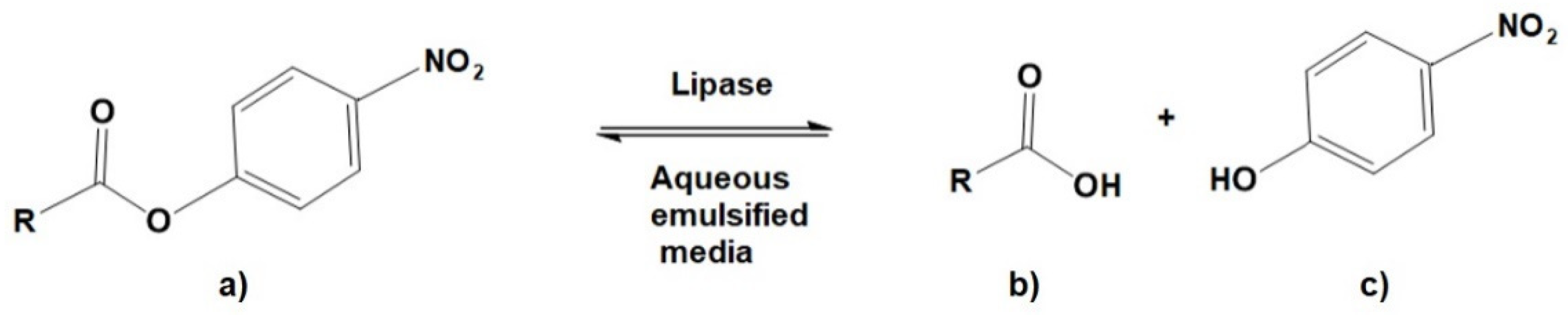
2.4. Protein Determinations and Lipase Activity Assays

Lipase enzymatic activity was determined by spectrophotometric quantification of pNP hydrolyzed from the correspondent emulsified pNP ester. First, the emulsified pNP ester was prepared by mixing one volume of Triton X-100 with 4 volumes of isopropanol, then 34 µmol of the pNP ester were dissolved in 2.00 mL of this solution to prepare a substrate solution. Finally, one volume of the substrate solution was mixed with 10 volumes of phosphate buffer (50 mM, pH 7.0, 550 ppm gum arabic) [33].
The enzymatic reaction took place by mixing 250 µL of emulsified pNP ester with 50 µL of enzyme solution at 40 °C in 300 µL microplate wells for 5 min. The amount of pNP released by the enzyme was estimated at 410 nm against a standard curve performed under identical conditions. As the UV/Vis absorbance of pNP is dependent on its phenol dissociation, measurements at 317 and 347 nm wavelengths were also considered in some specific cases for analytic determinations [33,34,35]. Protein concentration was estimated by the Bradford method [36] using bovine serum albumin as standard. One enzymatic unit (U/mg) was defined as the amount (mg) of protein that hydrolyzed one micromole of pNP ester per minute under the above-described conditions. Absorbance was measured using a Synergy HT Multi-Detection 96-well Microplate Reader (BioTek Instruments, Winooski, VT, USA). In the lipase activity assays absorbance was measured after 5 min of hydrolysis reaction.

2.5. Coffee Plantation Microorganism Sampling and Screening

A random sampling of microorganisms was undertaken during the dry season in a coffee plantation located at Santa Lucia experimental station property of Universidad Nacional (UNA, 10°01′21″ N, 84°06’40″ W), Heredia, Costa Rica. Sampling was performed with the help of a sterile ribbed spatula or by hand using sterile nitrile gloves. Soil and vegetable samples were collected from 12 points and used to inoculate culture plates. The collected samples were incubated at room temperature (24 ± 3 °C) for 48 h. After the incubation process, plates with a large mixed microbial load were subjected to a purification process to obtain pure strains. After the purification process of the sampled microorganisms, 130 fungal strains were obtained, no bacterial strains were isolated.

2.6. Fungal Extracellular Lipases Production and Screening

Fungal strains with lipase activity were preserved from PDA plates at −20 °C. The growth process was performed in triplicate for each of the strains. For lipase production, preserved samples were initially grown in PDA in an incubator at room temperature (24 ± 3 °C) for 12 days, then transferred into minimum mineral media enriched with olive oil agar plates and grown for 12 days. From the initial 130 fungal strains, only 51 of them were able to survive in this lipid-enriched medium, which is why they were identified as the only ones capable of secreting lipases into the extracellular medium and selected for further experiments. Finally, 10 out of 51 were chosen according to two criteria: biomass formation rate and morphological diversity.
Strains that grew on plates enriched with olive oil were transferred to a lipase-inducing liquid medium for 12 days. Samples were taken for DNA strain characterization as described below. Then, clarified extracellular lipases (CEL) were harvested by two sequential filtrations, first under vacuum and second with a 0.20 µm syringe membrane filter. CEL was stored at 4 °C. CEL was not passed through further purification treatments to develop potential agro-industrial applications for coffee biomass.
Enzyme activity was measured using one short-chain carbon ester, p-nitrophenyl butyrate (pNPB for esterase activity), and one long-chain carbon ester p-nitrophenyl palmitate (pNPP for lipase activity) as substrates for the CEL of the following strains: Penicillium crustosum (2-8A), Fusarium sp. (2-5B-1), Aspergillus sp. (2-5B-2), Fusarium sp. (2-5A-1), Fusarium sp. (2-5E), Mucor microsporus Namyslowski (2-2), Rhizopus sp. (12-C), Cunninghamella echinulata (2-2A), Penicillium rubens (C-2), and Aspergillus sp. (SL14D). In addition, an Aspergillus niger commercial lipase was used as a reference for this stage. After this screening, the most promising strain was used for further tests, other species were discarded. The reactions were followed under the conditions previously described in Section 2.4. using a Synergy HT Multi-Detection 96-well Microplate Reader (BioTek Instruments, Winooski, VT, USA).

2.7. Strain Characterization: DNA Extraction

A protocol based on lysis with cetyltrimethylammonium bromide (CTAB) by ethanol precipitation was executed, modified from Stewart [37]. Briefly, fresh mycelium (50 mg) was placed in a 1.5 mL centrifuge tube with 400 µL of sodium chloride-Tris-EDTA (STE) buffer (1.75 M NaCl, 125 mM Tris, 25 mM EDTA) and processed with an automatic cell disruptor (MM400, Retsch GmbH, Haan, Germany) at 30 Hz for 4 min using four 3 mm stainless steel grinding balls. Then, 100 µL of 10% CTAB, 0.6% v/v β- mercaptoethanol, and 0.05 mg/mL proteinase K were added. The samples were incubated at 60 °C for one hour. The tubes were mixed by inversion every 15 min. Then, 500 µL of chloroform:isoamyl alcohol solution (24:1) was added and mixed by inversion, following centrifugation at 15,000 rpm for 10 min at 4 °C. Immediately, the supernatant containing the DNA was transferred to a new 1.5 mL tube. One volume of isopropanol was added and mixed carefully by inversion. The samples were left for one hour on ice to precipitate the DNA. Then, they were centrifuged at 15,000 rpm for 20 min at 4 °C. The supernatant was removed, and the DNA was washed with 500 µL 70% ethanol. After another centrifugation at 15,000 rpm for 2 min, the ethanol was removed. The tubes were allowed to dry uncovered for 15 min at room temperature (25 °C). The DNA was resuspended in 50 µL of 10 mM Tris (pH 8.0) buffer and incubated at 65 °C for 1 h. The DNA obtained was visualized on agarose gels and quantified by spectrophotometry (Nanodrop 2000).

2.8. Strain Characterization: DNA Barcoding and Lipase Sequencing

Fungal isolates were identified by DNA sequencing of the nuclear ribosomal internal transcribed spacer (ITS) region. As described previously, genomic DNA was isolated from fungal tissue with a standard CTAB extraction method [37]. Polymerase chain reaction (PCR) amplification was performed with ITS1 and ITS4 primers [38] using DreamTaq PCR Master Mix (Thermo Scientific, Carlsbad, CA, USA) following the manufacturer’s recommendations. Direct Sanger sequencing of amplicons was undertaken using the same primers as in PCR (Macrogen Inc., Seoul, Republic of Korea). Amplicon sequencing data pre-processing and bioinformatic analysis were undertaken using Geneious Prime 2019.2.3 (Biomatters, Auckland, New Zealand). Taxonomical assignments were completed with the IDTAXA Classifier [39] and the RDP Naive Bayesian rRNA Classifier [40] using the UNITE [41] and Warcup v2 [42] databases, and with the BLAST tool [43] using the NCBI fungal ITS RefSeq database and the optimal identity thresholds predicted to discriminate fungal species [44]. A 99.6% identity threshold to BLAST best hit was used to identify isolates, data available in the Supplementary Information. Fungal lipases were amplified from genomic DNA using degenerate primers (Supplementary Table S1) designed with Primaclade [45] based on sequence alignments of publicly available Aspergillus, Fusarium, Penicillium, and Mucor lipases. BLAST was used to perform DNA and protein sequence similarity searches against the NCBI nucleotide collection database and the Lipase Engineering Database (LED) [46] MUSCLE [47] was used for creating multiple alignments of lipase sequences. Gene prediction was undertaken using the Augustus web server [48]. bThe CD-Search tool [49] was used to detect conserved functional domains in predicted protein sequences, while DeepLoc [50] was used to predict the subcellular localization. DNA sequences were submitted to GenBank (accession numbers MN737511-MN737550, MN747984-MN747992, and OL840776-OL840780).

2.9. Substrate, pH, and Temperature Effect on Lipase Activity

Isolate 2-1A CEL, identified as Fusarium sp., was chosen as the most promising biocatalyst for the study. Then, enzyme activity was measured at different pH values considering the pNP absorbance is highly dependent on the pH, in this case, activity tests were measured at 317 nm (protonated pNP), 347 nm (isosbestic point of absorbance), and 410 nm (unprotonated pNP) in a Synergy HT Multi-Detection 96-well Microplate Reader (BioTek Instruments, Winooski, VT) [33,34,35]. Buffer solutions were: 100 mM citric acid/disodium phosphate buffer solutions with pH of 3.00–6.00; 100 mM disodium phosphate buffer solutions, pH 6.50–8.50, and 100 mM sodium carbonate buffer solution with pH 9.00–11.00. Optimal pH was estimated at 7.00, the lipase activity was measured at this pH value, and at temperatures 25, 30, 35, 40, 45, 50, 55, and 60 °C. The last analysis was based on the carbon chain for saturated fatty acids esters recognition. For this purpose, Fusarium sp. CEL were assayed against the following substrates: pNPA (C-2), pNPB (C-4), pNPO (C-8), pNPD (C-12), pNPM (C-14), pNPP (C-16), and pNPS (C-18). These substrates were at the same concentration, 1 mM, in an emulsified aqueous media as previously described in Section 2.4.

2.10. Kinetic Parameters

Fusarium sp. CEL were kinetically studied using 0.1, 0.2, 0.4, 0.6, and 0.8 mM of pNPP (C-16) as substrate. Enzyme rate was measured at each concentration to determine Km and Vmax using Lineweaver–Burk calculations. The amount of converted substrate was obtained spectrophotometrically by the equivalent pNP product released under the previously indicated conditions (Synergy HT Multi-Detection 96-well Microplate Reader, BioTek Instruments, Winooski, VT, USA).

2.11. Enzyme Stability for Freezing and Lyophilization

To know basic handling and storage conditions of CEL from Fusarium sp., stability to freezing/thawing, and lyophilization stability were investigated. Five CEL samples (3.0 mL) were frozen for 24 h at −20 °C and then thawed to estimate the enzyme stability to this condition, five CEL samples (3.0 mL) were frozen for 24 h and then lyophilized to estimate their stability, and five CEL samples (3.0 mL) were stored at 4 °C as a positive control, then lipase activity was measured in the three groups. The average activity was calculated, and the standard deviation was reported for each group.

2.12. Determination of Ionic Strength Effect on Enzyme Activity

The effect of salinity on the enzyme activity was determined by incubating the enzyme in 0, 10, 20, 40, 80, 100, 200, 400, and 800 ppm of NaCl solutions and measuring the residual activity under these conditions. The measurements were made using the same pNPP emulsified medium adjusting the salinity to the specified concentrations of NaCl.

2.13. Lipase Application in Biomass Degradation

Lipase’s ability to hydrolyze phenolic acids bound to cell walls from coffee biomass was assayed as follows. Solid coffee waste was dried by lyophilization and ground in a blade mill with a 1 mm sieve. This solid (2 g) was subsequently washed five times with 500 mL of hot water (85 °C) to eliminate any free organic compound. Then, the suspension was centrifuged at 10,000 rpm for 15 min at 4 °C. The supernatant was filtered with a 0.20 µm syringe membrane filter, and the cleanliness was determined by UV/VIS and UHPLC-DAD methods. The liquid was lyophilized again and used for enzymatic biomass degradation. The solid (100 mg) was mixed with 200 µL of 736 µg/mL CEL solution, 1.80 mL of pH 7 buffer solution, and 3 mL of distilled water in a test tube with a screw cap. The reaction was kept at 40 °C for 7 days. Degradation products were analyzed by UHPLC-DAD using water as a blank.

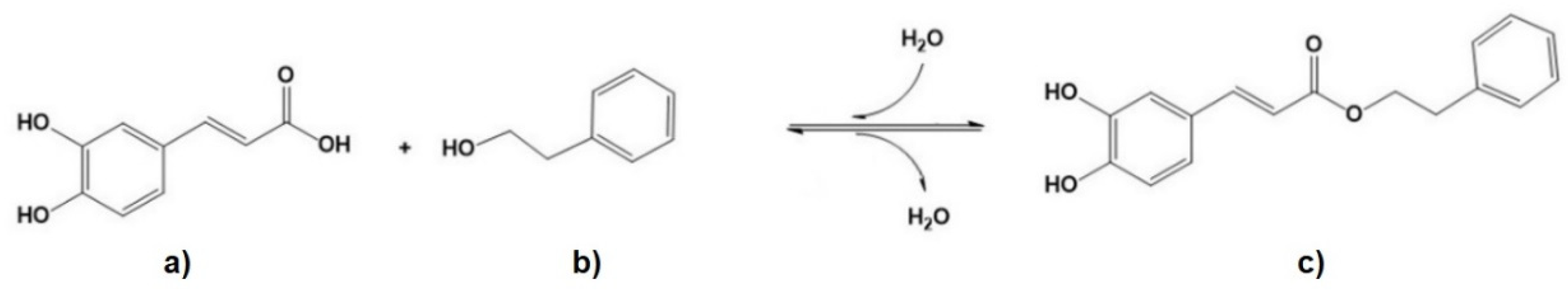
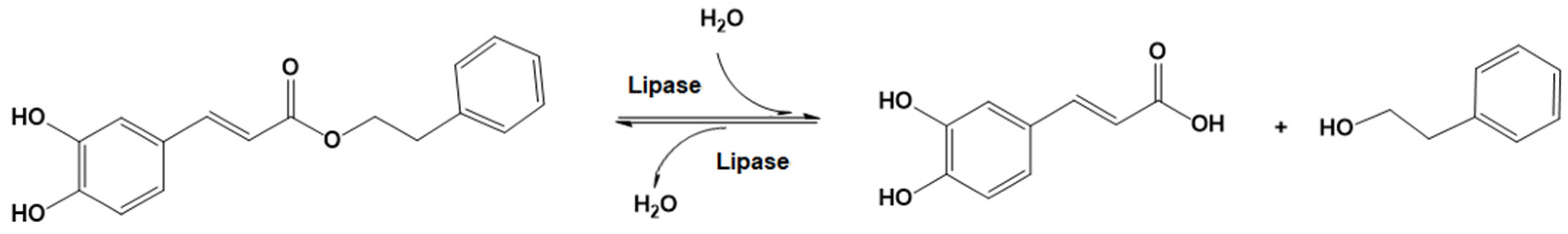
2.14. Lipase-Catalyzed CAPE Hydrolysis

In order to determine the enzyme ability to recognize CAPE as substrate, a 200 ppm stock solution was prepared as follows: 20 mg of CAPE, 4 mL of isopropyl alcohol, 50 µL of Triton X-100, and pH 7 buffer (with 0.5 mg/mL of gum arabic) solution was added to reach a final volume of 100 mL. In total, 5 mL of the substrate solution was placed in a screw cap test tube along with 300 µL of CEL (736 µg/mL). Water was used as blank and commercial Aspergillus niger as reference. The reaction was monitored, collecting samples at 1, 2, 3, 6, 12, 24, and 48 h. The reaction was quenched by freezing rapidly at −30 °C and then heating at 90 °C for 15 min. UHPLC-DAD analysis was performed using an Ultimate 3000 UHPLC-DAD-CAD system (Thermo Scientific, Sunnyvale, CA, USA). The separation was performed on an Acclaim C18 Polar Advantage II column (5 µm, 120 Å, 4.6 × 250). The solvents were (A) water/trifluoroacetic acid (0.1%), (B) methanol, and (C) acetonitrile. The gradient was linear, and the proportions of solvents were as follows: 0–5 min 85% A and 15% B, 5–8 min 85–10% A, 15–80% B and 0–10% C, 8–12 min 10% A, 80–40% B and 10–50% C, and 12–15 min 100% C. The injection volume was 10 µL, and the column temperature was kept at 40 °C. The flow rate was 1 mL/min. The detection was performed at 327 nm for CAPE and caffeic acid (CA) as a hydrolysis product. The concentrations were estimated from calibration curves (peaks area) constructed with CAPE and CA.

2.15. Statistical Analysis

Multiple comparisons among several groups such as enzymatic activity in short carbon chain and long carbon chain substrates and thermal treatments were performed considering significant differences at p < 0.05 (one-way ANOVA and post-hoc Tukey test). The results are presented as means and standard deviations. The results are represented according to their significant differences using different letters in the graphs.

3. Results

3.1. Fungal DNA Barcoding and Lipase Sequencing

ITS-based DNA barcoding of 51 fungal isolates revealed 12 different genera. Most isolates were assigned to Aspergillus (10 isolates), Penicillium (9 isolates), and Fusarium (10 isolates) (Supplementary Table S2). Putative lipase encoding sequences were successfully amplified in 14 isolates, and their coding sequences predicted by AU-GUSTUS presented high similarity (79–100% identity) to other cDNA sequences described as lipases, probable triacylglycerol lipase precursors, alpha/beta-hydrolases, and hypothetical/uncharacterized proteins available in the NCBI nucleotide collection database. Predicted proteins produced significant alignments to the homologous families abH23.01 (Rhizomucor mihei lipase-like) and abH23.02 (Saccharomyces lipase-like) in the LED database (Supplementary Table S3). According to CD-Search and DeepLoc tools, all predicted proteins contained the active site flap/lid and the nucleophilic elbow, typical features conserved in lipases [51], and were predicted to be extracellular and soluble. Further characterization, such as heterologous expression in Pichia pastoris and lipase activity assays of purified recombinant proteins, is needed to determine the usefulness of these putative lipases for CAPE hydrolysis.

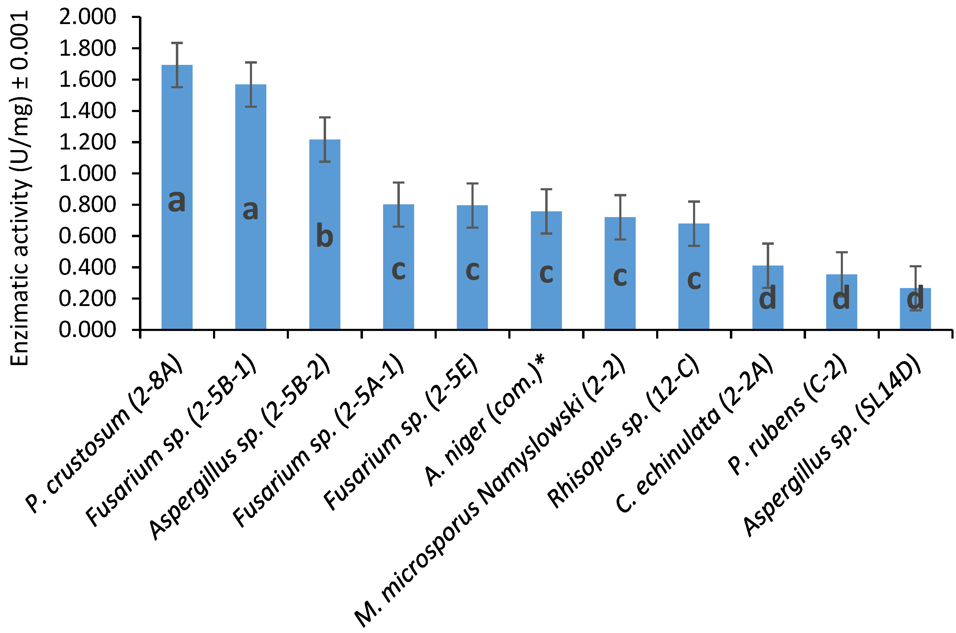
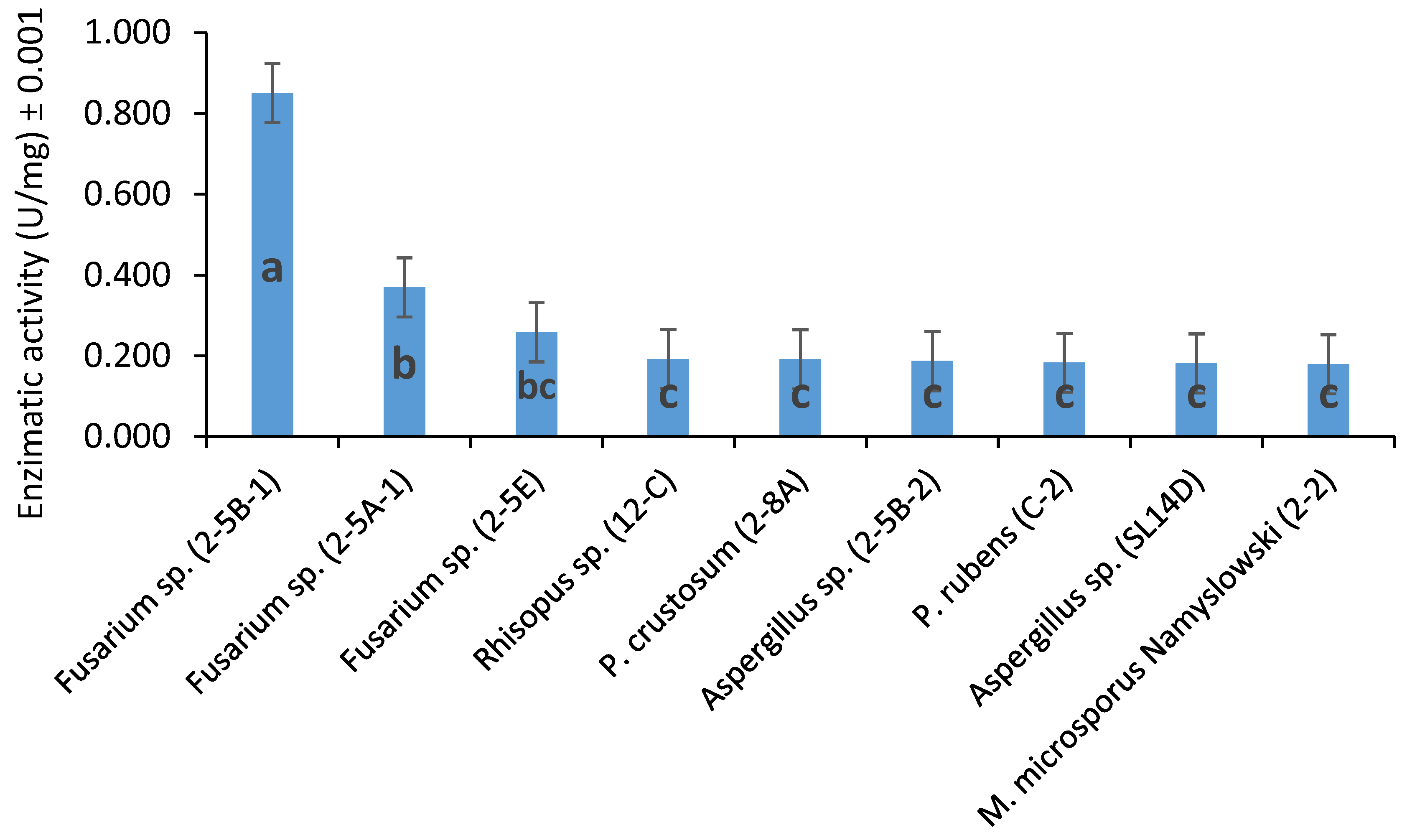
3.2. Fungal Extracellular Lipases and Esterases Screening

Lipase and esterase activities were quantified by using p-nitrophenyl esters as substrates. The reaction occurred in an emulsified media where the ester was hydrolyzed, releasing the correspondent carboxylic acid and pNP as products, where pNP concentration was determined spectrophotometrically (see Scheme 3), molar amount of pNP is the same molar amount of hydrolyzed pNPP.
The screening of the 10 fungal strains showed that all of them have both lipase activity with long-chain carbon ester (pNPP) and esterase activity with short-chain carbon ester (pNPB). P. crustosum and Fusarium sp. were the most active on long-chain ester (Figure 2), and while there were no significant differences between them, they differed significantly from the rest. Only three CEL obtained were significantly more active on pNPP than A. niger commercial lipase.
All the extracts showed less activity on short-chain esters such as pNPB-C4 (Figure 3) which means that, in general, their effects are mainly as lipases and not as esterases. Combining the results from both substrates, the CEL of Fusarium sp. is the most promising for biochemical characterization and further biotransformation analysis on biomass.

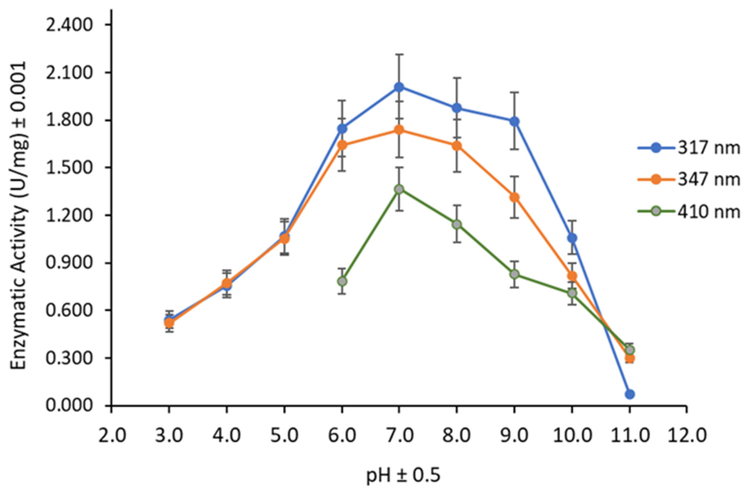
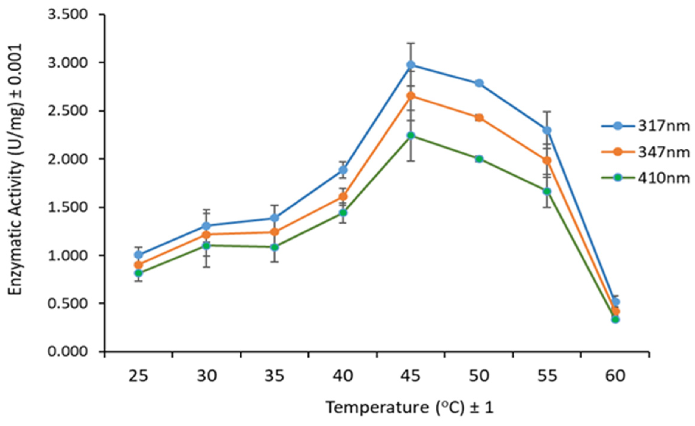
3.3. Substrate, pH, and Temperature Effect on Lipase Activity

The pH analysis was carried out with buffer solutions at pH 3.0–11.0, using pNPP at 40 °C. As shown in Figure 4, the optimal pH was 7.0 and the trends were similar among the different wavelengths employed.
To determine the optimal temperature, enzymatic activity was measured at temperatures from 25 to 60 °C, using pNPP at pH 7.0. The optimal temperature was 45 °C (Figure 5). Then, enzymatic activity was assayed under optimized conditions using substrates with carbon chains of 4–18 carbon atoms. The substrates with carbon chains equal to or above 8 carbon atoms seemed to be the more appropriate for this lipase, showing a higher performance towards the C-16 carbon chain (Figure 6). In this sense, palmitic acid esters are the substrates more related to the Fusarium sp. CEL. On the other hand, short carbon chain substrates (such as C-2 and C-4) showed less than 50% of relative activity, confirming that lipase instead of esterase activity is the main feature of Fusarium sp. CEL.

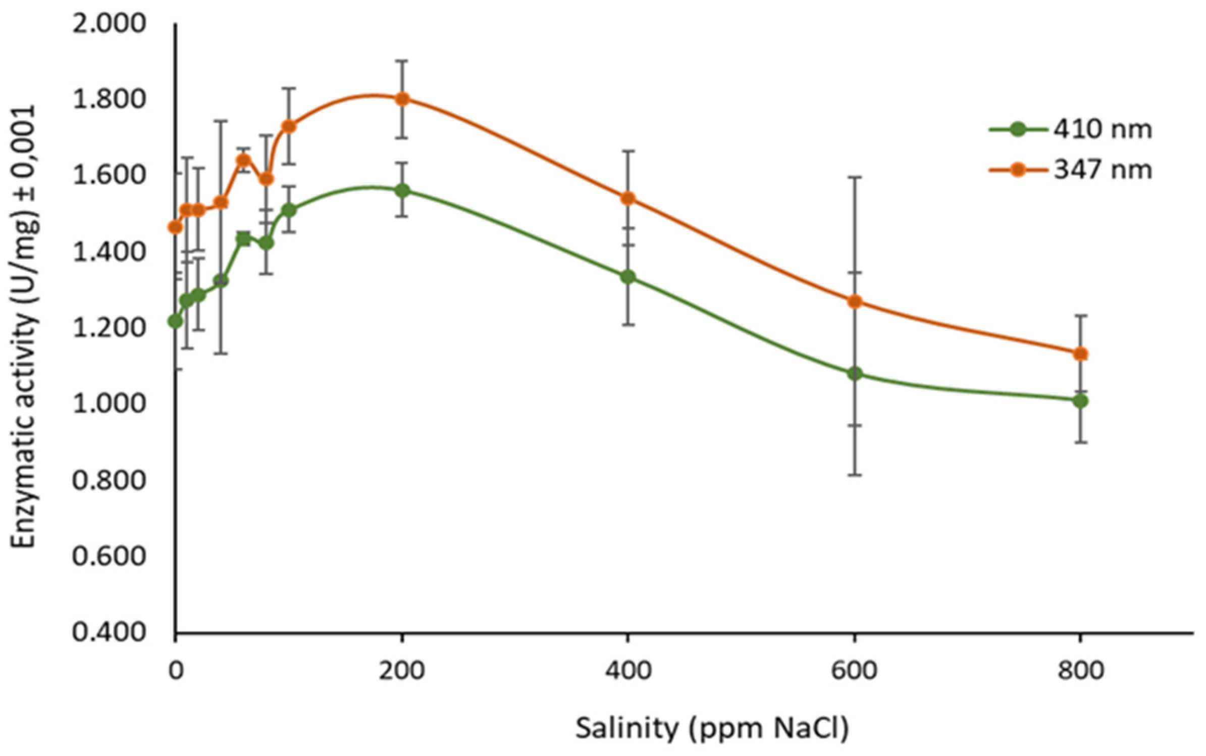
3.4. Determination of Ionic Strength Effect on Enzyme Activity

The effect of the ionic strength on Fusarium sp. CEL activity was measured in solutions with 0–800 ppm of NaCl, using pNPP at pH 7.00 and 45 °C. The optimum NaCl concentration determined for enzymatic activity was 200 ppm, which means that the enzyme needs small quantities of salt (Figure 7). A more saline medium (i.e., 800 ppm NaCl) reduced the enzymatic activity to 65% of maximum relative activity. On the other hand, a more diluted medium also affected the enzyme catalytic behavior reducing its activity to 80% of maximum relative activity.

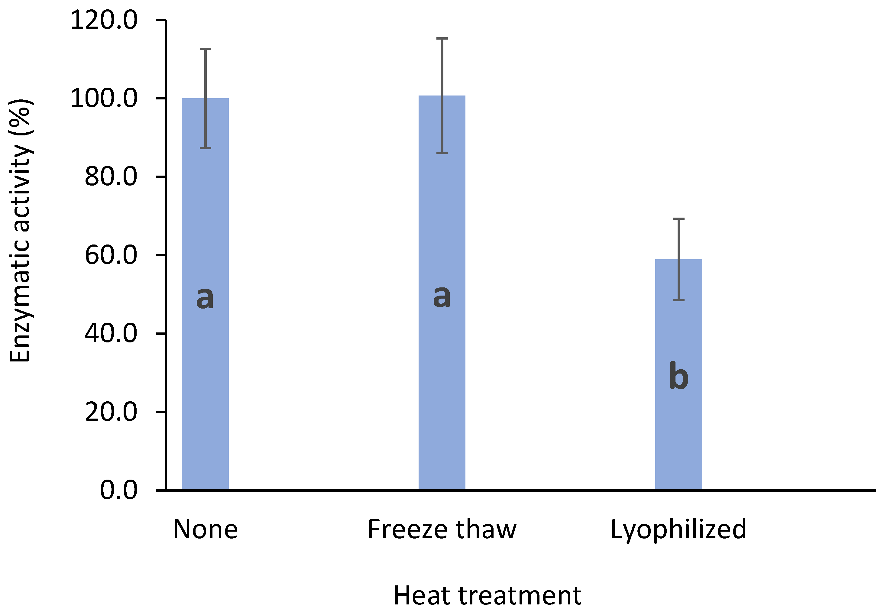
3.5. Enzyme Stability after Freeze/Thawing and Lyophilization

The thermal stability of CEL from Fusarium sp. was tested using enzymatic activity assays under optimized conditions after freeze/thawing and lyophilization treatments. The enzymatic activity of CEL was decreased by lyophilization by 40% (Figure 8). On the other hand, the activity was retained after one cycle of freeze/thawing.
This result shows the robustness of the enzymes contained in the Fusarium sp. CEL. In this regard, some lipases, i.e., Sporobolomyces ruberrimus, decrease their enzymatic activity when stored at 4 and 10 °C rather than at room temperature [52]. On the other hand, the stability of CEL from Fusarium sp. represents an advantage since it allows it to freeze, and even to lyophilize, which may allow to use it in synthesis reactions in non-aqueous environments.

3.6. Lipase Applications in Biomass Degradation

Fusarium sp. lipases did not show capabilities on the degradation of coffee biomass under the conditions tested. Thus, it is necessary for future applications to take into consideration to use these lipases in combination with other organisms and enzymatic systems.

3.7. Vmax and Km Kinetic Constants

The Km and Vmax values of Fusarium sp. lipase extract calculated from a Lineweaver–Burk plot (Figure 9) using pNPP as hydrolysis substrate were 0.0032 mM and 299.8 μmol min−1 mg−1, respectively. As shown in the plot, there was a linear response with R2 = 0.9984.
The Km was lower than those reported for A. niger F044 (7.37 mM) [53] and Spirulina platensis (0.02 mM) [54]. This difference could be related to the low specificity towards long-chain pNP substrates (Figure 6). The Vmax value of Fusarium sp. lipase extract was significantly higher than Nomuraea rileyi MJ (18.9 μmol min−1 mg−1) [55] and Fusarium solani FSI (1.7 μmol min−1 mg−1) [56]. The studies pointed out were carried out with purified lipases, for that reason differences are expected. The aim of this work is to develop applications in biomass transformation for future industrial purposes, and therefore purification was not considered necessary, because this step would increase manufacture costs for the catalyst. At this regard, many of the commercially available enzymes are a non-purified catalyst, and their activity is highly stable and reproducible.

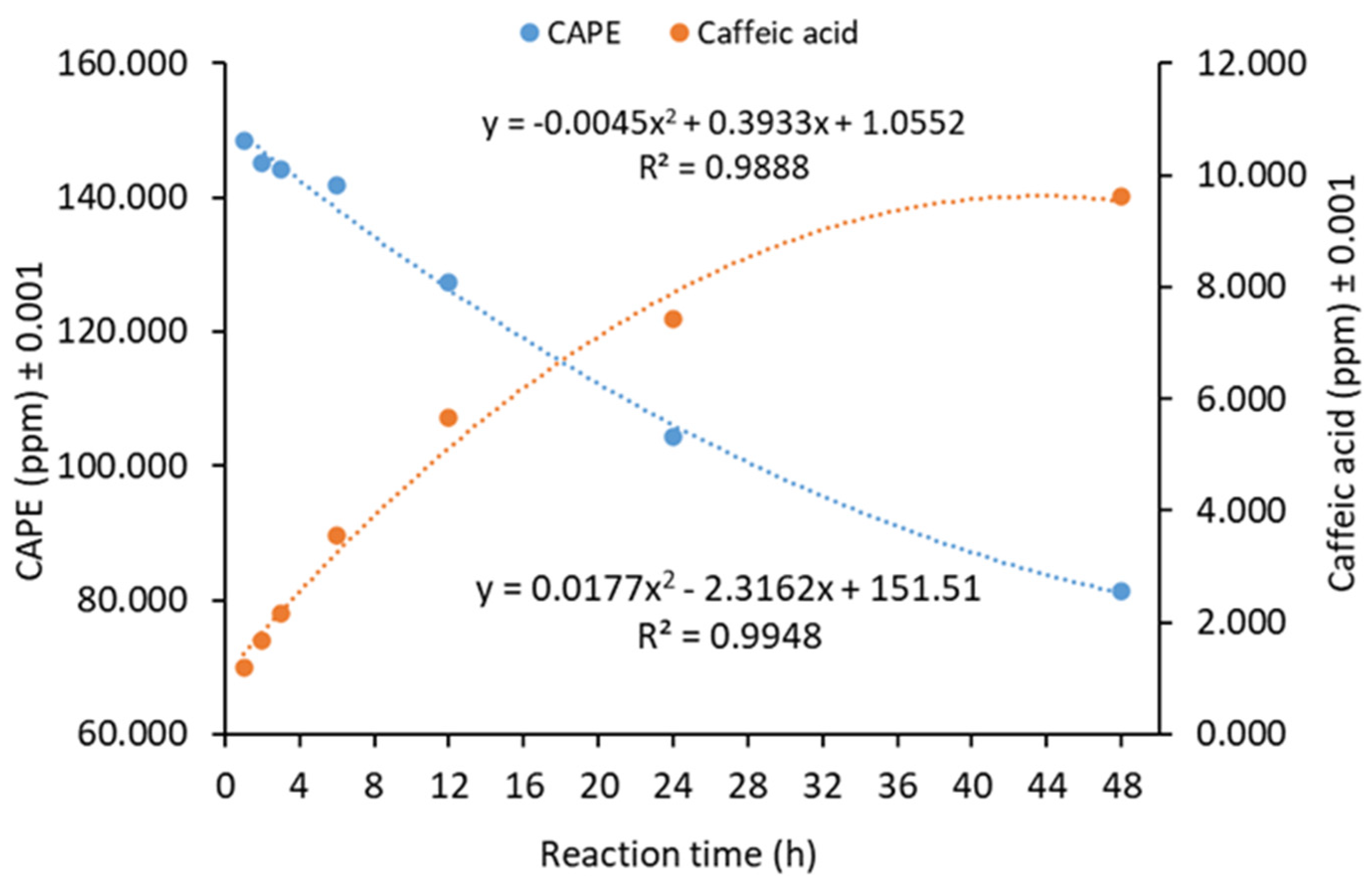
3.8. Kinetics of Lipase-Catalyzed CAPE Hydrolysis

Ester hydrolysis is a reversible reaction that is not affected by a catalyst. Nevertheless, the amount of water in the reaction media shifted the reaction equilibrium towards the hydrolysis of CAPE. In this sense, the hydrolysis of CAPE by Fusarium sp. clarified extracellular lipases was investigated under aqueous media, releasing caffeic acid and phenethyl alcohol as products (see Scheme 4).
The kinetic study of CAPE degradation under optimized conditions by Fusarium sp. clarified extracellular lipases was developed. As shown in Figure 10, CAPE concentration decreased while caffeic acid concentration increased. As a result, extracellular enzymes can hydrolyze CAPE in percentages close to 50% after 48 h of reaction.

4. Discussion

As shown in Figure 4, lipases from Fusarium sp. are affected by the pH, which is the expected behavior with a Gaussian tendency in most enzymes [55]. The most favorable pH value was 7.0. The CEL are relatively stable between pH values 6.0–9.0, but the activity decreases with extremely high or low pH values. This pattern of lipase activity at different pH values shown in Figure 4 is probably the result of changes in the shape of the enzyme’s active site [57].
Meanwhile, Figure 5 shows that lipases from Fusarium sp. worked better at intermediate temperatures and reached their optimum at 45 °C. It is a higher value when compared to the Pseudomonas sp. lipase, which has hydrolytic activity at 35 °C [58]. Lipases have a wide range of optimum temperatures depending on which microorganism they were obtained from. While some lipases function at low temperatures, others are more active at higher temperatures [59].
When considering the activity on different substrates, as shown in Figure 6, CEL obtained from Fusarium sp. can hydrolyze the linkage in many pNP esters, showing bond specificity. Fusarium sp. CEL can hydrolyze short, intermediate, and long-chain esters, being less active on C-2 and C-4 esters, showing a predominant lipase over esterase activity. The evidence also shows lipase promiscuity, a well-known phenomenon [60]. This property of recognizing different substrates is beneficial for this study. The enzymes showed different degrees of specificity towards the substrate due to its molecular recognition mechanism, and it operates through structural complementarity. The function of extracellular enzymes in biomass transformation requires moderate substrate specificity because the esters from coffee wastes can be very diverse in terms of carbon chain length and conformational structure.
CAPE is considered a critical nutraceutical compound, that can be obtained from bee propolis [16,18,61,62]. Regarding lipase-catalyzed CAPE hydrolysis, in Figure 10, the trend shown in the plot is expected with a progressive decrease in the concentration of the substrate and an increase in the products evidenced by caffeic acid. In addition, the enzymatic hydrolysis reaction of CAPE would allow obtaining significant amounts of caffeic acid, an essential antioxidant of natural origin [63,64,65,66]. Although hydrolysis is usually carried out in acidic or basic media at relatively high temperatures, the use of Fusarium sp. CEL allows this bioprocess to be carried out in a more environmentally friendly condition.
The fact of carrying out this type of reaction under mild conditions (45 °C) suggests that these enzymes can perform the reverse reaction, CAPE synthesis. Because ester hydrolysis is a thermodynamically controlled reaction (Scheme 4), catalysts do not affect the equilibrium. Thus, CAPE synthesis (reverse reaction) with Fusarium sp. CEL, using caffeic acid from coffee biomass, can be engineered as a potential industrial application for Fusarium sp. CEL. Other types of bioconversions could also be possible among CAPE derivatives, for example, esterification and alcoholysis reactions. Lipases can catalyze these reactions due to their promiscuity [60]. Further studies may be developed to optimize reaction conditions for the CAPE synthesis through esterification and alcoholysis reactions. Hence, the CAPE synthesis and transformation, using caffeic acid from coffee biomass, is a potential industrial application for this enzyme.
Recently, prediction models for the ultrasound-enhanced synthesis of CAPE using a commercial immobilized lipase from Candida antarctica, Novozym® 435, from caffeic acid and phenethyl alcohol were developed. The authors predicted an optimum reaction time of 9.6 h for CAPE synthesis, an enzyme amount of 2938 PLU, a substrate molar ratio of 1:71, and ultrasonic power of 2 W/cm2 with the molar conversion up to 96%. The experiment was performed using the optimal conditions predicted by the models, and as a result, a molar conversion of 93% was achieved [67].
Fusarium sp. CEL showed no effect on the degradation of coffee biomass under the conditions tested. This result could be related to the optimal pH determined for Fusarium sp. CEL (7.00) and to the acidic coffee biomass that affected the enzyme structure. Thus, future applications must be combined with other microorganisms and enzymatic systems or mild chemical pretreatments for biomass, such as addition of weak bases. Biomass degradation is a slow and complex process that can take several weeks and even months. Therefore, as a hypothesis for future studies, it is advisable to perform a more extended assay than the one studied here.

5. Conclusions

The coffee plantation environments are a good source of fungal extracellular lipases that can have biotechnological applications in synthesizing valuable bioactive compounds and the sustainable management of coffee biomass.
Aspergillus sp., Rhizopus sp., Fusarium sp., Penicillium rubens, Penicillium crustosum, Papiliotrema flavescens, Cunninghamella echinulata, and Mucor microsporus Namyslowski might be considered as part of the fungal strains present in the coffee plantation environment. They grew in lipase inducer media, which indicates they have metabolic pathways to degrade lipids as the sole carbon source.
Fusarium sp. CEL were the most active lipases from the microorganisms investigated in this study, with a broad range of carbon chain lengths. However, other species such as Penicillium crustosum and Penicillium rubens presented important lipase activity, which might be considered for further studies.
Fusarium sp. CEL displayed optimal activity at 45 °C, pH 7.00, and 200 ppm of NaCl. At the same time, the substrates with carbon chains equal to or above 8 carbon atoms seemed to be the more appropriate for this lipase, showing a higher performance towards the C-16 carbon chain, confirming that lipase instead of esterase activity is the main feature of Fusarium sp. CEL.
Fusarium sp. CEL presented high stability against freezing and thawing processes, which allow preserving enzyme solutions at −20 °C. Nevertheless, the enzyme is affected by lyophilization, noticeably reducing its activity, which suggests the best way to preserve CEL is as enzyme solutions (even frozen solutions) but not as lyophilized powder.
A chromatographic methodology for quantifying CAPE hydrolysis produced by lipase-catalyzed reactions was developed, and we determined optimal reaction and quenching conditions.
Fusarium sp. CEL recognize CAPE as substrate in a hydrolysis reaction. They can bio-convert CAPE to caffeic acid and phenethyl alcohol under mild conditions and short times (pH 7.0, 45 °C, and 48 h). As a thermodynamically controlled reaction, CAPE synthesis (reverse reaction), starting from caffeic acid sourced from coffee biomass, can be engineered as a potential industrial application for Fusarium sp. CEL. This potential can be used later in studies of transformations for coffee biomass available compounds as well as in other types of derivatives of high added value or in the production of bioactive compounds.

Supplementary Materials

The following are available online at https://www.mdpi.com/article/10.3390/biomass2020005/s1, Table S1: Degenerate primers designed for fungal lipases, Table S2: ITS-based DNA barcoding of fungal isolates, Table S3: Predicted protein alignments to the homologous families in the LED and NCBI nr databases.

Author Contributions

Conceptualization, M.S.-B., L.R.V.-P. and R.S.-L.; methodology, L.R.V.-P., R.S.-L., J.C.-R., D.P.-A. and F.S.-C.; resources, M.S.-B., L.R.V.-P. and R.S.-L.; writing—original draft preparation, M.S.-B., R.S.-L., F.S.-C. and L.R.V.-P.; writing—review and editing, M.S.-B., F.S.-C. and R.S.-L.; supervision and project administration, M.S.-B.; funding acquisition, M.S.-B., F.S.-C. and R.S.-L. All authors have read and agreed to the published version of the manuscript.

Funding

This research was funded by the grant number FI-260B-17 from Ministerio de Ciencia, Tecnología y Telecomunicaciones and Consejo Nacional de Investigaciones Científicas y Tecnológicas (CONICIT) from Costa Rica (Project UNA-SIA-250-18). Institutional Funds for Academic Development (FIDA) from Universidad Nacional (UNA) of Costa Rica for the development of this work as part of project SIA 252-15 were also granted.

Institutional Review Board Statement

Not applicable.

Informed Consent Statement

Not applicable.

Data Availability Statement

The nucleotide sequence data presented in this study are openly available in GenBank, under accession numbers MN737511-MN737550, MN747984-MN747992, and OL840776-OL840780. Biodiversity prospecting permits R-CM-001-2016-OT (2016–2019) and R-CM-002-2019-OT (2019–2022), issued by the Costa Rican National Board for Biodiversity Management (CONAGEBIO by its initials in Spanish), can be verified at CONAGEBIO website: https://www.conagebio.go.cr/Conagebio/public/permisosOtorgados.html, accessed on 14 December 2021.

Acknowledgments

The authors thank the support of the undergrad students Elias Quesada Carrillo and Nicole Hernández Herrera from Industrial Bioprocess Engineering who assisted in the sampling and general lab tasks for the microorganism screening and mycologist Steffany Orozco Cayasso from the phytopathology laboratory at Agricultural Sciences School of Universidad Nacional, Costa Rica for her support on some fungal morphological identification.

Conflicts of Interest

The authors declare no conflict of interest.

References

  1. International Coffee Organization. Trade Statistics. Total Production by All Exporting Countries. 2020. Available online: https://www.ico.org/prices/po-production.pdf (accessed on 14 December 2021).
  2. Esquivel, P.; Jiménez, V.M. Functional properties of coffee and coffee by-products. Food Res. Int. 2012, 46, 488–495. [Google Scholar] [CrossRef]
  3. Farah, A.; dos Santos, T.F. The Coffee Plant and Beans: An Introduction. Coffee Health Dis. Prev. 2015, 5–10. [Google Scholar] [CrossRef]
  4. Oliveira, L.S.; Franca, A.S. An Overview of the Potential Uses for Coffee Husks. In Coffee in Health and Disease Prevention; Elsevier: Amsterdam, The Netherlands, 2015; pp. 281–291. ISBN 9780124095175. [Google Scholar] [CrossRef]
  5. Low, J.H.; Rahman, W.A.W.A.; Jamaluddin, J. Structural elucidation of tannins of spent coffee grounds by CP-MAS 13C NMR and MALDI-TOF MS. Ind. Crops Prod. 2015, 69, 456–461. [Google Scholar] [CrossRef] [Green Version]
  6. Janissen, B.; Huynh, T. Chemical composition and value-adding applications of coffee industry by-products: A review. Resour. Conserv. Recycl. 2017, 128, 110–117. [Google Scholar] [CrossRef]
  7. Franklin, G.; Dias, A. Chlorogenic acid participates in the regulation of shoot, root and root hair development in Hypericum perforatum. Plant Physiol. Biochem. 2011, 49, 835–842. [Google Scholar] [CrossRef] [PubMed]
  8. Bressani, R. The By-Products of Coffee Berries. In Coffee Pulp: Composition, Technology and Utilization; Braham, J.E., Bresani, R., Eds.; International Development Research Centre: Ottawa, ON, Canada, 1979; p. 95. [Google Scholar]
  9. Torres-Mancera, M.T.; Cordova-López, J.; Rodríguez-Serrano, G.; Roussos, S.; Ramírez-Coronel, M.A.; Favela-Torres, E.; Saucedo-Castañeda, G. Enzymatic extraction of hydroxycinnamic acids from coffee pulp. Food Technol. Biotechnol. 2011, 49, 369–373. [Google Scholar]
  10. Torres-Mancera, M.T.; Baqueiro-Peña, I.; Figueroa-Montero, A.; Rodríguez-Serrano, G.; González-Zamora, E.; Favela-Torres, E.; Saucedo-Castañeda, G. Biotransformation and improved enzymatic extraction of chlorogenic acid from coffee pulp by filamentous fungi. Biotechnol. Prog. 2013, 29, 337–345. [Google Scholar] [CrossRef] [PubMed]
  11. Baeza, G.; Amigo-Benavent, M.; Sarriá, B.; Goya, L.; Mateos, R.; Bravo, L. Green coffee hydroxycinnamic acids but not caffeine protect human HepG2 cells against oxidative stress. Food Res. Int. 2014, 62, 1038–1046. [Google Scholar] [CrossRef] [Green Version]
  12. Cho, A.-S.; Jeon, S.-M.; Kim, M.-J.; Yeo, J.; Seo, K.-I.; Choi, M.-S.; Lee, M.-K. Chlorogenic acid exhibits anti-obesity property and improves lipid metabolism in high-fat diet-induced-obese mice. Food Chem. Toxicol. 2010, 48, 937–943. [Google Scholar] [CrossRef]
  13. dos Santos, M.D.; Almeida, M.C.; Lopes, N.P.; de Souza, G.E.P. Evaluation of the Anti-inflammatory, Analgesic and Antipyretic Activities of the Natural Polyphenol Chlorogenic Acid. Biol. Pharm. Bull. 2006, 29, 2236–2240. [Google Scholar] [CrossRef] [Green Version]
  14. Zhou, Y.; Ruan, Z.; Zhou, L.; Shu, X.; Sun, X.; Mi, S.; Yang, Y.; Yin, Y. Chlorogenic acid ameliorates endotoxin-induced liver injury by promoting mitochondrial oxidative phosphorylation. Biochem. Biophys. Res. Commun. 2016, 469, 1083–1089. [Google Scholar] [CrossRef] [PubMed]
  15. Magoni, C.; Bruni, I.; Guzzetti, L.; Dell’Agli, M.; Sangiovanni, E.; Piazza, S.; Regonesi, M.E.; Maldini, M.; Spezzano, R.; Caruso, D.; et al. Valorizing coffee pulp by-products as anti-inflammatory ingredient of food supplements acting on IL-8 release. Food Res. Int. 2018, 112, 129–135. [Google Scholar] [CrossRef]
  16. Grunberger, D.; Banerjee, R.; Eisinger, K.; Oltz, E.M.; Efros, L.; Caldwell, M.; Estevez, V.; Nakanishi, K. Preferential cytotoxicity on tumor cells by caffeic acid phenethyl ester isolated from propolis. Experientia 1988, 44, 230–232. [Google Scholar] [CrossRef] [PubMed]
  17. Xiang, D.; Wang, D.; He, Y.; Xie, J.; Zhong, Z.; Li, Z.; Xie, J. Caffeic acid phenethyl ester induces growth arrest and apoptosis of colon cancer cells via the β-catenin/T-cell factor signaling. Anti-Cancer Drugs 2006, 17, 753–762. [Google Scholar] [CrossRef]
  18. Omene, C.O.; Wu, J.; Frenkel, K. Caffeic Acid Phenethyl Ester (CAPE) derived from propolis, a honeybee product, inhibits growth of breast cancer stem cells. Investig. New Drugs 2011, 30, 1279–1288. [Google Scholar] [CrossRef] [PubMed] [Green Version]
  19. Fischer, V.; Touraud, D.; Kunz, W. Eco-friendly one pot synthesis of caffeic acid phenethyl ester (CAPE) via an in-situ formed deep eutectic solvent. Sustain. Chem. Pharm. 2016, 4, 40–45. [Google Scholar] [CrossRef]
  20. Touaibia, M.; Guay, M. Natural Product Total Synthesis in the Organic Laboratory: Total Synthesis of Caffeic Acid Phenethyl Ester (CAPE), A Potent 5-Lipoxygenase Inhibitor from Honeybee Hives. J. Chem. Educ. 2011, 88, 473–475. [Google Scholar] [CrossRef]
  21. Zhang, P.; Tang, Y.; Li, N.-G.; Zhu, Y.; Duan, J.-A. Bioactivity and Chemical Synthesis of Caffeic Acid Phenethyl Ester and Its Derivatives. Molecules 2014, 19, 16458–16476. [Google Scholar] [CrossRef] [Green Version]
  22. A Bakar, P.N.M.; Gonawan, F.N.; Kamaruddin, A.H. Parameters study of lipase-transesterification reaction of ethyl caffeate and glycerol in deep eutectic solvent (DES). IOP Conf. Series Mater. Sci. Eng. 2018, 440, 012004. [Google Scholar] [CrossRef]
  23. Ha, S.H.; Van Anh, T.; Koo, Y.-M. Optimization of lipase-catalyzed synthesis of caffeic acid phenethyl ester in ionic liquids by response surface methodology. Bioprocess Biosyst. Eng. 2013, 36, 799–807. [Google Scholar] [CrossRef]
  24. Wang, J.; Li, J.; Zhang, L.; Gu, S.; Wu, F. Lipase-catalyzed Synthesis of Caffeic Acid Phenethyl Ester in Ionic Liquids: Effect of Specific Ions and Reaction Parameters. Chin. J. Chem. Eng. 2013, 21, 1376–1385. [Google Scholar] [CrossRef]
  25. Gu, S.; Wang, J.; Wei, X.; Cui, H.; Wu, X.; Wu, F. Enhancement of Lipase-catalyzed Synthesis of Caffeic Acid Phenethyl Ester in Ionic Liquid with DMSO Co-solvent. Chin. J. Chem. Eng. 2014, 22, 1314–1321. [Google Scholar] [CrossRef]
  26. Kotogán, A.; Németh, B.; Vágvölgyi, C.; Papp, T.; Takó, M. Screening for extracellular lipase enzymes with transesterification capacity in Mucoromycotina strains. Food Technol. Biotechnol. 2014, 52, 73–82. [Google Scholar]
  27. Rajapriya, G.; Morya, V.; Mai, N.L.; Koo, Y.-M. Aspergillus niger whole-cell catalyzed synthesis of caffeic acid phenethyl ester in ionic liquids. Enzym. Microb. Technol. 2018, 111, 67–73. [Google Scholar] [CrossRef]
  28. Anastas, P.; Eghbali, N. Green Chemistry: Principles and Practice. Chem. Soc. Rev. 2010, 39, 301–312. [Google Scholar] [CrossRef]
  29. Sheldon, R.A. Fundamentals of green chemistry: Efficiency in reaction design. Chem. Soc. Rev. 2011, 41, 1437–1451. [Google Scholar] [CrossRef] [Green Version]
  30. Asamblea Legislativa de la República de Costa Rica. Ley de Biodiversidad N°7788. La Gaceta N° 101 del 27 de Mayo de 1998; San José, Costa Rica, 1998; p. 40. Available online: http://www.pgrweb.go.cr/scij/Busqueda/Normativa/Normas/nrm_texto_completo.aspx?param2=NRTC&nValor1=1&nValor2=39796&strTipM=TC (accessed on 14 December 2021).
  31. The Contracting Parties. Convention on Biological Diversity; Rio de Janeiro, Brazil, 1992; p. 31. Available online: https://www.cbd.int/doc/legal/cbd-en.pdf (accessed on 14 December 2021).
  32. Costa Rican National Board for Biodiversity Management (Comisión Nacional de Gestión de la Biodiversidad). Granted Biopropective Permits. Available online: https://www.conagebio.go.cr/Conagebio/public/permisosOtorgados.html (accessed on 14 December 2021).
  33. Syedd-León, R.; Sandoval-Barrantes, M.; Trimiño-Vásquez, H.; Villegas-Peñaranda, L.R.; Rodríguez-Rodríguez, G. Revisiting the fundamentals of p-nitrophenol analysis for its application in the quantification of lipases activity. A graphical update. Uniciencia 2020, 34, 31–43. [Google Scholar] [CrossRef]
  34. Peng, Y.; Fu, S.; Liu, H.; Lucia, L.A. Accurately Determining Esterase Activity via the Isosbestic Point of p-Nitrophenol. BioResources 2016, 11, 10099–10111. [Google Scholar] [CrossRef]
  35. Fourage, L.; Helbert, M.; Nicolet, P.; Colas, B. Temperature Dependence of the Ultraviolet–Visible Spectra of Ionized and Un-ionized Forms of Nitrophenol: Consequence for the Determination of Enzymatic Activities Using Nitrophenyl Derivatives—A Warning. Anal. Biochem. 1999, 270, 184–185. [Google Scholar] [CrossRef]
  36. Bradford, M.M. A rapid and sensitive method for the quantitation of microgram quantities of protein utilizing the principle of protein-dye binding. Anal. Biochem. 1976, 72, 248–254. [Google Scholar] [CrossRef]
  37. Stewart, C.N., Jr. Rapid DNA extraction from plants. In Fingerprinting Methods Based on Arbitrarily Primed PCR; Springer: Berlin/Heidelberg, Germany, 1997. [Google Scholar]
  38. White, T.J.; Bruns, T.; Lee, S.; Taylor, J. Amplification and direct sequencing of fungal ribosomal RNA genes for phylogenetics. In PCR Protocols; Elsevier: Amsterdam, The Netherlands, 1990; pp. 315–322. [Google Scholar]
  39. Murali, A.; Bhargava, A.; Wright, E.S. IDTAXA: A novel approach for accurate taxonomic classification of microbiome sequences. Microbiome 2018, 6, 140. [Google Scholar] [CrossRef] [PubMed]
  40. Wang, Q.; Garrity, G.M.; Tiedje, J.M.; Cole, J.R. Naïve Bayesian Classifier for Rapid Assignment of rRNA Sequences into the New Bacterial Taxonomy. Appl. Environ. Microbiol. 2007, 73, 5261–5267. [Google Scholar] [CrossRef] [PubMed] [Green Version]
  41. Nilsson, R.H.; Larsson, K.-H.; Taylor, A.F.S.; Bengtsson-Palme, J.; Jeppesen, T.S.; Schigel, D.; Kennedy, P.; Picard, K.; Glöckner, F.O.; Tedersoo, L.; et al. The UNITE database for molecular identification of fungi: Handling dark taxa and parallel taxonomic classifications. Nucleic Acids Res. 2019, 47, D259–D264. [Google Scholar] [CrossRef] [PubMed]
  42. Deshpande, V.; Wang, Q.; Greenfield, P.; Charleston, M.; Porras-Alfaro, A.; Kuske, C.R.; Cole, J.R.; Midgley, D.J.; Tran-Dinh, N. Fungal identification using a Bayesian classifier and the Warcup training set of internal transcribed spacer sequences. Mycologia 2016, 108, 1–5. [Google Scholar] [CrossRef]
  43. Altschul, S.F.; Gish, W.; Miller, W.; Myers, E.W.; Lipman, D.J. Basic local alignment search tool. J. Mol. Biol. 1990, 215, 403–410. [Google Scholar] [CrossRef]
  44. Vu, D.; Groenewald, M.; De Vries, M.; Gehrmann, T.; Stielow, B.; Eberhardt, U.; Al-Hatmi, A.; Groenewald, J.Z.; Cardinali, G.; Houbraken, J.; et al. Large-scale generation and analysis of filamentous fungal DNA barcodes boosts coverage for kingdom fungi and reveals thresholds for fungal species and higher taxon delimitation. Stud. Mycol. 2019, 92, 135–154. [Google Scholar] [CrossRef]
  45. Gadberry, M.D.; Malcomber, S.T.; Doust, A.N.; Kellogg, E. Primaclade--a flexible tool to find conserved PCR primers across multiple species. Bioinformatics 2004, 21, 1263–1264. [Google Scholar] [CrossRef] [Green Version]
  46. Fischer, M. The Lipase Engineering Database: A navigation and analysis tool for protein families. Nucleic Acids Res. 2003, 31, 319–321. [Google Scholar] [CrossRef]
  47. Edgar, R.C. MUSCLE: Multiple sequence alignment with high accuracy and high throughput. Nucleic Acids Res. 2004, 32, 1792–1797. [Google Scholar] [CrossRef] [Green Version]
  48. Stanke, M.; Steinkamp, R.; Waack, S.; Morgenstern, B. AUGUSTUS: A web server for gene finding in eukaryotes. Nucleic Acids Res. 2004, 32, W309–W312. [Google Scholar] [CrossRef] [Green Version]
  49. Marchler-Bauer, A.; Bo, Y.; Han, L.; He, J.; Lanczycki, C.J.; Lu, S.; Chitsaz, F.; Derbyshire, M.K.; Geer, R.C.; Gonzales, N.R.; et al. CDD/SPARCLE: Functional classification of proteins via subfamily domain architectures. Nucleic Acids Res. 2017, 45, D200–D203. [Google Scholar] [CrossRef] [PubMed] [Green Version]
  50. Armenteros, J.J.A.; Sønderby, C.K.; Sønderby, S.K.; Nielsen, H.; Winther, O. DeepLoc: Prediction of protein subcellular localization using deep learning. Bioinformatics 2017, 33, 3387–3395. [Google Scholar] [CrossRef] [PubMed]
  51. Kohno, M.; Funatsu, J.; Mikami, B.; Kugimiya, W.; Matsuo, T.; Marita, Y. The Crystal Structure of Lipase II from Rhizopus niveus at 2.2 A Resolution. J. Biochem. 1996, 120, 505–510. [Google Scholar] [CrossRef] [PubMed]
  52. Ferraz, L.R.; Oliveira, D.D.S.D.; Silva, M.F.; Rigo, E.; Di Luccio, M.; Oliveira, J.V.; de Oliveira, D.; Treichel, H. Production and partial characterization of multifunctional lipases by Sporobolomyces ruberrimus using soybean meal, rice meal and sugarcane bagasse as substrates. Biocatal. Agric. Biotechnol. 2012, 1, 243–252. [Google Scholar] [CrossRef]
  53. Shu, Z.-Y.; Yang, J.-K.; Yan, Y.-J. Purification and characterization of a lipase from Aspergillus niger F044. Chin. J. Biotechnol. 2007, 23, 96–101. [Google Scholar] [CrossRef]
  54. Demir, B.S.; Tükel, S.S. Purification and characterization of lipase from Spirulina platensis. J. Mol. Catal. B Enzym. 2010, 64, 123–128. [Google Scholar] [CrossRef]
  55. Supakdamrongkul, P.; Bhumiratana, A.; Wiwat, C. Characterization of an extracellular lipase from the biocontrol fungus, Nomuraea rileyi MJ, and its toxicity toward Spodoptera litura. J. Invertebr. Pathol. 2010, 105, 228–235. [Google Scholar] [CrossRef]
  56. Maia, M. Effect of culture conditions on lipase production by Fusarium solani in batch fermentation. Bioresour. Technol. 2001, 76, 23–27. [Google Scholar] [CrossRef]
  57. Geoffry, K.; Achur, R.N. Screening and production of lipase from fungal organisms. Biocatal. Agric. Biotechnol. 2018, 14, 241–253. [Google Scholar] [CrossRef]
  58. Rashid, N.; Shimada, Y.; Ezaki, S.; Atomi, H.; Imanaka, T. Low-Temperature Lipase from Psychrotrophic Pseudomonas sp. Strain KB700A. Appl. Environ. Microbiol. 2001, 67, 4064–4069. [Google Scholar] [CrossRef] [Green Version]
  59. Treichel, H.; Oliveira, D.; Mazutti, M.; Di Luccio, M.; Oliveira, J.V. A Review on Microbial Lipases Production. Food Bioprocess Technol. 2009, 3, 182–196. [Google Scholar] [CrossRef]
  60. Kapoor, M.; Gupta, M.N. Lipase promiscuity and its biochemical applications. Process Biochem. 2012, 47, 555–569. [Google Scholar] [CrossRef]
  61. Wu, J.; Omene, C.; Karkoszka, J.; Bosland, M.; Eckard, J.; Klein, C.; Frenkel, K. Caffeic acid phenethyl ester (CAPE), derived from a honeybee product propolis, exhibits a diversity of anti-tumor effects in pre-clinical models of human breast cancer. Cancer Lett. 2011, 308, 43–53. [Google Scholar] [CrossRef] [PubMed] [Green Version]
  62. Ishida, Y.; Gao, R.; Shah, N.; Bhargava, P.; Furune, T.; Kaul, S.C.; Terao, K.; Wadhwa, R. Anticancer Activity in Honeybee Propolis: Functional Insights to the Role of Caffeic Acid Phenethyl Ester and Its Complex With γ-Cyclodextrin. Integr. Cancer Ther. 2018, 17, 867–873. [Google Scholar] [CrossRef] [Green Version]
  63. Gülçin, I. Antioxidant activity of caffeic acid (3,4-dihydroxycinnamic acid). Toxicology 2006, 217, 213–220. [Google Scholar] [CrossRef]
  64. Sato, Y.; Itagaki, S.; Kurokawa, T.; Ogura, J.; Kobayashi, M.; Hirano, T.; Sugawara, M.; Iseki, K. In vitro and in vivo antioxidant properties of chlorogenic acid and caffeic acid. Int. J. Pharm. 2010, 403, 136–138. [Google Scholar] [CrossRef]
  65. Norata, G.D.; Marchesi, P.; Passamonti, S.; Pirillo, A.; Violi, F.; Catapano, A.L. Anti-inflammatory and anti-atherogenic effects of cathechin, caffeic acid and trans-resveratrol in apolipoprotein E deficient mice. Atherosclerosis 2007, 191, 265–271. [Google Scholar] [CrossRef]
  66. Huang, M.T.; Smart, R.C.; Wong, C.Q.; Conney, A.H. Inhibitory effect of curcumin, chlorogenic acid, caffeic acid, and ferulic acid on tumor promotion in mouse skin by 12-O-tetradecanoylphorbol-13-acetate. Cancer Res. 1988, 48, 5941–5946. Available online: http://www.ncbi.nlm.nih.gov/pubmed/3139287 (accessed on 19 November 2018). [PubMed]
  67. Chen, H.-C.; Chen, J.-H.; Chang, C.; Shieh, C.-J. Optimization of ultrasound-accelerated synthesis of enzymatic caffeic acid phenethyl ester by response surface methodology. Ultrason. Sonochem. 2011, 18, 455–459. [Google Scholar] [CrossRef] [PubMed]
Figure 1. (a) Schematic representation of coffee fruit anatomy; (b) coffee tree with fruits.
Biomass 02 00005 g001
Scheme 1. Chemical structures of some antioxidant and bioactive compounds present in coffee biomass: (a) chlorogenic acid; (b) caffeic acid; (c) ferulic acid; (d) p-coumaric acid.
Biomass 02 00005 sch001
Scheme 2. Esterification of caffeic acid with phenethyl alcohol to produce CAPE: (a) caffeic acid; (b) phenethyl alcohol; (c) caffeic acid phenethyl ester (CAPE).
Biomass 02 00005 sch002
Scheme 3. Chemical representation of lipase/esterase activity using p-nitrophenyl ester as substrate: (a) p-nitrophenyl ester; (b) carboxylic acid; (c) p-nitrophenol.
Biomass 02 00005 sch003
Figure 2. Enzymatic activity of clarified extracellular lipases from 10 fungal strains isolated from coffee plantations on 1 mM pNPP, pH 7 at 40 °C for 5 min read at 410 nm. * A. niger (com.) = A. niger commercial lipase. Different letters indicate significant differences (p < 0.05). Note: for detailed information about DNA pairwise identity of isolates see Table S2 in Supplementary Information.
Biomass 02 00005 g002
Figure 3. Enzymatic activity of clarified extracellular lipases from fungal strains isolated from coffee plantations on 1 mM pNPB, pH 7 at 40 °C for 5 min read at 410 nm. Different letters indicate significant differences (p < 0.05). Note: for detailed information about DNA pairwise identity of isolates see Table S2 in Supplementary Information.
Biomass 02 00005 g003
Figure 4. Enzymatic activity of clarified extracellular lipases from Fusarium sp. isolated from coffee plantations on 1 mM pNPP at different pH values and 40 °C for 5 min.
Biomass 02 00005 g004
Figure 5. Effect of temperature on Fusarium sp. clarified extracellular lipases activity with 1 mM pNPP at pH 7.0 and 45 °C for 5 min.
Biomass 02 00005 g005
Figure 6. Effect of carbon chain length of the pNP ester (1 mM) on the Fusarium sp. clarified extracellular lipases activity at pH 7.0 and 45 °C.
Biomass 02 00005 g006
Figure 7. Optimum salinity in hydrolysis of 1 mM pNPP with clarified extracellular lipases from Fusarium sp. at pH 7.0 and 45 °C.
Biomass 02 00005 g007
Figure 8. Change in the clarified extracellular lipases activity of Fusarium sp. after freeze/thawing and lyophilization, read at 410 nm, pH 7 and 45 °C. Different letters indicate significant differences (p < 0.05).
Biomass 02 00005 g008
Figure 9. Lineweaver–Burk plot for Fusarium sp. clarified extracellular lipases using pNPP as substrate at pH 7.0 and 45 °C.
Biomass 02 00005 g009
Scheme 4. Lipase-catalyzed CAPE hydrolysis generating caffeic acid and phenethyl alcohol as products.
Biomass 02 00005 sch004
Figure 10. Concentration of substrate CAPE and product caffeic acid in the hydrolysis reaction catalyzed by the Fusarium sp. clarified extracellular lipases at pH 7 and 45 °C.
Biomass 02 00005 g010
Publisher’s Note: MDPI stays neutral with regard to jurisdictional claims in published maps and institutional affiliations.

