Article

Antioxidant Enzyme Activities as Biomarkers of Cu and Pb Stress in Centella asiatica

1
Department of Biology, Faculty of Science, Universiti Putra Malaysia, UPM, Serdang 43400, Selangor, Malaysia
2
Laboratory of Vaccines and Biomolecules, Institute of Bioscience, Universiti Putra Malaysia, UPM, Serdang 43400, Selangor, Malaysia
3
Department of Microbiology, Faculty of Biotechnology and Biomolecular Sciences, Universiti Putra Malaysia, UPM, Serdang 43400, Selangor, Malaysia
4
Faculty of Science, Technology, Engineering and Mathematics, INTI International University, Nilai 71800, Negeri Sembilan, Malaysia
5
Faculty of Health and Life Sciences, INTI International University, Persiaran Perdana BBN, Nilai 71800, Negeri Sembilan, Malaysia
6
Centre for Pre-University Study, Level, 6, Unity Building, MAHSA University, Bandar Saujana Putra, Jenjarom 42610, Selangor, Malaysia
7
Natural Resources and Environment Board, Petra Jaya, Kuching 93050, Sarawak, Malaysia
8
Graduate School of Maritime Sciences, Faculty of Maritime Sciences, Kobe University, Kobe 658-0022, Japan
9
Department of Biology, Faculty of Science, University of Tabuk, Tabuk P.O. Box 741, Saudi Arabia
10
Indian River Research and Education Center, IFAS, University of Florida, Fort Pierce, FL 34945, USA
11
Shrimp Research Center, Iranian Fisheries Science Research Institute, Agricultural Research, Education and Extension Organization (AREEO), Bushehr 7516989177, Iran
12
Department of Mathematics and Statistics, Faculty of Science, Universiti Putra Malaysia, UPM, Serdang 43400, Selangor, Malaysia
13
Department of Earth Sciences, National Cheng-Kung University, No 1, University Road, Tainan City 701, Taiwan
*
Author to whom correspondence should be addressed.
Stresses 2021, 1(4), 253-265; https://doi.org/10.3390/stresses1040018
Submission received: 28 September 2021 / Revised: 25 October 2021 / Accepted: 30 October 2021 / Published: 5 November 2021
(This article belongs to the Special Issue Responses and Defense Mechanisms against Toxic Metals)

Abstract

:
The present study investigated the antioxidant enzyme activities (AEA) of ascorbate peroxidase (APX), catalase (CAT), guaiacol peroxidase (GPX), and superoxide dismutase (SOD) as biomarkers of Cu and Pb stress by using Centella asiatica grown in an experimental hydroponic condition. The results showed (i) higher accumulations of Cu and Pb in the roots of C. asiatica than those in the leaves, (ii) synergistic effects of Cu and Pb stress at higher metal-level exposures, and (iii) Cu and Pb stress triggered the increment of APX, CAT, GPX, and SOD levels in both the leaves and roots of C. asiatica. The increment of four AEA indicated that C. asiatica underwent oxidative stress caused by the production of reactive oxygen species when the plant was exposed to Cu and Pb. In order to prevent damages caused by Cu and Pb stress, the AEA system was heightened in C. asiatica, in which APX, CAT, GPX, and SOD can be used as biomarkers of Pb and Cu stress in the plant.

1. Introduction

High levels metal accumulation in plants can cause metal stress in their cells. Metal stress can act as a catalyst in the production of reactive oxygen species (ROS) molecules, which induce oxidative stress in plants [1]. Plant cells can produce harmful ROS molecules, such as superoxide anion (O2−), hydrogen peroxide (H2O2), hydroxyl radical (·OH), and singlet oxygen (1O2), as their by-products [2]. These ROS molecules may oxidize and damage cellular components, particularly biomolecules, including proteins, enzymes, lipids and nucleic acids [3,4].
Cu and Pb can cause oxidative stress by disrupting cellular redox homeostasis in plant cells [5]. This disruption involves the rise of cellular levels of ROS in plant cells that can disturb metabolic pathways [6] through changes in biochemical properties and physiological alterations. Hence, the efficiency of a plant’s antioxidant defence system is extremely critical for its tolerance mechanisms to metal stress. Plants usually activate their antioxidant defence mechanisms to diminish oxidative injury caused by metal stress [7].
Plants are capable of eliminating ROS molecules by producing differential antioxidant enzyme activities (AEA) [8]. The activity of ROS in plants is generally moderated by two defensive antioxidant systems. The primary antioxidant system includes enzyme-like ascorbate peroxidase (APX, EC 1.11.1.11), catalase (CAT, EC 1.11.1.6), guaiacol peroxidase (GPX, EC 1.11.1.7), and superoxide dismutase (SOD, EC 1.15.1.1), alongside scavenger enzymes like glutathione S-transferase (GST). The second system includes non-enzymatic, low-molecular-mass antioxidants such as ascorbate, glutathione, carotenoids, tocopherols, and proline [2,9]. Both systems are functionally interconnected, and they can protect plant cells by detoxification of ROS in order to reduce oxidative damage under metal stress [10].
Metalloenzyme SOD is vital in plant stress tolerance, and it can remove O2 by catalysing its dismutation. It catalyses the dismutation of superoxide molecules to H2O2 and oxygen (O2) (O2 + O2 + 2H+ → 2H2O2 + O2) [7]. The displacement of O2 decreases the formation of OH through the metal-catalysed Haber Weiss-type reaction [4]. APX is assumed to play the foremost essential role in scavenging ROS in plant cells. It is involved in scavenging H2O2 in water-water and ASH-GSH cycles. It reduces H2O2 to water by using ascorbate as an electron donor (H2O2 + AA → 2H2O + DHA) [7].
CAT can be a tetrameric haem-containing enzyme with the potential to dismutate H2O2 to H2O and O2. It catalyses the removal of H2O2 to water and O2 (H2O2 → H2O + 1/2O2) [7]. GPX decomposes indole-3-acetic acid (IAA) and plays a role in the biosynthesis of lignin. It defends plant cells against metal stresses by consuming H2O2 (H2O2 + GSH → H2O + GSSG) [2].
Centella asiatica (family: Umbelliferae) is grown in abundance in many countries around the world, including Malaysia [11]. It is a perennial herb with many useful medicinal properties and uses [12,13]. It can be used as a biomonitor due to its sedentary lifeform, abundant quantity, ease of identification, and large size, which provides sufficient tissue for metal analysis [14,15].
Previously, Yap et al. [11] reported on the effect of metal-contaminated soils on metal accumulations in C. asiatica without studying its antioxidants. There are limited studies on antioxidants in relation to metal stress in C. asiatica except for a few studies reported by Ong et al. [15,16,17] and Yap et al. [18]. However, none of the studies investigated AEA as biomarkers of Cu and Pb stress. Therefore, the objective of this study was to investigate the AEA of APX, CAT, GPX, and SOD as biomarkers of Cu and Pb stress by using C. asiatica grown in an experimental hydroponic condition.

2. Materials and Methods

2.1. Experimental Hydroponic Design

Young C. asiatica plants were grown in a greenhouse at University Agricultural Park, Universiti Putra Malaysia for one month before being transferred into an experimental hydroponic solution. The experiment was conducted in the greenhouse under a light-weight density of 2500 Lux [15,17]. The cleaned roots of the plants were soaked in modified Hoagland nutrient solution before being acclimatized for one week [19].
After one week, the acclimatized C. asiatica plants were exposed to three exposure levels of Pb and Cu treatments (Table 1). The metals were prepared from Pb(NO3)2 and Cu(II)SO4 (Analytical grade, Merck). For this metal exposure study, higher concentrations (mg/L) of Pb (Pb-1, 0.20; Pb-2, 0.40 and Pb-3, 0.60) than Cu (Cu-1, 0.10; Cu-2, 0.20 and Cu-3, 0.30) were used because Pb is less toxic compared to Cu [20]. The combination of Cu and Pb was also conducted with increasing levels of both metal exposures, as indicated in Table 1. The plants with only Hoagland solution were used as controls for comparison between the treatment groups. The toxicity tests lasted for 20 days in the greenhouse, where solutions (pH 5.8) were changed every 10 days in every treatment with five plants in duplicate. The hydroponic experiment was repeated twice.

2.2. Metal Analysis

After 20 days of the exposure experiment, the whole plants were harvested. The roots and leaves were carefully separated from the plants for the analysis of Cu and Pb. The roots and leaves were pooled from five plants from each treatment. They were dried separately at 105 °C in an oven for 72 h. Triplicates of the dried samples were digested in 10 mL of concentrated nitric acid (HNO3, AnalaR grade, BDH 69%). After dilution, the digested solutions were filtered into acid-washed pill boxes. They were later determined for Cu and Pb by using an air-acetylene flame atomic absorption spectrophotometer (FAAS, Perkin Elmer Model AAnalyst 800; Perkin Elmer LLC, Norwalk, CT, USA).
To avoid possible contamination during metal analysis, all equipment and glassware was acid-washed in 10% nitric acid solution. They were soaked in acid solution for 48 h and later rinsed with double-distilled water before use. The recovery rate of Cu and Pb was 79.5 and 74.5%, respectively, compared to Lagarosiphon major (NR.60) certified reference materials (CRM) value.

2.3. Analysis of Antioxidant Enzyme Activities

After 20 days of the exposure experiment, C. asiatica plants were harvested and immersed in 20 m/moll Na2-EDTA for 15 min [21]. The roots and leaves were separated and washed three times with double-deionized water [15,17]. For AEA activity determination, the plant samples were stored in an ice box (5 °C) after being harvested to ensure optimal enzyme activities. All the solutions for antioxidant enzymes were freshly prepared to ensure optimal antioxidant enzyme reactions.
The leaves and roots were selected to investigate the changes in antioxidant levels caused by the metals. The enzyme extraction was performed as described by Mishra et al. [22]. Duplicates of about 0.2 g of each fresh tissue (leaves and roots) were homogenized in an ice-cooled mortar with 5 mL of 100 mM potassium phosphate buffer (pH 7.0) containing 0.1 mM EDTA and 1% (w/v) polyvinylpyrrolidone. The homogenate was transferred to a 1.5 mL Eppendorf tube and centrifuged at 15,000× g for 15 min at 4 °C [22]. The supernatant was used for AEA determination.
The GPX activity was determined by using a modified method as described by Hemeda and Klein [23]. As for APX activity, the method described by Nakano and Asada [24] was used. The SOD activity was determined from the inhibition of the photochemical reduction in nitroblue tetrazolium [25]. Lastly, CAT activity was assayed based on the method described by Aebi [26]. The activities of CAT, GPX, SOD, and APX were determined by using an UV-spectrophotometer with absorbances at 240 nm, 470 nm, 560 nm, and 290 nm, respectively.

2.4. Statistical Analysis
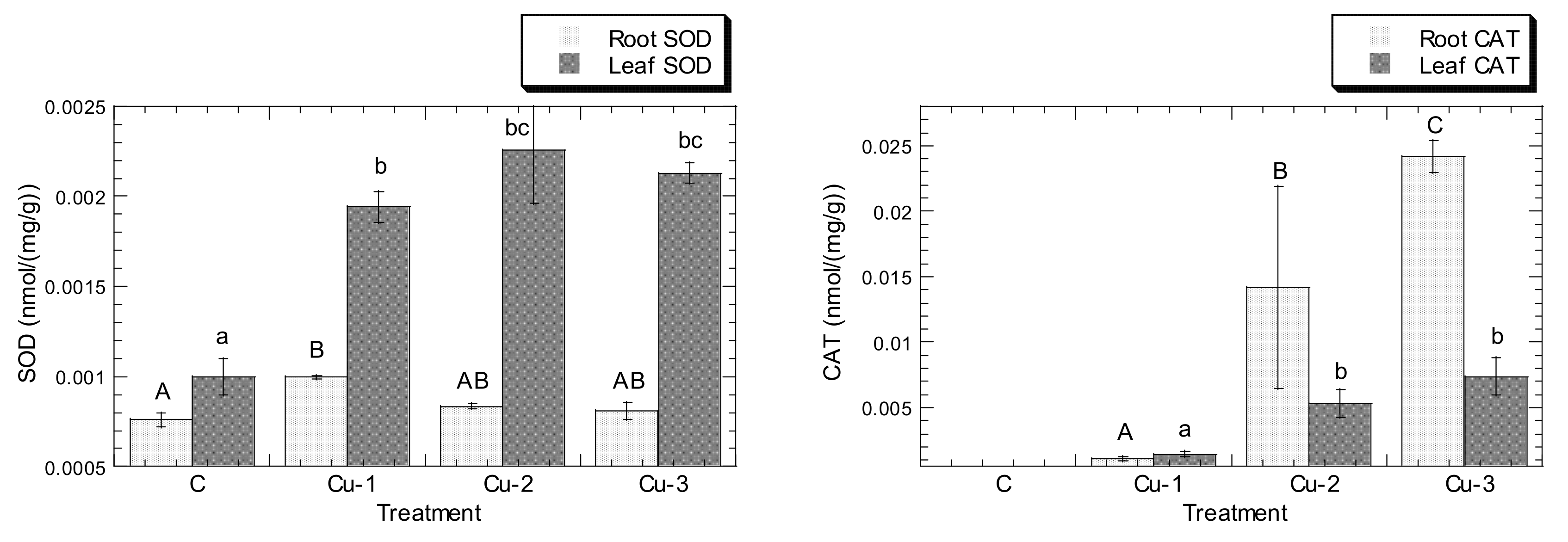
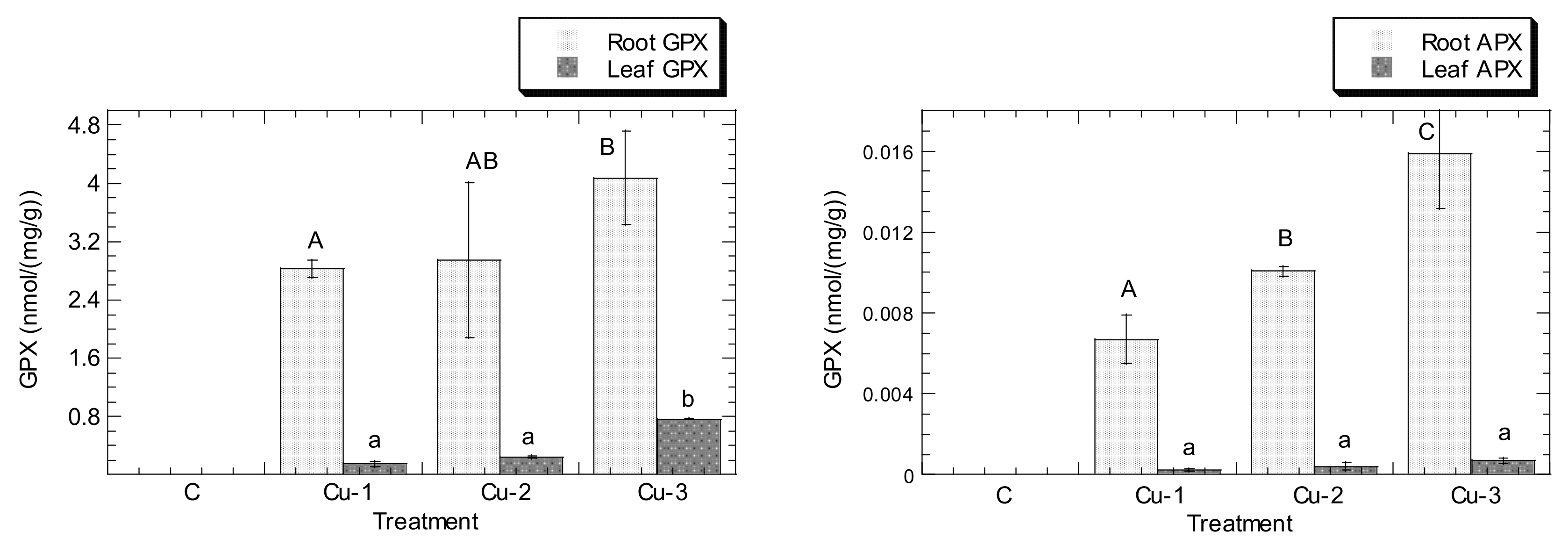
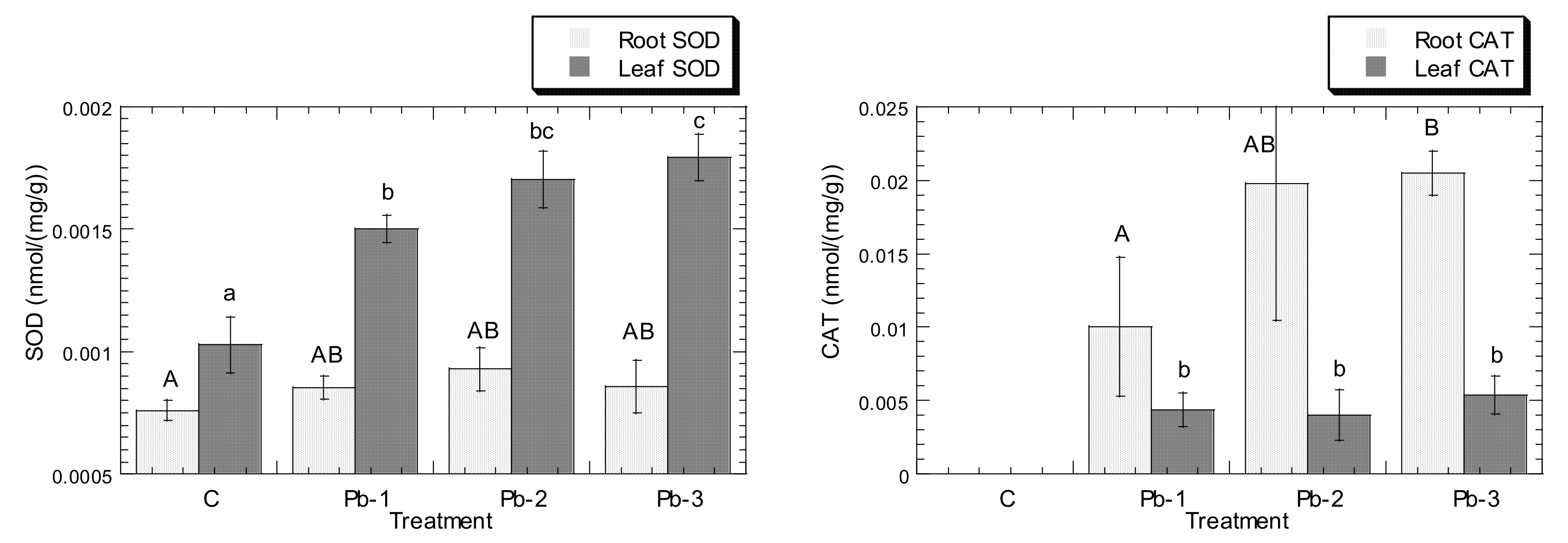
All graphical bar charts were plotted using KaleidaGraph (Version 3.08, Sygnergy Software, Eden Prairie, MN, USA). Statistical analysis was performed using the Student-Newman-Keuls (SNK) post hoc test and the Statistical Package for the Social Sciences (SPSS Statistics for Windows, version 17.0; SPSS Inc., Chicago, IL, USA).

3. Results

The effects of Cu and Pb stress on the accumulation of both metals in the roots and leaves of C. asiatica are presented in Figure 1.
For Cu in the leaves, the levels increased from 11.8 (control) to 31.1 mg/kg dry weight in Cu-3 treatment, with a significant increase (p < 0.05) at Cu-1, Cu-2, and Cu-3 when compared to the control. However, when the Cu stress was added with Pb exposure, the Cu levels in the leaves increased significantly (p < 0.05) to 50.9 mg/kg dry weight (41.9%) for Cu-2 and to 46.2 mg/kg dry weight (32.8%) for Cu-3. However, there was no significant (p > 0.05) change in Cu-1.
With respect to Cu in the roots, the levels increased from 13.7 (control) to 43.7 mg/kg dry weight in Cu-1 treatment, with a significant increase (p < 0.05) at Cu-1, Cu-2, and Cu-3 when compared to the control. However, when the Cu stress was added with Pb exposure, the Cu levels in the leaves increased significantly (p < 0.05) to 43.1 mg/kg dry weight (10.6%) for Cu-2 and to 58.5 mg/kg dry weight (26.9%) for Cu-3. However, there was no significant (p > 0.05) change in Cu-1. This shows that Cu and Pb acted synergistically to Cu accumulation in the leaves and roots at Cu-2, and Cu-3.
With regard to Pb in the leaves, the levels increased from 8.88 (control) to 25.1 mg/kg dry weight in Pb-3 treatment, with a significant increase (p < 0.05) at Pb-1, Pb-2, and Pb-3 when compared to the control. However, when the Pb stress was added with Cu exposure, the Pb levels in the leaves increased significantly (p < 0.05) to 36.3 mg/kg dry weight (34.3%) for Pb-2 and to 33.4 mg/kg dry weight (25.9%) for Pb-3. However, there was no significant (p > 0.05) change in Pb-1. This shows that Cu and Pb acted synergistically to Pb accumulation in the leaves at Pb-2 and Pb-3.
For Pb in the roots, the levels increased from 12.8 (control) to 45.8 mg/kg dry weight in Pb-3 treatment, with a significant increase (p < 0.05) at Pb-1, Pb-2, and Pb-3 when compared to the control. However, when the Pb stress was added with Cu exposure, there was no significant (p > 0.05) change in Pb concentration in the roots for Pb-1, Pb-2, and Pb-3.
The effects of Cu exposures on the AEA in the roots and leaves of C. asiatica are presented in Figure 2. Overall, the roots accumulated higher levels of Cu than those in the leaves that served as the control and three exposures of Cu. Higher percentages of APX, CAT, and GPX were recorded in the roots than those in the leaves for Cu-1, Cu-2, and Cu-3. By contrast, the SOD percentage was higher in the leaves than that in the roots. Overall, the increased levels of Cu exposure triggered the increment of APX, CAT, GPX, and SOD levels in the leaves and roots.
In the roots, when compared to the control, the SOD levels increased from 6.59 to 16.5% for Cu-1 and Cu-2, respectively, but decreased to 12.1% for Cu-3. The CAT levels increased from 8.33 to 183% for the three levels of Cu exposure. The GPX levels increased from 475 to 676% for the three levels of Cu exposure. The APX levels increased from 49.1 to 140% for the three levels of Cu exposure. In general, the most activity was found in GPX, followed by CAT, APX, and SOD.
In the leaves, when compared to the control, the SOD levels increased from 94.4 to 117% for Cu-1 and Cu-2, respectively, but decreased to 110% for Cu-3. The CAT levels increased from 4.17 to 20.8% for the three levels of Cu exposure. The GPX levels increased from 90.7 to 456% for the three levels of Cu exposure. The APX levels increased from 5.97 to 17.9% for the three levels of Cu exposure. In general, the most activity was found in GPX, followed by SOD, CAT, and APX.
The effects of Pb exposures on the AEA in the roots and leaves of C. asiatica are shown in Figure 3. Overall, the roots accumulated significantly (p < 0.05) higher levels of Pb than those in the leaves of the control in the three exposure levels of Pb. Similar to Cu exposure, higher percentages of CAT, GPX, and APX were recorded in the roots than those in the leaves for Pb-1, Pb-2, and Pb-3. This is in contrast to SOD, in which the SOD percentage was higher in the leaves than that in the roots. Similar to Cu exposure, the increased levels of Pb exposure also triggered the increment of APX, CAT, GPX, and SOD levels in both the leaves and roots.
In the roots, when compared to the control, the SOD levels increased from 16.5 to 28.6% for Pb-1 and Pb-2, respectively, but decreased to 27.5% for Pb-3. The CAT levels increased from 75 to 158% for the three levels of Pb exposure. The GPX levels increased from 1067 to 1338% for Pb-1 and Pb-2, respectively, but decreased to 929% for Pb-3. The APX levels increased from 17.6 to 73.0% for the three levels of Pb exposure. In general, the most activity was found in GPX, followed by CAT, APX, and lastly, SOD.
In the leaves, when compared to the control, the SOD levels increased from 50.0 to 69.4% for the three levels of Pb exposure. The CAT levels decreased from 12.5 to 10.4% for Pb-1 and Pb-2, respectively, but increased to 14.6% for Pb-3. The GPX levels increased from 76.7 to 251% for the three levels of Pb exposure. The APX levels increased from 11.9 to 14.9% for the three levels of Pb exposure. In general, the most activity was found in GPX, followed by SOD, APX, and CAT.
The effects of combinations of Cu and Pb exposure on the AEA in the roots and leaves of C. asiatica are presented in Figure 4.
Higher percentages of GPX, APX, CAT, and SOD were recorded in the roots than in the leaves for CuPb-1, CuPb-2 and CuPb-3. In the roots, when compared to the control, the SOD levels increased from 30.8 to 55.0% for the three levels of CuPb exposure. The CAT levels increased from 75 to 125% for the three levels of CuPb exposure. The GPX levels decreased from 622 to 568% for CuPb-1 and CuPb-2, respectively, but increased to 628% for CuPb-3. The APX levels increased from 45.3 to 109% for CuPb-1 and CuPb-2, respectively, but decreased to 101% for CuPb-3. In general, the most activity was found in GPX, followed by CAT, APX, and SOD.
In the leaves, when compared to the control, the SOD levels increased from 8.45 to 30.6% for the three levels of CuPb exposure. The CAT levels decreased from 16.7 to 14.6% for CuPb-1 and CuPb-2, respectively, but increased to 29.2% for CuPb-3. The GPX levels increased from 110 to 351% for the three levels of CuPb exposure. The APX levels increased from 2.99 to 18.9% for the three levels of CuPb exposure. In general, the most activity was found in GPX, followed by SOD, APX, and CAT.
Overall, the roots accumulated higher levels of Cu than those in the leaves for the control, CuPb-1, and CuPb-3, except for CuPb-2 exposure. The increased levels of Cu exposure triggered the increment of APX, CAT, GPX, and SOD levels in both the leaves and roots. Overall, the roots accumulated higher levels of Pb than those in the leaves of the control and in the three exposures of CuPb. The increased levels of CuPb exposure triggered the increment of APX, CAT, GPX, and SOD levels in both the leaves and roots.

4. Discussion

4.1. Higher Accumulations of Cu and Pb in the Roots Than in the Leaves

The present experimental exposure results confirmed the accumulation of Cu and Pb in C. asiatica collected from the field was always higher in the roots than that in the leaves. This indicates that the roots were a storage site for Cu and Pb, regardless of their different functions. This could be due to a higher surface area of root hairs than that of the leaves for the absorption of metals [14]. Roots are the initial part of a plant that would absorb metals from habitat hydroponic waterbody before distributing them to the other parts of the plant.
The main reason why there were higher levels of Cu and Pb in the roots of C. asiatica could be due to a series of processes involved in the metal accumulation of Centella plants, including root uptake and root-to-shoot transport. The bioavailable levels of Cu and Pb in the Hoagland solution were absorbed at the root surface and transported to the cellular membrane of the root cells. The uptake of Cu and Pb into roots happened mainly through the apoplastic pathway and symplastic pathway [27]. The apoplastic pathway involves passive diffusion, while the symplastic pathway involves active transport against electrochemical potential gradients and concentration across the plasma membrane [28].
After passing into root cells, Cu and Pb ions formed complexes with various chelators, such as organic acids. These complexes were then trapped in the intracellular symplastic compartments, such as vacuoles, or the extracellular space, such as apoplastic cellular walls [29]. The metal ions sequestered inside the vacuoles may have been transferred into the stele and were translocated to the shoots through xylem vessels from the root [30]. By means of the apoplast or symplast, the metal ions were transported and later distributed in leaves, where the metal ions were sequestered in the extracellular compartments, such as cell walls or plant vacuoles. This would prevent accumulation of free ions of Cu and Pb in cytosols [28,31]. The other explanation could be attributed to the significantly (p < 0.05) higher levels of CAT, GPX, and APX in the roots than those in the leaves for the Cu, Pb and combined Cu and Pb stress, which will be further discussed in Section 4.3.

4.2. Synergistic Effects of Cu and Pb Stress at Higher Metal-Level Exposures

The present study showed that Cu and Pb acted synergistically to Cu accumulation in the leaves and roots at Cu-2, and Cu-3. Besides, Cu and Pb acted synergistically to Pb accumulation in the leaves only at Pb-2, and Pb-3. The combination of Pb and Cu stress resulted in non-significant (p > 0.05) change in Pb concentration in the roots for Pb-1, Pb-2, and Pb-3.
The more sensitive and toxic nature of Cu investigated in the present study could have been reduced with the addition of Pb by looking at the fact that synergistical effect to Cu accumulation (higher accumulation of Cu) was found in the leaves and roots for Cu-2, and Cu-3. The fact that Cu is presumably more toxic than Pb was based on the accumulation of Cu concentrations in the Cu treatments, in which the Cu concentrations were almost similar to those in Pb treatments. The ratio of Pb:Cu exposures in the experiment was always as 2:1. However, the present experimental results indicate that the accumulation of Pb and Cu in the leaves and roots was not in the ratio of 2:1. Instead, the ratios always showed almost 1:1 in both the accumulations of Pb and Cu, whether in leaves or roots, regardless of the Pb:Cu exposure ratios as 2:1. An [32] compared the acute toxicities of Cu and Pb to Sorghum bicolor, Cucumis sativus, Triticum aestivum, and Zea mays. Based on the EC50 values, the author found that Cu was more toxic to the plants than Pb. Bioaccumulations of Pb and Cu were observed in all test species, and they were concentration-dependent. Therefore, the differences in the toxicities of Pb and Cu in C. asiatica should be taken into account in biomonitoring and ecological risk assessments. Although Cu is an essential micronutrient for plant growth, it can be more toxic than nonessential Pb to biota when an elevated level of Cu is present in the plant’s habitat [32].
Ong et al. (2013) reported that Pb and Zn acted synergistically to Zn accumulation in leaves but antagonistically in roots of C. asiatica under experimental hydroponic conditions. By using high levels (2000 ppm) of Cu and Pb exposure, Nicholls and Mal [33] revealed that the treatment of weed Lythrum salicaria with Cu or Pb significantly reduced the growth and survival of the plants, and there was no synergistic relationship between the metals because of the extreme toxicity of the concentrations of Cu and Pb used in their study. Since this synergistic relationship was found in single Pb or Cu and combined Pb and Cu exposures, the levels of Cu and Pb exposure investigated in the present study were not considered toxic but stressful. There were also no observable symptoms of toxicity in the plant leaves.
It is postulated that the translocation of the essential Cu to the leaves occurs when there is a metabolism requirement. However, the translocation of nonessential Pb could face a toxicity consequence [34]. Generally, the toxic concentrations of Pb for plants are defined in the range of 30–300 mg/kg dw, whereas the toxicity limits for Cu are in the range of 20–100 mg/kg dw [35]. Therefore, in the present study, the ranges of concentrations of Pb (<50 mg/kg dw in leaves and roots) and Cu (<60 mg/kg dw in leaves and roots) in C. asiatica were below the toxic thresholds for both metals. Therefore, the present study, which employed three exposure levels of Cu and Pb, was considered stress- rather than toxicity-inducing.

4.3. Cu and Pb Stress Triggered the Increment of APX, CAT, GPX, and SOD Levels in Both Leaves and Roots

The increased levels of AEA in roots and leaves indicate that both Cu and Pb become a stress (rather than a toxin) in low or high Cu and Pb exposures. This could have increased the ROS levels in the subcellular compartments of the roots and leaves of C. asiatica [4]. Manan et al. [36] reported that CAT and APX were significantly higher in the wild fern Nephrolepis biserrata from soils contaminated by Zn, Pb, and Cu, when compared to the control. However, the higher SOD activity in the contaminated fern was not significantly different from the uncontaminated fern.
After exposure to Cu and Pb stress, the most activity in the roots of C. asiatica was GPX, followed by CAT, APX, and SOD. In the leaves of C. asiatica, the most activity was GPX, followed by SOD, APX, and CAT. All of the above results indicate that GPX has a higher affinity for H2O2 than SOD, CAT, and APX. This shows that GPX may have a more important role in removing ROS molecules during Cu and Pb stress [7]. In a pot experiment, Nadgórska-Socha et al. [37] reported that GPX in V. faba is a good defensive tool against Cu and Pb exposure. GPX activity in both roots and leaves in response metal stress due to Cu and Pb exposure was induced to a greater degree than CAT, SOD, and APX when compared to the control. This implies that a GPX-mediated reaction was favourably adopted by C. asiatica to scavenge H2O2, rather than tAPX-, CAT-, or SOD-mediated reactions.
The SOD activity in C. asiatica resulting from metal exposure was found to be the lowest in the roots. Lamhamdi et al. [9] reported a rise of SOD activity in wheat seedlings under Pb exposure. The lowest SOD increment in response to metal stress in the present study was supported by the study of Manan et al. [36]. They reported that SOD, APX, and CAT levels were 34%, 93%, and 223%, respectively, higher than those in the control (uncontaminated site). This shows that SOD activity could not be induced to a very high degree (>100%) because it may not be necessary for the survival of C. asiatica under Cu and Pb stress conditions [38].
In the present study, the CAT activity increased when the plants were exposed to Cu and Pb. This can be explained by the fact that CAT activity can remove high concentrations of H2O2 as an adaptive mechanism [39]. Enzymes of the ascorbate–glutathione cycle are localized mainly in chloroplasts, cytoplasm, and other cellular organelles. They play an important role in combating oxidative stress [40]. Kafel et al. [41] reported a rise of CAT activity in the above-ground parts of Philadelphus coronarius grown in a polluted environment.
In the present study, APX activity increased to high levels at three different exposure levels to Cu and Pb. The increased activity of APX can effectively scavenge H2O2 to guard against oxidative damage. By using aquatic duckweed, Spirodela polyrhiza, Upadhyay and Panda [42] demonstrated that the GPX activity was significantly increased in the mixture of Zn and Cu Zn treatment when compared to a single Zn treatment.
Israr et al. [1] reported a significant increase in three enzymes, including SOD and APX, in Sesbania drummondii seedlings when they were exposed to different metal-level treatments. Upon analysis of the consequences of soil metal contamination on Vicia faba, Nadgórska-Socha et al. [37] recommended the use of GPX activity and proline levels as biomarkers. Generally, GPX activity was higher in metal-stressed plants as compared with the control. Guo et al. [43] reported elevated GPX and SOD activities in the leaves and roots of barley with higher accumulations of three metals, including Cu.
The enhancement of four antioxidative activities investigated in the present study indicates that the leaves and roots of C. asiatica treated with Cu, Pb, and a combination of both metals appeared to be promising biomarkers of Cu and Pb stress.

5. Conclusions

Higher accumulations of Cu and Pb were observed in the roots of C. asiatica than in the leaves. Synergistic effects of Cu and Pb stress were found at higher metal-level exposures, and Cu and Pb stress triggered the increment of APX, CAT, GPX, and SOD levels in the leaves and roots of C. asiatica. The higher levels of Cu and Pb, especially in the roots of C. asiatica, reflect contamination of the hydroponic media by these two metals. The increments of the four AEA indicate that C. asiatica responded to Cu and Pb stress by producing ROS. The AEA are a significant antioxidant defensive system against Cu and Pb stress in C. asiatica, rendering higher tolerance to the toxicity caused by Pb and Cu stress. Collectively, APX, CAT, GPX, and SOD are good biomarkers of Pb and Cu stress in the roots and leaves of C. asiatica.

Author Contributions

Conceptualization, C.K.Y.; formal analysis, K.W.W.; funding acquisition, C.K.Y.; investigation, G.H.O.; methodology, G.H.O., R.N. and C.K.Y.; project administration, C.K.Y. and W.S.T.; resources, W.S.T., K.W.W., W.H.C. and M.H.I.; supervision, C.K.Y.; validation, W.S.T.; writing—original draft, C.K.Y.; writing—review & editing, W.S.T., K.W.W., W.H.C., R.N., M.H.I., W.C., F.B.E., H.O., K.A.A.-M., S.A.A.-S., M.S., M.M., W.J.L. and C.F.Y. All authors have read and agreed to the published version of the manuscript.

Funding

The present study was supported financially by the Fundamental Research Grant Scheme (FRGS), [Vote No.: 5524953] by the Ministry of Higher Education, Malaysia.

Institutional Review Board Statement

Not applicable.

Informed Consent Statement

Not applicable.

Data Availability Statement

Not applicable.

Conflicts of Interest

The authors declare no conflict of interest.

References

  1. Israr, M.; Jewell, A.; Kumar, D.; Sahi, S.V. Interactive Effects of Lead, Copper, Nickel and Zinc on Growth, Metal Uptake and Antioxidative Metabolism of Sesbania drummondii. J. Hazard. Mater. 2011, 186, 1520–1526. [Google Scholar] [CrossRef] [PubMed]
  2. Meena, M.; Samal, S. Alternaria Host-Specific (HSTs) Toxins: An Overview of Chemical Characterization, Target Sites, Regulation and Their Toxic Effects. Toxicol. Rep. 2019, 6, 745–758. [Google Scholar] [CrossRef]
  3. Kohmoto, K.; Otani, H. Host Recognition by Toxigenic Plant Pathogens. Experientia 1991, 47, 755–764. [Google Scholar] [CrossRef]
  4. Mittler, R.; Vanderauwera, S.; Gollery, M.; Van Breusegem, F. Reactive Oxygen Gene Network of Plants. Trends Plant Sci. 2004, 9, 490–498. [Google Scholar] [CrossRef] [PubMed]
  5. Meng, Q.; Zou, J.; Jiang, W.; Liu, D. Effect of Cu2+ Concentration on Growth, Antioxidant Enzyme Activity and Malondialdehyde Content in Garlic (Allium sativum L.). Acta Biol. Cracoviensia Ser. Bot. 2007, 49, 95–101. [Google Scholar]
  6. Hegedüs, A.; Erdei, S.; Horváth, G. Comparative Studies of H2O2 Detoxifying Enzymes in Green and Greening Barley Seedlings under Cadmium Stress. Plant Sci. 2001, 160, 1085–1093. [Google Scholar] [CrossRef]
  7. Gupta, V.K.; Sharma, S.K. Plants as Natural Antioxidants. Nat. Prod. Radiance 2006, 5, 9. [Google Scholar]
  8. Verma, S.; Dubey, R.S. Lead Toxicity Induces Lipid Peroxidation and Alters the Activities of Antioxidant Enzymes in Growing Rice Plants. Plant Sci. 2003, 164, 645–655. [Google Scholar] [CrossRef]
  9. Lamhamdi, M.; Bakrim, A.; Aarab, A.; Lafont, R.; Sayah, F. Lead Phytotoxicity on Wheat (Triticum aestivum L.) Seed Germination and Seedlings Growth. Comptes Rendus Biol. 2011, 334, 118–126. [Google Scholar] [CrossRef]
  10. Tang, L.; Kwon, S.-Y.; Kim, S.-H.; Kim, J.-S.; Choi, J.S.; Cho, K.Y.; Sung, C.K.; Kwak, S.-S.; Lee, H.-S. Enhanced Tolerance of Transgenic Potato Plants Expressing Both Superoxide Dismutase and Ascorbate Peroxidase in Chloroplasts against Oxidative Stress and High Temperature. Plant Cell Rep. 2006, 25, 1380–1386. [Google Scholar] [CrossRef] [PubMed]
  11. Idris, F.N.; Mohd Nadzir, M. Comparative Studies on Different Extraction Methods of Centella asiatica and Extracts Bioactive Compounds Effects on Antimicrobial Activities. Antibiotics 2021, 10, 457. [Google Scholar] [CrossRef] [PubMed]
  12. Seevaratnam, V.; Banumathi, P.; Premalatha, M.R.; Sundaram, S.; Arumugam, T. Functional Properties of Centella asiatica (L.): A Review. Int. J. Pharm. Pharm. Sci. 2012, 4, 8–14. [Google Scholar]
  13. Sarumathi, A.; Anbu, S.; Saravanan, N. Antibacterial Activity of Centella asiatica (Linn.) Leaves. Int. J. Chem. Pharm. Sci. 2013, 1, 393–395. [Google Scholar]
  14. Yap, C.K.; Fitri, M.; Yazdani, M.; Tan, S. Effects of Metal-Contaminated Soils on the Accumulation of Heavy Metals in Different Parts of Centella asiatica: A Laboratory Study. Sains Malays. 2010, 39, 347–352. [Google Scholar]
  15. Ong, G.H.; Yap, C.K.; Maziah, M.; Tan, S. Synergistic and Antagonistic Effects of Zinc Bioaccumulation with Lead and Antioxidant Activities in Centella asiatica. Sains Malays. 2013, 42, 1549–1555. [Google Scholar]
  16. Ong, G.H.; Yap, C.K.; Marziah, M.; Tan, S. Accumulation of Heavy Metals and Antioxidative Enzymes of Centella asiatica in Relation to Metals of the Soils. Pertanika J. Trop. Agric. Sci. 2013, 36, 331–336. [Google Scholar]
  17. Ong, G.H.; Yap, C.K.; Maziah, M.; Tan, S.G. The Effect of Cu Exposure on the Bioaccumulation of Zn and Antioxidant Activities in Different Parts of Centella asiatica. Asian J. Microbiol. Biotechnol. Environ. Sci. 2011, 13, 387–392. [Google Scholar]
  18. Yap, C.K.; Ong, G.H.; Cheng, W.H.; Nulit, R.; Ali, K.; Al-Shami, S.A. Interactive effects of copper and lead on metal uptake and antoixidative metabolism of Centella asiatica under experimental hydroponic systems. In Advances in Hydroponics Research; Webster, D.J., Ed.; Nova Science Publishers: New York, NY, USA, 2017; pp. 57–72. [Google Scholar]
  19. Tang, Y.-T.; Qiu, R.-L.; Zeng, X.-W.; Ying, R.-R.; Yu, F.-M.; Zhou, X.-Y. Lead, Zinc, Cadmium Hyperaccumulation and Growth Stimulation in Arabis paniculata Franch. Environ. Exp. Bot. 2009, 66, 126–134. [Google Scholar] [CrossRef]
  20. Kopittke, P.M.; Blamey, F.P.C.; Asher, C.J.; Menzies, N.W. Trace Metal Phytotoxicity in Solution Culture: A Review. J. Exp. Bot. 2010, 61, 945–954. [Google Scholar] [CrossRef] [Green Version]
  21. Yang, X.; Long, X.; Ni, W.; Fu, C. Sedum Alfredii H: A New Zn Hyperaccumulating Plant First Found in China. Chin. Sci. Bull. 2002, 47, 1634–1637. [Google Scholar] [CrossRef]
  22. Mishra, S.; Srivastava, S.; Tripathi, R.D.; Govindarajan, R.; Kuriakose, S.V.; Prasad, M.N.V. Phytochelatin Synthesis and Response of Antioxidants during Cadmium Stress in Bacopa monnieri L. Plant Physiol. Biochem. PPB 2006, 44, 25–37. [Google Scholar] [CrossRef]
  23. Hemeda, H.M.; Klein, B.P. Effects of Naturally Occurring Antioxidants on Peroxidase Activity of Vegetable Extracts. J. Food Sci. 1990, 55, 184–185. [Google Scholar] [CrossRef]
  24. Nakano, Y.; Asada, K. Hydrogen Peroxide Is Scavenged by Ascorbate-Specific Peroxidase in Spinach Chloroplasts. Plant Cell Physiol. 1981, 22, 867–880. [Google Scholar] [CrossRef]
  25. Beauchamp, C.; Fridovich, I. Superoxide Dismutase: Improved Assays and an Assay Applicable to Acrylamide Gels. Anal. Biochem. 1971, 44, 276–287. [Google Scholar] [CrossRef]
  26. Aebi, H. Catalase in Vitro. Methods Enzymol. 1984, 105, 121–126. [Google Scholar] [CrossRef] [PubMed]
  27. Peer, W.A.; Baxter, I.R.; Richards, E.L.; Freeman, J.L.; Murphy, A.S. Phytoremediation and hyperaccumulator plants. In Molecular Biology of Metal Homeostasis and Detoxification: From Microbes to Man; Tamas, M.J., Martinoia, E., Eds.; Topics in Current Genetics; Springer: Berlin/Heidelberg, Germany, 2006; pp. 299–340. ISBN 978-3-540-31719-7. [Google Scholar]
  28. Yan, A.; Wang, Y.; Tan, S.N.; Mohd Yusof, M.L.; Ghosh, S.; Chen, Z. Phytoremediation: A Promising Approach for Revegetation of Heavy Metal-Polluted Land. Front. Plant Sci. 2020, 11, 359. [Google Scholar] [CrossRef] [PubMed]
  29. Ali, H.; Khan, E.; Sajad, M.A. Phytoremediation of Heavy Metals—Concepts and Applications. Chemosphere 2013, 91, 869–881. [Google Scholar] [CrossRef]
  30. Thakur, S.; Singh, L.; Wahid, Z.A.; Siddiqui, M.F.; Atnaw, S.M.; Din, M.F.M. Plant-Driven Removal of Heavy Metals from Soil: Uptake, Translocation, Tolerance Mechanism, Challenges, and Future Perspectives. Environ. Monit. Assess. 2016, 188, 206. [Google Scholar] [CrossRef]
  31. Tong, Y.P.; Kneer, R.; Zhu, Y.G. Vacuolar Compartmentalization: A Second-Generation Approach to Engineering Plants for Phytoremediation. Trends Plant Sci. 2004, 9, 7–9. [Google Scholar] [CrossRef]
  32. An, Y.-J. Assessment of Comparative Toxicities of Lead and Copper Using Plant Assay. Chemosphere 2006, 62, 1359–1365. [Google Scholar] [CrossRef]
  33. Nicholls, A.M.; Mal, T.K. Effects of Lead and Copper Exposure on Growth of an Invasive Weed, Lythrum salicaria L. (Purple Loosestrife). Ohio J. Sci. 2003, 103, 129–133. [Google Scholar]
  34. Alloway, B.J. Soil Factors Associated with Zinc Deficiency in Crops and Humans. Environ. Geochem. Health 2009, 31, 537–548. [Google Scholar] [CrossRef]
  35. Kabata-Pendias, A.; Pendias, H. Trace Elements in Soils and Plants, 3rd ed.; CRC Press: Boca Raton, FL, USA, 2001; ISBN 978-0-8493-1575-6. [Google Scholar]
  36. Manan, F.A.; Mamat, D.D.; Samad, A.A.; Ong, Y.S.; Ooh, K.F.; Chai, T.T. Heavy Metal Accumulation and Antioxidant Properties of Nephrolepis biserrata Growing in Heavy Metal-Contaminated Soil. Glob. NEST J. 2015, 17, 544–554. [Google Scholar]
  37. Nadgórska-Socha, A.; Kafel, A.; Kandziora-Ciupa, M.; Gospodarek, J.; Zawisza-Raszka, A. Accumulation of Heavy Metals and Antioxidant Responses in Vicia faba Plants Grown on Monometallic Contaminated Soil. Environ. Sci. Pollut. Res. Int. 2013, 20, 1124–1134. [Google Scholar] [CrossRef]
  38. Halliwell, B. Reactive Species and Antioxidants. Redox Biology Is a Fundamental Theme of Aerobic Life. Plant Physiol. 2006, 141, 312–322. [Google Scholar] [CrossRef] [Green Version]
  39. Reddy, A.M.; Kumar, S.G.; Jyothsnakumari, G.; Thimmanaik, S.; Sudhakar, C. Lead Induced Changes in Antioxidant Metabolism of Horsegram (Macrotyloma uniflorum (Lam.) Verdc.) and Bengalgram (Cicer arietinum L.). Chemosphere 2005, 60, 97–104. [Google Scholar] [CrossRef] [PubMed]
  40. Mhamdi, A.; Queval, G.; Chaouch, S.; Vanderauwera, S.; Van Breusegem, F.; Noctor, G. Catalase Function in Plants: A Focus on Arabidopsis Mutants as Stress-Mimic Models. J. Exp. Bot. 2010, 61, 4197–4220. [Google Scholar] [CrossRef] [PubMed] [Green Version]
  41. Kafel, A.; Nadgórska-Socha, A.; Gospodarek, J.; Babczyńska, A.; Skowronek, M.; Kandziora, M.; Rozpedek, K. The Effects of Aphis fabae Infestation on the Antioxidant Response and Heavy Metal Content in Field Grown Philadelphus coronarius Plants. Sci. Total Environ. 2010, 408, 1111–1119. [Google Scholar] [CrossRef] [PubMed]
  42. Upadhyay, R.; Panda, S.K. Zinc Reduces Copper Toxicity Induced Oxidative Stress by Promoting Antioxidant Defense in Freshly Grown Aquatic Duckweed Spirodela polyrhiza L. J. Hazard. Mater. 2010, 175, 1081–1084. [Google Scholar] [CrossRef]
  43. Guo, T.R.; Zhang, G.P.; Zhang, Y.H. Physiological Changes in Barley Plants under Combined Toxicity of Aluminum, Copper and Cadmium. Colloids Surf. B Biointerfaces 2007, 57, 182–188. [Google Scholar] [CrossRef]
Figure 1. Effects of Cu and Pb stress on the accumulation of Cu and Pb concentrations (mean ± SD, mg/kg dry weight) in the roots and leaves of Centella asiatica grown in an experimental hydroponic condition. Note: C = Control. Different uppercase and lowercase letters indicate the significant difference (SNK test, p < 0.05) in mean values.
Stresses 01 00018 g001
Figure 2. Effects of Cu exposures on the antioxidant enzyme activities (mean ± SD, nmol/(mg/g)) in the roots and leaves of Centella asiatica grown in an experimental hydroponic condition. Note: C = Control. The control values (C) of CAT, GPX, and APX were blanked by subtracting from those of the particular enzymes. For the concentrations of SOD, CAT, and APX, their actual values were multiplied by 1000. Different uppercase and lowercase letters indicate the significant difference (SNK test, p < 0.05) in mean values.
Stresses 01 00018 g002aStresses 01 00018 g002b
Figure 3. Effects of Pb exposure on the antioxidant enzyme activities (mean ± SD, nmol/(mg/g)) in the roots and leaves of Centella asiatica grown in an experimental hydroponic condition. Note: C = Control. The control values (C) of CAT, GPX, and APX were blanked by subtracting from those of the particular enzymes. For the concentrations of SOD, CAT, and APX, their actual values were multiplied by 1000. Different uppercase and lowercase letters indicate the significant difference (SNK test, p < 0.05) in mean values.
Stresses 01 00018 g003aStresses 01 00018 g003b
Figure 4. Effects of combinations of Cu and Pb exposure on the antioxidant enzyme activities (mean ± SD, nmol/(mg/g)) in the roots and leaves of Centella asiatica grown in an experimental hydroponic condition. Note: C = Control. The control values (C) of CAT, GPX, and APX were blanked by subtracting from those of the particular enzymes. For the concentrations of SOD, CAT, and APX, their actual values were multiplied by 1000. Different uppercase and lowercase letters indicate the significant difference (SNK test, p < 0.05) in mean values.
Stresses 01 00018 g004aStresses 01 00018 g004b
Table 1. Experimental hydroponic treatments of Cu and Pb concentrations (mg/L) investigated in the present study.
NoTreatmentsMetal Concentrations
1ControlNo metals added
2Cu-1Cu (0.10)
3Cu-2Cu (0.20)
4Cu-3Cu (0.30)
5Pb-1Pb (0.20)
6Pb-2Pb (0.40)
7Pb-3Pb (0.60)
8CuPb-1Cu (0.10) + Pb (0.20)
9CuPb-2Cu (0.20) + Pb (0.40)
10CuPb-3Cu (0.30) + Pb (0.60)
Publisher’s Note: MDPI stays neutral with regard to jurisdictional claims in published maps and institutional affiliations.

