Communication

Structure, Stability and Binding Properties of Collagen-Binding Domains from Streptococcus mutans

1
Department of Biomolecular Chemistry, Kyoto Prefectural University, Kyoto 606-8522, Kyoto, Japan
2
Department of Anatomy and Cell Biology, Osaka Medical and Pharmaceutical University, Takatsuki 569-8686, Osaka, Japan
3
Department of Bioscience and Biotechnology, Fukui Prefectural University, Fukui 910-1195, Fukui, Japan
4
Department of Biotechnology, College of Life Sciences, Ritsumeikan University, Kusatsu 525-8577, Shiga, Japan
5
Department of Pediatric Dentistry, Graduate School of Biomedical and Health Sciences, Hiroshima University, Hiroshima 734-8553, Hiroshima, Japan
6
Department of Pediatric Dentistry, Osaka University Graduate School of Dentistry, Suita 565-0871, Osaka, Japan
*
Author to whom correspondence should be addressed.
These authors contributed equally to this work.
Chemistry 2023, 5(3), 1911-1920; https://doi.org/10.3390/chemistry5030130
Submission received: 12 June 2023 / Revised: 24 July 2023 / Accepted: 25 August 2023 / Published: 1 September 2023
(This article belongs to the Section Crystallography)

Abstract

:
Collagen-binding proteins (CBP), Cnm and Cbm, from Streptococcus mutans are involved in infective endocarditis caused by S. mutans because of their collagen-binding ability. In this study, we focused on the collagen-binding domain (CBD), which is responsible for the collagen-binding ability of CBP, and analyzed its structure, binding activity, and stability using CBD domain variants. The CBD consists of the N1 domain, linker, N2 domain, and latch (N1-N2~) as predicted from the amino acid sequences. The crystal structure of the Cnm/CBD was determined at a 1.81 Å resolution. N1_linker_N2 forms a ring structure that can enfold collagen molecules, and the latch interacts with N1 to form a ring clasp. N1 and N2 have similar immunoglobulin folds. The collagen-binding activities of Cbm/CBD and its domain variants were examined using ELISA. N1-N2~ bound to collagen with KD = 2.8 μM, and the latch-deleted variant (N1-N2) showed weaker binding (KD = 28 μM). The linker-deleted variant (N1N2~) and single-domain variants (N1 and N2) showed no binding activity, whereas the domain-swapped variant (N2-N1~) showed binding ability, indicating that the two N-domains and the linker are important for collagen binding. Thermal denaturation experiments showed that N1-N2 was slightly less stable than N1-N2~, and that N2 was more stable than N1. The results of this study provide a basis for the development of CBD inhibitors and applied research utilizing their collagen-binding ability.

1. Introduction

Approximately 700 bacterial species are present in the human oral cavity [1]. Oral bacteria invade the blood vessels via bleeding during dental treatment and brushing [2,3]. In most healthy individuals, oral bacteria in blood vessels are cleared by the immune system; however, in patients with underlying diseases, oral bacteria in blood vessels may contribute to disease exacerbation [4]. Streptococcus mutans, a significant contributor to tooth decay, has been implicated in infective endocarditis and its complications, such as cerebral hemorrhage [5,6]. In particular, it has been revealed that special S. mutans expressing collagen-binding proteins (CBP) on the cell surface exhibit high pathogenicity [7,8]. Cnm and Cbm have been identified as CBPs in S. mutans, and are considered to be involved in these diseases [9,10]. It is thought that S. mutans with intravascular CBP adheres to the exposed collagen molecules at the site of vascular injury via CBP, inhibiting platelet aggregation and exacerbating bleeding [11]. Therefore, it is important to understand the molecular properties of CBPs to prevent and treat diseases associated with S. mutans.
Based on their amino acid sequences, Cnm and Cbm consist of a collagen-binding domain (CBD), repeating region, B-repeat, and LPXTG motif for cell wall adhesion [9,10]. The CBD is formed by the N1 domain, linker, N2 domain, and latch (Figure S1 in Supplementary Materials). The amino acid sequence similarity between Cnm/CBD and Cbm/CBD is 87%. In this study, we focused on CBD, and the N1 domain, linker, N2 domain, and latch which were denoted by N1, -, N2, and ~, respectively. Thus, the full length of the CBD was N1-N2~. Cnm and Cbm CBDs were produced using Escherichia coli and crystallized. Consequently, the Cnm/CBD crystal structure was obtained. Collagen-binding activity and thermal stability measurements were performed using Cbm/CBD and its domain variants. The results of this study will serve as basic data for the development of collagen-binding inhibitors and their applications in research.

2. Materials and Methods

2.1. Expression and Purification

pET-42a(+) vectors containing cnm and cbm were previously constructed [7,12,13]. The expression vector of Cnm/CBD was constructed from the pET-42a(+) vector containing cnm. The cDNA of Cbm/CBD was re-cloned into pET-28a(+) with the original NdeI and XhoI sites, and the resulting vector was used as a template to construct mutant expression vectors. CBDs and the variant regions (residue numbers) are shown in Table 1. The resultant plasmids were transformed into E. coli BL21-CodonPlus(DE3)-RIL. The E. coli organisms were grown in 2xYT medium containing 30 mg L−1 kanamycin at 37 °C up to OD600 = 0.5, and protein production was induced by the addition of IPTG (final concentration of 1 mM) and further cultured overnight at 18 °C. After that, cells were collected and were suspended in 50 mM Tris-HCl buffer, which was ultrasonicated on ice. Supernatants were obtained and purified using a Ni-NTA affinity column and 16/60 Superdex 200 column. The purity of the proteins was analyzed by SDS-PAGE on a 12% (w/v) polyacrylamide gel, followed by staining with Coomassie Brilliant Blue. The purified proteins were shown as single bands on SDS-PAGE.

2.2. Crystallization and Structure Determination

Purified Cnm/CBD and Cbm/CBD were concentrated to 25 mg mL−1 in 50 mM Tris-HCl buffer. The crystallization conditions were initially screened using crystallization kits from Hampton Research (Alise Viejo, CA, USA; Crystal Screen I and II) and Emerald Biostructures (Bainbridge Island, WA, USA; Wizard I, II, III and IV) by the sitting-drop vapor-diffusion method at 4 and 20 °C. The Cnm/CBD crystals appeared after three days in Wizard III No. 30 (25% PEG4000, 170 mM ammonium sulfate, 15% glycerol) at 20 °C. To improve the crystal quality, the crystallization conditions were further optimized. Diffraction-quality crystals were grown in 22% PEG6000, 100 mM ammonium sulfate, 10% glycerol at 20 °C with 14 mg mL−1 of the protein concentration. Unfortunately, no Cbm/CBD crystals were obtained.
Diffraction data of Cnm/CBD were collected at a wavelength of 1.0 Å at −100 °C (173 K) at beam line BL41XU in SPring-8 (Hyogo, Japan) and were processed with HKL2000 [14]. The structure was determined by the molecular replacement method using MOLREP in the CCP4 program suite [15]. The structure of Staphylococcus aureus Cna/CBD (PDB: 2F68) [16] was used as a starting model as is. Structural refinement was performed using REFMAC of the CCP4 suite [15] and the model was corrected using COOT [17]. Water molecules were added using COOT. Data collection and refinement statistics are shown in Table 2. The figures were prepared using PyMol (http://www.pymol.org (accessed on 1 June 2023)).

2.3. Collagen-Binding Assay

The collagen-binding activity was measured by ELISA. Briefly, Nunc MaxiSorp plates (Thermo Fisher Scientific, Tokyo, Japan) were coated with 1 μg of bovine dermis type I collagen (Nippi, Tokyo, Japan) in phosphate buffer in each well overnight at 4 °C. Wells were then blocked with 2% bovine serum albumin in PBS. After washing with PBS containing 0.05% Tween, protein samples (0–40 μM/0.1% BSA in PBS) were added to the wells and incubated for three hours at 37 °C. Wells were then washed with PBS-T and incubated with anti-His-tag mAb-HRP-DirecT (MBL, Tokyo, Japan) diluted 1/10,000 with 0.1% BSA in PBS-T for one hour at 37 °C. Peroxidase substrate (TMB) and peroxide solution (H2O2) (Thermo Fisher Scientific) were reacted for five minutes at room temperature as a chromogenic substrate solution, and 50 µL/well of 2 M H2SO4 was added to terminate the reaction. After a 30 min colorimetric reaction, the absorption at 450 nm was measured. Binding curves were generated using SigmaPlot, fitting the data to according the following equation:
y = B m a x · x K D + x
where y represents the measured absorption at 450 nm, Bmax represents the calculated maximal amplitude of the curve, x denotes the protein concentration, and KD is the concentration that gives one-half of the shift between maximum and minimum readings. Data are expressed as the mean ± SD of triplicate experiments.

2.4. Thermal Denaturation

Thermal denaturation was examined by CD monitoring at 218 nm. CD measurements were carried out on a J-725 automatic spectropolarimeter. The optical path length was 2 mm. The protein concentration was 0.17 mg mL−1 in 1 M GdnHCl in PBS. All experiments were carried out at a scan rate of 1 °C min−1. The mean molecular ellipticity, θ, in units of degrees square centimeters per decimole, was used. A nonlinear least-squares analysis was used to fit the data to
y = b n + a n T + b u + a u T   e x p H m R T T T m T m 1 + e x p H m R T T T m T m
where y is the observed CD signal at a given temperature [T], bn is the CD signal for the native state, bu is the CD signal for the unfolded state, an is the slope of the pretransition of the baseline, au is the slope of the post-transition of the baseline, ΔHm is the enthalpy of unfolding at the transition midpoint temperature (Tm), and R is the gas constant. Curve fitting was performed using SigmaPlot.

3. Results and Discussion

3.1. Structure of Cnm/CBD

The structure of Cnm/CBD was determined at a 1.81 Å resolution (Figure 1A). The electron density in the Cnm/CBD linker region was poor and the region could not be modeled, probably because of structural disorders. For Cna/CBD, the linker structure was not determined for the same reason [16]. The amino acid sequences of Cnm/CBD and Cna/CBD linkers are similar (Figure S1). Overlaying the structures of Cnm/CBD and Cna/CBD results in Figure 1B revealed that the structures of Cnm and Cna CBDs are very similar (RMSD = 1.213 Å). Here, the amino acid sequence similarity between Cnm/CBD and Cna/CBD is 54%. However, the position of the loop near the linker within the N1 domain differed. Therefore, the structures of Cnm/CBD and Cna/CBD-collagen complex (PDB:2F6A) [16] were superimposed (Figure 1C). As a result, it was found that Cnm/CBD and Cna/CBD in the Cna/CBD-collagen complex had similar structures (RMSD = 1.146 Å). This result suggests that the conformation of the Cnm/CBD in this crystal structure was similar to the collagen bound conformation of Cna/CBD.
The binding mechanism of collagen has been predicted from the Cna/CBD–collagen complex structure, and is known as the “Collagen Hug” model [16,18,19]. In this model, the N2 domain of CBD first binds to collagen, followed by the interaction of the N1-N2 linker with collagen, followed by binding between the N1 domain and collagen. Finally, an interaction occurs between the C-terminal latch of the N2 and N1 domains, completing the binding between CBD and collagen. As mentioned above, the structure of Cnm/CBD resembles that of Cna/CBD. In the Cnm/CBD structure, N1_linker_N2 forms a ring that can enfold collagen molecules, and the latch interacts with the N1 domain to form a ring clasp (Figure 1A). Therefore, Cnm/CBD may also bind to collagen in the collagen hug model. Furthermore, a model structure of Cbm/CBD was constructed by SWISS-MODEL (https://swissmodel.expasy.org (accessed on 24 July 2023)) based on the structure of Cnm/CBD (Figure S2 in Supplementary Materials). The structures of Cnm/CBD and model Cbm/CBD are very similar (RMSD = 0.093 Å), suggesting that Cbm/CBD, like Cnm/CBD, also binds to collagen by the collagen hug model.
Based on the structure of the Cna/CBD–collagen complex, an interaction site with collagen was reported, and the sites of interaction with collagen of Cnm/CBD and Cna/CBD were compared. Figure 1D shows the site thought to interact with collagen in the early stages of the collagen hug model. This is the boundary of the N2 domain with the linker. Y175 and F191 interact with proline in collagen via stacking. Y175 and F191 are also conserved in Cnm, Cbm, and Enterococcus faecalis Ace [18] CBDs. The conformations of Y175 and F191 in Cnm/CBD are similar to those in Cna/CBD, and this region is considered an important site for binding. On the other hand, N193, N223 and N278, which are thought to interact with collagen in Cna/CBD, are not conserved in Cnm/CBD (N193, D223 and M275) or Cbm/CBD (M194, D224 and V280), and further studies are required regarding the contribution of these residues to binding.
The N1 and N2 domains have similar immunoglobulin folds. Although there is no amino acid sequence homology between the N1 and N2 domains, their structures are relatively similar (RMSD = 6.65 Å), as shown in Figure 1E. To understand the structural collagen-binding properties of CBD, it is necessary to clarify the roles of the N-domain immunoglobulin fold, linker, and latch. Accordingly, domain variants of Cbm/CBD were constructed and their binding activity and stability were measured as described below.

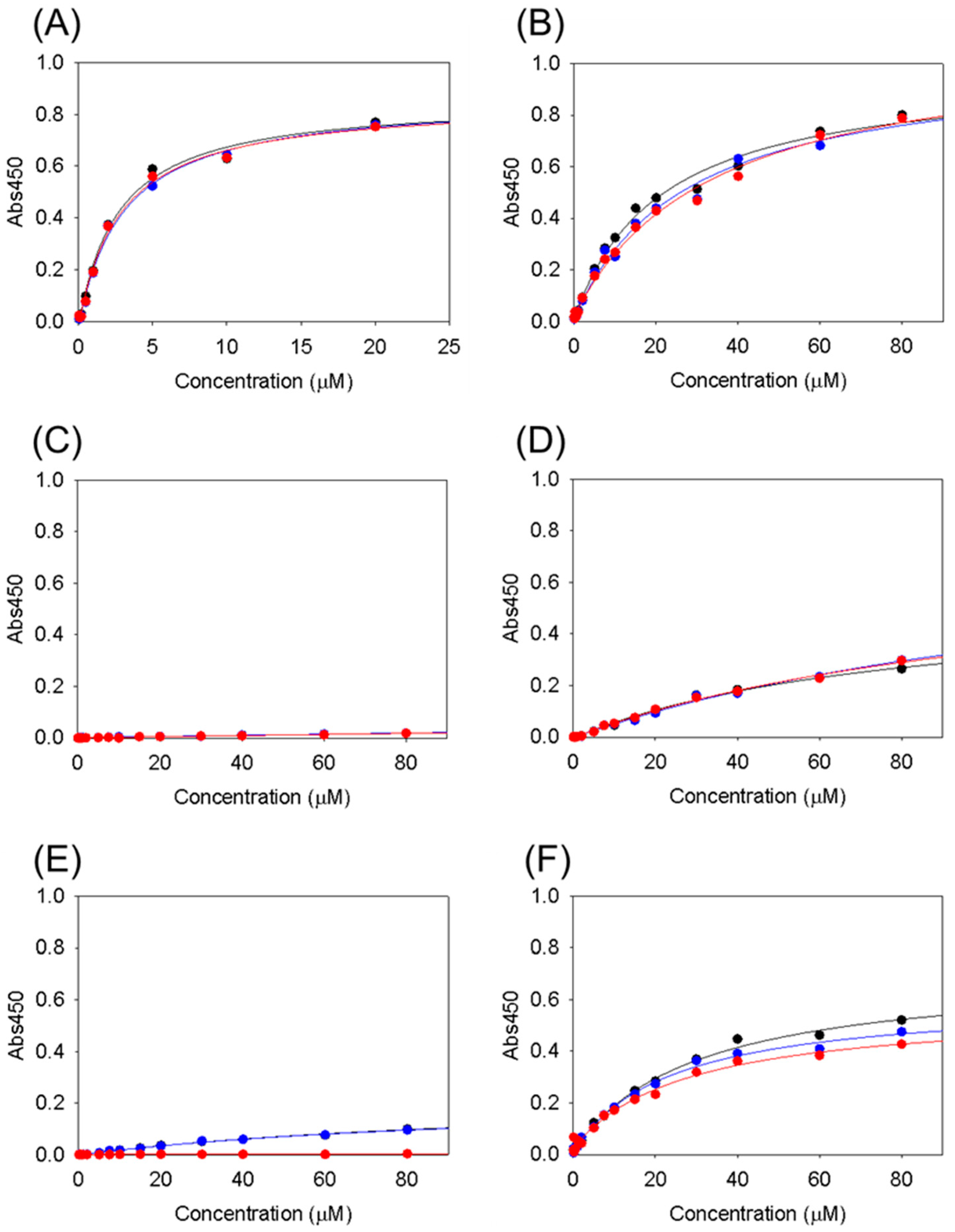
3.2. Collagen-Binding Activity of Cbm/CBD and Its Domain Variants

The binding activity of Cbm/CBD and its domain variants to type I collagen was measured using ELISA (Figure 2). The binding activity was evaluated by calculating the KD values (Table 3). Full-length CBD (N1-N2~) showed micromolar-order binding activity; however, several domain mutants lost binding activity.
First, the latch was verified. Cbm/CBD (N1-N2~) bound to collagen with a KD = 2.8 μM, and the latch-deleted variant (N1-N2) showed weaker binding (KD = 28 μM). This result indicates that the latch is not necessary for binding to collagen but enhances binding. In other words, the latch strengthens binding to collagen by connecting the N2 domain to the N1 domain.
Next, the roles of the two N-domains and the linkers connecting them were examined. The linker-deleted variant (N1N2~) and single-domain variants (N1 and N2) showed no binding activity. Structural analysis suggested the importance of binding between the N2 domain and collagen, as described above. However, the N2 domain alone could not bind, suggesting the need to wrap the collagen with the linker followed by the N1 domain. Therefore, we created a variant (N2-N1~) in which the N1 and N2 domains were exchanged. The N1 and N2 domains had an immunoglobulin fold and similar structures. Surprisingly, the domain-swapped variant (N2-N1~) had binding ability (KD = 27 μM), implying that the two N-domains and the linker are important for collagen binding. In the domain-swapped variant N2-N1~ structure, there is no latch interaction site in the N2 domain; therefore, the latch following the N1 domain cannot interact with the N2 domain, and there was no specific interaction with collagen by Y175 and F191 in the N2 domain, as described above. In addition, the binding activity of N2-N1~ was similar to that of N1-N2. These results indicate that the morphology of the two N-domains and the linker connecting them are required for collagen binding.

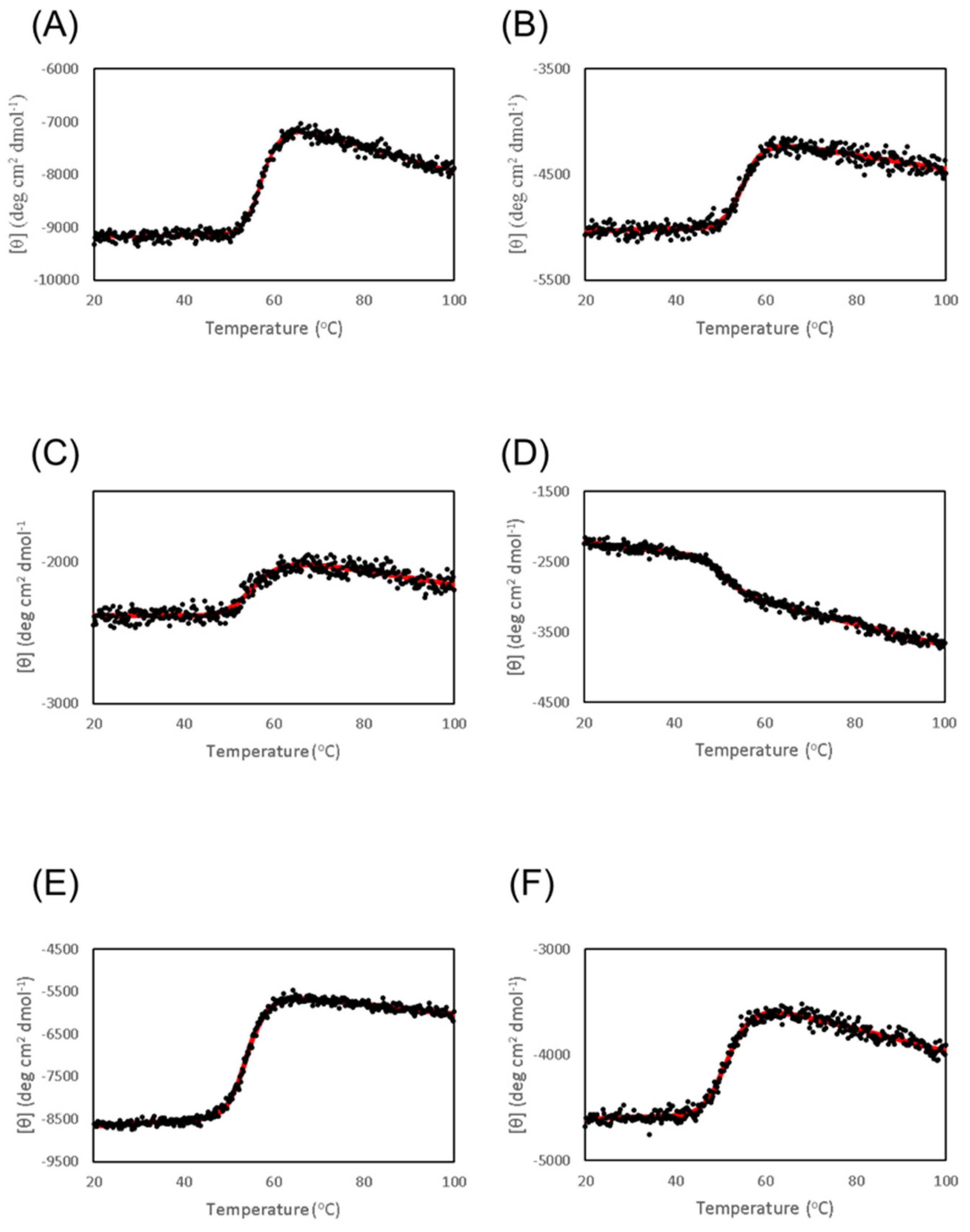
3.3. Thermal Stability of Cbm/CBD and Its Domain Variants

To determine the stability of each CBD domain, thermal denaturation experiments on Cbm/CBD and its domain variants were performed using CD (Figure 3). The thermal denaturation curve of Cbm/CBD showed a single step denaturation with a Tm value of 57.6 °C (Table 4), and thus failing to assess the stability of the N1 and N2 domains. Therefore, we performed denaturation experiments on the N1 and N2 domain variants and found that the denaturation curve of the N2 variant showed a similar change to that of N1-N2~, whereas the change in the CD signal of the N1 variant was different from those of the N1-N2~ and N2 variants, and the amount of change was also smaller. This result suggests that during N1-N2 denaturation, N2 domain denaturation was predominantly observed. Further verification of this phenomenon using other detection methods would be needed. The Tm for the N1 variant was 50.7 °C, while that for the N2 variant was 54.2 °C, indicating that the N2 domain is more stable than the N1 domain.
The latch-deficient variant (N1-N2) was slightly less stable than the N1-N2 variant. This indicates that the latch contributes to the stabilization of the CBD by interacting with the N1 domain. The Tm values of the linker-deleted variant (N1N2~) and the domain-swapped variant (N2-N1~) were 56.0 and 51.9 °C, respectively, which may reflect denaturation of the N2 domain.

4. Conclusions

In this study, to understand the binding mechanism and interaction mode of Cnm and Cbm (CBDs) with collagen, we performed structural analyses, binding assays, and stability measurements of the CBD and domain variants. Structural analysis of Cnm/CBD revealed the formation of a ring structure that enclosed the collagen molecules. A comparison with the Cna/CBD–collagen complex suggested the collagen hug model and an important site for the N2 domain in collagen binding. Binding assays using domain variants showed that the N-domain_linker_N-domain configuration is required for collagen binding. Thermal denaturation experiments revealed that the N2 domain is more stable than the N1 domain. The results of the structural and stability analyses indicated that the N2 domain plays a slightly more important role in the structure than the N1 domain in the CBD. In contrast, the importance of the ring morphology formed by the two N-domains and the linker was demonstrated with respect to function. In the future, the basic knowledge obtained here will be effectively utilized for the development of Cnm and Cbm inhibitors and for applied research that utilizes the collagen-binding ability of CBDs.

Supplementary Materials

The following supporting information can be downloaded at: https://www.mdpi.com/article/10.3390/chemistry5030130/s1, Figure S1: The amino acid sequence alignments of the CBDs of Cnm, Cbm, and Can; Figure S2: The model structure of Cbm/CBD.

Author Contributions

Conceptualization, A.H. and K.T.; Methodology, A.H., A.M., S.-i.T., T.Y. and H.M. (Hiroyoshi Matsumura); Investigation, A.N. and H.M. (Hikaru Matsui); Resources, R.N. and K.N.; Writing—Original Draft Preparation, K.T.; Writing—Review and Editing, all authors; Supervision, K.T.; Project Administration, A.H. and S.-i.T.; Funding Acquisition, A.H. and S.-i.T. All authors have read and agreed to the published version of the manuscript.

Funding

This study was supported by JSPS KAKENHI, Grant Numbers JP15K15749, JP18K09735, JP22K10233, and JP21K05386.

Data Availability Statement

Crystallographic data for Cnm/CBD has been deposited at the PBD under 8JL8.

Conflicts of Interest

The authors declare no conflict of interest.

References

  1. Aas, J.A.; Paster, B.J.; Stokes, L.N.; Olsen, I.; Dewhirst, F.E. Defining the normal bacterial flora of the oral cavity. J. Clin. Microbiol. 2005, 43, 5721–5732. [Google Scholar] [CrossRef] [PubMed]
  2. Debelian, G.J.; Olsen, I.; Tronstad, L. Systemic diseases caused by oral microorganisms. Endod. Dent. Traumatol. 1994, 10, 57–65. [Google Scholar] [CrossRef] [PubMed]
  3. Roberts, G.J.; Lucas, V.S.; Omar, J. Bacterial endocarditis and orthodontics. J. R. Coll. Surg. Edinb. 2000, 45, 141–145. [Google Scholar] [PubMed]
  4. Seymour, R.A.; Lowry, R.; Whitworth, J.M.; Martin, M.V. Infective endocarditis, dentistry and antibiotic prophylaxis; time for a rethink? Br. Dent. J. 2000, 189, 610–616. [Google Scholar] [CrossRef] [PubMed]
  5. Nakatani, S.; Ohara, T.; Ashihara, K.; Izumi, C.; Iwanaga, S.; Eishi, K.; Okita, Y.; Daimon, M.; Kimura, T.; Toyoda, K.; et al. JCS 2017 Guideline on Prevention and Treatment of Infective Endocarditis. Circ. J. 2019, 83, 1767–1809. [Google Scholar] [CrossRef] [PubMed]
  6. Kim, S.L.; Gordon, S.M.; Shrestha, N.K. Distribution of streptococcal groups causing infective endocarditis: A descriptive study. Diagn. Microbiol. Infect. Dis. 2018, 91, 269–272. [Google Scholar] [CrossRef] [PubMed]
  7. Nakano, K.; Hokamura, K.; Taniguchi, N.; Wada, K.; Kudo, C.; Nomura, R.; Kojima, A.; Naka, S.; Muranaka, Y.; Thura, M.; et al. The collagen-binding protein of Streptococcus mutans is involved in haemorrhagic stroke. Nat. Commun. 2011, 2, 485. [Google Scholar] [CrossRef] [PubMed]
  8. Inenaga, C.; Hokamura, K.; Nakano, K.; Nomura, R.; Naka, S.; Ohashi, T.; Ooshima, T.; Kuriyama, N.; Hamasaki, T.; Wada, K.; et al. A Potential new risk factor for stroke: Streptococcus mutans with collagen-binding protein. World Neurosurg. 2018, 113, e77–e81. [Google Scholar] [CrossRef] [PubMed]
  9. Sato, Y.; Okamoto, K.; Kagami, A.; Yamamoto, Y.; Igarashi, T.; Kizaki, H. Streptococcus mutans strains harboring collagen-binding adhesin. J. Dent. Res. 2004, 83, 534–539. [Google Scholar] [CrossRef] [PubMed]
  10. Nomura, R.; Nakano, K.; Naka, S.; Nemoto, H.; Masuda, K.; Lapirattanakul, J.; Alaluusua, S.; Matsumoto, M.; Kawabata, S.; Ooshima, T. Identification and characterization of a collagen-binding protein, Cbm, in Streptococcus mutans. Mol. Oral Microbiol. 2012, 27, 308–323. [Google Scholar] [CrossRef] [PubMed]
  11. Avilés-Reyes, A.; Miller, J.H.; Lemos, J.A.; Abranches, J. Collagen-binding proteins of Streptococcus mutans and related streptococci. Mol. Oral. Microbiol. 2017, 32, 89–106. [Google Scholar] [CrossRef]
  12. Matsumoto-Nakano, M.; Fujita, K.; Ooshima, T. Comparison of glucan-binding proteins in cariogenicity of Streptococcus mutans. Oral Microbiol. Immunol. 2007, 22, 30–35. [Google Scholar] [CrossRef] [PubMed]
  13. Nomura, R.; Otsugu, M.; Naka, S.; Teramoto, N.; Kojima, A.; Muranaka, Y.; Matsumoto-Nakano, M.; Ooshima, T.; Nakano, K. Contribution of the interaction of Streptococcus mutans serotype k strains with fibrinogen to the pathogenicity of infective endocarditis. Infect. Immun. 2014, 82, 5223–5234. [Google Scholar] [CrossRef] [PubMed]
  14. Otwinowski, Z.; Minor, W. Processing of X-ray diffraction data collected in oscillation mode. Methods Enzymol. 1997, 276, 307–326. [Google Scholar] [PubMed]
  15. Winn, M.D.; Ballard, C.C.; Cowtan, K.D.; Dodson, E.J.; Emsley, P.; Evans, P.R.; Keegan, R.M.; Krissinel, E.B.; Leslie, A.G.; McCoy, A.; et al. Overview of the CCP4 suite and current developments. Acta Crystallogr. D Biol. Crystallogr. 2011, 67, 235–242. [Google Scholar] [CrossRef] [PubMed]
  16. Zong, Y.; Xu, Y.; Liang, X.; Keene, D.R.; Höök, A.; Gurusiddappa, S.; Höök, M.; Narayana, S.V. A ‘Collagen Hug’ model for Staphylococcus aureus CNA binding to collagen. EMBO J. 2005, 24, 4224–4236. [Google Scholar] [CrossRef] [PubMed]
  17. Emsley, P.; Lohkamp, B.; Scott, W.G.; Cowtan, K. Features and development of coot. Acta Crystallogr. D Biol. Crystallogr. 2010, 66, 486–501. [Google Scholar] [CrossRef] [PubMed]
  18. Liu, Q.; Ponnuraj, K.; Xu, Y.; Ganesh, V.K.; Sillanpää, J.; Murray, B.E.; Narayana, S.V.; Höök, M. The Enterococcus faecalis MSCRAMM ACE binds its ligand by the Collagen Hug model. J. Biol. Chem. 2007, 282, 19629–19637. [Google Scholar] [CrossRef] [PubMed]
  19. Herman-Bausier, P.; Valotteau, C.; Pietrocola, G.; Rindi, S.; Alsteens, D.; Foster, T.J.; Speziale, P.; Dufrêne, Y.F. Mechanical strength and inhibition of the Staphylococcus aureus collagen-binding protein Cna. mBio 2016, 7, e01529-16. [Google Scholar] [CrossRef] [PubMed]
Figure 1. (A) Structure of Cnm/CBD with N1 (green) and N2 (yellow) domains. N and C represent the N- and C-terminal regions. The β-strand at the C-terminal indicates the latch region (yellow). The linker region is represented by a dashed line because it is missing. (B) Superposition of Cnm/CBD and Cna/CBD (gray: 2F68). (C) Superposition of Cnm/CBD- and Cna/CBD-collagen complexes (gray: 2F6A). The collagen chains are represented by purple, cyan, and white lines. (D) Superposition of Cnm/CBD- and Cna/CBD-collagen complexes near the N2 domain and collagen chains. (E) Superposition of N1 and N2 domains of Cnm/CBD.
Chemistry 05 00130 g001
Figure 2. Binding curves measured by ELISA of (A) Cbm/CBD (N1-N2~), (B) N1-N2, (C) N1N2~, (D) N1, (E) N2, and (F) N2-N1~. The red, black, and blue circles and lines represent individual measurements. The lines are the best-fit curves according to Equation (1).
Chemistry 05 00130 g002
Figure 3. Thermal denaturation curves of (A) Cbm/CBD (N1-N2~), (B) N1-N2, (C) N1N2~, (D) N1, (E) N2 and (F) N2-N1~. The red lines are the best-fit curves according to Equation (2).
Chemistry 05 00130 g003
Table 1. Residue numbers of CBDs and the variants.
VariantResidue Numbers
CnmCnm/CBD (N1-N2~)30–325
CbmCbm/CBD (N1-N2~)31–330
N1-N231–320
N1N2~31–163/172–330
N131–163
N2172–320
N2-N1~172–320/164–171/31–163/321–330
Table 2. Data collection and refinement statistics of Cnm/CBD.
Cnm/CBD
Data collection
  Space groupP41212
  Unit-cell parameters
    a, b, c (Å)40.91, 40.91, 364.89
    α, β, γ (deg)90, 90, 90
  Resolution range (Å)50.0–1.81 (1.84–1.81)
  Reflections measured518,784
  Unique reflections53,762
  Completeness (%)99.4 (98.8)
  Redundancy9.7 (8.2)
  Rmerge (%)9.3 (84.5)
  Rmeas (%)9.8 (90.0)
  Rpim (%)2.9 (29.6)
  CC1/2 (%)94.5 (73.6)
  I/σ27.2 (2.1)
Refinement
  Resolution limits (Å)45.61–1.81
  Rwork/Rfree0.222/0.247
  B-factors (Å2)
    Protein33.0
    Sulfate ion/glycerol/water57.7/53.2/39.2
  R.m.s deviations from ideal values
    Bond lengths (Å)/bond angles (deg)0.015/2.01
Ramachandran plot
    Favored (%)97.21
    Allowed (%)2.79
    Disallowed (%)0
Table 3. KD values for collagen binding of Cbm/CBD and its domain variants measured by ELISA.
KD (μM)
Cbm/CBD (N1-N2~)2.8 ± 0.2
N1-N228 ± 6.5
N1N2~ND
N1ND
N2ND
N2-N1~27 ± 2.9
Errors represent the standard deviation (n = 3).
Table 4. Tm values of Cbm/CBD and its domain variants measured by CD.
Tm (°C)
Cbm/CBD (N1-N2~)57.6 ± 0.1
N1-N255.2 ± 0.1
N1N2~56.0 ± 0.3
N150.7 ± 0.3
N254.2 ± 0.1
N2-N1~51.9 ± 0.1
The errors represent those from a nonlinear least-squares fit using Equation (2).
Disclaimer/Publisher’s Note: The statements, opinions and data contained in all publications are solely those of the individual author(s) and contributor(s) and not of MDPI and/or the editor(s). MDPI and/or the editor(s) disclaim responsibility for any injury to people or property resulting from any ideas, methods, instructions or products referred to in the content.

