Review

An Electrochemist Perspective of Microbiologically Influenced Corrosion

by
Daniel J. Blackwood
Department of Materials Science & Engineering, National University of Singapore, Singapore 117579, Singapore
Corros. Mater. Degrad. 2020, 1(1), 59-76; https://doi.org/10.3390/cmd1010005
Submission received: 23 July 2018 / Revised: 6 August 2018 / Accepted: 7 August 2018 / Published: 9 August 2018

Abstract

:
Microbiologically influenced corrosion (MIC) is a major concern in a wide range of industries, with claims that it contributes 20% of the total annual corrosion cost. The focus of this present work is to review critically the most recent proposals for MIC mechanisms, with particular emphasis on whether or not these make sense in terms of their electrochemistry. It is determined that, despite the long history of investigating MIC, we are still a long way from really understanding its fundamental mechanisms, especially in relation to non-sulphate reducing bacterial (SRB) anaerobes. Nevertheless, we do know that both the cathodic polarization theory and direct electron transfer from the metal into the cell are incorrect. Electrically conducting pili also do not appear to play a role in direct electron transfer, although these could still play a role in aiding the mass transport of redox mediators. However, it is not clear if the microorganisms are just altering the local chemistry or if they are participating directly in the electrochemical corrosion process, albeit via the generation of redox mediators. The review finishes with suggestions on what needs to be done to further our understanding of MIC.

1. Introduction

Microbiologically influenced corrosion (MIC) is a type of corrosion where microorganisms are the main perpetrators in the degradation process of a wide range of metals and alloys, and is a major concern to a wide range of industries, from oil and gas to aviation [1,2]. It has been estimated that MIC constitutes about 20% of the total annual corrosion cost [3], although there has never been any real numbers presented to support this figure. Nevertheless, notable MIC-related cases include the 2006 Prudhoe Bay oil spill and the corrosion of deep-sea tsunami early warning systems [4]. A wide range of microorganisms has been associated with MIC, inducing protozoa, algae, archaeons, diatoms and fungi, but bacteria, especially sulphate-reducing bacteria (SRB), have received the most attention because these are economically the most destructive [5]. In addition to directly causing corrosion, microorganisms can metabolize certain corrosion inhibitors [6], as well as lubricating oils, with the latter leading to fretting and fatigue corrosion.
There have been a number of good reviews on MIC over the years [5,7,8]. However, despite this volume of work, we are still not certain of the fundamental interactions between microorganisms and metallic substrates that ultimately lead to the observed rapid corrosion. Therefore, the major focus of this present work will be to review critically the most recent proposals for MIC mechanisms, with particular emphasis on whether or not these make sense in terms of their electrochemistry.

2. A Brief History of Microbiologically Influenced Corrosion (MIC)

Microbiological Influenced Corrosion has a long history, the effect of microbes on corrosion being noted by Gaines in 1910 [9]. However, it was probably only after the 1934 work of Kühr and Vlugt [10], on the graphitization of cast irons in anaerobic soils, that significant research was first devoted to this field. Nevertheless, industry was not totally convinced that MIC was a real phenomenon, so for the years leading up to the early 1970s research on MIC was mainly focused on the corrosion of steels by SRB (e.g., Desulfovibrio species) and validating the now largely discredited cathodic depolarization mechanism [11,12,13,14,15]. The 1980s and 1990s marked the period where industrial attention to MIC problems increased significantly and consequently spurred further studies. There were a number of intensive works that focused on understanding the complex interactions that occur at the biologically conditioned interface between the metal and the biofilm, with microbiological and material characterisation techniques being coupled with electrochemical monitoring programmes [5,16,17,18,19,20,21,22].
Moving into the 21st century, with improved accuracy and a wider range of sophisticated techniques becoming available, especially with advances in microbiological identification techniques, researchers are now capable of analysing in great detail the microbial populations found in marine environments and petroleum reservoirs, revealing that populations are much more diverse than solely containing SRB [23,24,25,26,27]. As a result, it has been increasingly evident that the phenomenon of MIC in anaerobic environments is not simply the result of the action of SRB, but heavily influenced by the presence of other microorganisms found commonly in industrial environments. These include nitrate-reducing bacteria (NRB), acid-producing bacteria (APB), sulphur-oxidizing bacteria (SOB), iron-oxidizing bacteria (IOB), iron-reducing bacteria (IRB) and methanogens, an archaea species [7,8,28,29,30,31,32,33,34,35,36]. Some actions of these microorganisms are also suggested to be protective against the corrosion of steels [37,38,39].

3. The Biofilm

MIC is caused by sessile cells within a biofilm adhered to the metal’s surface, rather than planktonic (free-swimming) cells in the solution. The main driving forces for biofilm formation, as opposed to remaining planktonic, are: access to uninterrupted nutrient sources; adhesion as a way to remain in a favourable environment; shielding from harmful environmental conditions; and benefiting from cooperative relationships with other species [40]. Typically, when a metal is immersed into seawater or any other nature water, the attachment of organic and inorganic molecules occurs within a few hours and a biofilm forms within days after immersion, with the latter consisting of bacteria, diatoms and micro-organisms in an extra-cellular polymeric substances [41,42]. The strength of the attachment is a strong function of the surface roughness and surface energy of the material on which the biofilm develops [43,44]. Porous weld-joints, crevices, grain boundaries and scratched edges are better sites for colonization than smooth surfaces, which are also the sites most prone to abiotic corrosion [45,46]. Corrosion can start in the early stages of biofilm formation and, once formed, biofilms have the capacity to locally alter the concentrations of dissolved oxygen, chloride and other ionic species as well as pH levels, all of which impact corrosion rates [47,48].
Unfortunately, the nature of the microorganisms that comprise the biofilm are likely to be vastly different from their planktonic counterparts. This causes difficulties in assessing the degree of threat posed by the local microbiological community, as it is easy to collect solution samples for analysis but much harder to take swabs from the inside walls of a long pipeline. This is a task made more complicated by the fact that the ecology of the biofilm can be altered by small oil leaks, which allow hydrocarbon-degrading microorganisms to thrive and in turn provide the nutrients required by corrosive species [27]. Even if the microorganisms are non-corrosive in themselves, the presence of a biofilm will impede the diffusion of chemical species across it and this can lead to the formation of differential oxygen concentration cells. This is especially likely if the biofilm contains voids or channels surrounding micro-colonies, through which oxygen diffusion is faster or at least not consumed by the microorganisms. As a result, the anaerobic zones under respiring colonies become the anodes and the surrounding volume works as the cathode [48]. Naturally, differential oxygen cells could also form if the biofilm only partially covers the metals surface. However, in many cases a biofilm that consists of non-corroding microorganisms may actually reduce corrosion rates, since it acts as a diffusion barrier layer [8,49]. For example, Pedersen and Hermansson [50] found that axenic cultures of Pseudomonas S9 and Serratia marcescens EF190 decreased the corrosion rate of SIS 1156 (UNS G10080) carbon steel.
Although a biofilm can consist of just a single species, most naturally occurring biofilms contain consortia of multiple species, the actions of which may combine to generate much more serious corrosion problems than the single species biofilms that are usually found in laboratory conditions [51,52]. For example, as well as being corrosive in their own right, aerobic acid-producing bacteria can produce metabolic products, such as acetates, which act as a carbon food source for SRB that can then also significantly accelerate the corrosion rate [5,53]. Even if the aerobes are not corrosive themselves, they will still consume oxygen allowing an anaerobic environment to develop at the biofilm/metal interface where the SRB or other corrosive anaerobic microorganisms can thrive. However, the situation is complicated by the fact that in a consortium the corrosive SRB have to compete for resources with other non-corrosive anaerobic microorganisms. For example, Duan et al., [54] reported accelerated corrosion of carbon steel in a single SRB culture, but the corrosion was inhibited when a mixed anaerobic bacteria culture was tested. This highlights the great difficulty of replicating the MIC rates recorded in the field under laboratory conditions. Furthermore, in their recent work, Deutzmann et al. [55] suggested that certain extracellular enzymes, held in the biofilm matrix, could also lead to corrosion that further complicates matters.

4. MIC Mechanisms

The conditions under which MIC occurs can be broadly classified into two categories, aerobic and anaerobic, with the latter being thought to be economically the most destructive. Here it is worth defining what is meant by aerobic and anaerobic environments. From the corrosion perspective, a good way to define “anaerobic” is the condition where it is necessary for water reduction to occur in order to supply the cathodic current required to support the corrosion reaction. Likewise, a condition is defined as “aerobic” when the concentration of dissolved oxygen is sufficiently high to support corrosion process without the need for water reduction [56]. Note that these definitions for anaerobic and aerobic conditions are metal-specific, as they are dependent on the corrosion rate rather than the bulk dissolved oxygen concentration, i.e., a single environment can appear as anaerobic to an active metal, yet aerobic to a passive one. This is perfectly acceptable in corrosion science, as we are interested in the environment at the electrode/electrolyte interface. In addition, these definitions are not universal as there can be other reducible species in the environment in addition to dissolved oxygen and protons. Nevertheless, these definitions do allow one to categorize an environment as being aerobic or anaerobic by comparing the corrosion potential to the value for the reversible hydrogen electrode; positive to RHE means aerobic, negative means anaerobic. Note that there are many reports that claim to be working under anaerobic conditions but quote corrosion potentials positive of this limit, meaning that the test environment almost certainly contains sufficient residual oxygen for its reduction to be the source of cathodic current regardless of the authors’ claims of deoxygenation.
The mechanisms of MIC in aerobic environments are well covered in the recent review by Little and Lee [57]. These include direct action of acidic by-products of the metabolic reactions [8], such as Thiobacillus that can oxidize sulphur compounds to produce 2% sulphuric acid [58,59] and the development of a form of crevice corrosion that arises when iron-oxidizing bacteria convert soluble ferrous ions to non-soluble ferric oxides/hydroxides that form tubercles on the metals surface [60,61,62]. The mechanism in the case of manganese oxidizers attacking stainless steels is believed to be that the microbial oxidation of the Mn2+ leads to the deposition of MnO2 on the alloy’s surface, and the subsequent reduction of Mn(IV) to Mn(III/II) drives the localized corrosion in a fashion akin to the well-known manganese battery systems [63,64]. The bacteria then re-oxidize any Mn2+ ions released such that the cycle perpetuates and it has been reported that this mechanism can cause corrosion to occur with as little as 10–20 ppb Mn2+ in the bulk solution [57,65]. This is also the mechanism behind microbial fuel cells based on mangamese oxidizing bacteria [66]. Interestingly, carbon steels are less vulnerable to this form of attack, as they form a much thicker insulating oxide that cuts the electrical contact between the MnO2 and the substrate metal.
Under anaerobic environments, a range of mechanisms have been proposed to explain MIC, most based on SRB, but none of which are truly satisfactory. The most important of these mechanisms can be divided into four groups based on: (i) cathodic depolarization theory; (ii) attack by sulphides; (iii) direct electron uptake by the microorganisms, including via redox mediators; and (iv) the impact of microorganisms on the mineralogy of surface films.

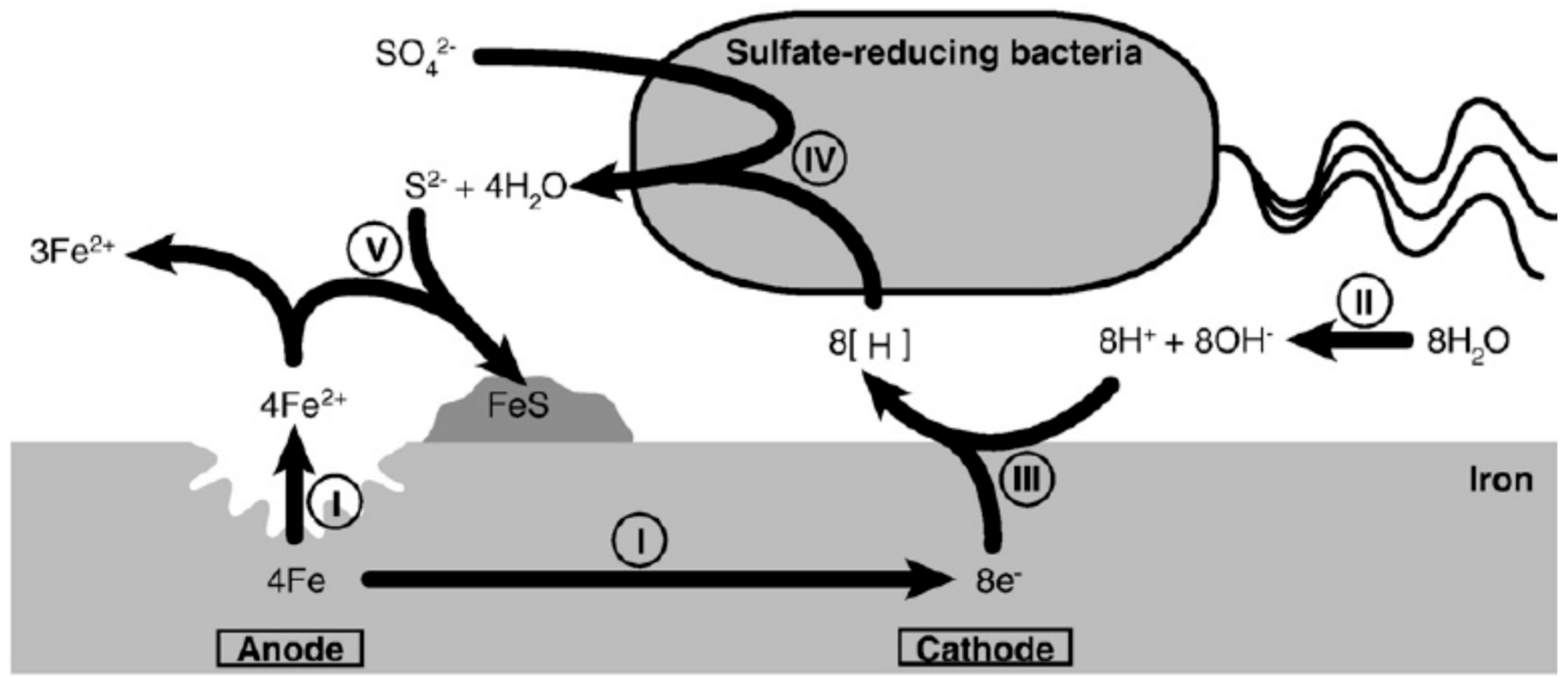
4.1. Cathodic Depolarization Theory (CDP)

Cathodic depolarization theory (CDP) has its roots in the early work of Kühr and Vlugt [10], who suggested that the accelerated corrosion was due to the consumption of cathodic hydrogen by sulphate-reducing bacteria and its hydrogenase enzyme. The removal of the hydrogen was thought to accelerate the cathodic reaction, thereby depolarizing the open-circuit potential (i.e., causing a positive shift) leading to increased corrosion rates [67]. Figure 1 presents a schematic illustration of the CDP theory due to SRB, with the depolarization step being SRB assisted reduction of sulphate in which the adsorbed hydrogen atoms are removed from the surface [5]:
SO 4 2 + 8 H ads S 2 +   4 H 2 O
Much of the support for CDP theory came from claims that MIC only occurred with types of SRB that were capable of utilizing the hydrogen produced in the cathodic supporting reaction. The mechanism is easy to understand and is thus widely quoted, especially in the microbiological community [68,69]. However, from the electrochemical standpoint CDP theory has a number of serious problems that undermine its credibility.
To start with, it is well established that at a metal’s surface hydrogen evolution occurs via the adsorption (Volmer reaction; 120 mV per decade of current density) and desorption (Tafel or Heyrovsky reactions; 30 and 40 mV per decade of current density respectively) of hydrogen and the nature of the rate determining step can be determined from the cathodic Tafel slope, βc [70]. Over 30 years ago, Frankenthal and Milner [71] used Tafel slope measurements to demonstrate that the rate determining step for hydrogen evolution on carbon steels in seawater is the adsorption step rather than the desorption step required in the cathodic depolarization mechanism. This means that the removal of the hydrogen from the surface should have no effect on the rate-limiting step and not accelerate the cathodic reaction.
Likewise, Smart et al. [72] investigated the effects of hydrogen overpressure on the corrosion of carbon steel in 3.5% NaCl and found that the reaction was not cathodically limited. Some of these data are reproduced in Table 1. It can be seen that not only are the βc values >120 mV per decade of current density, the corrosion potential, Ecorr, shifted to more positive values with increasing hydrogen overpressure, whereas if the corrosion was cathodically limited it would be expected to follow the response of the reversible hydrogen electrode (Equation (2)) and shift negative by 30 mV per decade increase in hydrogen overpressure. The carbon steel’s corrosion current density was also found to increase with, or be unaffected by, the hydrogen overpressure; in contrast, CDP theory would predict a slower corrosion rate. The same authors, revealed that the anaerobic corrosion rate of carbon steel in Swedish granitic groundwaters at 90 °C, increased with increasing hydrogen overpressure as confirmed by weight loss measurements [73].
E = 0.00 0.0591 pH 0.0295 Log [ P H 2 ]
Furthermore, it has often been shown that hydrogen-consuming SRB do not affect corrosion rates any more than their non-hydrogen consuming counterparts [31,74,75]. After investigating a range of microorganisms (acetogens, sulphate-reducing bacteria and methanogens) isolated from oil facilities, Mori et al. [35] concluded that hydrogen consumption did not strongly stimulate iron corrosion. It is thus clear that the CDP theory, based on removal of adsorbed hydrogen, is not a mechanism that contributes to MIC.

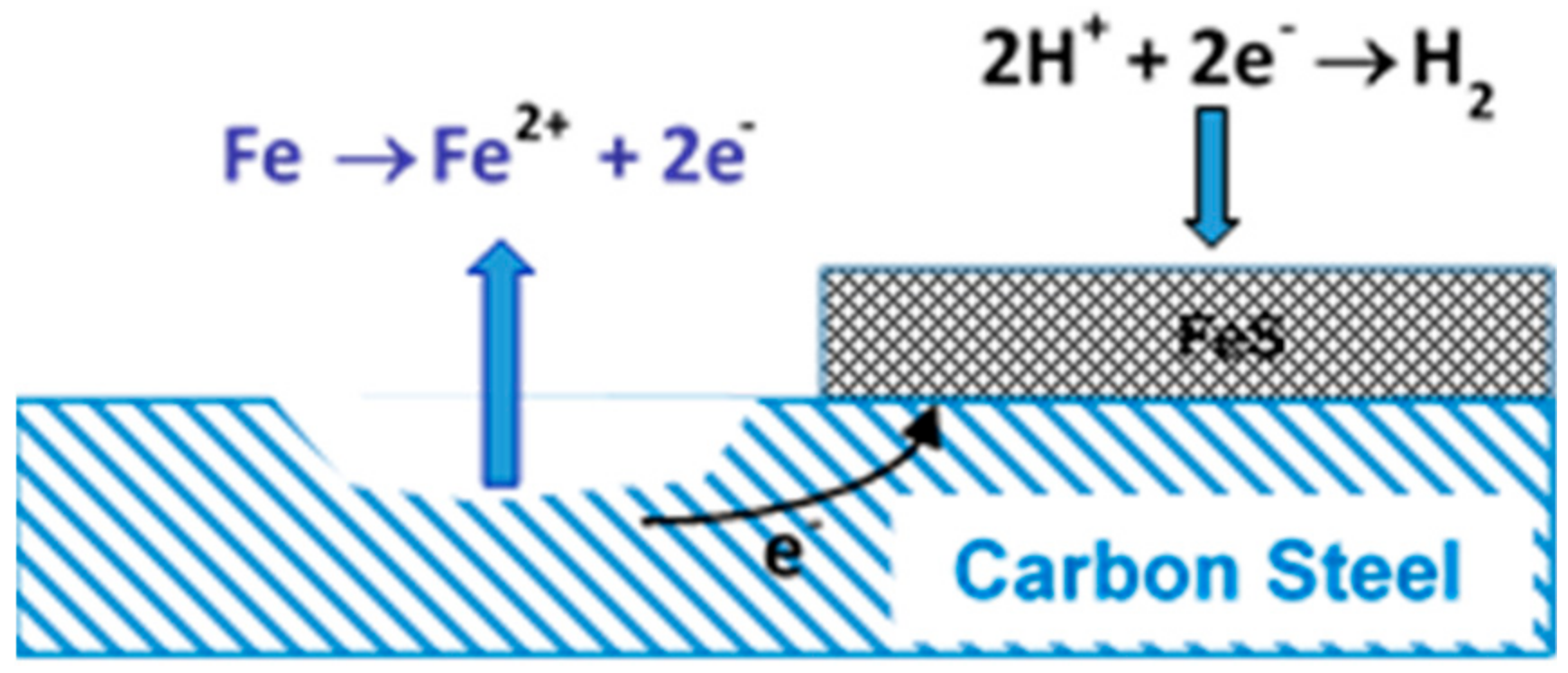
4.2. Attack by Sulphide

Despite the problems with the CDP theory, cathodic depolarization of the corrosion potential is regularly observed in MIC experiments using SRB, implying that the kinetics of the cathodic reactions are indeed increased. This led authors, such as Costello et al. [76] and King and Miller [77], to conclude that the observed depolarization is due to the dissolved hydrogen sulphide, produced by the bacteria, causing the development of a conductive iron sulphide layer. Since certain forms of iron sulphide are better hydrogen catalysts than bare steel, less overpotential will be required for this reaction to consume the electrons generated by the steel’s corrosion and so a positive shift in the corrosion potential will be observed (cathode depolarization). That is, the MIC mechanism is thought to be akin to galvanic corrosion and the role of SRB is now reduced to simply aiding in the production of a large surface area conductive iron sulphide cathode (Figure 2).
It has also been hypothesized that electrically conductive FeS can bind protons to its surface and that this facilitates electron transfer between the iron’s surface and the SRB cells, although the actual mechanism of this last step is unclear [78]. However, the nature of the iron sulphide films is very dependent on the conditions under which it forms, with mackinawite, pyrrhotite, pyrite and non-stoichiometric forms all being reported to form on steel substrates exposed to SRB [79]. In some cases, when these sulphide films are well adhered and insulating, they are actually act as a physical barrier against further corrosion [80,81].
An alternative suggestion for the role of the H2S produced by SRB is that it decreases the local pH, thus providing more protons for cathodic reduction. However, this is unlikely as in contrast to abiotic H2S corrosion, where H2S is directly supplied to the system, SRB actually do not produce H2S directly [82]. Instead the final metabolic product is HS (Reaction 3), which then combines with protons in the solution to form H2S, such that a localized increase in pH is expected, not a decrease [83]. Therefore, unlike its abiotic equivalent, the H2S biologically produced by SRB does not contribute to additional protons for cathodic reduction.
SO 4 2 + 9 H +   + 8 e   HS + 4 H 2 O
The metabolically produced sulphide has also been suggested to provide an additional cathodic reduction reaction, since the redox reaction between H2S and Fe to form FeS is more thermodynamically favorable than that between H2O and Fe to form either FeO or Fe(OH)2 [84,85,86]:
H 2 S + Fe H 2 + FeS               Δ G 0   =   72.5   kJ   mol 1   per   mole   of   Fe
H 2 O + Fe H 2 + FeO                 Δ G 0 = 14.3 kJ   mol 1   per   mole   of   Fe
2 H 2 O + Fe H 2 + Fe ( OH ) 2           Δ G 0   =   15.6   kJ   mol 1   per   mole   of   Fe
Finally, in situations where oxygen intermittently reaches the metal/electrolyte interface, as could occur when a mixed biofilm sloughs off, the FeS may play the role of a catalyst for the reduction of oxygen. In unpublished work for the United Kingdom Atomic Energy Authority, the present author observed that when an FeS film produced by SRB was coupled to bare carbon steel via a zero resistance ammeter, the galvanic corrosion current density increased significantly on allowing oxygen into the environment, signifying increased corrosion of the carbon steel. However, this effect lasted for less than an hour, as in the presence of oxygen the FeS was passivated by a layer of FeOOH. This is consistent with the work of Lee et al. [87], who reported much higher corrosion rates when carbon steel exposed to SRB was cycled between aerobic and anaerobic conditions than observed under either continuous aerobic or continuous anaerobic environments. Such anaerobic/aerobic cycling could easily occur naturally as biofilms periodically sluff-off to expose the underling FeS layer to the dissolved oxygen in the bulk solution.
The iron sulphide extended cathode mechanism is attractive for explaining MIC caused by SRB and is probably correct, although much work needs to be done to understand the electronic properties of the FeS formed if predictive models are ever to be developed. Nevertheless, a major problem clearly exists with any mechanisms involving the sulphide, produced by the SRB, in that it cannot explain MIC caused by anaerobic organisms which do not produce H2S, such as methanogens and nitrate reducing bacteria [26,35,88,89,90].

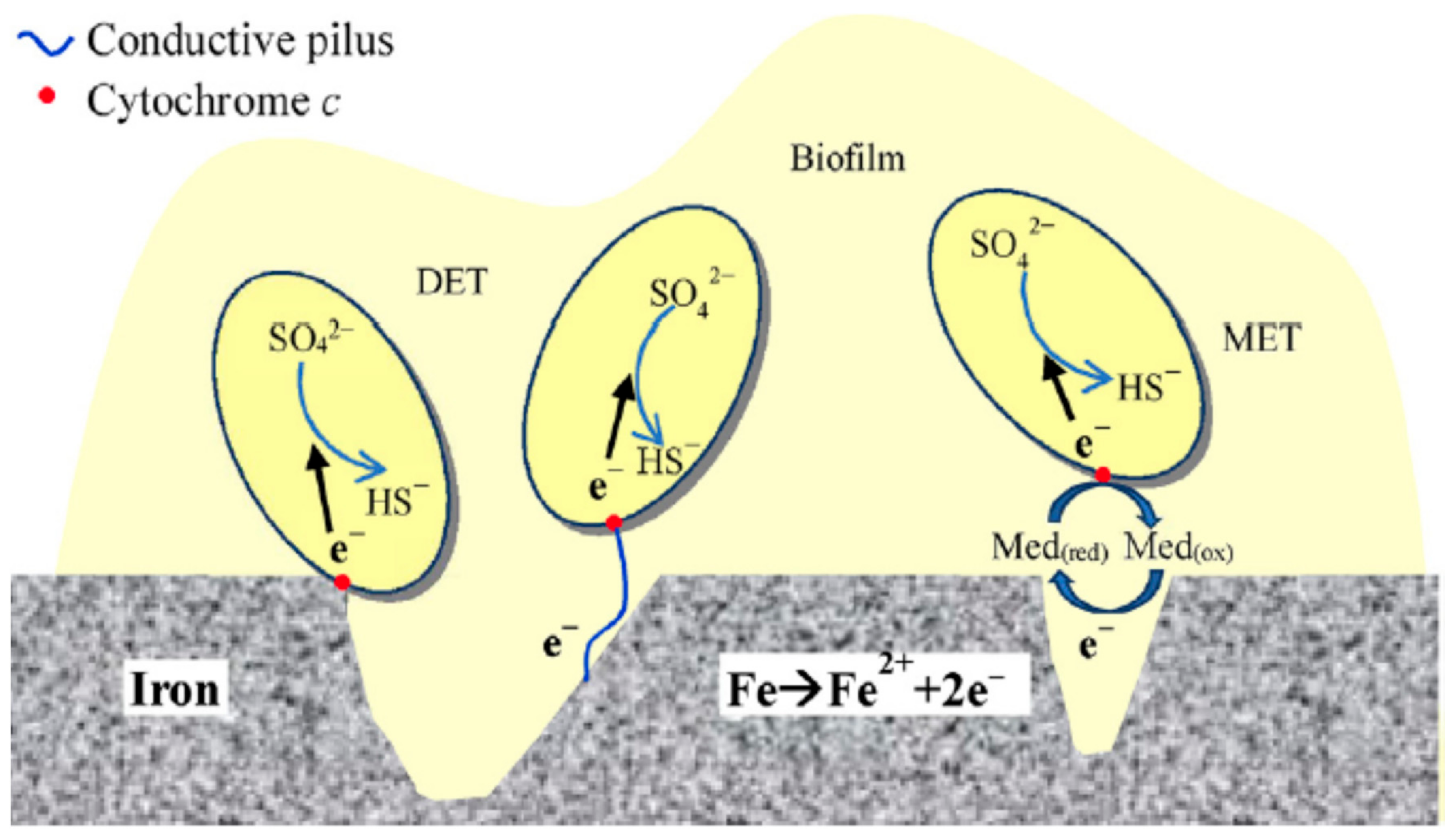
4.3. ‘Direct’ Electron Uptake

Serious consideration of “direct” electron uptake as a possible MIC mechanism arose from the work of Dinh et al. [31]. These authors demonstrated that some strains of SRB and methanogens that are unable to utilize the hydrogen generated by the abiotic anaerobic corrosion of steels as the intermediate for electron transfer displayed much higher corrosion rates than their hydrogen-utilizing counterparts. It was thus proposed that these microorganisms were facilitating the removal of electrons from the iron via a mechanism that did not involve hydrogen as the cathodic reaction. Venzlaff et al. [91] conducted electrochemical potentiodynamic measurements on the corrosion on iron by SRB, and came to similar conclusions to Dinh et al. [31], i.e., that non-H2 consuming bacteria resulted in higher corrosion rates, seemingly confirming a biological catalysis role rather than an abiotic cathodic effect of FeS. Several other authors have also claimed “direct” corrosion on carbon steel by SRB, methanogens and both iron-reducing (IRB) and iron-oxidizing bacteria (IOB) [33,35,92,93]. This class of mechanisms has recently been reviewed by Li et al. [94].
Gu et al. [82] used bioenergetics to argue why “direct” MIC is thermodynamically favourable, proposing the biocatalytic cathodic sulphate reduction (BCSR) theory. The central part of this theory is that the electrons released by the metal’s corrosion are transported across the bacterium’s cell wall and utilized in the cytoplasm; e.g., for sulphate reduction in the case of SRB. However, how the electrons were transported across the cell membrane was not explained; in normal metabolic processes, the charge is transported into the cytoplasm by soluble ions, such as lactate, as solvated free electrons do not exist in an aqueous solution.
Later, Xu and Gu [95] showed that when the SRB species Desulfovibrio vulgaris was starved of lactate, (its electron donor), it greatly accelerated the corrosion of C1018 carbon steel. These authors interpreted this as evidence that the starved SRB switch to elemental iron as the electron donor to produce energy for maintenance. Similar accelerated MIC under energy starvation conditions has been reported for both methanogens [90] and nitrate-reducing bacteria [96], indeed in these cases it seems that these microorganisms are only corrosive when starved of their normal energy sources. Unfortunately, it is likely that in real industrial applications microorganisms will be under stressed conditions and thus at their most corrosive, so perhaps this partly explains why MIC rates measured under laboratory conditions are often much lower than those reported in the field. However, the question arises of how the iron is able to act as the electron donor, which returns to the central problem of the BCSR theory, which is; how are electrons transported into the cell’s cytoplasm?
To answer this question, Xu et al. [97] adapted the concept of extracellular electron transfer, previously used in research into microbial fuel cells, in which the microorganisms serve as some sort of a bio-cathode [98]. Following Xu et al.’s approach leads to four possibilities for the electron transport process [90,92,99,100]:
(i)
direct contact between the cell and the metal substrate;
(ii)
conductive pili—microorganisms producing extended flagellum or pili to be in contact with the electrode surface to conduct electrons;
(iii)
endogenous mediators—redox molecules being produced by the microorganism itself, for example riboflavin and ferredoxin;
(iv)
exogenous mediators—chemicals added externally to the system, for example ferrocyanide.
The first two would be direct electron transfer, while the last two require redox mediators (Figure 3).
From the standpoint of an electrochemist, there is a major problem with the two direct electron transfer mechanisms, which is the distance over which the electron would have to be transferred in a single step. Charge transfer at the electrode/electrolyte interface usually takes place across the inner Helmholtz layer, which is typically 1 nm thick, where the electric field that drives the transfer is of the order of 107 V cm−1 (Figure 4); Bockris and Reddy considered this enormous electric field to be the “essence of electrochemistry” [101]. Furthermore, Gray and Winkler [102] calculated, via a Landau–Zener treatment, that direct electron transfer between aqueous species would not occur over distances greater than 2 nm.
For the first mechanism, electron transfer by direct contact, the thickness of the cell membrane, which is 7.5 to 10 nm thick [103], represents the absolute minimum distance over which the electron transfer would have to occur in a single step. This is already well above the maximum single jump distance calculated by Gray and Winkler [102]. In addition, the electrons would be required to reach either the mitochondria or the cell’s nucleus to take part in the metabolic reaction, which means travelling a distance of hundreds of nanometres if not microns. Such a distance would be inconceivably large for a single electron transfer step, so the direct electron transfer mechanism by contact can be eliminated. Further evidence against a direct electron transfer by contact mechanism comes from the fact that very few proteins, which are far smaller than single cell organisms, can approach sufficiently close to solid electrodes for direct electron transfer to their redox centres to take place [104].
The second direct electron transfer mechanism, via conductive pili, requires further discussion. Sherar et al. [105] reported that SRB cells only formed pili to attach to a carbon steel substrate when starved of an organic carbon source. Xu et al. [106] noted that these were also the conditions that gave accelerated MIC and theorized that in response to being starved of an organic carbon source, the SRB used the pili to transfer electrons from the steel into the cell’s cytoplasm for the sulphate reduction reaction. The pili are thought to behave as microbial nanowires, using the concept of extracellular electron transfer; however, the actual mechanism of the electrical conduction that underlines this process is vague, but probably involves conjugated systems [107,108,109]. It is also worth noting that it is experimentally difficult to distinguish electronic and ionic conduction in an electrochemical system, so any claims of high electrical conductivity need to be treated with caution. Furthermore, the distances over which these conductivity measurements have been made are very short, less than 3 nm for spectroscopic and electrochemical measurements and less than 10 nm for dried proteins, although the latter still retain the tightly bound water molecules so the measured conductivity could still have a large ionic component [110]. The small distances over which these conductivity measurements are made are consistent with the fact that conjugation lengths in biological molecules are much shorter than in artificial conducting polymers. This is why there are no reliable reports of biological systems with electrical conductivities in the same range as these organic semiconductors over any practical length scale; with the possible exception of DNA [111]. Therefore, an extended conjugation is also not a viable mechanism for electronic transport through pili that are of the order of one micron in length.
In order to transport electrons along the entire length of the pilius, some form of electron hopping would be required. Gray and Winkler [102] have shown that hopping could allow electron transfer in biological systems to exceed the 2 nm single step limit, but only if the free-energy changes for endergonic intermediate steps are no more than 0.2 eV, and even then the distances are below 10 nm. Perhaps, it could be argued that electrons can hop from one biological conjugated chain to the next one, as is known to occur in conducting polymers. However, this chain-to-chain hopping contributes the majority of the resistance of a conducting polymer [112] and thus the orders of magnitude more hops that would be required in a biological system (due to the far shorter conjugation) again make this suggestion implausible. Even if it were possible, there would still be the question of how the electric field required to drive this hopping process would be generated. An alternative model for the electron transport in the pili could be the Z-scheme of photosynthesis [113], but this is really a series of redox mediators, allowing much of the transport to be ionic rather than electronic, so it is not a direct electron transfer mechanism.
This leaves only the last two electron transport mechanisms, both of which rely on redox mediators acting as shuttles between the metallic substrate and the cell’s cytoplasm. This concept is well established in electrochemistry, being the basis of dye-sensitized solar cells [114] and bioelectrochemical investigations into proteins [115]. Furthermore, it seems likely that this redox mediator is predominately utilized by the microbes when they are deprived of their usual metabolic route. That is the overall mechanism has the form:
X   m e t a b o l i c   r e a c t i o n X + + e
Fe + 2 X +   Fe 2 + + 2 X
where X is the redox mediator species and the electron is not free but associated with some metabolite. The biological nature of the metabolic reaction is beyond the scope of the present review, but it has to be sufficient to keep the microbes alive. This was demonstrated by Tan et al. [90] for the case of starved methanogens, which upon the introduction of a H2/CO2 mixture into the headspace resumed methane production and at the same time stopped participating in the corrosion of the carbon steel substrates.
The mediator species X must have a redox potential positive of the corrosion potential, which in the case of carbon steels at 25 °C means positive of the reaction:
Fe   Fe 2 + + 2 e   E =   0.44 + 0.0295 Log   [ Fe 2 + ]
Assuming a ferrous concentration of 10−6 M to mark the onset of corrosion (as used in Pourbaix diagrams, albeit a somewhat arbitrary value), yields a potential for Reaction (9) of −0.617 V vs. SHE. In practice, the measured corrosion potentials of carbon steel samples in anaerobic neutral environments are within 100 millivolts negative of the reversible hydrogen potential, which at pH 7 is −0.414 V vs. SHE.
The first candidate to consider for the redox mediator X would be the cathodic hydrogen produced by the non-MIC related anaerobic corrosion of the carbon steel. If, in the absence of any suitable carbon-based energy source, the microbes oxidized hydrogen to protons a decrease in the pH of the culture media would occur, which would cause an increase in the corrosion rate. This mechanism is unlikely, as there appear to be no reports that the removal of usual energy sources causes microbes to change the pH of their environment. However, pH measurements tend to be reported for the bulk environment, which may not reflect the conditions under the biofilm. Note that this is not the same mechanism as CDP theory, as the biological consumption of the hydrogen occurs at different locations and leads to different environmental conditions.
Returning to the nature of the redox mediator, as well as having a redox potential that is positive of the measured corrosion potential, to sustain corrosion in the long-term its redox chemistry needs to be highly reversible; otherwise it would be consumed and the accelerated corrosion would stop. However, the redox mediator does not have to one of the species in the original culture media, but instead it could one that is a product of the metabolic reactions. Also if the redox mediator is being continuously regenerated in the metabolic reaction, it does not need to be present in high concentrations, so its detection is likely to be difficult. Tan et al. [90] identified several possible candidates for the corrosion-causing redox mediator used by methanogens under starved conditions, such as: the Co(I)/Co(II) couple in vitamin B12 present in the culture media; hydroquinone oxidoreductase (F420-H2); nicotinamide adenine dinucleotide (NADH); and flavin adenine dinucleotide (FAD). Likewise, Eschbach et al. [116] demonstrated that in the absence of alternative energy systems, Pseudomonas aeruginosa generate pyruvate, which in addition to having a redox potential of −0.19 V vs. SHE, has a low pKa value of 2.5, meaning it could also lower the local pH beneath the biofilm. Table 2 summarizes the redox reactions for all these potential mediators [117,118,119].
Finally, note that the biological literature often uses the term “direct electron transfer” for the case when the redox mediators are endogenous, since no external redox mediator was added to the described experiments (Figure 5) [120]. However, from the standpoint of an electrochemist the exogenous and endogenous situations are identical, with nether being considered “direct” electron transfer as they both involve a redox mediator. This is only one example of the differences between the terminologies used by microbiologists and electrochemists, which sometimes hampers the necessary collaborative efforts required to investigate MIC successfully. The need to form multidisciplinary teams to determine the significance of molecular microbiological data in respect to corrosion was recently highlighted by Eckert and Skovhus [121].

4.4. Impact on Surface Films

In abiotic corrosion, it is well known that the nature of the film that forms on the metal’s surface has a strong impact on corrosion rates, causing both accelerated corrosion and passivation. The Pourbaix diagrams [122] for iron indicate that in near neutral environments under anaerobic conditions at ambient temperatures, carbon steels can be expected to be coated by either Fe3O4 of Fe(OH)2. However, Pourbaix only considered pure water systems, where in practice ferrous carbonates, phosphates, sulphides and other compounds are possible if the relevant anions are available. Whether or not these films are protective or aggressive depends on their mineralogy and this can be altered by what may appear to be only subtle changes to the local environment. For example, the adhesion of the ferric oxides is well known to vary dramatically over the pH range 4 to 10, given rise to a range of erosion–corrosion rates [123]. Likewise, as previously mentioned, Smart et al. [72,73] found that the corrosion rate of carbon steel in saline solutions generally increased with increasing hydrogen overpressures (Table 1), with this thought to be due to the excess hydrogen interfering with the formation of a protective magnetite layer via the Schikorr reaction:
3 Fe ( OH ) 2 Fe 3 O 4 + H 2 + 2 H 2 O
It is already well established that SRB can lead to the formation of non-protective iron sulphide films and that these films can act as extended cathodes to accelerate the corrosion process. So a question arises regarding the impact of other microbes on film formation. Unfortunately, little work has been done in this area, although X-ray diffraction (XRD) of the surface films formed during MIC investigations are often presented; however, these are of limited value as they provide only limited information about the films’ physical and electronic properties. Furthermore, correct analysis of these films is difficult, as it requires an anaerobic environment to be maintained throughout the sample preparation and characterization procedures.
A related mechanism has been proposed for the way in which iron-reducing bacteria (IRB) cause MIC. Under neutral aerobic environments, ferrous materials often produce protective ferric oxide films, but the environment beneath a mixed culture biofilm can be anaerobic regardless of the level of dissolved oxygen in the bulk solution. Thus, it has been proposed that under the anaerobic conditions at the biofilm/metal interface the IRB are able to reduce the normally insoluble iron (Ill) oxides to soluble iron (II) species, leading to the localized loss of the passive film and rapid corrosion of the underlying metal [124]. However, Beech and Sunner [125] found that the presence of IRB decreased corrosion rates due to their metabolic activity. This is consistent with the work of Lovley et al. [126] who showed that IRB reduced ferric oxides to magnetite (Fe3O4), which is known to be protective [73]. Nevertheless, Valencia-Cantero et al. [127] found that a consortium containing the IRB Geobacillus sp. G2 and SRB Desulfotomaculum sp. SRB-M, resulted in carbon steel corrosion rates that were tenfold higher than with just the SRB alone. Scanning electron microscopy (SEM) analysis showed that the IRB caused fracturing of mineral films that formed in the carbon steel’s surface, thereby increasing its corrosion.

5. Concluding Remarks

Despite the long history of investigating MIC, we are still a long way from really understanding its fundamental mechanisms, especially in relation to non-sulphate reducing anaerobes. As presented here from an electrochemist’s view, both the CDP theory and direct electron transfer from the metal into the cell are incorrect. Electrically conducting pili also do not appear to play a role in direct electron transfer, although these could still play a role in aiding the mass transport of redox mediators. However, it is not clear if the microorganisms are just altering the local chemistry or if they are participating directly in the electrochemical corrosion process, albeit via the generation of redox mediators. It is feasible that both are true, with the dominant mechanism depending on the actual microorganisms involved and the local environment.
Going forward there is still much work to be done if we are to develop our understanding to the level where we can predict the degree of threat and model the likely corrosion rates. In particular, detailed investigations into how microorganisms impact the mineralogy of surface films are required. This will not be easy as many in situ techniques either cannot penetrate the biofilm or involve the use of lasers that will disrupt the very biological behaviour that needs to be studied. Likewise, for ex situ techniques, strict anaerobic environments need to be maintained during both sample preparation and characterization stages. Identification of the corrosion redox mediators that certain microorganisms are thought to generate when under stressful conditions, if they do indeed exist, would be another useful step as their detection could provide an early warning of the onset of a MIC problem.
What is certain is that a full understanding of MIC will require close collaboration between microbiologists, corrosion engineers and electrochemists [121]. This will require patience between the team members, as what may be considered proven in one field may be disputed in another. The cause of such disputes is often going to be “language” and definitions, rather than points of science. For example, as discussed above, microbiologists and electrochemists differ over what constitutes “direct electron transfer” and this has led to arguments during conferences and seminars, but given calmer reflection, the two sides would realize they are in agreement.

Funding

This research is supported by the National Research Foundation, Prime Minister’s Office, Singapore under its Marine Science Research and Development Programme (Award No. MSRDP-P12).

Conflicts of Interest

The authors declare no conflict of interest.

References

  1. Kobrin, G. A Practical Manual on Microbiologically Influenced Corrosion; NACE International: Houston, TX, USA, 1993. [Google Scholar]
  2. Little, B.; Lee, J.; Ray, R. A review of ‘green’ strategies to prevent or mitigate microbiologically influenced corrosion. Biofouling 2007, 23, 87–97. [Google Scholar] [CrossRef] [PubMed] [Green Version]
  3. Flemming, H.C. Mikrobielle werkstoffzerstörung-grundlagen: Ökonomisch-technischer überblick. Mater. Corros. 1994, 45, 5–9. [Google Scholar] [CrossRef]
  4. Venkatesan, R.; Muthiah, M.A.; Murugesh, P. Unusual corrosion of instruments deployed in the deep sea for Indian tsunami early warning system. Mar. Technol. Soc. J. 2014, 48, 6–13. [Google Scholar] [CrossRef]
  5. Little, B.; Wagner, P.; Mansfeld, F. An overview of microbiologically influenced corrosion. Electrochim. Acta 1992, 37, 2185–2194. [Google Scholar] [CrossRef]
  6. Iofa, Z.A.; Batrakov, V.V.; Cho Ngok, B. Influence of anion adsorption on the action of inhibitors on the acid corrosion of iron and cobalt. Electrochim. Acta 1964, 9, 1645–1653. [Google Scholar] [CrossRef]
  7. Stott, J.F.D. What progress in the understanding of microbially induced corrosion has been made in the last 25 years? A personal viewpoint. Corros. Sci. 1993, 35, 667–673. [Google Scholar] [CrossRef]
  8. Videla, H.A.; Herrera, L.K. Microbiologically influenced corrosion: Looking to the future. Int. Microbiol. 2005, 8, 169–180. [Google Scholar] [PubMed]
  9. Gaines, R.H. Bacterial Activity as a Corrosive Influence in the Soil. Ind. Eng. Chem. 1910, 2, 128–130. [Google Scholar] [CrossRef]
  10. Von Wolzogen Kühr, C.A.H.; Van der Vlugt, L.S. Graphitization of Cast Iron as an Electrobiochemical Process in Anaerobic Soils; Defense Technical Information Center: Fort Belvoir, VA, USA, 1964. [Google Scholar]
  11. Iverson, W.P. Direct evidence for the cathodic depolarization theory of bacterial corrosion. Science 1966, 151, 986–988. [Google Scholar] [CrossRef] [PubMed]
  12. Mara, D.D.; Williams, D.J.A. Polarization studies of pure Fe in the presence of hydrogenase-positive microbes—I. Non-photosynthetic bacteria. Corros. Sci. 1971, 12, 895–900. [Google Scholar] [CrossRef]
  13. Iverson, W.P. Corrosion of iron and formation of iron phosphide by Desulfovibrio Desulfuricans. Nature 1968, 217, 1265–1267. [Google Scholar] [CrossRef] [PubMed]
  14. Booth, G.H.; Tiller, A.K. Polarization studies of mild steel in cultures of sulphate-reducing bacteria. Trans. Faraday Soc. 1960, 56, 1689–1696. [Google Scholar] [CrossRef]
  15. Booth, G.H. Sulphur bacteria in relation to corrosion. J. Appl. Microbiol. 1964, 27, 174–181. [Google Scholar] [CrossRef]
  16. Ferris, F.G.; Schultze, S.; Witten, T.C.; Fyfe, W.S.; Beveridge, T.J. Metal interactions with microbial biofilms in acidic and neutral pH environments. Appl. Environ. Microbiol. 1989, 55, 1249–1257. [Google Scholar] [PubMed]
  17. Javaherdashti, R. A review of some characteristics of MIC caused by sulfate-reducing bacteria: Past, present and future. Anti-Corros. Methods Mater. 1999, 46, 173–180. [Google Scholar] [CrossRef]
  18. Watnick, P.; Kolter, R. Biofilm, city of microbes. J. Bacteriol. 2000, 182, 2675–2679. [Google Scholar] [CrossRef] [PubMed]
  19. Borenstein, S. Microbiologically Influenced Corrosion Handbook; Elsevier: Amsterdam, The Netherlands, 1994. [Google Scholar]
  20. Videla, H.A.; Characklis, W.G. Biofouling and microbially influenced corrosion. Int. Biodeterior. Biodegrad. 1992, 29, 195–212. [Google Scholar] [CrossRef]
  21. Mansfeld, F.; Little, B. A technical review of electrochemical techniques applied to microbiologically influenced corrosion. Corros. Sci. 1991, 32, 247–272. [Google Scholar] [CrossRef]
  22. Walsh, D.; Pope, D.; Danford, M.; Huff, T. The effect of microstructure on microbiologically influenced corrosion. JOM J. Miner. Met. Mater. Soc. 1993, 45, 22–30. [Google Scholar] [CrossRef]
  23. Magot, M.; Ollivier, B.; Patel, B.K.C. Microbiology of petroleum reservoirs. Antonie Van Leeuwenhoek 2000, 77, 103–116. [Google Scholar] [CrossRef] [PubMed]
  24. Mitterer, R.M. Methanogenesis and sulfate reduction in marine sediments: A new model. Earth Planet. Sci. Lett. 2010, 295, 358–366. [Google Scholar] [CrossRef]
  25. Magot, M. Indigenous microbial communities in oil fields. In Petroleum Microbiology; Oliver, B., Magot, B., Eds.; American Society of Microbiology: Washington, DC, USA, 2005; pp. 21–34. [Google Scholar]
  26. Larsen, J.; Rasmussen, K.; Pedersen, H.; Sørensen, K.; Lundgaard, T.; Skovhus, T.L. Consortia of MIC bacteria and archaea causing pitting corrosion in top side oil production facilities. In Proceedings of the Corrosion 2010, Paper No 10252. San Antonio, TX, USA, 14–18 March 2010. [Google Scholar]
  27. Vigneron, A.; Alsop, E.B.; Chambers, B.; Lomans, B.P.; Head, I.M.; Tsesmetzis, N. Complementary microorganisms in highly corrosive biofilms from an offshore oil production facility. Appl. Environ. Microbiol. 2016, 82, 2545–2554. [Google Scholar] [CrossRef] [PubMed]
  28. Javaherdashti, R. Microbiologically Influenced Corrosion: An Engineering Insight; Springer Science & Business Media: Berlin/Heidelberg, Germany, 2008. [Google Scholar]
  29. Usher, K.M.; Kaksonen, A.H.; Bouquet, D.; Cheng, K.Y.; Geste, Y.; Chapman, P.G.; Johnston, C.D. The role of bacterial communities and carbon dioxide on the corrosion of steel. Corros. Sci. 2015, 98, 354–365. [Google Scholar] [CrossRef]
  30. Usher, K.M.; Kaksonen, A.H.; Cole, I.; Marney, D. Critical review: Microbially influenced corrosion of buried carbon steel pipes. Int. Biodeterior. Biodegrad. 2014, 93, 84–106. [Google Scholar] [CrossRef]
  31. Dinh, H.T.; Kuever, J.; Mußmann, M.; Hassel, A.W.; Stratmann, M.; Widdel, F. Iron corrosion by novel anaerobic microorganisms. Nature 2004, 427, 829–832. [Google Scholar] [CrossRef] [PubMed]
  32. Zhang, T.; Fang, H.H.P.; Ko, B.C.B. Methanogen population in a marine biofilm corrosive to mild steel. Appl. Microbiol. Biotechnol. 2003, 63, 101–106. [Google Scholar] [CrossRef] [PubMed]
  33. Uchiyama, T.; Ito, K.; Mori, K.; Tsurumaru, H.; Harayama, S. Iron-corroding methanogen isolated from a crude-oil storage tank. Appl. Environ. Microbiol. 2010, 76, 1783–1788. [Google Scholar] [CrossRef] [PubMed]
  34. Boopathy, R.; Daniels, L. Effect of pH on anaerobic mild steel corrosion by methanogenic bacteria. Appl. Environ. Microbiol. 1991, 57, 2104–2108. [Google Scholar] [PubMed]
  35. Mori, K.; Tsurumaru, H.; Harayama, S. Iron corrosion activity of anaerobic hydrogen-consuming microorganisms isolated from oil facilities. J. Biosci. Bioeng. 2010, 110, 426–430. [Google Scholar] [CrossRef] [PubMed]
  36. Usher, K.M.; Kaksonen, A.H.; MacLeod, L.D. Marine rust tubercles harbour iron corroding archaea and sulphate reducing bacteria. Corros. Sci. 2014, 83, 189–197. [Google Scholar] [CrossRef]
  37. Kip, N.; van Veen, J.A. The dual role of microbes in corrosion. ISME J. 2015, 9, 542–551. [Google Scholar] [CrossRef] [PubMed]
  38. Videla, H.A.; Herrera, L.K. Understanding microbial inhibition of corrosion. A comprehensive overview. Int. Biodeterior. Biodegrad. 2009, 63, 896–900. [Google Scholar] [CrossRef]
  39. Herrera, L.K.; Videla, H.A. Role of iron-reducing bacteria in corrosion and protection of carbon steel. Int. Biodeterior. Biodegrad. 2009, 63, 891–895. [Google Scholar] [CrossRef]
  40. Jefferson, K.K. What drives bacteria to produce a biofilm? FEMS Microbiol. Lett. 2004, 236, 163–173. [Google Scholar] [CrossRef] [PubMed] [Green Version]
  41. Zobell, C.E.; Allen, E.C. The significance of marine bacteria in the fouling of submerged surfaces. J. Bacteriol. 1935, 29, 239–251. [Google Scholar] [PubMed]
  42. Dempsey, M.J. Marine bacterial fouling: A scanning electron microscope study. Mar. Biol. 1981, 61, 305–315. [Google Scholar] [CrossRef]
  43. Li, K.; Whitfield, M.; van Vliet, K.J. Beating the bugs: Roles of microbial biofilms in corrosion. Corros. Rev. 2013, 31, 73–84. [Google Scholar] [CrossRef]
  44. Callow, M.E.; Fletcher, R.L. The influence of low surface energy materials on bioadhesion—A review. Int. Biodeterior. Biodegrad. 1994, 34, 333–334. [Google Scholar] [CrossRef]
  45. Eashwar, M.; Dexter, S. Relation of Bacterial Settlement Patterns to Anodic Activity on Stainless Steel Weldments. In Proceedings of the Corrosion 1999, San Antonio, TX, USA, Paper No 174. 25–30 April 1999. [Google Scholar]
  46. Little, B.; Wagner, P.; Jacobus, J. The Impact of Sulfate-Reducing Bacteria on Welded Copper Nickel Seawater Piping Systems; Report No. DTIC ADA204956; Defense Technical Information Center: Fort Belvoir, VA, USA, 1988. [Google Scholar]
  47. Tiller, A.K. Aspect of microbial corrosion. In Corrosion Processes; Parkins, R.N., Ed.; Applied Science Publication: London, UK, 1982; pp. 115–159. [Google Scholar]
  48. Little, B.; Wagner, P.; Mansfeld, F. Microbiologically influenced corrosion of metals and alloys. Int. Mater. Rev. 1991, 36, 253–272. [Google Scholar] [CrossRef]
  49. Characklis, W. Bioengineering report: Fouling biofilm development: A process analysis. Biotechnol. Bioeng. 1981, 23, 1923–1960. [Google Scholar] [CrossRef] [Green Version]
  50. Pedersen, A.; Hermansson, M. The effects on metal corrosion by Serratia marcescens and a Pseudomonas SP. Biofouling 2009, 313–322. [Google Scholar]
  51. Pope, D.H.; Pope, R.M. Guide for the Monitoring and Treatment of Microbiologically Influenced Corrosion in the Natural Gas Industry; Gas Research Institute: Des Plaines, IL, USA, 1998. [Google Scholar]
  52. Little, B.J.; Lee, J.S.; Ray, R.I. The influence of marine biofilms on corrosion: A concise review. Electrochim. Acta 2008, 54, 2–7. [Google Scholar] [CrossRef] [Green Version]
  53. Meyer, B. Approaches to prevention, removal and killing of biofilms. Int. Biodeterior. Biodegrad. 2003, 51, 249–253. [Google Scholar] [CrossRef]
  54. Duan, J.; Wu, S.; Zhang, X.; Huang, G.; Du, M.; Hou, B. Corrosion of carbon steel influenced by anaerobic biofilm in natural seawater. Electrochim. Acta 2008, 54, 22–28. [Google Scholar] [CrossRef]
  55. Deutzmann, J.S.; Sahin, M.; Spormann, A.M. Extracellular enzymes facilitate electron uptake in biocorrosion and bioelectrosynthesis. mBio 2015, 6, e00496-15. [Google Scholar] [CrossRef] [PubMed]
  56. Blackwood, D.J.; Seah, K.H.W.; Teoh, S.H. Corrosion of metallic implants. In Engineering Materials for Biomedical Applications; Teoh, S.H., Ed.; World Scientific: Singapore, 2004; Chapter 3; pp. 3.1–3.56. [Google Scholar]
  57. Little, B.J.; Lee, J.S. Microbiologically influenced corrosion: An update. J. Int. Mater. Rev. 2014, 59, 384–393. [Google Scholar] [CrossRef]
  58. Gu, T.; Galicia, B. Can acid producing bacteria be responsible for very fast MIC pitting? In Proceedings of the Corrosion 2012, Salt Lake City, UT, USA, 11–15 March 2012. Paper No 1214. [Google Scholar]
  59. Parker, C.D. Species of sulphur bacteria associated with the corrosion of concrete. Nature 1947, 159, 439–440. [Google Scholar] [CrossRef] [PubMed]
  60. Ray, R.I.; Lee, J.S.; Little, B.J. Iron-oxidizing bacteria: A review of corrosion mechanisms in fresh water and marine environments. In Proceedings of the Corrosion 2010, Paper No 10218. San Antonio, TX, USA, 14—18 March 2010. [Google Scholar]
  61. Little, B.; Lee, J.; Ray, R. Recent Developments in the Identification of Mechanisms for Microbiologically Influenced Corrosion; Report No. DTIC A537292; Defense Technical Information Center: Fort Belvoir, VA, USA, 2010. [Google Scholar]
  62. Banfield, J.F.; Welch, S.A.; Zhang, H.; Ebert, T.T.; Penn, R.L. Aggregation-based crystal growth and microstructure development in natural iron oxyhydroxide biomineralization products. Science 2000, 289, 751–754. [Google Scholar] [CrossRef] [PubMed]
  63. Linhardt, P. Microbially influenced corrosion of stainless steel by manganese oxidizing microorganisms. Mater. Corros. 2004, 55, 158–163. [Google Scholar] [CrossRef]
  64. Linhardt, P. Twenty years of experience with corrosion failures caused by manganese oxidizing microorganisms. Mater. Corros. 2010, 61, 1034–1039. [Google Scholar] [CrossRef]
  65. Sly, L.I.; Hodgkinson, M.C.; Arunpairojana, V. Deposition of manganese in a drinking water distribution system. Appl. Environ. Microbiol. 1990, 56, 628–639. [Google Scholar] [PubMed]
  66. Rhoads, A.; Beyenal, H.; Lewandowski, Z. Microbial fuel cell using anaerobic respiration as an anodic reaction and biomineralized manganese as a cathodic reactant. Environ. Sci. Technol. 2005, 39, 4666–4671. [Google Scholar] [CrossRef] [PubMed]
  67. Iverson, W.P. Direct Evidence for the Cathodic Depolarization Theory of Bacterial Corrosion; Defense Technical Information Center: Fort Belvoir, VA, USA, 1965; Report No. DTIC AD0476409. [Google Scholar]
  68. Bryant, R.D.; Jansen, W.; Boivin, J.; Laishley, E.J.; Costerton, J.W. Effect of hydrogenase and mixed sulfate-reducing bacterial populations on the corrosion of steel. Appl. Environ. Microbiol. 1991, 57, 2804–2809. [Google Scholar] [PubMed]
  69. Bryant, R.D.; Laishley, E.J. The effect of inorganic phosphate and hydrogenase on the corrosion of mild steel. Appl. Environ. Microbiol. 1993, 38, 824–827. [Google Scholar] [CrossRef]
  70. Trasatti, S. Work function, electronegativity, and electrochemical behaviour of metals: III. Electrolytic hydrogen evolution in acid solutions. J. Electroanal. Chem. 1972, 39, 163–184. [Google Scholar] [CrossRef]
  71. Frankenthal, R.P.; Milner, P.C. Technical note: Hydrogen evolution kinetics on a high-carbon steel and on tin in seawater. Corrosion 1986, 42, 51–53. [Google Scholar] [CrossRef]
  72. Smart, N.R.; Blackwood, D.J.; Werme, L. Anaerobic corrosion of carbon steel and cast iron in artificial groundwaters: Part 1—Electrochemical aspects. Corrosion 2002, 58, 547–559. [Google Scholar] [CrossRef]
  73. Smart, N.R.; Blackwood, D.J.; Werme, L. Anaerobic corrosion of carbon steel and cast iron in artificial groundwaters: Part 2—Gas generation. Corrosion 2002, 58, 627–637. [Google Scholar] [CrossRef]
  74. Spruit, C.J.P.; Wanklyn, J.N. Iron sulphide ratios in corrosion by sulphate-reducing bacteria. Nature 1951, 168, 951–952. [Google Scholar] [CrossRef] [PubMed]
  75. Hardy, J.A. Utilization of cathodic hydrogen by sulphate-reducing bacteria. Br. Corros. J. 1983, 18, 190–193. [Google Scholar] [CrossRef]
  76. Costello, J.A. Cathodic depolarization by sulfate-reducing bacteria. S. Afr. J. Sci. 1974, 70, 202–204. [Google Scholar]
  77. King, R.A.; Miller, J.D.A.; Smith, J.S. Corrosion of mild steel by iron sulphides. Br. Corros. J. 1973, 8, 137–141. [Google Scholar]
  78. Enning, D.; Venzlaff, H.; Garrelfs, J.; Dinh, H.T.; Meyer, V.; Mayrhofer, K.; Hassel, A.W.; Stratmann, M.; Widdel, F. Marine sulfate-reducing bacteria cause serious corrosion of iron under electroconductive biogenic mineral crust. Environ. Microbiol. 2012, 14, 1772–1787. [Google Scholar]
  79. Jia, R.; Tan, J.L.; Jin, P.; Blackwood, D.J.; Xu, D.; Gu, T. Effects of biogenic H2S on the microbiologically influenced corrosion of C1018 carbon steel by sulfate reducing Desulfovibrio vulgaris biofilm. Corros. Sci. 2018, 130, 1–11. [Google Scholar] [CrossRef]
  80. Shoesmith, D.W.; Taylor, P.; Bailey, M.G.; Owen, D.G. The formation of ferrous monosulfide polymorphs during the corrosion of iron by aqueous hydrogen sulfide at 21 °C. J. Electrochem. Soc. 1980, 127, 1007–1015. [Google Scholar] [CrossRef]
  81. Zheng, Y.; Ning, J.; Brown, B.; Young, D.; Nešić, S. Mechanistic study of the effect of iron sulphide layers on hydrogen sulphide corrosion of carbon steel. In Proceedings of the Corrosion 2015, Paper No 5933. Dallas, TX, USA, 15–19 March 2015. [Google Scholar]
  82. Gu, T.; Zhao, K.; Nešić, S. A new mechanistic model for MIC based on a biocatalytic cathodic sulfate reduction theory. In Proceedings of the Corrosion 2009, Atlanta, GA, USA, 22–26 March 2009. Paper No 09390. [Google Scholar]
  83. Elliott, P.; Ragusa, S.; Catcheside, D. Growth of sulfate-reducing bacteria under acidic conditions in an upflow anaerobic bioreactor as a treatment system for acid mine drainage. Water Res. 1998, 32, 3724–3730. [Google Scholar] [CrossRef]
  84. Morse, J.W.; Millero, F.J.; Cornwell, J.C.; Rickard, D. The chemistry of the hydrogen sulfide and iron sulfide systems in natural waters. Earth-Sci. Rev. 1987, 24, 1–42. [Google Scholar] [CrossRef]
  85. Sun, W.; Nešić, S.; Young, D.; Woollam, R.C. Equilibrium expressions related to the solubility of the sour corrosion product mackinawite. Ind. Eng. Chem. Res. 2008, 47, 1738–1742. [Google Scholar] [CrossRef]
  86. Enning, D.; Garrelfs, J. Corrosion of iron by sulfate-reducing bacteria: New views of an old problem. Appl. Environ. Microbiol. 2014, 80, 1226–1236. [Google Scholar] [CrossRef] [PubMed]
  87. Lee, J.S.; Ray, R.I.; Little, B.J.; Lemieux, E.J. Evaluation of deoxygenation as a corrosion control measure for ballast tanks. Corrosion 2005, 61, 1173–1188. [Google Scholar] [CrossRef]
  88. Iino, T.; Ito, K.; Wakai, S.; Tsurumaru, H.; Ohkuma, M.; Harayama, S. Iron Corrosion Induced by Nonhydrogenotrophic Nitrate-Reducing Prolixibacter sp. Strain MIC1-1. Appl. Environ. Microbiol. 2015, 81, 1839–1846. [Google Scholar] [CrossRef] [PubMed]
  89. Jia, R.; Yang, D.; Xu, D.; Gu, T. Electron transfer mediators accelerated the microbiologically influence corrosion against carbon steel by nitrate reducing Pseudomonas aeruginosa biofilm. Bioelectrochemistry 2017, 118, 38–46. [Google Scholar] [CrossRef] [PubMed]
  90. Tan, J.L.; Goh, P.C.; Blackwood, D.J. Influence of H2S-producing chemical species in culture medium and energy source starvation on carbon steel corrosion caused by methanogens. Corros. Sci. 2017, 119, 102–111. [Google Scholar] [CrossRef]
  91. Venzlaff, H.; Enning, D.; Srinivasan, J.; Mayrhofer, K.J.J.; Hassel, A.W.; Widdel, F.; Stratmann, M. Accelerated cathodic reaction in microbial corrosion of iron due to direct electron uptake by sulfate-reducing bacteria. Corros. Sci. 2013, 66, 88–96. [Google Scholar] [CrossRef] [Green Version]
  92. Kato, S. Microbial extracellular electron transfer and its relevance to iron corrosion. Microb. Biotechnol. 2016, 9, 141–148. [Google Scholar] [CrossRef] [PubMed] [Green Version]
  93. Mand, J.; Park, H.S.; Okoro, C.; Lomans, B.P.; Smith, S.; Chiejina, L.; Voordouw, G. Microbial methane production associated with carbon steel corrosion in a Nigerian oil field. Front. Microbiol. 2015, 6, 1538. [Google Scholar] [CrossRef] [PubMed]
  94. Li, Y.; Xu, D.; Chen, C.; Li, X.; Jia, R.; Zhang, D.; Sand, W.; Wang, F.; Gu, T. Anaerobic microbiologically influenced corrosion mechanisms interpreted using bioenergetics and bioelectrochemistry: A review. J. Mater. Sci. Technol. 2018, 34, 1713–1718. [Google Scholar] [CrossRef]
  95. Xu, D.; Gu, T. Carbon source starvation triggered more aggressive corrosion against carbon steel by the Desulfovibrio vulgaris biofilm. Int. Biodeterior. Biodegrad. 2014, 91, 74–81. [Google Scholar] [CrossRef]
  96. Jia, R.; Yang, D.; Xu, J.; Xu, D.; Gu, T. Microbiologically influenced corrosion of C1018 carbon steel by nitrate reducing Pseudomonas aeruginosa biofilm under organic carbon starvation. Corros. Sci. 2017, 127, 1–9. [Google Scholar] [CrossRef]
  97. Xu, D.; Li, Y.; Gu, T. Mechanistic modeling of biocorrosion caused by biofilms of sulfate reducing bacteria and acid producing bacteria. Bioelectrochemistry 2016, 110, 52–58. [Google Scholar] [CrossRef] [PubMed]
  98. Zhou, M.; Wang, H.; Hassett, D.J.; Gu, T. Recent advances in microbial fuel cells (MFCs) and microbial electrolysis cells (MECs) for wastewater treatment, bioenergy and bioproducts. J. Chem. Technol. Biotechnol. 2013, 88, 508–518. [Google Scholar] [CrossRef]
  99. Torres, C.I.; Marcus, A.K.; Lee, H.S.; Parameswaran, P.; Krajmalnik-Brown, R.; Rittmann, B.E. A kinetic perspective on extracellular electron transfer by anode-respiring bacteria. FEMS Microbiol. Rev. 2010, 34, 3–17. [Google Scholar] [CrossRef] [PubMed] [Green Version]
  100. Zhang, P.; Xu, D.; Li, Y.; Yang, K.; Gu, T. Electron mediators accelerate the microbiologically influenced corrosion of 304 stainless steel by the Desulfovibrio vulgaris biofilm. Bioelectrochemistry 2015, 101, 14–21. [Google Scholar] [CrossRef] [PubMed]
  101. Bockris, J.; Reddy, A.K.N. Modern Electrochemistry; Plenum Press: New York, NY, USA, 1970; Volume 2, Chapter 11; 630p. [Google Scholar]
  102. Gray, H.B.; Winkler, J.R. Long-Range Electron Transfer. Proc. Natl. Acad. Sci. USA 2005, 102, 3534–3539. [Google Scholar] [CrossRef] [PubMed]
  103. Hine, R. “Membrane.” The Facts on File Dictionary of Biology, 3rd ed.; Checkmark: New York, NY, USA, 1999; 198p. [Google Scholar]
  104. Eddowes, M.J.; Hill, H.A.O. Novel method for the investigation of the electrochemistry of metalloproteins: Cytochrome. J. Chem. Soc. Chem. Com. 1977, 45, 771–772. [Google Scholar] [CrossRef]
  105. Sherar, B.W.A.; Power, I.M.; Keech, P.G.; Mitlin, S.; Southam, G.; Shoesmith, D.W. Characterizing the effect of carbon steel exposure in sulfide containing solutions to microbially induced corrosion. Corros. Sci. 2011, 53, 955–960. [Google Scholar] [CrossRef]
  106. Xu, D.; Li, Y.; Song, F.; Gu, T. Laboratory investigation of microbiologically influenced corrosion of C1018 carbon steel by nitrate reducing bacterium Bacillus licheniformis. Corros. Sci. 2013, 77, 385–390. [Google Scholar] [CrossRef]
  107. Reguera, G.; McCarthy, K.D.; Mehta, T.; Nicoll, J.S.; Tuominen, M.T.; Lovley, D.R. Extracellular electron transfer via microbial nanowires. Nature 2005, 435, 1098–1101. [Google Scholar] [CrossRef] [PubMed]
  108. Gorby, Y.A.; Yanina, S.; McLean, J.S.; Rosso, K.M.; Moyles, D.; Dohnalkova, A.; Beveridge, T.J.; Chang, I.S.; Kim, B.H.; Kim, K.S. Electrically conductive bacterial nanowires produced by Shewanella oneidensis strain MR-1 and other microorganisms. Proc. Natl. Acad. Sci. USA 2006, 103, 11358–11363. [Google Scholar] [CrossRef] [PubMed] [Green Version]
  109. Lovley, D.R. Happy together: Microbial communities that hook up to swap electrons. ISME J. 2017, 11, 327–336. [Google Scholar] [CrossRef] [PubMed]
  110. Amdursky, N.; Marchak, D.; Sepunaru, L.; Pecht, I.; Sheves, M.; Cahen, D. Electronic Transport via Proteins. Adv. Mater. 2014, 26, 7142–7161. [Google Scholar] [CrossRef] [PubMed]
  111. Fink, H.W.; Schönenberger, C. Electrical conduction through DNA molecules. Nature 1999, 398, 407–410. [Google Scholar] [CrossRef] [PubMed]
  112. Buck, R.P. Electron hopping in one dimension: Mixed conductor membranes. J. Phys. Chem. 1988, 92, 4196–4200. [Google Scholar] [CrossRef]
  113. Witt, H.T.; Müller, A.; Rumberg, B. Oxidized cytochrome and chlorophyll in photosynthesis. Nature 1961, 192, 967–969. [Google Scholar] [CrossRef] [PubMed]
  114. Xie, Z.B.; Midya, A.; Loh, K.P.; Adams, S.; Blackwood, D.J.; Wang, J.; Zhang, X. Highly efficient dye-sensitized solar cells using phenothiazine derivative organic dyes. Prog. Photovoltaics 2010, 18, 573–581. [Google Scholar] [CrossRef]
  115. Harper, A.; Anderson, M.R. Electrochemical glucose sensors—Developments using electrostatic assembly and carbon nanotubes for biosensor construction. Sensors 2010, 10, 8248–8274. [Google Scholar] [CrossRef] [PubMed]
  116. Eschbach, M.; Schreiber, K.; Trunk, K.; Buer, J.; Jahn, D.; Schobert, M. Long-term anaerobic survival of the opportunistic pathogen Pseudomonas aeruginosa via pyruvate fermentation. J. Bacteriol. 2004, 186, 4596–4604. [Google Scholar] [CrossRef] [PubMed]
  117. Schumacher, W.; Holliger, C.; Zehnder, A.J.B.; Hagen, W.R. Redox chemistry of cobalamin and iron-sulfur cofactors in the tetrachloroethene reductase of Dehalobacter restrictus. FEBS Lett. 1997, 409, 421–425. [Google Scholar] [CrossRef] [Green Version]
  118. Müller, V.; Blaut, M.; Gottschalk, G. Bioenergetics of methanogenesis. In Methanogenesis: Ecology, Physiology, Biochemistry & Genetics; Ferry, J.G., Ed.; Springer: Dordrecht, The Netherlands, 1993; pp. 360–406. [Google Scholar]
  119. Loach, P.A. Oxidation-reduction potentials, absorbance bands and molar absorbance of compounds used in biochemical studies. In Handbook of Biochemistry and Molecular Biology, 4th ed.; Lundlad, R.L., MacDonald, F.M., Eds.; CRC Press: Boca Raton, FL, USA, 2010; pp. 557–563. [Google Scholar]
  120. Pandit, S.; Chandrasekhar, K.; Kakarla, R.; Kadier, A.; Jeevitha, V. Basic Principles of Microbial Fuel Cell: Technical Challenges and Economic Feasibility. In Microbial Applications; Kalia, V., Kumar, P., Eds.; Springer: Cham, Switzerland, 2017; Volume 1, pp. 165–188. [Google Scholar]
  121. Eckert, R.; Skovhus, T.L. Advances in the application of molecular microbiological methods in the oil and gas industry and links to microbiologically influenced corrosion. Int. Biodeterior. Biodegrad. 2018, 126, 169–176. [Google Scholar] [CrossRef]
  122. Pourbaix, M. Atlas of Electrochemical Equilibria in Aqueous Solutions; National Association of Corrosion Engineers: Houston, TX, USA, 1974. [Google Scholar]
  123. Fontana, M.G. Corrosion Engineering; McGraw Hill: New York, NY, USA, 1986; 94p. [Google Scholar]
  124. Obuekwe, C.O.; Westlake, D.W.S.; Plambeck, J.A.; Cook, F.D. Corrosion of mild steel in cultures of ferric iron reducing bacterium isolated from crude oil I. polarization characteristics. Corrosion 1981, 37, 461–467. [Google Scholar] [CrossRef]
  125. Beech, I.B.; Sunner, J. Biocorrosion: Towards understanding interactions between biofilms and metals. Curr. Opin. Biotechnol. 2004, 15, 181–186. [Google Scholar] [CrossRef] [PubMed]
  126. Lovley, D.R.; Stolz, J.F.; Nord, G.L.; Philips, E.J.P. Anaerobic production of magnetite by a dissumlatory iron-reducing microorganism. Nature 1987, 330, 252–254. [Google Scholar] [CrossRef]
  127. Valencia-Cantero, E.; Peña-Cabriales, J.J. Effects of iron-reducing bacteria on carbon steel corrosion Induced by Thermophilic sulfate-reducing consortia. J. Microbiol. Biotechnol. 2014, 24, 280–286. [Google Scholar] [CrossRef] [PubMed]
Figure 1. Schematic illustration of microbiologically influenced corrosion (MIC) via the cathodic depolarization mechanism for sulphate-reducing bacteria (SRB) at an iron electrode. Note that this mechanism is no longer considered as correct. (Figure reproduced with permission from Mori et al. [35].
Cmd 01 00005 g001
Figure 2. Simple schematic of how FeS films produced by actions of SRB lead to a form of galvanic corrosion.
Cmd 01 00005 g002
Figure 3. Schematic illustration of “direct” and mediator electron transfer mechanisms proposed for MIC by sessile SRB cells, “Med(red)” and “Med(ox)” denote the reduced and oxidized forms of the mediator. Figure reproduced with permission from Li et al. [94].
Cmd 01 00005 g003
Figure 4. Schematic of the metal/electrolyte interface. Charge transfer takes place across the inner Helmholtz, which is typically 1 nm thick and the potential drop is around 1 V giving rise to the fields of the order of 107 V cm−1. It is this enormous field that drives all electrochemical processes.
Cmd 01 00005 g004
Figure 5. Different mechanisms proposed for the electron transfer in microbial fuel cells. From the electrochemical standpoint these are all redox mediated; methyl viologen, phenazine and cytochrome C are all redox mediators even if the latter resides within or on the walls of the microorganisms. Figure reproduced with permission from Pandit et al. [120].
Cmd 01 00005 g005
Table 1. Impact of hydrogen overpressure on the corrosion of carbon steel in 3.5% NaCl at different temperatures. Data abstracted from Smart et al. [72].
GasPressureTempEcorrIcorrβcβa
(Atms)(°C)(mV vs. SHE)(µA.cm−2)(mV dec−1)(mV dec−1)
Argon124−5751.7−15671
Hydrogen124−6071.1−13271
1024−5673.5−19975
8524−5271.4−19459
Argon150−5931.2−12674
Hydrogen150−6130.98−14466
1050−5893.1−15867
8550−5674.3−20454
Argon180−5962.7−16846
Hydrogen180−5912.8−16147
1080−5887.2−18352
8580−55912−14950
Table 2. Redox potentials at pH 7 of various possible mediators.
Redox ReactionPotential V vs. SHE at pH 7
2 H + + 2 e   H 2 −0.4137 V
V i t B 12 [ C o ( I I ) ] + e V i t B 12 [ C o ( I ) ] −0.35 V
F 420 + H 2 F 420 H 2 −0.34 V
N A D + + H + + e N A D H −0.32 V
F A D + 2 H + + 2 e F A D H 2 −0.22 V
P y r u v a t e + 2 H + + 2 e L a c t a t e −0.19 V

