Automatic Characterization of Epiretinal Membrane in OCT Images with Supervised Training †
by
 , ,
, ,
Sergio Baamonde
1,2,*, 
Joaquim de Moura
1,2 ,
,
Jorge Novo
1,2,
Noelia Barreira
1,2 and
Marcos Ortega
1,2
1
VARPA Group, Department of Computer Science, University of A Coruña, 15071 A Coruña, Spain
2
CITIC-Research Center of Information and Communication Technologies, University of A Coruña, 15071 A Coruña, Spain
*
Author to whom correspondence should be addressed.
†
Presented at the XoveTIC Congress, A Coruña, Spain, 27–28 September 2018.
Proceedings 2018, 2(18), 1161; https://doi.org/10.3390/proceedings2181161
Published: 17 September 2018
(This article belongs to the Proceedings of XoveTIC Congress 2018)
Abstract
:This work presents an automatic method to characterize the presence or absence of the epiretinal membrane (ERM) in Optical Coherence Tomography (OCT) images. To this end, a predefined set of classifiers is used on multiple local-based feature vectors which represent the inner limiting membrane (ILM), the layer of the retina where the ERM can be present.
1. Introduction
Optical Coherence Tomography (OCT) is a non-invasive imaging technology which is able to obtain in vivo, cross-sectional and high-resolution images from within the retina. These benefits helped to establish the OCT technique as one of the most widely used techniques for medical imaging. OCT is used in the analysis of pathologies such as glaucoma, Age-related Macular Degeneration (AMD) or Diabetic Macular Edema (DME). Among other eye-related pathologies, OCT imaging can be used to detect the early presence of the epiretinal membrane (ERM) in the surface of the retina, which is crucial to avoid further deterioration, blurring or distortion of the central vision in the affected eye.
This work [1] presents a fully automatic methodology to identify the ERM presence in the OCT images. Other works are focused on the use of manual markers or supervised detections by the specialists, whereas this methodology faces a precise and automatic identification of the region of interest and classification of the points inside this area without the need of any external input.
2. Methodology
The identification of the region of interest (ROI) is done by means of a deformable model which adapts its contour to the ILM layer, area where the ERM can be present.
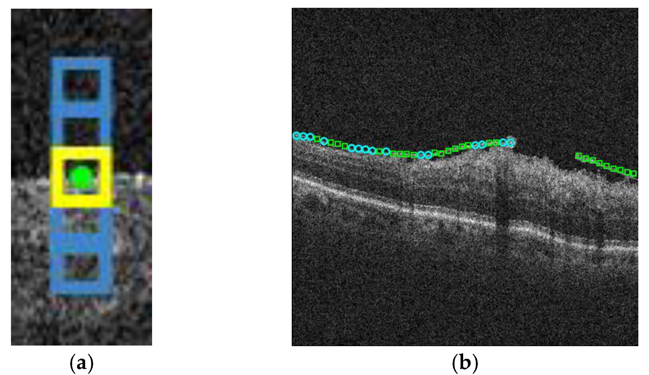
Once the ILM is identified precisely, we define a feature vector from a local window around each ILM point by applying a feature extraction procedure, as seen on Figure 1a.
Finally, the points of interest inside the ROI are classified using the obtained feature vectors to identify the presence or absence of the epiretinal membrane.
3. Experimental Results
This methodology was proved by using a dataset of 129 OCT images. 120 samples were equally taken from the complete dataset, highlighting zones with and without ERM presence. Multilayer perceptron, naive Bayes and random forest classifiers were tested to establish the validity of the proposal on top of refining the accuracy and quality of the results. Results (Figure 1b) show the areas with ERM presence, differentiating between areas where the ERM is next to the ILM and areas where the ERM is separated from the retina.
Acknowledgments
This work is supported by the Instituto de Salud Carlos III, Government of Spain and FEDER funds of the European Union through the PI14/02161 and the DTS15/00153 research projects and by the Ministerio de Economía y Competitividad, Government of Spain through the DPI2015-69948-R research project.
Conflicts of Interest
The authors declare no conflict of interest. The founding sponsors had no role in the design of the study; in the collection, analyses, or interpretation of data; in the writing of the manuscript, and in the decision to publish the results.
References
- Baamonde, S.; de Moura, J.; Novo, J.; Ortega, M. Automatic Detection of Epiretinal Membrane in OCT Images by Means of Local Luminosity Patterns. Adv. Comput. Intell. 2017, 10305, 222–235. [Google Scholar]
Figure 1.
(a) Vertical window around a ROI point. Central region surrounds the analyzed point. (b) Result from the classification process. The circles show the area where the ERM is placed on the ILM, whereas the squares represent the ERM that is separated from the retina.
Figure 1.
(a) Vertical window around a ROI point. Central region surrounds the analyzed point. (b) Result from the classification process. The circles show the area where the ERM is placed on the ILM, whereas the squares represent the ERM that is separated from the retina.
Publisher’s Note: MDPI stays neutral with regard to jurisdictional claims in published maps and institutional affiliations. |
© 2022 by the authors. Licensee MDPI, Basel, Switzerland. This article is an open access article distributed under the terms and conditions of the Creative Commons Attribution (CC BY) license (https://creativecommons.org/licenses/by/4.0/).
Share and Cite
MDPI and ACS Style
Baamonde, S.; Moura, J.d.; Novo, J.; Barreira, N.; Ortega, M. Automatic Characterization of Epiretinal Membrane in OCT Images with Supervised Training. Proceedings 2018, 2, 1161. https://doi.org/10.3390/proceedings2181161
AMA Style
Baamonde S, Moura Jd, Novo J, Barreira N, Ortega M. Automatic Characterization of Epiretinal Membrane in OCT Images with Supervised Training. Proceedings. 2018; 2(18):1161. https://doi.org/10.3390/proceedings2181161
Chicago/Turabian StyleBaamonde, Sergio, Joaquim de Moura, Jorge Novo, Noelia Barreira, and Marcos Ortega. 2018. "Automatic Characterization of Epiretinal Membrane in OCT Images with Supervised Training" Proceedings 2, no. 18: 1161. https://doi.org/10.3390/proceedings2181161