Paediatric Salmonellosis—Differences between Tropical and Sub-Tropical Regions of Queensland, Australia


Abstract
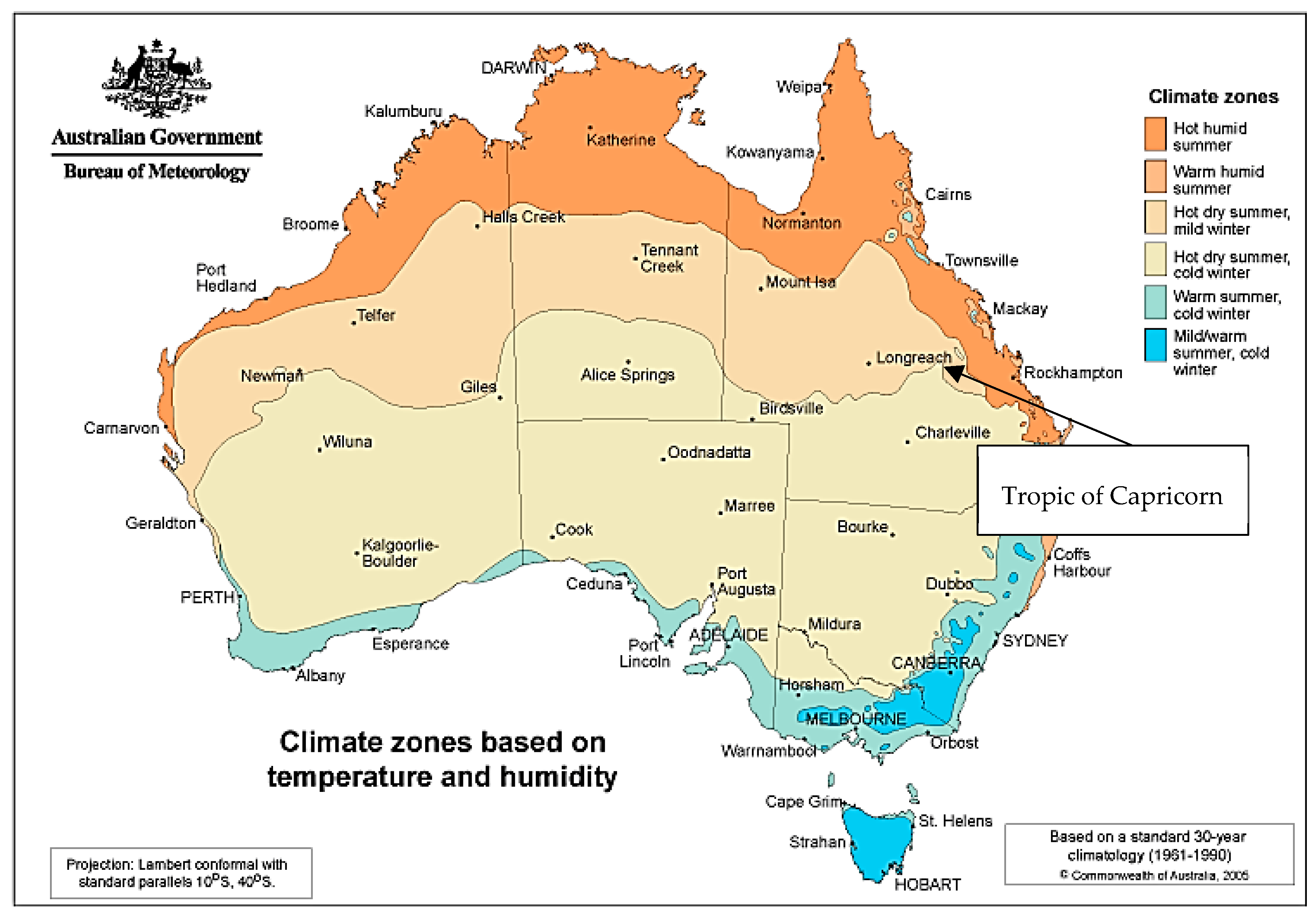
:1. Introduction
2. Methods
3. Results
3.1. Age Distribution
3.2. Faecal Specimens
3.3. Blood Cultures
3.4. Cerebrospinal Fluid (CSF)
3.5. Serovar Distribution
3.6. Antibiotic Susceptibility Testing
4. Discussion
Author Contributions
Funding
Acknowledgments
Conflicts of Interest
Appendix A

| Serovar | Tropical | Sub-tropical | Total |
|---|---|---|---|
| Aberdeen | 147 | 310 | 457 |
| Abony | 3 | 0 | 3 |
| Adelaide | 7 | 29 | 36 |
| Agona | 17 | 49 | 66 |
| Amsterdam | 1 | 0 | 1 |
| Anatum | 52 | 28 | 80 |
| Arizonae | 6 | 2 | 8 |
| Bahrenfeld | 6 | 1 | 7 |
| Ball | 2 | 2 | 4 |
| Bareilly | 0 | 3 | 3 |
| Beaudesert | 1 | 0 | 1 |
| Bergedorf | 1 | 0 | 1 |
| Birkenhead | 2 | 274 | 276 |
| Blockley | 0 | 1 | 1 |
| Bovismorbificans | 18 | 36 | 54 |
| Braenderup | 3 | 1 | 4 |
| Brazzaville | 0 | 1 | 1 |
| Bredeney | 8 | 14 | 22 |
| Breukelen | 3 | 0 | 3 |
| Bukavu | 1 | 1 | 2 |
| Cerro | 1 | 4 | 5 |
| Chailey | 30 | 9 | 39 |
| Charity | 0 | 2 | 2 |
| Chester | 112 | 145 | 257 |
| Concord | 0 | 1 | 1 |
| Corvallis | 5 | 25 | 30 |
| Dan | 2 | 0 | 2 |
| Derby | 1 | 0 | 1 |
| Eastbourne | 12 | 14 | 26 |
| Emek | 1 | 0 | 1 |
| Emmastad | 2 | 1 | 3 |
| Enteritidis | 73 | 50 | 123 |
| Falkensee | 1 | 0 | 1 |
| Ferruch | 0 | 1 | 1 |
| Give | 4 | 5 | 9 |
| Hadar | 1 | 0 | 1 |
| Havana | 12 | 20 | 32 |
| Heidelberg | 62 | 55 | 117 |
| Hessarek | 0 | 1 | 1 |
| Hvittingfoss | 173 | 154 | 327 |
| Infantis | 8 | 29 | 37 |
| Isangi | 0 | 1 | 1 |
| Java | 2 | 0 | 2 |
| Javiana | 0 | 42 | 42 |
| Johannesburg | 7 | 3 | 10 |
| Kentucky | 0 | 2 | 2 |
| Kiambu | 4 | 26 | 30 |
| Kimberley | 1 | 0 | 1 |
| Kinondoni | 1 | 0 | 1 |
| Kottbus | 6 | 8 | 14 |
| Lansing | 18 | 7 | 25 |
| Lindern | 1 | 0 | 1 |
| Litchfield | 20 | 33 | 53 |
| Livingstone | 0 | 1 | 1 |
| Mbandaka | 6 | 9 | 15 |
| Meleagridis | 1 | 0 | 1 |
| Mgulani | 101 | 24 | 125 |
| Mikawasima | 0 | 2 | 2 |
| Mississippi | 0 | 5 | 5 |
| Montevideo | 19 | 20 | 39 |
| Muenchen | 47 | 149 | 196 |
| Muenster | 0 | 1 | 1 |
| Mugulani | 1 | 0 | 1 |
| Napoli | 0 | 1 | 1 |
| Newport | 3 | 14 | 17 |
| Ohio | 1 | 2 | 3 |
| Ohlstedt | 8 | 9 | 17 |
| Onderstepoort | 1 | 2 | 3 |
| Oranienburg | 10 | 8 | 18 |
| Orientalis | 14 | 38 | 52 |
| Orion | 38 | 5 | 43 |
| Oslo | 4 | 7 | 11 |
| Panama | 0 | 7 | 7 |
| Para B Biovar Java | 44 | 56 | 100 |
| Paratyphi A | 1 | 7 | 8 |
| Paratyphi B | 4 | 2 | 6 |
| Poona | 9 | 5 | 14 |
| Potsdam | 3 | 61 | 64 |
| Ramatgam | 1 | 0 | 1 |
| Reading | 54 | 49 | 103 |
| Rissen | 0 | 1 | 1 |
| Rubislaw | 4 | 12 | 16 |
| Saintpaul | 327 | 399 | 726 |
| Salford | 0 | 1 | 1 |
| Sandiego | 14 | 3 | 17 |
| Schwarzengrund | 1 | 2 | 3 |
| Senftenberg | 1 | 0 | 1 |
| Singapore | 5 | 18 | 23 |
| Stanley | 7 | 12 | 19 |
| Tennessee | 2 | 3 | 5 |
| Thompson | 36 | 4 | 40 |
| Typhi | 15 | 9 | 24 |
| Typhimurium | 222 | 1348 | 1570 |
| Uganda | 6 | 0 | 6 |
| Untyped | 307 | 469 | 776 |
| Urbana | 6 | 0 | 6 |
| Virchow | 460 | 704 | 1164 |
| Virginia | 1 | 1 | 2 |
| Wandsworth | 11 | 1 | 12 |
| Wangata | 1 | 28 | 29 |
| Warneri | 1 | 0 | 1 |
| Waycross | 63 | 246 | 309 |
| Welikade | 143 | 5 | 148 |
| Weltevreden | 70 | 24 | 94 |
| Yarrabah | 1 | 0 | 1 |
| Zanzibar | 37 | 45 | 82 |
| Zehlendorf | 2 | 2 | 4 |
| Total | 2951 | 5211 | 8162 |
References
- Graham, S.M.; English, M. Non-typhoidal salmonellae: A management challenge for children with community-acquired invasive disease in tropical African countries. Lancet 2009, 373, 267–269. [Google Scholar] [CrossRef]
- Date, K.A.; Bentsi-Enchill, A.D.; Fox, K.K.; Abeysinghe, N.; Mintz, E.D.; Khan, M.I.; Sahastrabuddhe, S.; Hyde, T.B. Typhoid Fever surveillance and vaccine use—South-East Asia and Western Pacific regions, 2009–2013. MMWR 2014, 63, 855–860. [Google Scholar] [PubMed]
- Wain, J.; Hendriksen, R.S.; Mikoleit, M.L.; Keddy, K.H.; Ochiai, R.L. Typhoid fever. Lancet 2015, 385, 1136–1145. [Google Scholar] [CrossRef]
- Crump, J.A. Updating and refining estimates of typhoid fever burden for public health action. Lancet Glob Health. 2014, 2, e551–e553. [Google Scholar] [CrossRef] [Green Version]
- Food Safety Australia and New Zealand. Salmonella (non-typhoidal). 2013. Available online: https://www.foodstandards.gov.au/publications/Documents/Salmonella(non-typhoidal).pdf (accessed on 21 November 2018).
- Mogasale, V.; Maskery, B.; Ochiai, R.L.; Lee, J.S.; Mogasale, V.V.; Ramani, E.; Kim, Y.E.; Park, J.K.; Wierzba, T.F. Burden of typhoid fever in low-income and middle-income countries: A systematic, literature-based update with risk-factor adjustment. Lancet Glob Health. 2014, 2, e570–e580. [Google Scholar] [CrossRef]
- Crump, J.A.; Ramadhani, H.O.; Morrissey, A.B.; Msuya, L.J.; Yang, L.Y.; Chow, S.C.; Morpeth, S.C.; Reyburn, H.; Njau, B.N.; Shaw, A.V.; et al. Invasive bacterial and fungal infections among hospitalized HIV-infected and HIV-uninfected children and infants in northern Tanzania: Paediatric invasive infections in Tanzania. Trop. Med. Int. Health 2011, 16, 830–837. [Google Scholar] [CrossRef]
- Arndt, M.B.; Mosites, E.M.; Tian, M.; Forouzanfar, M.H.; Mokhdad, A.H.; Meller, M.; Ochiai, R.L.; Walson, J.L. Estimating the burden of paratyphoid a in Asia and Africa. PLoS Neglect Trop. D 2014, 8, e2925. [Google Scholar] [CrossRef]
- Asmar, B.I.; Abdel-Haq, N. Nontyphoidal Salmonella infection in children: Relation to bacteremia, age, and infecting serotype. Infect. Dis.-Nor. 2016, 48, 147–151. [Google Scholar] [CrossRef] [PubMed]
- Biggs, H.M.; Lester, R.; Nadjm, B.; Mtove, G.; Todd, J.E.; Kinabo, G.D.; Philemon, R.; Amos, B.; Morrissey, A.B.; Reyburn, H.; et al. Invasive Salmonella infections in areas of high and low malaria transmission intensity in Tanzania. Clin. Infect. Dis. 2014, 58, 638–647. [Google Scholar] [CrossRef]
- Majowicz, S.E.; Musto, J.; Scallan, E.; Angulo, F.J.; Kirk, M.; O’Brien, S.J.; Jones, T.F.; Fazil, A.; Hoekstra, R.M. The Global Burden of Nontyphoidal Salmonella Gastroenteritis. Clin. Infect. Dis. 2010, 50, 882–889. [Google Scholar] [CrossRef] [Green Version]
- Ao, T.T.; Feasey, N.A.; Gordon, M.A.; Keddy, K.H.; Angulo, F.J.; Crump, J.A. Global burden of invasive nontyphoidal Salmonella disease. Emerg Infect. Dis. 2015, 21. [Google Scholar] [CrossRef] [PubMed]
- National Notifiable Disease Surveillance System. Australia’s notifiable disease status, 2014: Annual report of the National Notifiable Diseases Surveillance System: Part 4: Australian Government Department of Health. 2016. Available online: http://www.health.gov.au/internet/main/publishing.nsf/content/cda-cdi4001e3.htm (accessed on 21 November 2018).
- Er, J.; Wallis, P.; Maloney, S.; Norton, R. Paediatric bacteraemias in tropical Australia: Paediatric bacteraemias in Australia. J. Paediatr. Child H 2014. [Google Scholar] [CrossRef] [PubMed]
- Kirk, M.D.; Pires, S.M.; Black, R.E.; Caipo, M.; Crump, J.A.; Devleesschauwer, B. World Health Organization Estimates of the Global and Regional Disease Burden of 22 Foodborne Bacterial, Protozoal, and Viral Diseases, 2010: A Data Synthesis. PLoS Med. 2015, 12, e1001921. [Google Scholar]
- Queensland Health. Salmonella Infection 2010. Available online: https://www.health.qld.gov.au/cdcg/index/salmonella (accessed on 23 November 2018).
- Hsu, R.B.; Lin, F.Y. Risk factors for bacteraemia and endovascular infection due to non-typhoid salmonella: A reappraisal. QJMed 2005, 98, 821–827. [Google Scholar] [CrossRef]
- Dhanoa, A.; Fatt, Q.K. Non-typhoidal Salmonella bacteraemia: Epidemiology, clinical characteristics and its’ association with severe immunosuppression. Ann. Clin. Microb. Anti 2009, 8. [Google Scholar] [CrossRef] [PubMed]
- Crum-Cianflone, N.F. Salmonellosis and the gastrointestinal tract: More than just peanut butter. Curr. Gastr. Rep. 2008, 10, 424–431. [Google Scholar] [CrossRef]
- Beaverton. The Produce Contamination Problem: Causes and Solutions, 2nd ed.; Ringgold Inc.: Bristol, UK, 2014. [Google Scholar]
- Brander, R.L.; Walson, J.L.; John-Stewart, G.C.; Naulikha, J.M.; Ndonye, J.; Kipkemoi, N.; Rwigi, D.; Singa, B.O.; Pavlinac, P.B. Correlates of multi-drug non-susceptibility in enteric bacteria isolated from Kenyan children with acute diarrhea. PLoS Neglect. Trop. D 2017, 11, e0005974. [Google Scholar] [CrossRef]
- Tian, L.; Zhu, X.; Chen, Z.; Liu, W.; Li, S.; Yu, W.; Zhang, W.; Xiang, X.; Sun, Z. Characteristics of bacterial pathogens associated with acute diarrhea in children under 5 years of age: A hospital-based cross-sectional study. BMC Infect Dis. 2016, 16, 253. [Google Scholar] [CrossRef]
- Zhang, H.; Pan, F.; Zhao, X.; Wang, G.; Tu, Y.; Fu, S.; Wang, J.; Pan, J.; Song, J.; Wang, W.; et al. Distribution and antimicrobial resistance of enteric pathogens in Chinese paediatric diarrhoea: A multicentre retrospective study, 2008-2013. Epidemiol. Infect. 2015, 143, 2512. [Google Scholar] [CrossRef] [PubMed]
- Chiyangi, H.; Muma, J.B.; Malama, S.; Manyahi, J.; Abade, A.; Kwenda, G.; Matee, M.I. Identification and antimicrobial resistance patterns of bacterial enteropathogens from children aged 0-59 months at the University Teaching Hospital, Lusaka, Zambia: A prospective cross sectional study. BMC Infect. Dis. 2017, 17, 117. [Google Scholar] [CrossRef]
- EUCAST: Clinical breakpoints. Available online: http://www.eucast.org/clinical_breakpoints/ (accessed on 15 November 2018).
- Institute CaLS. Performance Standards for Antimicrobial Susceptibility Testing; Twentieth Informational Supplement. CLSI Document M100-S20; Clinical and Laboratory Standards Institute: Wayne, PA, USA, 2016. [Google Scholar]
- Ford, L.; Glass, K.; Veitch, M.; Wardell, R.; Polkinghorne, B.; Dobbins, T.; Lal, A.; Kirk, M.D. Increasing Incidence of Salmonella in Australia, 2000-2013. PLoS ONE 2016, 11, e0163989. [Google Scholar] [CrossRef] [PubMed]
- Li, R.; Wang, Y.; Shen, J.; Wu, C. Development of a Novel Hexa-plex PCR Method for Identification and Serotyping of Salmonella Species. Foodborne Pathog Dis. 2014, 11, 75–77. [Google Scholar] [CrossRef] [PubMed]
- Ashbolt, R.; Kirk, M.D. Salmonella Mississippi infections in Tasmania: The role of native Australian animals and untreated drinking water. Epidemiol. Infect. 2006, 134, 1257–1265. [Google Scholar] [CrossRef] [PubMed]
- Gibney, K.B.D.; Cheng, A.C.P.; Hall, R.F.; Leder, K.P. Sociodemographic and geographical inequalities in notifiable infectious diseases in Australia: A retrospective analysis of 21 years of national disease surveillance data. Lancet Infect. Dis. 2016, 17, 86–97. [Google Scholar] [CrossRef]
- Milazzo, A.; Giles, L.C.; Zhang, Y.; Koehler, A.P.; Hiller, J.E.; Bi, P. Factors Influencing Knowledge, Food Safety Practices and Food Preferences During Warm Weather of Salmonella and Campylobacter Cases in South Australia. Foodborne Pathog Dis. 2017, 14, 125–131. [Google Scholar] [CrossRef]
- Williams, S.; Patel, M.; Markey, P.; Muller, R.; Benedict, S.; Ross, I.; Heuzenroeder, M.; Davos, D.; Cameron, S.; Krause, V. Salmonella in the tropical household environment—Everyday, everywhere. J. Infection. 2015, 71, 642–648. [Google Scholar] [CrossRef]
- Hall, G.; Hanigan, I.C.; Dear, K.B.G.; Vally, H. The influence of weather on community gastroenteritis in Australia. Epidemiol. Infect. 2011, 139, 927–936. [Google Scholar] [CrossRef]
- Zhang, Y.; Bi, P.; Hiller, J.E. Climate variations and Salmonella infection in Australian subtropical and tropical regions. Sci. Total Environ. 2010, 408, 524–530. [Google Scholar] [CrossRef]
- Fearnley, E.J.; Lal, A.; Bates, J.; Stafford, R.; Kirk, M.D.; Glass, K. Salmonella source attribution in a subtropical state of Australia: Capturing environmental reservoirs of infection. Epidemiol. Infect. 2018, 146, 1903–1908. [Google Scholar] [CrossRef]
- Messer, R.D.; Warnock, T.H.; Heazlewood, R.J.; Hanna, J.N. Salmonella meningitis in children in far north Queensland. J. Paediatr. Child. H 1997, 33, 535–538. [Google Scholar] [CrossRef]
- Callaway, Z.; Thomas, A.; Melrose, W.; Buttner, P.; Speare, R. Salmonella Virchow and Salmonella Weltevreden in a random survey of the Asian house gecko, Hemidactylus frenatus, in houses in Northern Australia. Vector-Borne Zoonot. 2011, 11, 621–625. [Google Scholar] [CrossRef] [PubMed]
- Murphy, D.; Oshin, F. Reptile-associated salmonellosis in children aged under 5 years in South West England. Arch. Dis Child. 2015, 100, 364–365. [Google Scholar] [CrossRef] [PubMed]
- Finlay, F.; Furnell, C.; Ridley, P. Salmonella in pets: The risk to children. Community Pract. 2015, 88, 27–28. [Google Scholar]
- Sauteur, P.M.M.; Relly, C.; Hug, M.; Wittenbrink, M.M.; Berger, C. Risk Factors for Invasive Reptile-Associated Salmonellosis in Children. Vector-Borne Zoonot. 2013, 13, 419–421. [Google Scholar] [CrossRef] [PubMed] [Green Version]
- Munnoch, S.A.; Ward, K.; Sheridan, S.; Fitzsimmons, G.J.; Shadbolt, C.T.; Piispanen, J.P.; Wang, Q.; Ward, T.J.; Worgan, T.L.; Oxenford, C.; et al. A multi-state outbreak of Salmonella Saintpaul in Australia associated with cantaloupe consumption. Epidemiol. Infect. 2009, 137, 367–374. [Google Scholar] [CrossRef]
- Freeman, R.; Dabrera, G.; Lane, C.; Adams, N.; Browning, L.; Fowler, T.; Gorton, R.; Peters, T.; Mather, H.; Ashton, P.; et al. Association between use of proton pump inhibitors and non-typhoidal salmonellosis identified following investigation into an outbreak of Salmonella Mikawasima in the UK, 2013. Epidemiol. Infect. 2016, 144, 968. [Google Scholar] [CrossRef]
- Doorduyn, Y.; Van Den Brandhof, W.E.; Van Duynhoven, Y.T.H.P.; Wannet, W.J.B.; Van Pelt, W. Risk factors for Salmonella Enteritidis and Typhimurium (DT104 and non-DT104) infections in The Netherlands: Predominant roles for raw eggs in Enteritidis and sandboxes in Typhimurium infections. Epidemiol. Infect. 2006, 134, 617–626. [Google Scholar] [CrossRef]
- Moffatt, C.R.M.; Musto, J.; Pingault, N.; Miller, M.; Stafford, R.; Gregory, J.; Polkinghorne, B.G.; Kirk, M.D. Salmonella Typhimurium and Outbreaks of Egg-Associated Disease in Australia, 2001 to 2011. Foodborne Pathog. Dis. 2016, 13, 379–385. [Google Scholar] [CrossRef]
- Ashdown, L.R.; Ryan, P.J. Invasive disease due to Salmonella virchow: A north Queensland problem. Med. J. Australia 1990, 153, 330–332. [Google Scholar]
- Gastrointestinal tract infections: Acute gastroenteritis. In Therapeutic Guidelines, Antibiotic, 15th ed.; Therapeutic Guidelines Ltd.: West Melbourne, Australia, 2014; pp. 338–339.


| Antibiotic | Tropical Region | Sub-Tropical Region | ||||
|---|---|---|---|---|---|---|
| Number Susceptible | Number Tested | Percentage Susceptible (95% C.I.) | Number Susceptible | Number Tested | Percentage Susceptible (95% C.I.) | |
| Ampicillin | 2579 | 2653 | 97.21 (96.58,97.84) | 4322 | 4544 | 95.11 (94.49, 95.74) |
| Trimethoprim/Sulfamethoxazole | 2539 | 2563 | 99.06 (98.69, 99.44) | 4350 | 4458 | 97.58 (97.13, 98.03) |
© 2019 by the authors. Licensee MDPI, Basel, Switzerland. This article is an open access article distributed under the terms and conditions of the Creative Commons Attribution (CC BY) license (http://creativecommons.org/licenses/by/4.0/).
Share and Cite
Berger, D.; Smith, F.; Sabesan, V.; Huynh, A.; Norton, R. Paediatric Salmonellosis—Differences between Tropical and Sub-Tropical Regions of Queensland, Australia. Trop. Med. Infect. Dis. 2019, 4, 61. https://doi.org/10.3390/tropicalmed4020061
Berger D, Smith F, Sabesan V, Huynh A, Norton R. Paediatric Salmonellosis—Differences between Tropical and Sub-Tropical Regions of Queensland, Australia. Tropical Medicine and Infectious Disease. 2019; 4(2):61. https://doi.org/10.3390/tropicalmed4020061
Chicago/Turabian StyleBerger, Daria, Felicity Smith, Vana Sabesan, Aimee Huynh, and Robert Norton. 2019. "Paediatric Salmonellosis—Differences between Tropical and Sub-Tropical Regions of Queensland, Australia" Tropical Medicine and Infectious Disease 4, no. 2: 61. https://doi.org/10.3390/tropicalmed4020061