
In-Situ Comparative Study of Eucalyptus, Basil, Cloves, Thyme, Pine Tree, and Tea Tree Essential Oil Biocide Efficacy


 ,
,  , and
, and
Abstract
:1. Introduction
2. Materials and Methods
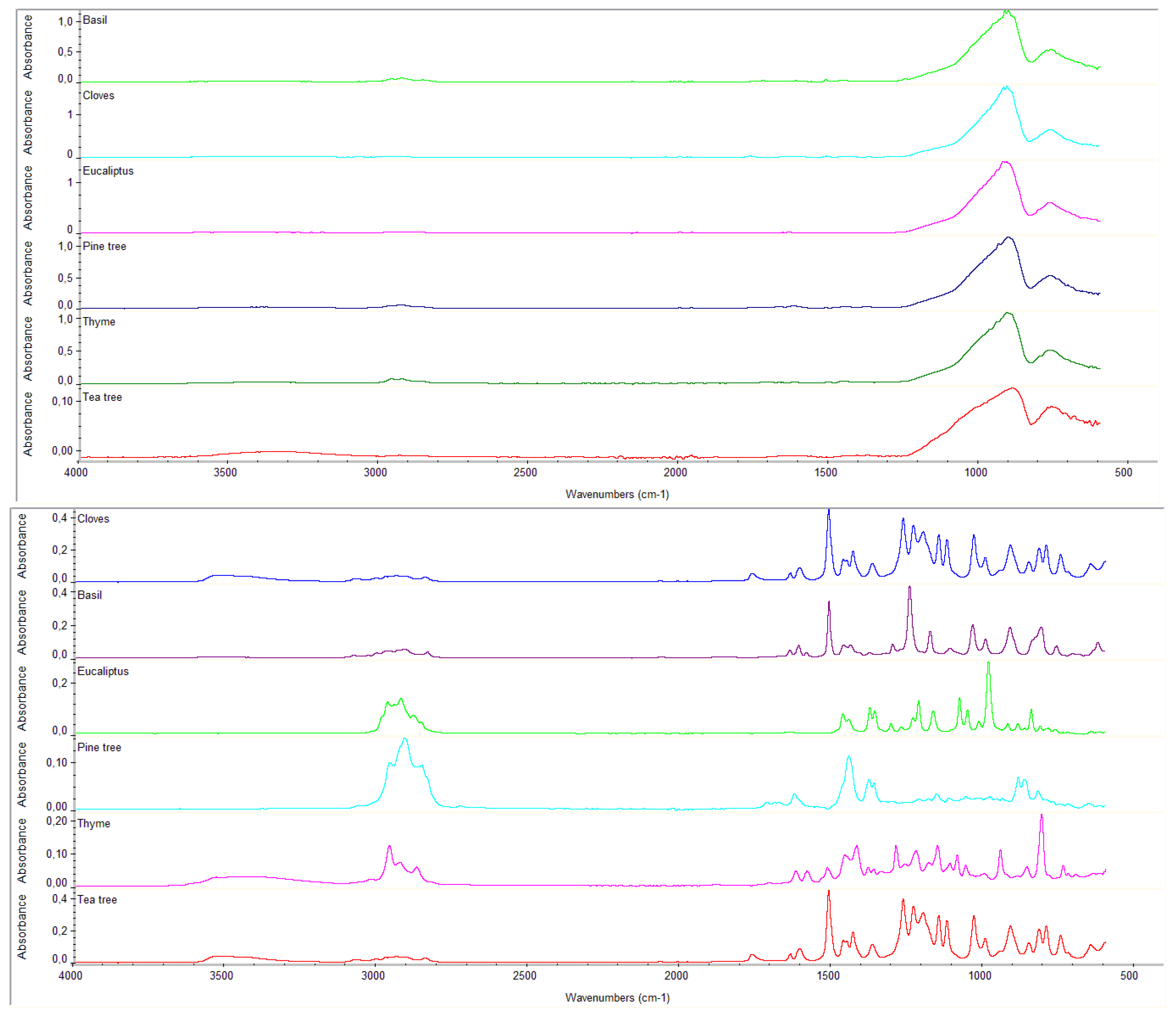
2.1. FTIR Analysis
2.2. Ultraviolet Induced Luminescence (UVL)
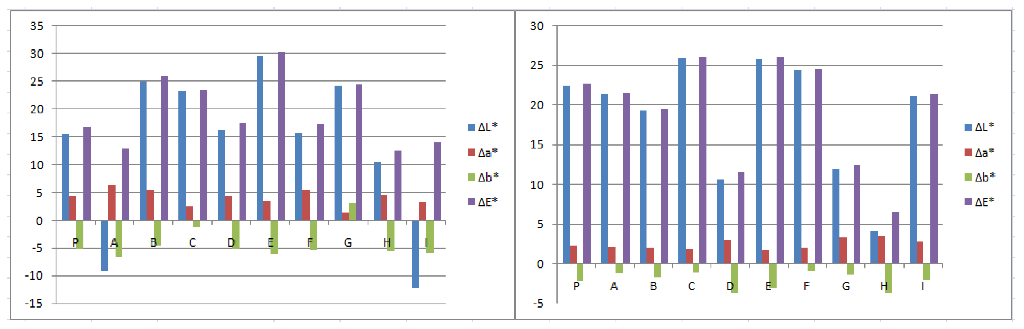
2.3. Spectro-Colorimetric Analysis
2.4. Adenosine Triphosphate (ATP) Test
3. Results and Discussions
3.1. Analysis of Residue Using FTIR Analysis
3.2. Ultraviolet Induced Luminescence (UVL)
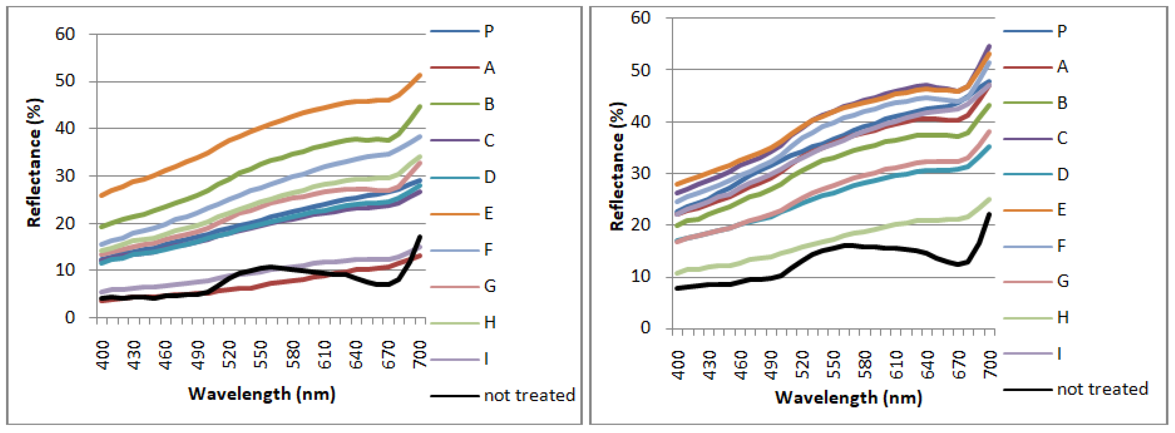
3.3. Spectro Colorimetric Analysis
3.4. Adenosine Triphosphate (ATP) Test
4. Conclusions
Author Contributions
Funding
Conflicts of Interest
References
- Veneranda, M.; Blanco-Zubiaguirre, L.; Roselli, G.; Di Girolami, G.; Castro, K.; Madariaga, J.M. Evaluating the exploitability of several essential oils constituents as a novel biological treatment against cultural heritage biocolonization. Microchem. J. 2018, 138, 1–6. [Google Scholar] [CrossRef]
- Mascaro, M.E.; Pellegrino, G.; Palermo, A.M. Analysis of Biodeteriogens on Architectural Heritage. An Approach of Applied Botany on a Gothic Building in Southern Italy. Sustainability 2022, 14, 34. [Google Scholar] [CrossRef]
- Gaylarde, C.C. Influence of Environment on Microbial Colonization of Historic Stone Buildings with Emphasis on Cyanobacteria. Heritage 2020, 3, 1469–1482. [Google Scholar] [CrossRef]
- Charola, A.E.; McNamara, C.; Koestler, R.J. Biocolonization of Stone: Control and Preventive Methods Proceedings from the MCI Workshop Series A; Smithsonian Institution Scholarly Press: Washington, DC, USA, 2011; Volume 2, pp. 1–116. [Google Scholar]
- Fierascu, I.; Ion, R.M.; Radu, M.; Dima, S.O.; Bunghez, I.R.; Avramescu, S.M.; Fierascu, R.C. Comparative study of antifungal effects of natural extracts and essential oils. Rev. Roum. Chim. 2014, 59, 207–211. [Google Scholar]
- Cappitelli, F.; Cattò, C.; Villa, F. The Control of Cultural Heritage Microbial Deterioration. Microorganisms 2020, 8, 1542. [Google Scholar] [CrossRef] [PubMed]
- Liu, X.; Koestlker, R.J.; Warscheid, T.; Katayama, Y.; Gu, J. Microbial deterioration and sustainable conservation of stone monuments and buildings. Environ. Sci. Eng. 2020, 3, 991–1004. [Google Scholar] [CrossRef]
- Ranalli, G.; Zanardini, E. Biocleaning on Cultural Heritage: New frontiers of microbial biotechnologies. J. Appl. Microbiol. 2021, 131, 583–603. [Google Scholar] [CrossRef]
- Macchia, A.; Sacco, F.; Morello, M.; Prestileo, F.; La Russa, F.M.; Ruffolo, S.; Luvidi, L.; Settimo, G.; Rivaroli, L.; Laurenzi Tabasso, M.; et al. Chemical Exposure in Cultural Heritage Restoration: Questionnaire to Define the State of Art, Atti del 30° Convegno Internazionale Scienza e Beni Culturali “Quale Sostenibilità per il Restauro? Bressanone, Arcadia Ricerche: Venezia, Italy, 2014; pp. 529–539. [Google Scholar]
- Young, M.E.; Alakomi, H.L.; Fortune, I.; Gorbushina, A.A.; Krumbein, W.E.; Maxwell, I.; McCullagh, C.; Robertson, P.; Saarela, M.; Valero, J.; et al. Development of a biocidal treatment regime to inhibit biological growths on cultural heritage: BIODAM. Environ. Geol. 2008, 56, 631–641. [Google Scholar] [CrossRef]
- Hrubec, T.C.; Seguin, R.P.; Xu, L.; Cortopassi, G.A.; Datta, S.; McDonald, V.A.; Healy, C.A.; Musse, N.A.; Anderson, T.C.; Williams, R.T. Altered toxicological endpoints in humans from common quaternary ammonium compound disinfectant exposure. Toxicol. Rep. 2021, 8, 646–656. [Google Scholar] [CrossRef]
- Zhang, C.; Cui, F.; Zeng, F.C.; Jiang, M.; Yang, Z.; Yu, Z.; Zhu, M.; Shen, L. Quaternary ammonium compounds (QACs): A review on occurrence, fate and toxicity in the environment. Sci. Total Environ. 2015, 518–519, 352–362. [Google Scholar] [CrossRef]
- Rotolo, V.; Barresi, G.; Di Carlo, E.; Giordano, A.; Lombardo, G.; Crimi, E.; Costa, E.; Bruno, M.; Palla, F. Plant extract as green potential strategies to control the biodeterioration of cultural heritage. Int. J. Conserv. Sci. 2016, 7, 839–846. [Google Scholar]
- Tabata, A.; Nagamune, H.; Maeda, T.; Murakami, K.; Miyake, Y.; Kourai, H. Correlation between Resistance of Pseudomonas aeruginosa to Quaternary Ammonium Compounds and Expression of Outer Membrane Protein OprR. Antimicrob. Agents Chemother. 2003, 47, 2093–2099. [Google Scholar] [CrossRef] [PubMed] [Green Version]
- Genova, C.; Fuentes, E.; Sanmartín, P.; Favero, G.; Prieto, B. Phytochemical Compounds as Cleaning Agents on Granite Colonized by Phototrophic Subaerial Biofilms. Coatings 2020, 10, 295. [Google Scholar] [CrossRef] [Green Version]
- Kalemba, D.; Kunicka, A. Antibacterial and Antifungal Properties of Essential Oils. Curr. Med. Chem. 2003, 10, 813–829. [Google Scholar] [CrossRef]
- Fernandez, F.; Germinario, S.; Basile, R.; Monttagno, R.; Kapetanaki, K.; Gobakis, K.; Kolokotsa, D.; Lagou, A.M.; Dania, P.; Enna, M.T.; et al. Development of Eco-Friendly and Self-Cleaning Lime-Pozzolan Plasters for Bio-Construction and Cultural Heritage. Buildings 2020, 10, 172. [Google Scholar] [CrossRef]
- Di Turo, F.; Medeghini, L. How Green Possibilities Can Help in a Future Sustainable Conservation of Cultural Heritage in Europe. Sustainability 2021, 13, 3609. [Google Scholar] [CrossRef]
- Pons-Valladares, O.; Nikolic, J. Sustainable Design, Construction, Refurbishment and Restoration of Architecture: A Review. Sustainability 2020, 12, 9741. [Google Scholar] [CrossRef]
- DiVito, M.; Sclocchi, M.C.; Girolamo, A.; Mondello, F. Application of Essential Oils in Cultural Heritage: State of the Art. Antimicrob. Act. Essent. Oils 2015. [Google Scholar]
- Hüsnü, K.; Başer, C.; Demirci, F. Chemistry of Essential Oils 4. Flavors Fragr. 2007, 4, 43–86. [Google Scholar]
- Popa, R.M.; Fetea, F.; Socaciu, C. ATR-FTIR-MIR Spectrometry and Pattern Recognition of Bioactive Volatiles in Oily versus Microencapsulated Food Supplements: Authenticity, Quality, and Stability. Molecules 2021, 26, 4837. [Google Scholar] [CrossRef]
- Fidanza, M.R.; Caneva, G. Natural biocides for the conservation of stone cultural heritage: A review. J. Cult. Herit. 2019, 38, 271–286. [Google Scholar] [CrossRef]
- Ramsey, J.T.; Shropshire, B.C.; Nagy, T.R.; Chambers, K.D.; Li, Y.; Korach, K.S. Essential Oils and Health. Yale J. Biol. Med. 2020, 93, 291–305. [Google Scholar]
- Wells, R.; Truong, F.; Adal, A.M.; Sarker, L.; Mahmoud, S.S. Lavandula Essential Oils: A Current Review of Applications in Medicinal, Food, and Cosmetic Industries of Lavender. Nat. Prod. Commun. 2018, 13, 1403–1417. [Google Scholar] [CrossRef] [Green Version]
- Hamid, A.A.; Aiyelaagbe, O.; Usman, L.A. Essential oils: Its medicinal and pharmacological uses. Int. J. Curr. Res. 2011, 3, 86–98. [Google Scholar]
- Ferrentino, G.; Morozova, K.; Horn, C.; Scampicchio, M. Extraction of Essential Oils from Medicinal Plants and their Utilization as Food Antioxidants. Curr. Pharm. Des. 2020, 26, 519–541. [Google Scholar] [CrossRef] [PubMed]
- Hamdoon, A.M.; Kamal, A.Q.; Hussein, M.A.; Mohsen, S.A.; Omar, K.; Salman, A.A.M. Bio-Evaluation of the Wound Healing Activity of Artemisia judaica L. as Part of the Plant’s Use in Traditional Medicine; Phytochemical, Antioxidant, Anti-Inflammatory, and Antibiofilm Properties of the Plant’s Essential Oils. J. Antioxid. 2022, 11, 332. [Google Scholar]
- Axinte, L.; Cuzman, O.A.; Feci, E.; Palanti, S.; Tiano, P. Cinnamaldehyde, a potential active agent for the conservation of wood and stone religious artifacts. Eur. J. Sci. Theol. 2011, 7, 25–34. [Google Scholar]
- Palla, F.; Bruno, M.; Mercurio, F.; Tantillo, A.; Rotolo, V. Essential Oils as Natural Biocides in Conservation of Cultural Heritage. Molecules 2020, 25, 730. [Google Scholar] [CrossRef] [Green Version]
- Nakagawa, S.; Hillebrand, G.G.; Nunez, G. Rosmarinuso cinalis L. (Rosemary) Extracts Containing Carnosic Acid and Carnosolare Potent Quorum Sensing Inhibitors of Staphylococcus aureus. Virulence Antibiot. 2020, 9, 149. [Google Scholar] [CrossRef] [Green Version]
- Candela, R.G.; Maggi, F.; Lazzara, G.; Rosselli, S.; Bruno, M. The essential oil of Thymbra capitata and its application as a biocide on stone and derived surfaces. Plants 2019, 8, 300. [Google Scholar] [CrossRef] [Green Version]
- Borrego, S.; Vald’es, O.; Vivar, I.; Lavin, P.; Guiamet, P.; Battistoni, P.; G’omezde Saravia, S.; Borges, P. Essential Oils of Plants as Biocides against Microorganisms Isolated from Cuban and Argentine Documentary Heritage. ISRN Microbiol. 2012, 1–8. [Google Scholar] [CrossRef] [PubMed] [Green Version]
- Hammer, K.A.; Carson, C.F.; Riley, T.V. Antimicrobial activity of essential oils and other plant extracts. J. Appl. Microbiol. 1999, 86, 985–990. [Google Scholar] [CrossRef] [PubMed] [Green Version]
- Rakotonirainy, M.S.; Lavédrine, L. Screening for antifungal activity of essential oils and related compounds to control the biocontamination in libraries and archives storage areas. Int. Biodeterior. Biodegrad. 2005, 55, 141–147. [Google Scholar] [CrossRef]
- Gatenby, S.; Townley, P. Preliminary research into the use of the essential oil of Meleleuca alternifolia (teatreeoil) inmuseum conservation. AICCM Bull. 2003, 28, 67–70. [Google Scholar] [CrossRef]
- Stupar, M.; Lj Grbić, M.; Džamić, A.; Unković, N.; Ristić, M.; Jelikić, A.; Vukojević, J. Antifungal activity of selected essential oils and biocide benzalkonium chloride against the fungi isolated from cultural heritage objects. S. Afr. J. Bot. 2014, 93, 118–124. [Google Scholar] [CrossRef]
- Bachir, R.G.; Benali, M. Antibacterial activity of the essential oils from the leaves of Eucalyptus globulus against Escherichia coli and Staphylococcus aureus. Asian Pac. J. Trop. Biomed. 2012, 2, 739–742. [Google Scholar] [CrossRef] [Green Version]
- Amri, I.; Gargouri, S.; Hamrouni, L.; Hanana, M.; Fezzani, T.; Jamoussi, B. Chemical composition, phytotoxic and antifungal activities of Pinus pinea essential oil. J. Pest Sci. 2012, 85, 199–207. [Google Scholar] [CrossRef]
- Oxenham, S.K.; Svoboda, K.P.; Walters, D.R. Antifungal Activity of the Essential Oil of Basil (Ocimum basilicum). J. Phytopathol. 2005, 153, 174–180. [Google Scholar] [CrossRef]
- Zrira, S.; Ghanmi, M. Chemical Composition and Antibacterial Activity of the Essential of Cedrus atlantica (Cedar wood oil). J. Essent. Oil Bear. Plants 2016, 19, 1267–1272. [Google Scholar] [CrossRef]
- Available online: https://www.exentiae.it/en/biotersus/ (accessed on 14 March 2022).
- Available online: https://www.ibixbiocare.it/it/prodotti/essenzio (accessed on 14 March 2022).
- Marasco, A.; Nocerino, S.; Pinto, G.; Pollio, A.; Trojisi, G.; De Natale, A. Weathering of a Roman Mosaic—A Biological and Quantitative Study on In Vitro Colonization of Calcareous Tesserae by Phototrophic Microorganisms. PLoS ONE 2016, 11, e0164487. [Google Scholar] [CrossRef] [Green Version]
- Morar, M.I.; Fetea, F.; Rotar, A.M.; Nagy, M.; Semeniuc, C.A. Characterization of essential oils extracted from different aromatic plants by FTIR spectroscopy. Bull. UASVM Food Sci. Technol. 2017, 74, 37–38. [Google Scholar] [CrossRef] [Green Version]
- Sanmartin, P.; Vazquez-Nion, D.; Silva, B.; Prieto, B. Spectrophotometric color measurement for early detection and monitoring of greening on granite buildings. Biofouling J. Bioadhesion Biofilm Res. 2012, 28, 329–338. [Google Scholar] [CrossRef] [PubMed]
- Turek, C.; Stintzing, F.C. Stability of Essential Oils: A Review. Compr. Rev. Food Sci. Food Saf. 2013, 12, 40–53. [Google Scholar] [CrossRef]
- Devreux, G.; Santamaria, U.; Morresi, F.; Rodolfo, A.; Barbabietola, N.; Fratini, F.; Reale, R. Fitoconservazione. Trattamenti Alternativi Sulle Opere in Materiale Lapideo nei Giardini Vaticani. In Proceedings of the XIII Congresso Nazionale IGIIC—Lo Stato dell’Arte, Centro Conservazione e Restauro La Venaria Reale, Turin, Italy, 22–24 October 2015; pp. 1–11. [Google Scholar]


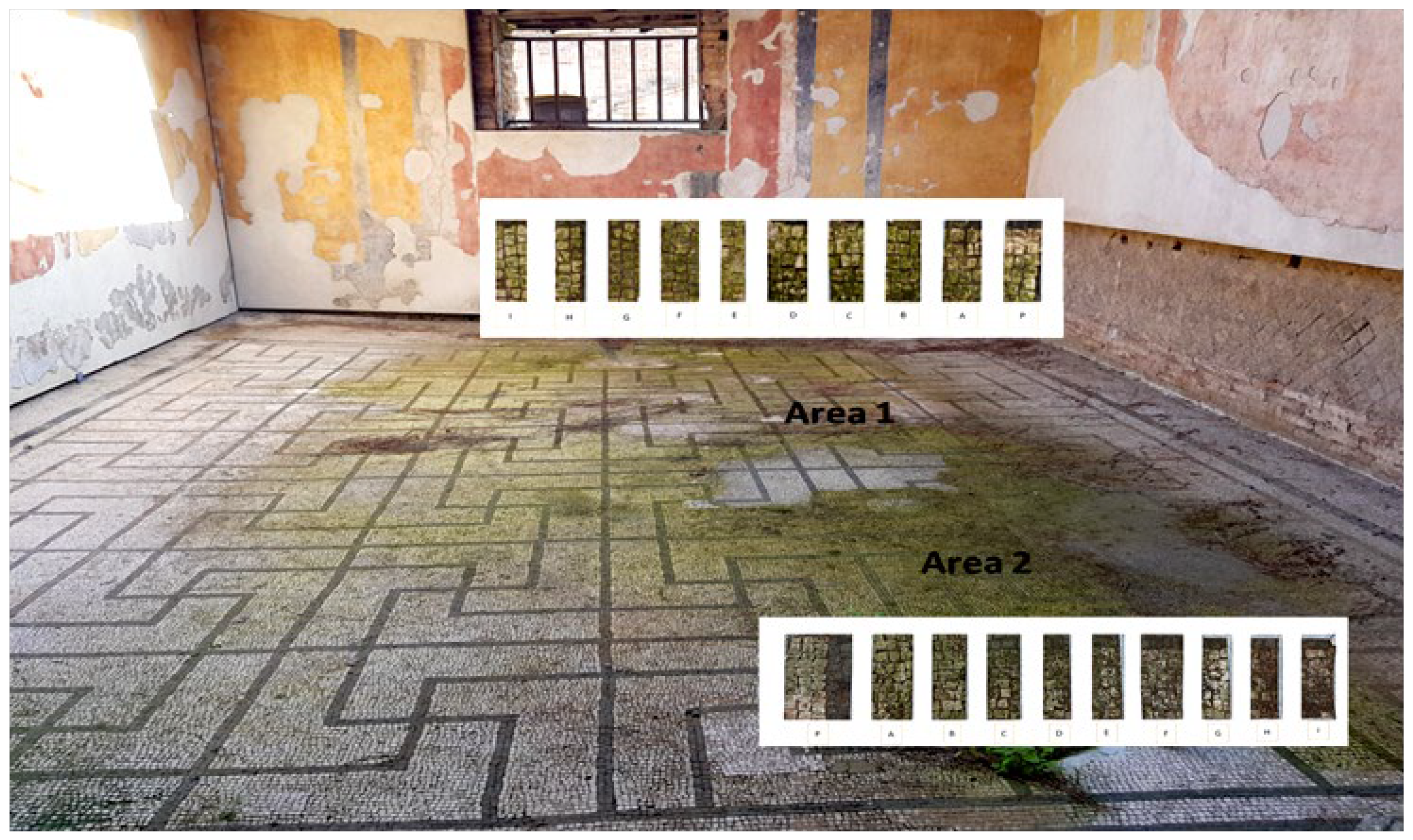
| Zones | Essential Oils | Composition | pH (Sd < 0.4) |
|---|---|---|---|
| A | Eucalyptus globulus | EO, NEVEK, Tween20 | 6.0 |
| B | Ocimum basilcum | EO, NEVEK, Tween20 | 6.0 |
| C | Essenzio | Extract of Thymus vulgaris and Origanum vulgare | 4.6 |
| D | Eugenia caryophyllus | EO, NEVEK, Tween20 | 6.0 |
| E | YOCOCOIL | NEVEK, Tween20, Pinus cembra L., Eucalyptus globulus, Thymus vulgaris, Eugenia caryophyllus, Ocimum basilicum | 6.4 |
| F | Biotersus | Cinnamonum zeylanicum, Eugenia caryophyllata, Corydothymus capitatus, Tween20 | 4.5 |
| G | Thymus vulgaris | EO, NEVEK, Tween20 | 6.5 |
| H | Pinus cembra L. | EO, NEVEK, Tween20 | 6.5 |
| I | Melaleuca alternifolia | EO, NEVEK, Tween20 | 6.5 |
| P | Preventol® RI50 | Quaternary ammonium salts | 6.7 |
| Biocides | Bands | Bands | Bands |
|---|---|---|---|
| Without Thickener (after Drying) | With Thickener (before Drying) | With Thickener (after Drying) | |
| Eucalyptus | 2928 | 2966-2943-2880-1464-1446-1359-1271-1233-1214-1166-1015-984-887-814-788-645 | 2927-1448-1370-1172-862 |
| Basil | 2925-1721-1511-1455-903 | 3077-3001-2963-2910-2834-1611-1584-1510-1463-1439-1300-1243-1176-1110-994-810-623 | 3417-2971-2439-2420-2322-2297-2182-2165-2108-2065-2022-1986-1917-1620-1462-1210-1174-1132-1091-946-859-820-806-788-742-699-683 |
| Cloves | 1765-1510 | 3521-3075-3003-2937-2842-1764-1606-1510-1462-1431-1265-1231-1199-1121-994-850-817-793-745-647 | 3425-2971-2896-2288-2190-2164-2050-1982-1457-1410-1263-876 |
| YOCOCOIL | 3454-2961-2919-2855- 2149-2048-2030-2002- 1976-1960-1735-1607-1511-1455-1351-1269-1249-1102-999-949-852-706-642-625 | 3348-2962-2924-1511-1449-1432-1267-1244-1214-1176-1121-989-840-807-741 | 3399-2971-2893-2325-2290-2189-2166-2105-2064-2050-1984-1563-1454-1263-1014-875-818-730-676 |
| Biotersus | 3338-2051 | 3399-2957-2924-2869-1734-1624-1513-1452-1429-1266-1234-1207-1178-1120-993-943-865-814-796-642 | 3364-2920-2322-2190-2166-2111-2077-2047-1983-1450-1354-1255-1019-888-818-658-631 |
| Thyme | 2959-2926-1457 | 3424-2960-2925-2869-1702-1619-1583-1519-1457-1419-1338-1289-1259-1224-1180-1112-1087-997-945-857-808-738 | 2960-2926-1457-1618-1583-1291-1257-809-740 |
| Pine tree | 3394-2929-1621-1446- 1379-936 | 2958-2909-2853-2725-1715-1688-1674-1624-1445-1377-1362-1300-1215-1182-1115-1022-1009-980-936-885-867-821-653 | 2921-1622-1446-1378-937 |
| Tea tree | 3340-889-864-799 | 3339-995-889-864-830-815-799-780 | 3340 |
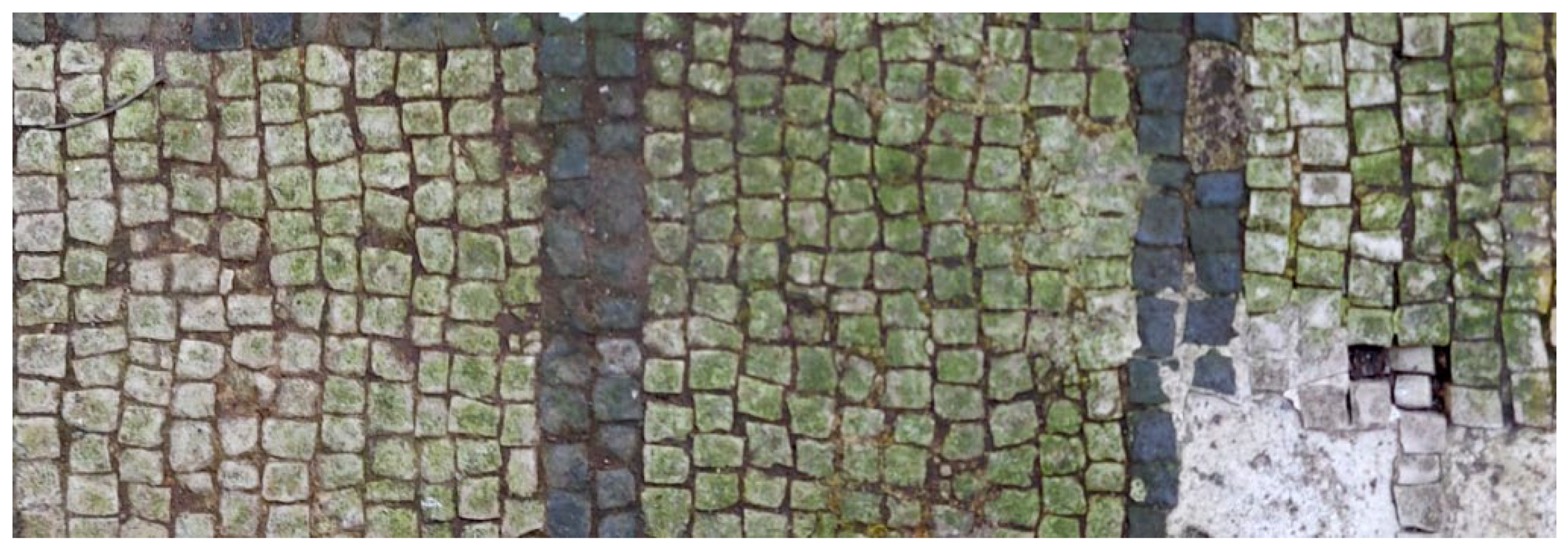
| Area 1 | Area 2 | |
|---|---|---|
| Before the treatment |  |  |
| After the biocide treatment |  |  |
| After the chemical and mechanical cleaning |  |  |
| Before the Biocide Treatment | After the Biocide Treatment | |
|---|---|---|
| Area 1 |  |  |
| Area 2 |  |  |
| Efficacy | P | A | B | C | D | E | F | G | H | I |
|---|---|---|---|---|---|---|---|---|---|---|
| Area 1 | ** | * | * | *** | ** | *** | ** | *** | *** | * |
| Area 2 | *** | *** | ** | * | * | ** | ** | *** | * | ** |
| Biocides | Area 1 | Area 2 | |||||||
|---|---|---|---|---|---|---|---|---|---|
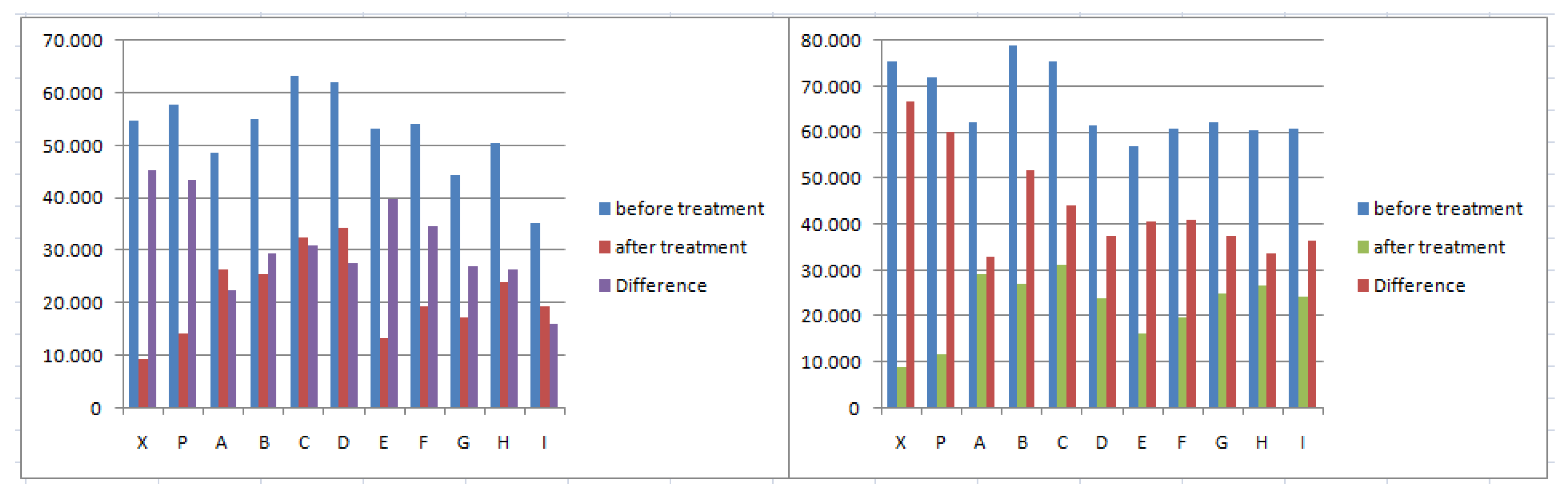
| Before | After | Difference | SD | Before | After | Difference | SD | ||
| X | Chemical and mechanical treatment | 54,678 | 9327 | 45,351 | <1102 | 75,346 | 8800 | 66,546 | 1310 |
| P | Preventol® RI50 | 57,617 | 14,261 | 43,356 | <1772 | 71,971 | 11,896 | 60,075 | 1001 |
| A | Eucalyptus | 48,765 | 26,291 | 22,474 | <1652 | 62,113 | 29,231 | 32,882 | 1641 |
| B | Basil | 54,891 | 25,447 | 29,444 | <1586 | 78,830 | 27,001 | 51,829 | 896 |
| C | Essenzio | 63,375 | 32,542 | 30,833 | <3206 | 75,344 | 31,244 | 44,100 | 1442 |
| D | Cloves | 61,932 | 34,341 | 27,591 | <1188 | 61,344 | 23,789 | 37,555 | 1204 |
| E | YOCOCOIL | 53,168 | 13,351 | 39,817 | <2325 | 56,793 | 16,101 | 40,692 | 2412 |
| F | Biotersus | 54,084 | 19,538 | 34,546 | <2988 | 60,668 | 19,567 | 41,101 | 2191 |
| G | Thyme | 44,263 | 17,311 | 26,952 | <692 | 62,231 | 24,878 | 37,353 | 1380 |
| H | Pine tree | 50,442 | 23,945 | 26,497 | <1247 | 60,323 | 26,578 | 33,745 | 1567 |
| I | Tea tree | 35,261 | 19,321 | 15,940 | <1281 | 60,641 | 24,080 | 36,561 | 2156 |
| Biocides | Area 1 | Area 2 | ||||||
|---|---|---|---|---|---|---|---|---|
| Vis | UV | CIELab | ATP | Vis | UV | CIELab | ATP | |
| Preventol® RI50 | x | x | xx | x | x | x | xx | |
| Eucalyptus | x | |||||||
| Basil | x | |||||||
| Essenzio | x | x | ||||||
| Cloves | ||||||||
| YOCOCOIL | x | x | x | x | x | x | x | |
| Biotersus | x | x | x | |||||
| Thyme | x | x | x | |||||
| Pine tree | x | x | ||||||
| Tea tree | x | x | x | |||||
Publisher’s Note: MDPI stays neutral with regard to jurisdictional claims in published maps and institutional affiliations. |
© 2022 by the authors. Licensee MDPI, Basel, Switzerland. This article is an open access article distributed under the terms and conditions of the Creative Commons Attribution (CC BY) license (https://creativecommons.org/licenses/by/4.0/).
Share and Cite
Macchia, A.; Aureli, H.; Prestileo, F.; Ortenzi, F.; Sellathurai, S.; Docci, A.; Cerafogli, E.; Colasanti, I.A.; Ricca, M.; La Russa, M.F. In-Situ Comparative Study of Eucalyptus, Basil, Cloves, Thyme, Pine Tree, and Tea Tree Essential Oil Biocide Efficacy. Methods Protoc. 2022, 5, 37. https://doi.org/10.3390/mps5030037
Macchia A, Aureli H, Prestileo F, Ortenzi F, Sellathurai S, Docci A, Cerafogli E, Colasanti IA, Ricca M, La Russa MF. In-Situ Comparative Study of Eucalyptus, Basil, Cloves, Thyme, Pine Tree, and Tea Tree Essential Oil Biocide Efficacy. Methods and Protocols. 2022; 5(3):37. https://doi.org/10.3390/mps5030037
Chicago/Turabian StyleMacchia, Andrea, Hélène Aureli, Fernanda Prestileo, Federico Ortenzi, Shaila Sellathurai, Antonella Docci, Eleonora Cerafogli, Irene Angela Colasanti, Michela Ricca, and Mauro Francesco La Russa. 2022. "In-Situ Comparative Study of Eucalyptus, Basil, Cloves, Thyme, Pine Tree, and Tea Tree Essential Oil Biocide Efficacy" Methods and Protocols 5, no. 3: 37. https://doi.org/10.3390/mps5030037