
In Vivo Evaluation of Wound Healing Efficacy of Gel-Based Dressings Loaded with Pycnogenol™ and Ceratothoa oestroides Extracts
 , ,
, ,
Abstract
:1. Introduction
2. Results and Discussion
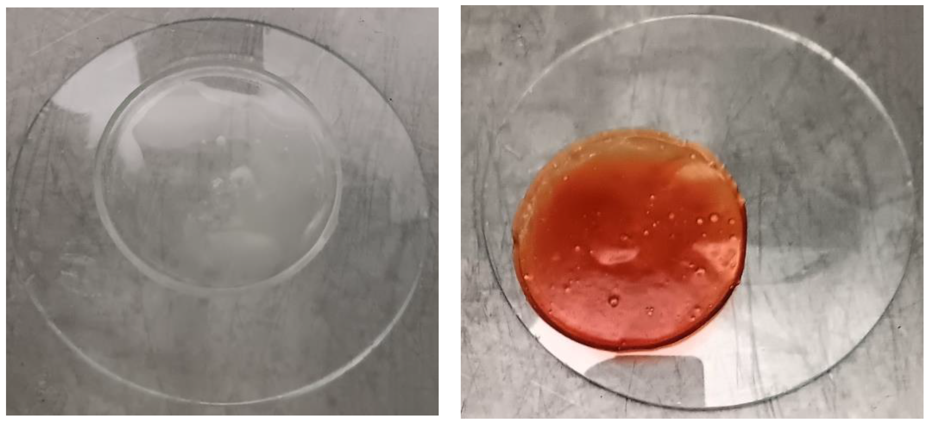
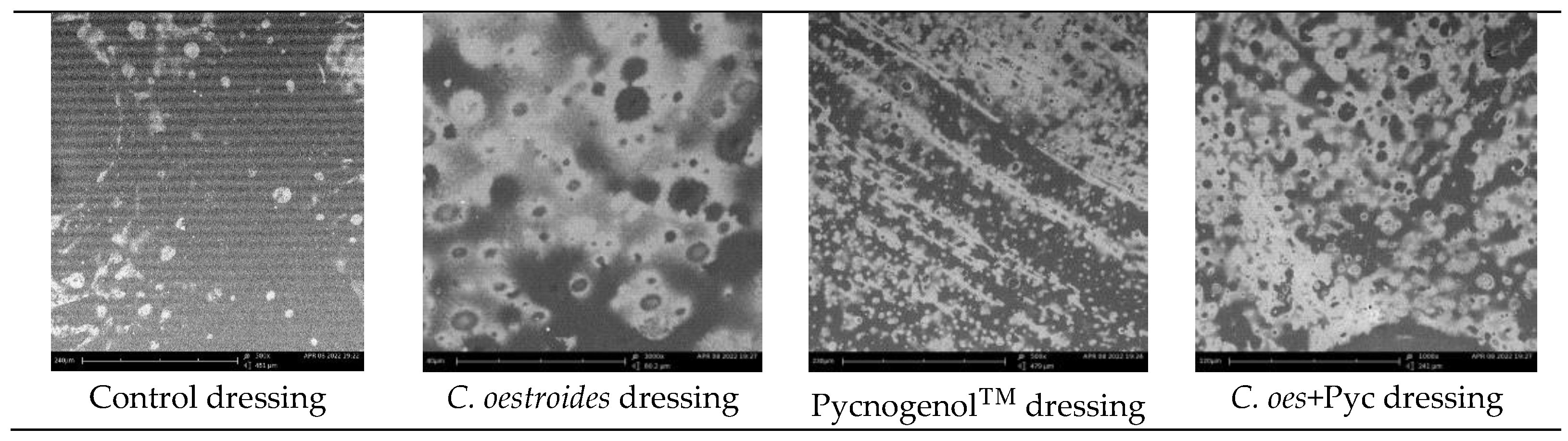
2.1. Dressings Characterization
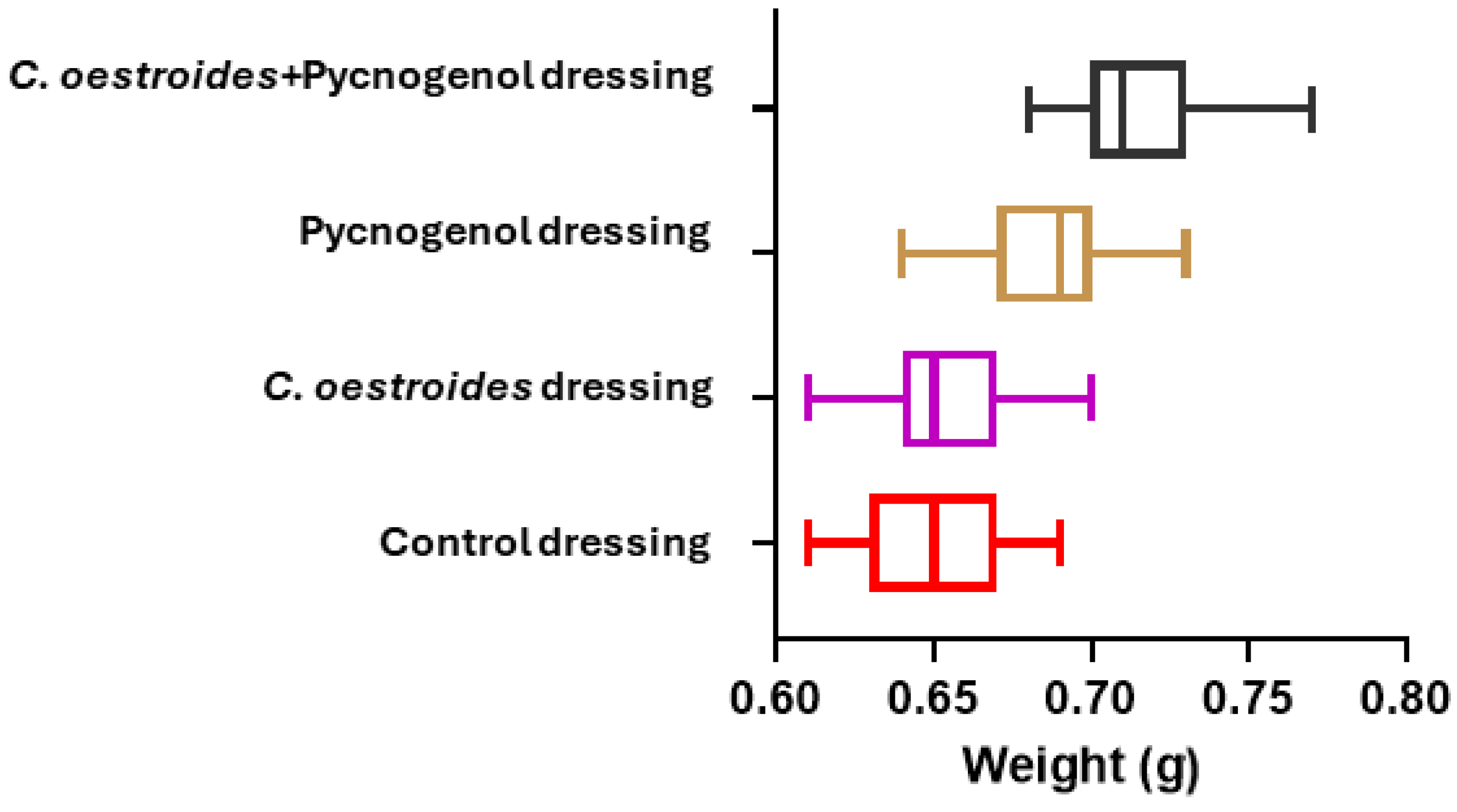
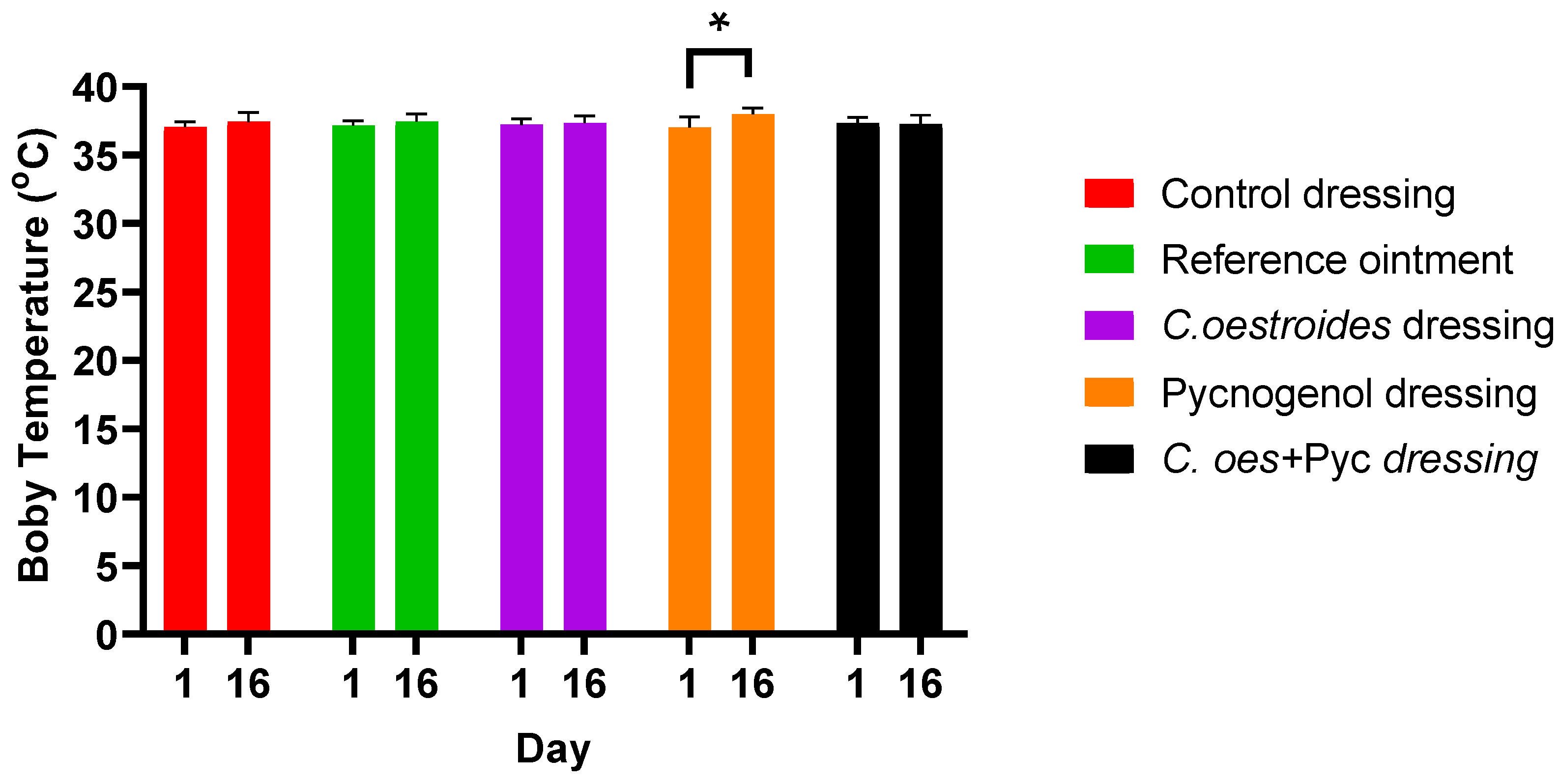
2.2. Weight and Temperature Measurements
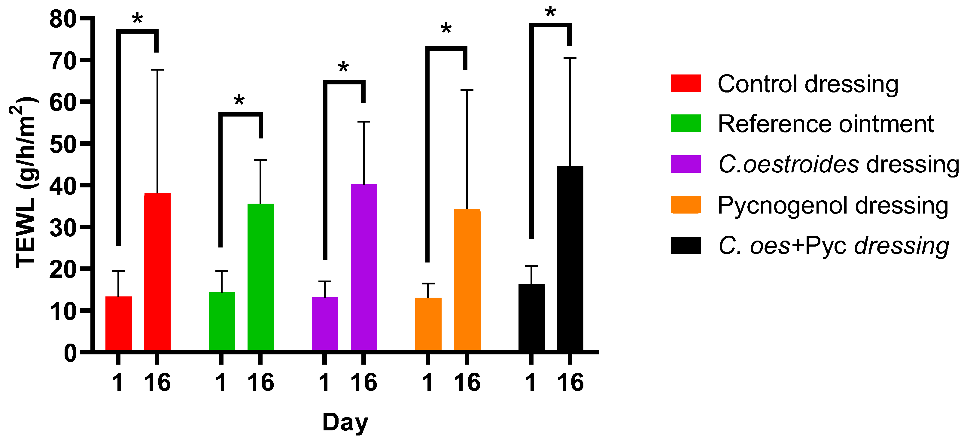
2.3. Transepidermal Water Loss
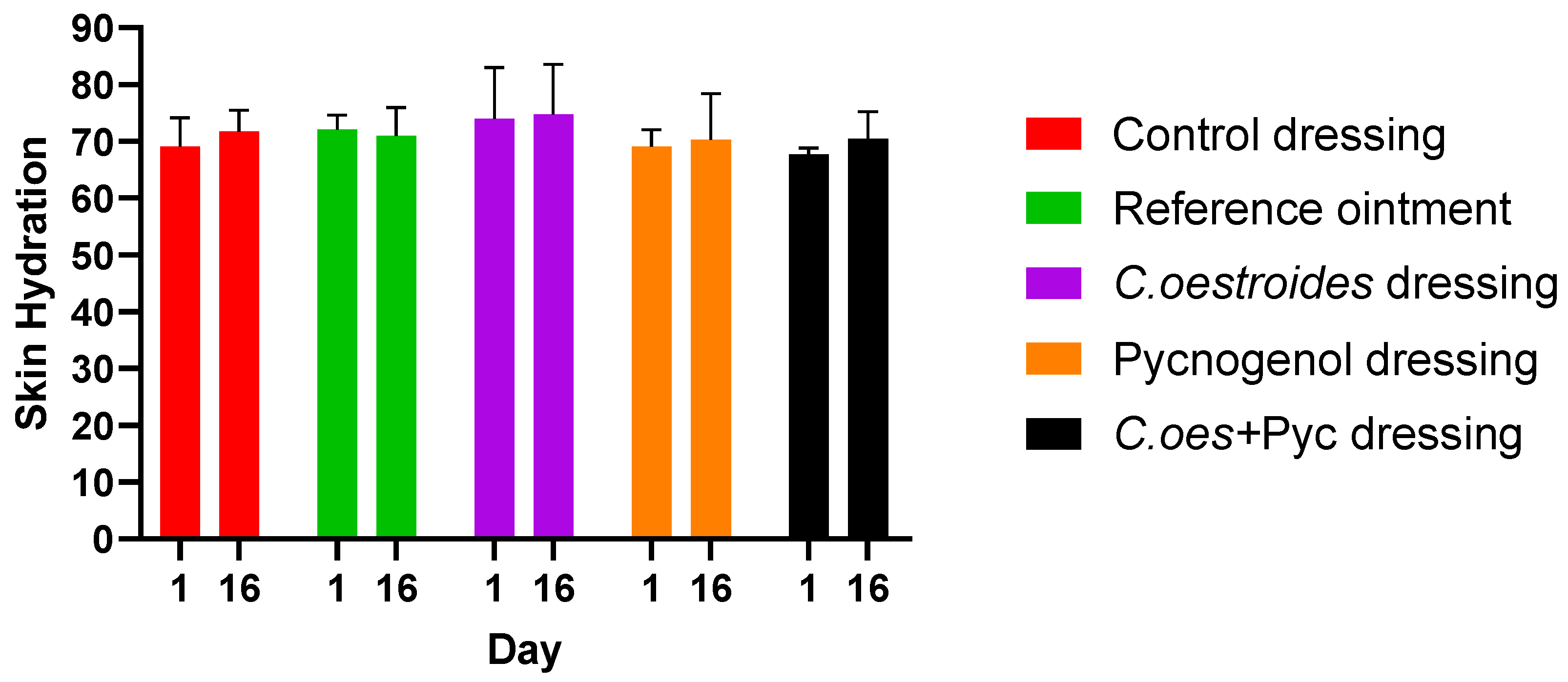
2.4. Hydration
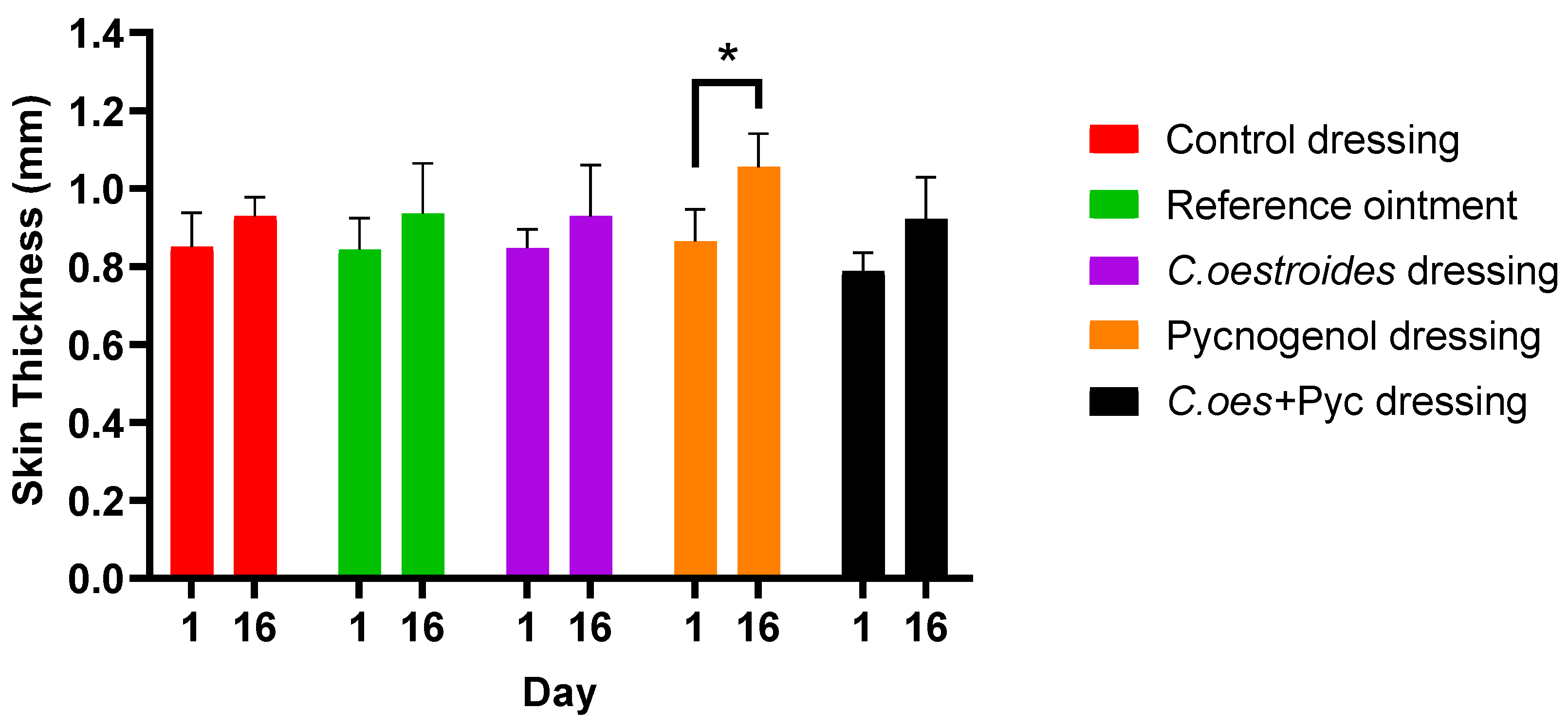
2.5. Skin Thickness
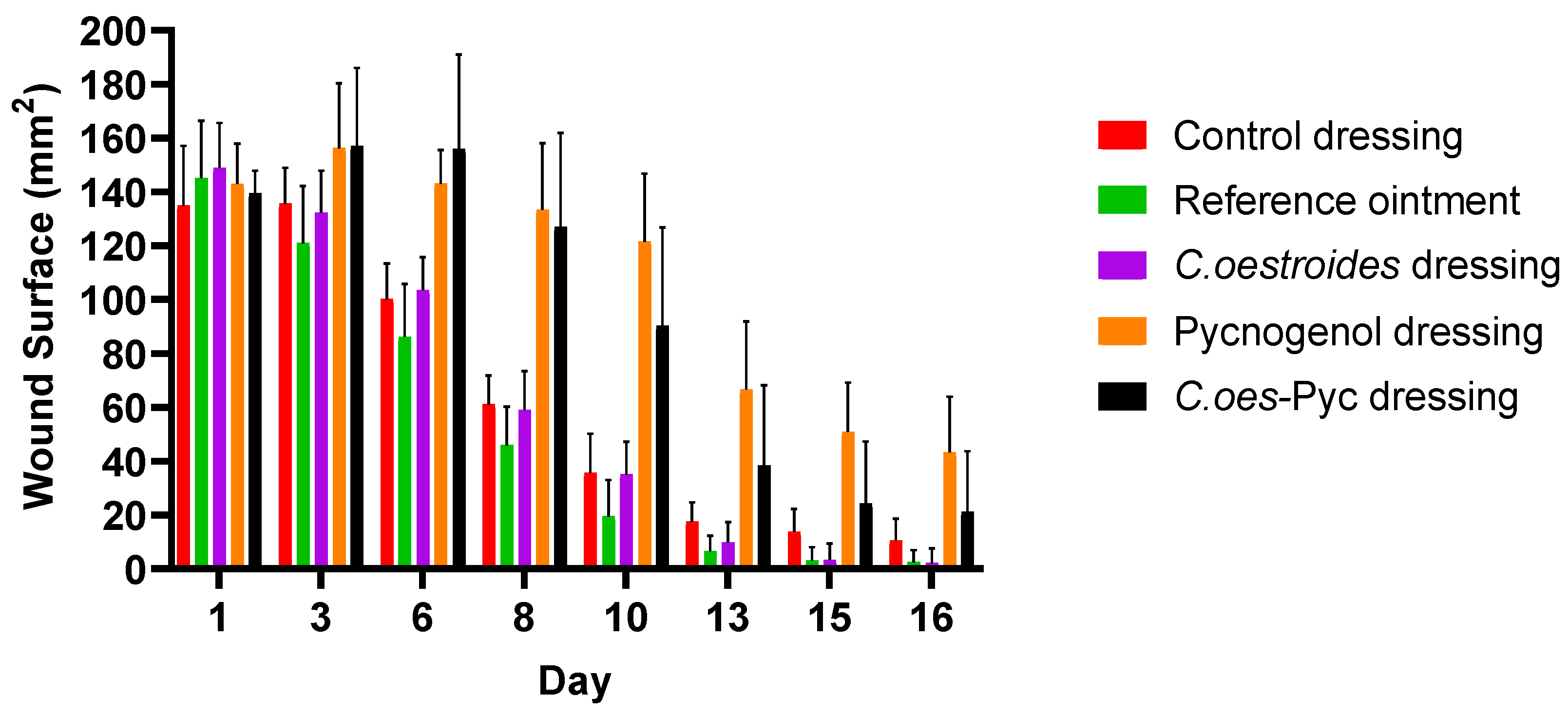
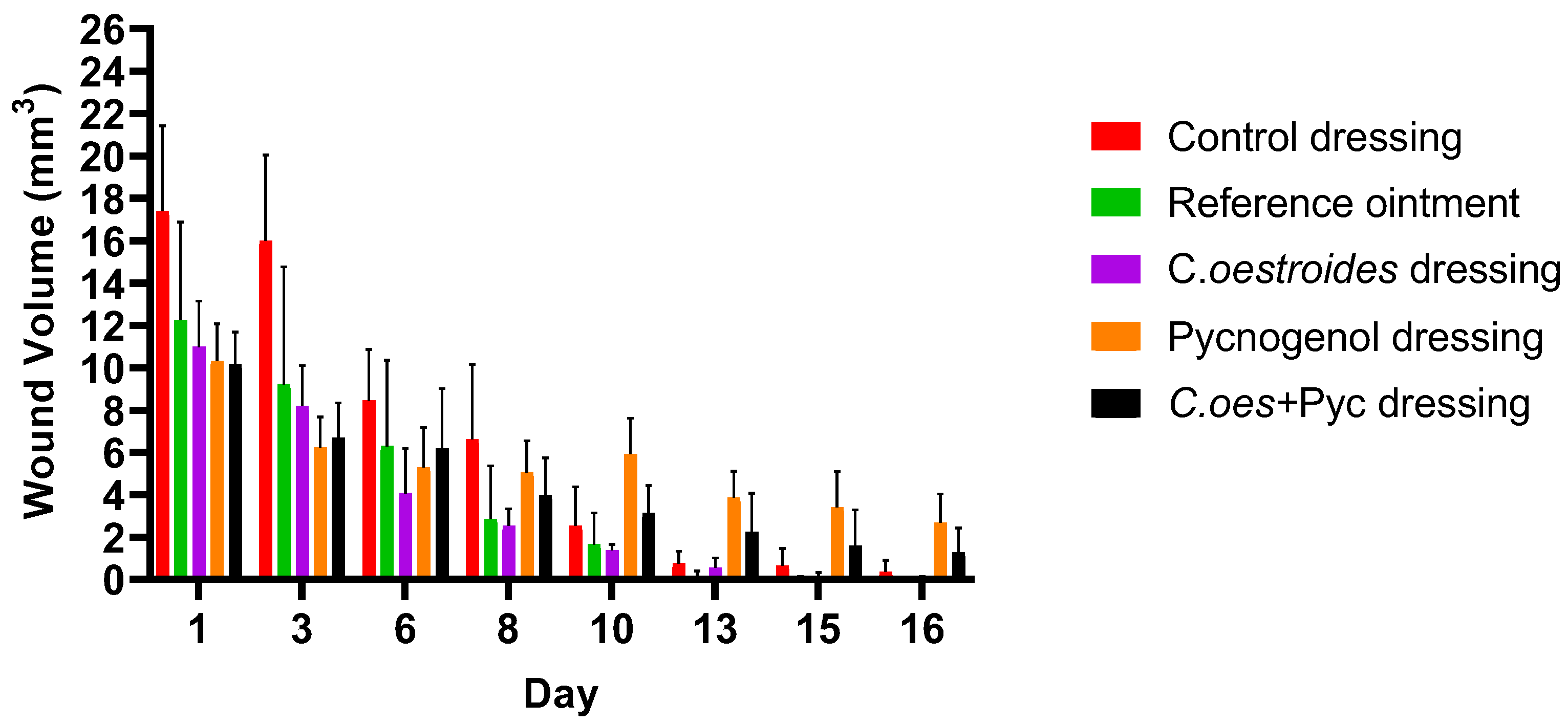
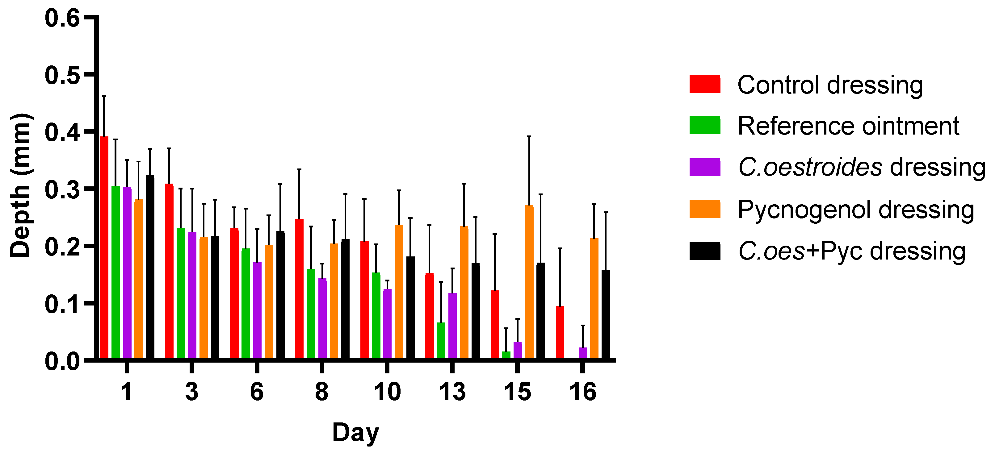
2.6. Wound Area, Volume, and Debt
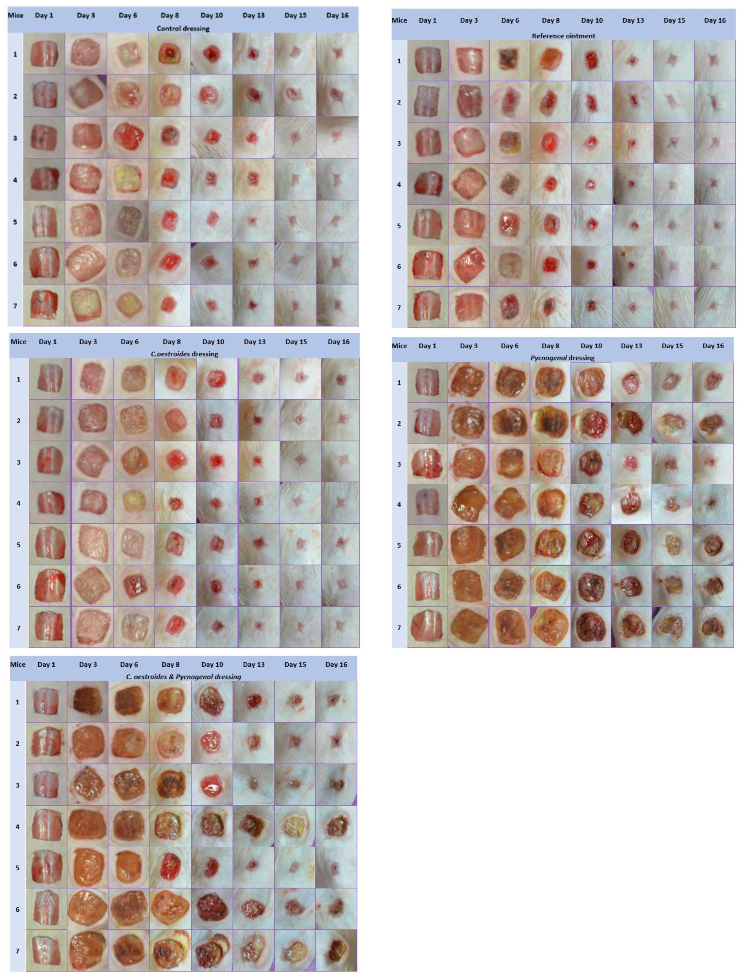
2.7. Photodocumentation
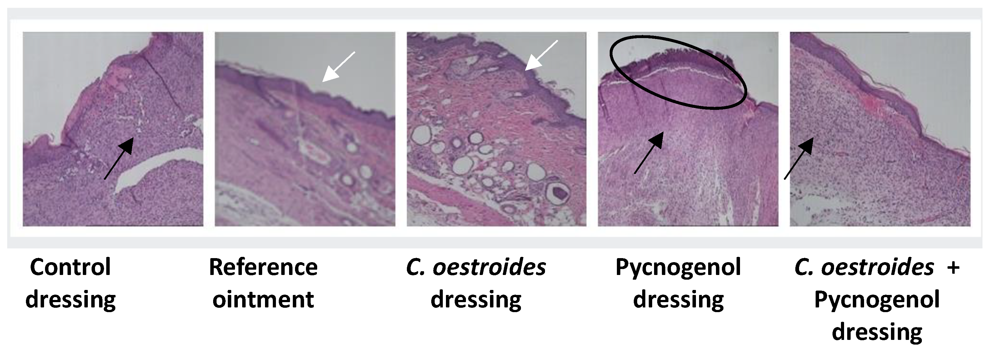
2.8. Histopathological Findings
2.9. Discussion
3. Conclusions
4. Materials and Methods
4.1. Reagents and Raw Materials
4.2. C. oestroides Olive Oil Extract Preparation
4.3. Preparation of the Dressings
4.4. Scanning Electron Microscopy (SEM)
4.5. Sterility Control
4.6. In Vivo Study Design and Animals
- Dressings composed only of excipients, serving as a control, without therapeutic agents.
- An ointment containing Ceratothoa oestroides extract, used as a positive control.
- Dressings loaded with Ceratothoa oestroides extract.
- Dressings loaded with Pycnogenol™
- Dressings incorporating a combination of both Ceratothoa oestroides and Pycnogenol™ extracts.
4.7. Wound Infliction
4.8. Wound Maintenance
4.9. Weight and Temperature Measurements
4.10. Photodocumentation
4.11. Evaluation of Transepidermal Water Loss (TEWL)
4.12. Evaluation of Hydration
4.13. Evaluation of Skin Thickness
4.14. Collection of Skin Samples—Histopathological Evaluation
4.15. Data Analysis
Author Contributions
Funding
Institutional Review Board Statement
Informed Consent Statement
Data Availability Statement
Acknowledgments
Conflicts of Interest
References
- Almadani, Y.H.; Vorstenbosch, J.; Davison, P.G.; Murphy, A.M. Wound Healing: A Comprehensive Review. Semin. Plast. Surg. 2021, 35, 141–144. [Google Scholar] [CrossRef]
- Kolimi, P.; Narala, S.; Nyavanandi, D.; Youssef, A.A.A.; Dudhipala, N. Innovative Treatment Strategies to Accelerate Wound Healing: Trajectory and Recent Advancements. Cells 2022, 11, 2439. [Google Scholar] [CrossRef] [PubMed]
- Senni, K.; Pereira, J.; Gueniche, F.; Delbarre-Ladrat, C.; Sinquin, C.; Ratiskol, J.; Godeau, G.; Fischer, A.-M.; Helley, D.; Colliec-Jouault, S. Marine polysaccharides: A source of bioactive molecules for cell therapy and tissue engineering. Mar. Drugs 2011, 9, 1664–1681. [Google Scholar] [CrossRef] [PubMed]
- Ramalingam, S.; Chandrasekar, M.J.N.; Nanjan, M.J. Plant-based Natural Products for Wound Healing: A Critical Review. Curr. Drug. Res. Rev. 2022, 14, 37–60. [Google Scholar] [CrossRef] [PubMed]
- Fan, B.; Dun, S.H.; Gu, J.Q.; Guo, Y.; Ikuyama, S. Pycnogenol Attenuates the Release of Proinflammatory Cytokines and Expression of Perilipin 2 in Lipopolysaccharide-Stimulated Microglia in Part via Inhibition of NF-κB and AP-1 Activation. PLoS ONE 2015, 10, e0137837. [Google Scholar] [CrossRef]
- Kyriazi, M.; Yova, D.; Rallis, M.; Lima, A. Cancer chemopreventive effects of Pinus Maritima bark extract on ultraviolet radiation and ultraviolet radiation 7, 12, dimethylbenz(a)anthracene induced skin carcinogenesis of hairless mice. Cancer Lett. 2006, 237, 234–241. [Google Scholar] [CrossRef] [PubMed]
- Blazsó, G.; Gábor, M.; Schönlau, F.; Rohdewald, P. Pycnogenol accelerates wound healing and reduces scar formation. Phytother. Res. 2004, 18, 579–581. [Google Scholar] [CrossRef] [PubMed]
- Dogan, E.; Yanmaz, L.; Gedikli, S.; Ersoz, U.; Okumus, Z. The Effect of Pycnogenol on Wound Healing in Diabetic Rats. Ostomy Wound Manage. 2017, 63, 41–47. [Google Scholar] [PubMed]
- Toledo, R.R.; Santos, M.E.R.C.; Schnaider, T.B. Effect of Pycnogenol on the Healing of Venous Ulcers. Ann. Vasc. Surg. 2017, 38, 212–219. [Google Scholar] [CrossRef] [PubMed]
- Belcaro, G.; Cesarone, M.R.; Errichi, B.M.; Ledda, A.; Di Renzo, A.; Stuard, S.; Dugall, M.; Pellegrini, L.; Rohdewald, P.; Ippolito, E.; et al. Venous ulcers: Microcirculatory improvement and faster healing with local use of Pycnogenol. Angiology 2005, 56, 699–705. [Google Scholar] [CrossRef] [PubMed]
- Meimeti, E.; Tentolouris, N.; Manes, C.; Loupa, C.; Provatopoulou, X.; Mostratos, D.; Vitsos, A.; Roussis, V.; Tzouvelekis, L.; Miriagou, V.; et al. Ointments containing Ceratothoa oestroides extract: Evaluation of their healing potential in the treatment of diabetic foot ulcers. Wound Repair. Regen. 2020, 28, 234–241. [Google Scholar] [CrossRef] [PubMed]
- Vitsos, A.; Tsagarousianos, C.; Vergos, O.; Stithos, D.; Mathioudakis, D.; Vitsos, I.; Zouni, P.; Kakolyri, A.; Meimeti, E.; Kyriazi, M.; et al. Efficacy of a Ceratothoa oestroides Olive Oil Extract in Patients With Chronic Ulcers: A Pilot Study. Int. J. Low. Extrem. Wounds. 2019, 18, 309–316. [Google Scholar] [CrossRef] [PubMed]
- Sofrona, E.; Tziveleka, L.A.; Harizani, M.; Koroli, P.; Sfiniadakis, I.; Roussis, V.; Rallis, M.; Ioannou, E. In Vivo Evaluation of the Wound Healing Activity of Extracts and Bioactive Constituents of the Marine Isopod Ceratothoa oestroides. Mar. Drugs 2020, 18, 219. [Google Scholar] [CrossRef] [PubMed]
- Meimeti, E.; Tentolouris, N.; Loupa, C.; Roussis, V.; Rallis, M. Marine Isopod Ceratothoa Oestroides Extract: A Novel Treatment for Diabetic Foot Ulcers? Case Report of an Immunosuppressed Patient. Med. Arch. 2019, 73, 131–133. [Google Scholar] [CrossRef]
- Meimeti, E.; Kafanas, A.; Pavlou, P.; Evangelatou, A.; Tsouparelou, P.; Kanellopoulos, S.; Kipouros, P.; Koliarakis, N.; Leonis, G.; Ioannou, E.; et al. Topical Treatment of Skin Injury Inflicted in Mice by X-Ray Irradiation. Skin. Pharmacol. Physiol. 2018, 31, 175–183. [Google Scholar] [CrossRef] [PubMed]
- Grigoropoulos, A.; Zouridaki, E.; Sgontzou, T. P 144—Clinical evaluation of wound healing capacity of isopod Ceratothoa oestroides oil extract. Free. Radic. Biol. Med. 2017, 108 (Suppl. S1), S67. [Google Scholar] [CrossRef]
- Meimeti, E.; Psarou, A.; Loupa, C.V.; Vitsos, A.; Tentolouris, N.; Roussis, V.; Rallis, M. Treatment of diabetic foot ulcer with ointment based on Ceratothoa oestroides extract and eosin—Case report. Int. J. Med. Pharm. Case Rep. 2017, 9, 1–6. [Google Scholar] [CrossRef]
- Zhang, M.; Zhao, X. Alginate hydrogel dressings for advanced wound management. Int. J. Biol. Macromol. 2020, 162, 1414–1428. [Google Scholar] [CrossRef] [PubMed]
- Zhang, H.; Cheng, J.; Ao, Q. Preparation of Alginate-Based Biomaterials and Their Applications in Biomedicine. Mar. Drugs 2021, 19, 264. [Google Scholar] [CrossRef] [PubMed]
- Aderibigbe, B.A.; Buyana, B. Alginate in Wound Dressings. Pharmaceutics 2018, 10, 42. [Google Scholar] [CrossRef] [PubMed]
- Lee, K.Y.; Mooney, D.J. Alginate: Properties and biomedical applications. Prog. Polym. Sci. 2012, 37, 106–126. [Google Scholar] [CrossRef]
- Obagi, Z.; Damiani, G.; Grada, A.; Falanga, V. Principles of Wound Dressings: A Review. Surg. Technol. Int. 2019, 35, 50–57. [Google Scholar] [PubMed]
- Trevithick, J.R.; Bantseev, V.; Hirst, M.; Dzialoszynski, T.M.; Sanford, E.S. Is pycnogenol a double-edged sword? Cataractogenic in vitro, but reduces cataract risk in diabetic rats. Curr. Eye Res. 2013, 38, 751–760. [Google Scholar] [CrossRef] [PubMed]
- Verlaet, A.; van der Bolt, N.; Meijer, B.; Breynaert, A.; Naessens, T.; Konstanti, P.; Smidt, H.; Hermans, N.; Savelkoul, H.F.; Teodorowicz, M. Toll-Like Receptor-Dependent Immunomodulatory Activity of Pycnogenol®. Nutrients 2019, 11, 214. [Google Scholar] [CrossRef] [PubMed]
- Percie du Sert, N.; Hurst, V.; Ahluwalia, A.; Alam, S.; Avey, M.T.; Baker, M.; Browne, W.J.; Clark, A.; Cuthill, I.C.; Dirnagl, U.; et al. The ARRIVE guidelines 2.0: Updated guidelines for reporting animal research. PLoS Biol. 2020, 18, e3000410. [Google Scholar] [CrossRef]
- Grada, A.; Mervis, J.; Falanga, V. Research Techniques Made Simple: Animal Models of Wound Healing. J. Investig. Dermatol. 2018, 138, 2095–2105.e1. [Google Scholar] [CrossRef] [PubMed]
- Zomer, H.D.; Trentin, A.G. Skin wound healing in humans and mice: Challenges in translational research. J. Dermatol. Sci. 2018, 90, 3–12. [Google Scholar] [CrossRef] [PubMed]
- Zhang, N.; Shi, K.; Hong, L.; Zhao, J.; Yu, J. Antera 3D camera: A novel method for evaluating the therapeutic efficacy of fractional CO2 laser for surgical incision scars. J. Cosmet. Dermatol. 2018, 17, 1041–1045. [Google Scholar] [CrossRef]
- Gardien, K.L.; Baas, D.C.; de Vet, H.C.; Middelkoop, E. Transepidermal water loss measured with the Tewameter TM300 in burn scars. Burns 2016, 42, 1455–1462. [Google Scholar] [CrossRef] [PubMed]
- Anthonissen, M.; Daly, D.; Peeters, R.; Van Brussel, M.; Fieuws, S.; Moortgat, P.; Flour, M.; Van den Kerckhove, E. Reliability of Repeated Measurements on Post-Burn Scars with Corneometer CM 825(®). Skin. Res. Technol. 2015, 21, 302–312. [Google Scholar] [CrossRef] [PubMed]


| Specimens’ 16th Day | Ιinflammation | Oedema | Hyperkeratosis | Wound Depth | Ulceration | Necrosis | Parakeratosis | Score |
|---|---|---|---|---|---|---|---|---|
| Control dressing | 2.25 | 2.5 | 2 | 3 | 1 | 0 | 0.5 | 11.25 |
| Reference ointment | 1 | 1 | 2 | 3 | 0 | 0 | 0 | 7 |
| C. oestroides dressing | 1 | 1 | 2 | 2.5 | 0 | 0 | 0 | 6.5 |
| Pycnogenol™ dressing | 3 | 3 | 3 | 3 | 1 | 1 | 1 | 15 |
| C. oestroides and Pycnogenol™ dressing | 3 | 3 | 3 | 3 | 1 | 1 | 1 | 15 |
| Ingredients | Control (in Grams) | Pycnogenol™ and C. oestroides (in Grams) | C. oestroides (in Grams) | Pycnogenol™ (in Grams) |
|---|---|---|---|---|
| Pycnogenol™ | - | 1.2 | - | 1.2 |
| Tapioca maltodextrin (Zorbit) | 1 | 1 | 1 | 1 |
| Sodium Alginate | 4 | 4 | 4 | 4 |
| Olive oil | 2.4 | - | - | 2.4 |
| Ceratothoa oestroides Olive oil extract | - | 2.4 | 2.4 | - |
| Wool fat (Lanolin) | 4.5 | 4.5 | 4.5 | 4,5 |
| Glycerin | 10 | 10 | 10 | 10 |
| Distilled water | 78.1 | 76.9 | 78.1 | 76.9 |
| Potassium hydroxide 50% Solution | .qs ad pH 6 | .qs ad pH 6 | .qs ad pH 6 | .qs ad pH 6 |
| Scoring Criteria for Histopathological Evaluation | ||||
|---|---|---|---|---|
| Inflammation | 0 (absence) | 1 (mild) | 2 (moderate) | 3 (heavy) |
| Oedema | 0 (absence) | 1 (mild) | 2 (moderate) | 3 (heavy) |
| Hyperkeratosis | 0 (absence) | 1 (mild) | 2 (moderate) | 3 (heavy) |
| Wound thickness | 0 (absence) | 1 (superficial) | 2 (moderate) | 3 (total) |
| Ulceration | 0 (absence) | 1 (presence) | ||
| Necrosis | 0 (absence) | 1 (presence) | ||
| Parakeratosis | 0 (absence) | 1 (presence) | ||
Disclaimer/Publisher’s Note: The statements, opinions and data contained in all publications are solely those of the individual author(s) and contributor(s) and not of MDPI and/or the editor(s). MDPI and/or the editor(s) disclaim responsibility for any injury to people or property resulting from any ideas, methods, instructions or products referred to in the content. |
© 2024 by the authors. Licensee MDPI, Basel, Switzerland. This article is an open access article distributed under the terms and conditions of the Creative Commons Attribution (CC BY) license (https://creativecommons.org/licenses/by/4.0/).
Share and Cite
Vitsos, A.; Ieronymaki, D.; Kostaki, M.; Almpani, C.; Barda, C.; Kikionis, S.; Sfiniadakis, I.; Dallas, P.; Rallis, M.C. In Vivo Evaluation of Wound Healing Efficacy of Gel-Based Dressings Loaded with Pycnogenol™ and Ceratothoa oestroides Extracts. Gels 2024, 10, 233. https://doi.org/10.3390/gels10040233
Vitsos A, Ieronymaki D, Kostaki M, Almpani C, Barda C, Kikionis S, Sfiniadakis I, Dallas P, Rallis MC. In Vivo Evaluation of Wound Healing Efficacy of Gel-Based Dressings Loaded with Pycnogenol™ and Ceratothoa oestroides Extracts. Gels. 2024; 10(4):233. https://doi.org/10.3390/gels10040233
Chicago/Turabian StyleVitsos, Andreas, Dimitra Ieronymaki, Maria Kostaki, Chara Almpani, Christina Barda, Stefanos Kikionis, Ioannis Sfiniadakis, Paraskevas Dallas, and Michail Christou Rallis. 2024. "In Vivo Evaluation of Wound Healing Efficacy of Gel-Based Dressings Loaded with Pycnogenol™ and Ceratothoa oestroides Extracts" Gels 10, no. 4: 233. https://doi.org/10.3390/gels10040233